Edited by:
Giuseppe Locatelli, University of Bern, Switzerland
Reviewed by:
Marina Romero-Ramos, Aarhus University, Denmark Ashley Harms, University of Alabama at Birmingham, United States Elisa Greggio, University of Padua, Italy
*Correspondence:
Arianna Bellucci [email protected]
Specialty section:
This article was submitted to Multiple Sclerosis and Neuroimmunology, a section of the journal Frontiers in Immunology
Received: 29 September 2020 Accepted: 27 January 2021 Published: 19 February 2021 Citation:
Bogale TA, Faustini G, Longhena F, Mitola S, Pizzi M and Bellucci A (2021) Alpha-Synuclein in the Regulation of Brain Endothelial and Perivascular Cells: Gaps and Future Perspectives. Front. Immunol. 12:611761. doi: 10.3389/fimmu.2021.611761
Alpha-Synuclein in the Regulation of
Brain Endothelial and Perivascular
Cells: Gaps and Future Perspectives
Tizibt Ashine Bogale
1, Gaia Faustini
1, Francesca Longhena
1, Stefania Mitola
2,3,
Marina Pizzi
1and Arianna Bellucci
1,3*
1Division of Pharmacology, Department of Molecular and Translational Medicine, University of Brescia, Brescia, Italy, 2Biotechnology Division, Department of Molecular and Translational Medicine, University of Brescia, Brescia, Italy, 3Laboratory for Preventive and Personalized Medicine, Department of Molecular and Translational Medicine, University of
Brescia, Brescia, Italy
Misfolded proteins, inflammation, and vascular alterations are common pathological
hallmarks of neurodegenerative diseases. Alpha-synuclein is a small synaptic protein
that was identified as a major component of Lewy bodies and Lewy neurites in
the brain of patients affected by Parkinson’s disease (PD), Lewy body dementia
(LBD), and other synucleinopathies. It is mainly involved in the regulation of synaptic
vesicle trafficking but can also control mitochondrial/endoplasmic reticulum (ER)
homeostasis, lysosome/phagosome function, and cytoskeleton organization. Recent
evidence supports that the pathological forms of α-synuclein can also reduce the release
of vasoactive and inflammatory mediators from endothelial cells (ECs) and modulates the
expression of tight junction (TJ) proteins important for maintaining the blood–brain barrier
(BBB). This hints that α-synuclein deposition can affect BBB integrity. Border associated
macrophages (BAMs) are brain resident macrophages found in association with the
vasculature (PVMs), meninges (MAMs), and choroid plexus (CPMs). Recent findings
indicate that these cells play distinct roles in stroke and neurodegenerative disorders.
Although many studies have addressed how α-synuclein may modulate microglia, its
effect on BAMs has been scarcely investigated. This review aims at summarizing the main
findings supporting how α-synuclein can affect ECs and/or BAMs function as well as their
interplay and effect on other cells in the brain perivascular environment in physiological
and pathological conditions. Gaps of knowledge and new perspectives on how this
protein can contribute to neurodegeneration by inducing BBB homeostatic changes in
different neurological conditions are highlighted.
Keywords: Parkinson’s disease, α-synuclein, endothelial cells, blood brain barrier, border-associated macrophages, perivascular cells
INTRODUCTION
Neurodegenerative diseases represent a relevant health burden, especially considering the growing
population of elderly subjects. Cerebrovascular disorders such as stroke are considered among
the major predisposing factors for the development of neurodegenerative diseases, including
Alzheimer’s disease (AD) and Parkinson’s disease (PD) (
1
,
2
). In particular, PD is the second most
common neurodegenerative disorder, affecting 2–3% of the population over the age of 65 years
(
3
). The lack of knowledge on the molecular underpinnings of
PD still limits the development of efficient therapies.
Protein aggregates enriched in insoluble α-synuclein fibrils
and loss of dopaminergic neurons in the nigrostriatal system
are key pathological features of this disorder (
4
,
5
). Of note, the
pathological deposition of insoluble α-synuclein at synapses is
believed to act as the primum movens for neuronal degeneration
in PD, as by hindering neurotransmitter release, it can trigger
synaptic failure (
6
–
8
). This event can then negatively impinge
on axonal projections, thus slowly flowing in a retrograde
neurodegenerative process culminating in neuronal cell death
(
6
–
8
). Additionally, α-synuclein-related neuroinflammation,
microglia activation, and vascular degeneration (
9
–
12
) have
been described as important players in disease pathogenesis.
This notwithstanding, whether α-synuclein communicates with
other neurovascular components such as border-associated
macrophages (BAMs) and vascular endothelial cells (ECs), which
are involved in the early phases of ischemic brain damage (
13
–
16
), remains to be explored.
Alpha-synuclein is a 14 kDa protein owning an undefined
structure in aqueous solutions (
17
). In neurons, the protein
regulates various processes including synaptic function,
mitochondrial homeostasis, autophagy/lysosomal functions, and
cytoskeletal reorganization (
8
,
18
–
24
). The diverse domains of
α-synuclein and its conformational plasticity allow the interaction
with a plethora of other proteins and lipid membranes
(
20
). Alpha-synuclein can also undergo post-translational
modifications as amino-terminal and carboxy-terminal nitration
and phosphorylation [e.g., Ser129 phosphorylation; (
25
–
27
)],
which in turn can impact its conformation and can lead to the
formation of toxic oligomers and fibrils (
20
). While oligomers
can affect membrane permeability as well as neuronal excitability
and engulf protein degradation systems (
28
–
30
), fibrils can
disrupt the integrity of intracellular organelles and induce
chronic inflammation (
28
,
31
). In the brain, α-synuclein is
expressed not only in the neuronal cells, but at lower levels
also in astrocytes, macrophages, and the microglia (
32
,
33
).
In the periphery, the protein is expressed in red blood cells
(
34
,
35
), platelets (
36
), and in other immune cells, such as T
cells, B cells, natural killer (NK) cells, and monocytes (
32
). It
has been found that α-synuclein can bind microglia cell surface
receptors, thus activating intracellular pathways mediating
the release of cytokines and upregulating of proinflammatory
genes (
10
,
37
). The protein can also regulate ECs function by
blocking the exocytosis of Weibel-Palade bodies (WPBs) (
38
)
and by downregulating the expression of tight junction (TJ)
proteins (
39
).
The deposition of α-synuclein insoluble aggregates named
Lewy bodies (LB) or glial cytoplasmic inclusions (GCI)
characterizes the brain of patients affected by PD and dementia
with LB (DLB) or multiple system atrophy (MSA), respectively
(
5
,
40
). For this reason, these disorders are commonly referred
to as synucleinopathies. Certain pathological strains of
α-synuclein, by moving between the brain cells and across the
blood–brain barrier (BBB) interfaces and acting as imprinting
templates for the pathological conformational shift of other
α-synuclein molecules, are believed to mediate the propagation of
pathological aggregates within the brain, from the periphery to
the brain, or from the brain to the periphery, with a prion-like
fashion (
41
,
42
).
This review focuses on how α-synuclein impacts vascular
ECs and BAMs regulation and crosstalk. Current gaps and
future perspectives in the context of neurological disorders are
also presented.
ALPHA-SYNUCLEIN FUNCTIONS IN THE
CENTRAL NERVOUS SYSTEM (CNS)
To date, the physiological function of α-synuclein has not been
fully disclosed, but we know that it controls neurotransmitter
release and synaptic plasticity, particularly inhibiting dopamine
overflow and modulating synaptic vesicles storage (
20
,
43
,
44
).
The full-length α-synuclein isoform consists of 140 amino
acids and its structure can be divided into three main regions.
The N-terminal part is essential for membrane binding (
45
–
47
) and includes the sites of main familial PD mutations, A30P,
A53T, and E46K (
18
,
20
), as well as for several post-translational
modifications (
48
). The central domain, called non-amyloid
component (NAC), is hydrophobic and highly aggregation-prone
(
49
), and is necessary and sufficient for α-synuclein fibrillation
(
50
). Finally, the C-terminal region is enriched in negative
charges (
51
) and can interact with the N-terminal domain to form
a compact aggregation-resistant structure (
52
).
Alpha-synuclein is described as an intrinsically disordered
protein as it can be found in monomeric form (
53
) or in
a stable tetramer (
54
) when purified at neutral pH. Rapid
environmental changes can induce the formation of partially
folded intermediates or kinetically trapped transition states
(
55
). Along aging, the high plasticity of α-synuclein, coupled
with post-translational modifications and protein enrichment
at synaptic sites, can promote in concert the formation of
high molecular weight soluble or insoluble aggregates, such as
oligomers, protofibrils, or fibrils (
20
,
21
). In PD, α-synuclein
deposition is thought to play a pathogenic role in triggering both
central and peripheral neurons degeneration, thus underlying the
onset of motor and non-motor symptoms, respectively (
56
,
57
).
Interestingly, both monomeric and aggregated α-synuclein can
be transferred from cell-to-cell (neuron-neuron, neuron-glia),
and also across the BBB, thus contributing to neuropathology
spreading (
58
). Endocytosis, carrier-mediated transports, and
tunneling nanotubes are described as the main mechanisms
for these exchanges (
59
,
60
). In addition, impairment in
glymphatic transport and lymphatic drainage pathways results
in the accumulation of α-synuclein in the brain parenchyma
and the progression of PD-like pathology in transgenic mouse
models (
61
). This is in line with evidence supporting that general
systemic circulation would act as a route for long-distance
transmission of endogenous α-synuclein (
62
).
Alpha-synuclein can also exert a physiological regulatory
action on intracellular organelles, including mitochondria
(
19
), endoplasmic reticulum (ER), mitochondria-ER associated
membranes (
63
), Golgi apparatus (
64
), and nuclei (
65
). Although
the nuclear localization of α-synuclein was the first to be reported,
its involvement in DNA repair mechanisms has been described
quite recently (
66
). Recent findings, showing reduced nucleus to
cytoplasmic transport in induced pluripotent stem cell
(iPSC)-derived neurons from familial patients with PD bearing A53T
mutation or multiplication of the α-synuclein gene locus SNCA
(
67
), support that the protein may also play a role in maintaining
nuclear membrane functions. Interestingly, reduced α-synuclein
DNA binding associates with transcription deregulation through
inhibition of cell cycle-related genes and the nuclear localization
of α-synuclein is modulated by its phosphorylation at Serine
129 (
68
).
The interplay between mitochondria and α-synuclein during
the progression of PD still constitutes an issue to be solved, as
the exact contribution of mitochondrial deficits and α-synuclein
aggregation to dopaminergic neurons degeneration has yet to be
clearly elucidated (
69
,
70
). Indeed, the aggregation of α-synuclein
induces neural deficits, but it is also evident that mitochondrial
dysfunctions are crucial events in the pathogenesis of PD (
71
,
72
).
Notably, the observation that α-synuclein is increased following
a stroke, and that its induction is involved in the response to
post-stroke brain damage, reinforces the idea that the protein
can act as a pivotal regulator of neuronal resilience to injury
(
35
,
73
–
75
).
Numerous studies have shown that α-synuclein accumulation
and aggregation can activate neuroinflammation (
76
–
79
), in
agreement with the evidence showing increased levels of tumor
necrosis factor alpha (TNFα), interleukin-1β (IL-1β), and IL-6
in the brains of patients with PD (
80
–
82
). In particular, reactive
microglia have been found in PD brains (
83
,
84
) and in transgenic
mouse models of PD, and can be activated by α-synuclein
pathological deposition (
85
,
86
). The main mechanisms involved
in α-synuclein aggregates-related microglia response are the
activation of nod-like receptor (NLR) pyrin domain containing
3 (NLRP3) or caspase 1 inflammasome and nuclear factor-κB
(NF-κB) signaling (
87
). Moreover, the nicotinamide adenine
dinucleotide phosphate (NADPH) oxidase (NOX) pathway has
been found to modulate the migration of microglial cells exposed
to the protein (
88
,
89
). Of note, lipopolysaccharide (LPS) and
IL-1β increase the expression of α-synuclein in human macrophages
(
90
,
91
), while murine macrophages are activated by full length
α-synuclein in vitro and in vivo (
92
,
93
). Finally, the expression
of α-synuclein in peripheral blood mononuclear cells (PBMCs)
(
94
) and its modulation in PD brains support that α-synuclein
may be implicated in the modulation of systemic inflammatory
responses, even though its exact contribution is to be further
investigated (
32
).
ALPHA-SYNUCLEIN IN ECs
Endothelial cells constitute a distinct cell population coating
the innermost lining of blood and lymphatic vessels (
95
,
96
).
These cells are known to exhibit differential gene expression,
morphology, and function across the vascular tree and organs of
the body (
97
,
98
). However, to which extent such heterogeneity
impacts the endothelial dysfunctions in neurodegenerative
diseases such as PD remains unclear. Cerebral ECs exert multiple
functions, including the formation of the BBB, the regulation of
immune cells trafficking and vascular hemostasis, and the control
of cell migration and proliferation (
95
). In the CNS, ECs organize
and maintain the BBB through anatomical and molecular
interactions with neurons, pericytes, astrocytes, microglia, and
perivascular macrophages in the neurovascular unit (NVU)
[(
99
–
103
); Figure 1]. Moreover, ECs and astrocytes secrete and
deposit basement membranes (BM) that provide additional
barrier functions [(
104
); Figure 1]. Interestingly, studies in stroke
and PD models showed that BBB disruption leads to enhanced
neuroinflammation and accumulation of toxic forms of
α-synuclein, which in turn could promote the progression of
neuronal loss by impacting on diverse components of the BBB
[(
73
,
105
); Figure 1].
The expression of α-synuclein in vascular ECs supplying
the brain and peripheral organs has been known for a long
time (
96
). In the normal human brain, a gradient distribution
appears to exist, where α-synuclein is present in higher levels
in ECs of leptomeningeal vessels, while intra-parenchymal and
capillary ECs show lower and no expressions, respectively (
106
).
Nonetheless, the existence of such graded expression in PD, its
functional relevance, and regulation have not been elucidated
yet. Conversely, ECs lines, including those derived from cerebral
micro-vessels, exhibit low endogenous α-synuclein levels when
compared to neurons (
38
,
39
,
106
).
Interestingly,
transmitted-electron
microscopy
studies
addressing subcellular localization in ECs, identified α-synuclein
near WPBs, elongated intracellular granules that contain
chemokines, cytokines, and adhesive molecules which are rapidly
released into the extracellular space by agonists and modulate
ECs response to stimuli (
38
). Pathological conditions such as
hypoxia, ischemia, inflammation, and oxidative stress increase
α-synuclein levels, its aggregation in neurons, and to some extent in
non-neuronal cells in vivo and in vitro (
35
,
73
,
74
,
90
,
107
–
109
).
However, similar stimuli failed to upregulate α-synuclein levels
in ECs (
106
,
110
), supporting the need for a better understanding
of the mechanisms regulating its expression in these cells.
Interestingly, wild-type and mutant α-synuclein inhibit the
agonist-induced-release of von Willebrand factor (vWF) and
P-selectin translocation from WPBs in ECs (
38
). These processes
enable ECs to control vascular homeostasis during inflammatory
response and thrombosis [(
111
,
112
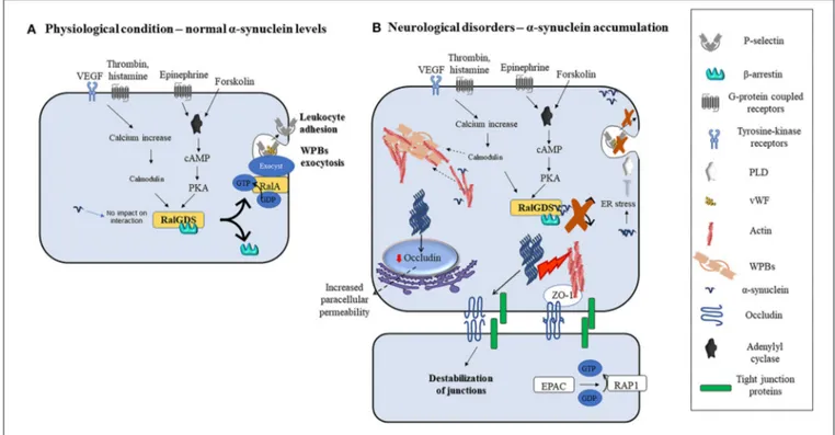
); Figure 2]. Indeed, agonists
such as thrombin, vascular endothelial growth factors (VEGF),
histamine, and superoxide can induce an increase in intracellular
calcium levels. Subsequently, calcium binds and activates
calmodulin which then triggers the translocation of Ral specific
guanine exchange factor (RalGDS), from cytosol to plasma
membrane and activates membrane-bound RalA (a small
GTPase and substrate for RalGDS) by exchanging GDP with
GTP [(
112
); Figure 2A]. Afterward, the RalA-GTP interacts with
and assembles exocyst, a multi-protein complex important in
targeting vesicles to membranes (
113
), to promote the exocytosis
of WPBs (Figure 2A). In parallel, forskolin, or epinephrine can
increase cyclic adenosine monophosphate (cAMP) levels thus
inducing protein kinase A activation, which also causes RalGDS
membrane translocation [(
38
,
112
); Figure 2A]. Upon activation
of these pathways, α-synuclein binds to both RalGDS and
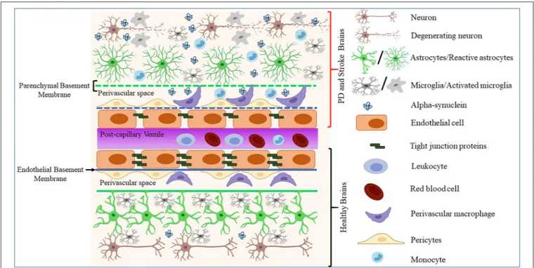
FIGURE 1 | Schematic representation of how pathological α-synuclein-accumulation occurring in PD or stroke can disrupt the physiological homeostasis of the BBB by affecting its diverse components. The BBB is composed of microvascular endothelial cells, pericytes, astrocytes, and BM components deposited by ECs (endothelial BM) and astrocytes (parenchymal BM). More recently, perivascular macrophages and vessel-associated microglia were found to play a role in the maintenance and repair of BBB whose disruption is detected in various neurological disorders including stroke and PD. This could result in BM damage (dotted lines), downregulation of TJ proteins, abnormal accumulation, and spreading of toxic forms of proteins such as α-synuclein, activation of glial cells and PVMs, and infiltration of peripheral leukocytes and monocytes, leading to neuronal degeneration.
β-arrestin, thus enhancing their interaction and inhibiting their
dissociation and translocation to the plasma membrane, thereby
preventing WPBs exocytosis [(
38
); Figure 2B]. Moreover, it
may be also feasible to foresee that exocytosis might also be
prevented by inactivation of phospholipase D (PLD) due to
α-synuclein overexpression-related ER stress (
114
) or enhanced
polymerization of actin filaments by α-synuclein [(
18
,
115
,
116
);
Figure 2B
], that by immobilizing WPBs would avoid
their release.
Alpha-synuclein is present in the cerebrospinal fluid (CSF)
and the blood and transported across the BBB (
59
,
117
–
120
). In
particular, α-synuclein can be transferred by multiple transport
mechanisms including carriers such as lipoprotein
receptor-related protein-1 (LRP-1) (
59
) or extracellular vesicles (EVs)
(
121
). Exosome-derived α-synuclein induces oligomerization of
endogenous soluble protein in recipient cells and contributes
to intercellular propagation of pathology. The CSF of patients
with PD show α-synuclein containing exosomes derived
from various cells including microglia and exert different
functions (
122
,
123
). In line with this, erythrocyte-derived
exosomes containing α-synuclein from patients with PD induce
microglial activation in vivo and in vitro, thus suggesting
that erythrocyte-derived extravasated α-synuclein may play a
role in disease pathogenesis (
121
). These evidences support
that further studies are needed to understand how
exosome-associated physiological or pathological forms of the protein
may impact on brain immune cells and ECs function and thus
on BBB integrity.
Alpha-synuclein-induced inflammation might contribute first
to the stimulation of rapid ECs response, which by driving the
contraction of ECs, leads to the formation of gaps between
them. This reshaping of ECs alters the continuous ECs layer
mediating the improvement of its paracellular permeability and
induces the activation of ECs. Consequently, the induction of
proinflammatory molecules production and release from ECs
increases the local blood flow. These events, in conjunction
with the ECs layer alteration, prompt BBB dysfunction, leading
to the extravasation of protein-rich exudates as well as to
the recruitment and activation of circulating leukocytes, that
further promote neuroinflammation (
124
). In particular, it may
be feasible that the chronic upregulation of TNF-α and IL-1β
associated with α-synuclein deposition, observed in patients with
PD and animal models (
82
), might induce sustained activation of
ECs. The consequent activation of NF-κB and activator protein
1 (AP-1) and the production of vascular cell adhesion molecule
1 (VCAM 1) and intercellular adhesion molecule 1 (ICAM 1)
(
124
), would thus set the stage for enhanced neuroinflammation,
BBB injury, and neurodegeneration (Figure 2). On this line, the
mechanisms linking α-synuclein deposition to endothelial injury
warrants further investigation.
Evidence supports that α-synuclein preformed fibrils (pffs)
downregulate the expression of occludin and of zonula occludens
FIGURE 2 | Modulation of ECs function by α-synuclein in physiological or pathological conditions. (A) Various agonists bind to endothelial cells G-protein coupled receptors (thrombin, superoxide, histamine, and epinephrine) or tyrosine kinase receptors (VEGF) to activate different intracellular pathways that lead to the release of contents of WPBs and translocation of P-selectin leading to leukocyte adhesion. Thrombin, VEGF, histamine, and superoxide lead to intracellular calcium increase and calmodulin activation, while forskolin and epinephrine increase intracellular cAMP and activate PKA. Calmodulin and PKA then trigger the translocation of RalGDS from cytosol to plasma membrane that activates membrane-bound RalA by exchanging GDP with GTP. This results in WPBs exocytosis. (B) Alpha-synuclein inhibits the release of contents of WPBs in vitro by various putative mechanisms which include binding to RalGDS and β-arrestin to prevent their dissociation, and hence exchange of RalA, blocking PLD activity, and immobilization of WPBs through enhanced actin polymerization. Fibrillary α-synuclein destabilizes TJ thereby affecting ECs paracellular permeability. This may occur through the downregulation of occludin and ZO-1.
1 (ZO-1) (Figure 2B). As a consequence, the transport across
intercellular junctions between ECs could be improved (
39
).
However, α-synuclein pffs do not trigger endothelial dysfunction
or release of proinflammatory cytokines from ECs in culture
(
39
), supporting that these cells are less vulnerable to α-synuclein
toxicity. On the other hand, activation of Ras homologous
guanosine triphosphate phosphatase (RhoGTPases) leads to
distinct effects on the ECs’ barrier function depending on
the type of GTPase activated (
125
,
126
). For instance, excess
activation of RhoA by thrombin or VEGF induces the formation
of stress fibers which destabilizes intercellular junctions and
downregulates the expression of eNOS, thereby promoting
paracellular permeability and endothelial dysfunction [(
126
);
Figure 2
]. A recent study on a human brain-chip modeling
the substantia nigra (SN) showed that α-synuclein fibrils can
induce increased paracellular permeability (
127
). Interestingly,
transcriptomic analysis of the ECs in the brain-chip revealed the
upregulation of genes involved in inflammation, oxidative stress,
autophagy, efflux system, and extracellular matrix deposition
and the downregulation of genes that encode for TJ proteins
(
127
). Conversely, the overexpression of A30P mutated
α-synuclein has been found to upregulate collagen IV α2 chain
(COL4A2), a major constituent of BMs in vivo and in vitro (
22
),
further supporting that α-synuclein changes may impact the BBB
integrity also by affecting this component. However, whether
and how α-synuclein influences these pathways to regulate ECs
functions at the BBB or the secretion and assembly of other BM
elements, their degrading enzymes, or interaction with receptor
proteins and other neighboring cells still needs to be addressed.
Likewise, since endothelial dysfunction might, in turn, alter the
transport of α-synuclein between the brain and vasculature, thus
promoting its accumulation and the progression of α-synuclein
pathology, studies addressing whether and how BBB dysfunction
may impact PD progression could bring new insights into our
basic understanding of the pathophysiology of this disorder.
Indeed, brains of patients with PD show evidence of
endothelial degeneration, downregulation of TJ proteins,
and even angiogenesis (
39
,
105
,
128
,
129
). These changes
were observed mostly in the SN, locus coeruleus (LC), and
caudate putamen (CP), brain regions where
α-synuclein-induced degeneration is prominent, and to a lesser extent in
the cerebral cortex (
105
,
128
,
129
). Moreover, pathological
alterations in the capillary BM including collagen deposition
and thickening are evident in PD brains (
12
,
128
,
130
). It is
thus plausible to speculate that such changes may reduce the
efficiency of the exchange of molecules between the brain and
vasculature, rendering neurons vulnerable to oxidative stress and
accumulating cellular waste products.
FIGURE 3 | Alpha-synuclein perivascular immunoreactivity in postmortem sections from sporadic patients with PD. For these experiments, sections from three patients with PD (PD16, disease duration 18 years; PD45, disease duration 19 years; and PD121, disease duration 4 years) and three healthy controls (CO28, CO34, and CO48), kindly supplied by the Parkinson’s UK Brain Bank, were analyzed. Briefly, sections were treated for antigen retrieval with 10 mM sodium citrate (20′
at 95◦C) and 10% formic acid (15′
at RT). After 1 h incubation at room temperature (RT) with blocking solution (2% w/vol bovine serum albumin, 3% vol/vol normal goat serum, 0.3% Triton X-100 diluted in PBS 0.1 M pH 7.4) the 5-µm slices were subjected to either single α-synuclein (Sin211 MA5-12272 Thermo Fisher Scientific, Waltham, USA; dilution 1:500) or double laminin α2 (4H8-2, abcam ab11576; dilution 1:100)/α-synuclein (Sin211, MA5-12272 Thermo Fisher; dilution 1:500) immunolabeling according to previously described protocols (137,138). Single α-synuclein immunopositive signal was revealed by Blue Alkaline Phosphatase (Vector Laboratories, Burlingame, CA) acquired by using a 40X objective, while for double immunolabeling laminin α2 was revealed by brown 3,3-diaminobenzidine (DAB) and α-synuclein by violet (Nickel supplemented) DAB (Vector laboratories) and acquired by a 100X objective. All the images were acquired by using an inverted light microscope (Olympus BX41; Olympus, Milan, Italy). (A–C) Representative images of perivascular α-synuclein immunolabeling (blue, arrows) in the TEC of three sporadic PD cases (PD 16, PD 121, and PD 45). (D–F) Images from the TEC of two of the healthy controls analyzed (CO28 and CO48). (D,E) The absence of α-synuclein immunolabeling in control brains, while (F) shows a representative image from a negative control for the immunostaining performed without the addition of the primary antibody on an adjacent section of CO48. (G,H) Representative images showing the presence of α-synuclein violet immunolabeling at the outer (G, arrows) and inner (H, arrows) side of laminin α2-positive perivascular BM in the brain of a sporadic patient with PD (PD45). Images are representative of the TEC (G) and CP (H). (I) Representative image showing the absence of α-synuclein accumulation around laminin α2-immunolabeling in the proximity of a vessel of the CP of a healthy control (CO34). Scale bar: (A–F) 40 µm; (G–I) 25 µm.
Angiogenesis is a well-recognized adaptive response to
cerebral hypoxia or ischemia and is regulated by BM proteins
and their integrin receptors (
131
). Interestingly, the integrin
receptor αvβ is upregulated in angiogenic vessels (
131
,
132
) and
in cerebral vessels of patients with PD and incidental LB disease
(iLBD) (
129
), suggesting that the immature nascent vessels
generated in PD brains, could contribute to neuroinflammation
by facilitating the infiltration of peripheral immune cells
and inflammatory or toxic factors (
129
). Consistently,
co-localization of areas of leakage of an intravascular tracer with
β3 integrin-expressing new vessels, indicating the presence of
both angiogenesis and compromised BBB, has been observed
in toxin-induced animal model of PD (
132
). Based on
Braak’s PD staging (
56
), patients with iLBD may represent
an early disease stage where LB is restricted to LC and
SN (
129
). Therefore, the presence of angiogenesis in patients
with iLBD and PD supports that α-synuclein related vascular
dysfunction might precede or/and contribute to the progression
of neuroinflammation and neurodegeneration. This is further
substantiated by findings showing that α-synuclein-related
angiogenesis and downregulation of TJ proteins are not
necessarily related to inflammation (
39
,
129
). Indeed, recent
findings indicate that dysfunction in BBB accompanied by
pathological activation of pericytes precedes the onset of
neuronal degeneration in a mouse model of PD (
133
), thus
supporting that vascular dysfunction may be an early pathogenic
events leading to neuronal damage. Furthermore, VEGF plays a
protective role in PD through a direct effect on dopaminergic
neurons (
134
) or via the canonical VEGF receptor (VEGFR2)
pathway (
135
). VEGF released by activated astrocytes and
microglia acts in a paracrine fashion to modulate ECs structure
and function both in PD and DLB patients and in animal
models of PD (
136
). This notwithstanding, whether α-synuclein
is involved in the upregulation of VEGF in ECs remains to
be investigated.
Interestingly, in postmortem sections of the trans-entorhinal
(TEC) cortex (Figures 3A–C) and CP (not shown) from sporadic
patients with PD, we observed perivascular accumulation of
α-synuclein immunoreactivity (in blue) in correspondence of
some vessels. Conversely, the brains of healthy or negative
controls did not exhibit this feature (Figures 3D–F). By double
immunolabeling of laminin α2 (brown) and α-synuclein (violet),
we found that in the PD brains, α-synuclein-positive perivascular
staining could be identified either in the outer (Figure 3G) and
inner (Figure 3H) sides of the perivascular basement membrane,
while in control brains, it did not show α-synuclein positivity in
the proximity of laminin α2 staining (Figure 3I).
Although further studies are needed to corroborate
whether and how α-synuclein deposition affects these
cells, these findings, when coupled to the aforementioned
noxious effects exerted by pathological α-synuclein on ECs,
support that perivascular accumulation of the protein, by
inducing ECs activation may compromise brain vessels
integrity exacerbating astrocyte and microglia activation
thus promoting neurodegeneration and BBB disruption
(Figure 4).
ALPHA-SYNUCLEIN IN BAMs AND OTHER
PERIVASCULAR CELLS
Border-associated macrophages are a subset of CNS myeloid
cells (macrophages) that like microglia originate prenatally in
the yolk sac (
139
), invade the brain during the early prenatal
period, and localize in the choroid plexus, perivascular, and
leptomeningeal spaces. BAMs form stable populations with
the sole exception of the choroid plexus macrophages that
exchange with peripheral monocytes (
139
,
140
). Indeed, BAMs
can be anatomically distinguished into perivascular macrophages
(PVM), meningeal macrophages (MAM), and choroid plexus
associated macrophages (CPM) (
139
,
140
). In healthy brains,
PVMs are involved in the regulation of BBB permeability and
phagocytosis of pathogens but can also promote the entrance of
peripheral immune cells into the brain (
102
). Under pathological
conditions such as cerebral amyloid angiopathy (CAA), AD
(
141
) and PD (
141
), PVMs can participate in the clearance
of toxic amyloid-β and α-synuclein. Consistently, the depletion
of PVMs using clodronate-containing liposomes in a mouse
model of PD resulted in the increased expression of VCAM 1,
FIGURE 4 | Possible downstream effects of pathological α-synuclein-induced activation of ECs on microglia, astrocytes, and neurons. Alpha-synuclein accumulation-induced ECs activation could promote BBB disruption and peripheral immune cell infiltration, improve microglia and astrocytes activation and exacerbate oxidative stress, thus promoting neuronal degeneration.
the infiltration of T cells, and the propagation of α-synuclein
pathology (
142
). Moreover, in rats that underwent transient
ischemia followed by reperfusion injury, BAMs were involved
in promoting peripheral immune cell infiltration and vascular
permeability without impacting the extent of ischemic damage,
thus suggesting that additional studies are needed to fully
understand the modulatory role of these cells in cerebrovascular
dysfunctions and neuroinflammation (
13
).
Border associated macrophages are involved in immune
surveillance and support the entrance of peripheral immune
cells into the CNS under pathological conditions (
143
,
144
).
In rodents, monkeys, and humans, PVMs express the mannose
receptor CD206 (
145
) and the scavenger receptor CD163,
which under physiological conditions is expressed on tissue
macrophages, with the exception of microglia and some
monocytes (
146
–
148
). Recent research reports, dissecting the
molecular signature of brain macrophages in mice at the
single-cell level, reported a clear segregation of BAMs from microglia,
identified a BAMs core gene signature, and even showed
heterogeneity within BAMs (
149
–
152
). In stroke animal models
and patients, a unique transcriptional signature of BAMs, their
local proliferation and migration in the brain parenchyma, have
been detected (
14
).
It is now believed that BAMs play a role in immune
function, BBB integrity, and lymphatic clearance (
139
,
140
).
Currently, the identification of BAMs mainly relies on the use of
anatomical studies aimed at disclosing their localization in the
brain thanks to the use of few reliable molecular markers (
139
,
140
). However, this approach is not applicable in the presence
of inflammatory conditions or tissue injury when peripheral
monocytes/macrophages enter the brain and reside in the same
location and express similar molecular markers (
139
). Despite
these limitations, remarkable progresses have been made to
fully characterize and understand their role in the normal and
diseased brain.
Alpha-synuclein is expressed by microglia and peripheral
monocytes/macrophages in a lower amount compared to
neurons (
32
), but the expression in BAMs has not been
described yet. Since BAMs share similar ontogeny and molecular
and immunologic characteristics with microglia (
139
,
140
),
they might exhibit analogous changes and activation states
to α-synuclein stimulation (
153
,
154
). Indeed, it has been
described that BAMs and microglia display multiple similarities
such as the expression of myeloid-specific markers. Among
them, ionized calcium-binding adaptor molecule 1(Iba1),
F4/80 (mouse) or EMR1 (human), chemokine receptors,
scavenger receptors, receptor tyrosine kinases, Integrins, pattern
recognition receptors (PRRs), and cytokines receptors (
155
).
α-synuclein is known to induce inflammatory response and
migration of microglia (
32
,
89
,
156
). For instance, previous
studies showed that α-synuclein induces NOX2 activation in
microglia by binding to toll-like receptor 2 (TLR-2) and CD11b
leading to microglia-mediated neuronal toxicity (
157
). Similarly,
α-synuclein binds to TLR-4 and activates NF-kB signaling
which then induces a selective autophagy pathway named
synucleinphagy and release of exosomes containing the protein,
thus contributing to the intercellular spread of α-synuclein
pathology (
154
).
Interestingly, monomeric α-synuclein can also impact
microglia polarization by conferring an anti-inflammatory
profile to these cells through the interaction with extracellular
signal-regulated kinase (ERK) and the recruitment of the
ERK/NF-κB, and peroxisome proliferator-activated receptor
γ
(PPARγ) pathways (
158
). It may thus be feasible that these
pathways may also be activated upon exposure of BAMs
to α-synuclein.
It is worth mentioning that pericytes can also play a
role in the formation, maintenance, and regulation of BBB
(
159
). Additionally, pericytes (
60
) and astrocytes (
160
) can
mediate the transfer of α-synuclein between cells of the NVU,
suggesting a possible role of non-neuronal cells in α-synuclein
pathology spreading in PD (
161
). In vitro studies also showed
that α-synuclein activates pericytes which in turn release
proinflammatory mediators that can mediate BBB dysfunction
(
162
). Early pericyte activation associated with BBB leakage has
been recently described in a human α-synuclein
overexpression-based mouse model of PD (
133
), thus further supporting that
vascular pathology can constitute a relevant pathophysiological
aspect of PD.
Peripheral immune cells such as lymphocytes have also been
involved in the pathogenesis of PD. Indeed, studies in the brains
of patients with PD and animal models showed that T cells
with upregulated expression of the ICAM 1 receptor lymphocyte
function-associated antigen-1 (LFA1) can promote leukocyte
infiltration (
163
). Alpha-synuclein-specific T-cell reactivity has
been found to be higher in early PD while decreasing in patients
with late-stage disease (
164
). When considering the increase of
α-synuclein in animal models of stroke (
73
), where Treg cells
interact with ICAM1 on inflamed microvessels and platelets
promoting vascular dysfunction (
165
), this evidence suggests
that the accumulation of α-synuclein occurring following brain
ischemia could very well-boost these pathogenic processes.
Several studies showed that α-synuclein aggregates can be
detected in reactive astrocytes in the brains of patients with PD
and animal models (
33
) suggesting a role of these cells in the
clearance and /or spreading of α-synuclein toxicity in the brain
parenchyma and NVU. Even though the role of endogenous
α-synuclein in astrocytes remains to be fully explored, in disease
states, α-synuclein activates astrocytes by interacting with PRRs
such as TLR-4 (
33
). Activated astrocytes can in turn uptake and
degrade the protein via the endosomal-lysosomal pathway and
contribute to non-cell autonomous degeneration (
166
,
167
).
Recent evidence suggests that α-synuclein can be removed
from the brain via extracellular space drainage pathway which
includes glymphatic transport and meningeal lymphatic system
(
61
), whose reduction lead to the accumulation of toxic
forms of amyloid-β in the brain parenchyma of AD rodent
models (
168
–
170
). Similarly, a recent study in a transgenic PD
mouse model overexpressing human A53T mutated α-synuclein
showed that blockage of the deep cervical lymph node reduces
glymphatic transport of an intraventricular tracer and promotes
the accumulation of α-synuclein and its aggregation in SN, thus
leading to the progression of α-synuclein pathology (
61
).
Taken together, impairment in these systems results in the
accumulation of toxic proteins in the brain and contributes to the
progression of neurodegenerative diseases. The close association
of BAMs to perivascular and lymphatic drainage systems in the
brain when coupled to the detection of α-synuclein aggregates
in the perivascular space of a PD mouse model (
61
) supports
that understanding of whether and how these cells contribute
to the clearance of α-synuclein along these pathways deserves
ad-hoc investigation.
In addition to this, it is plausible that the increase in
α-synuclein levels observed following ischemia and spinal cord
injury (
35
,
73
,
74
,
171
) could result in a chemotactic gradient
for microglia migration and activation (
89
) contributing to
brain damage. Consistently, inhibition of α-synuclein induction
following ischemia or spinal cord injury reduces secondary
neuronal injury, inflammatory response, and improves
neurological outcomes (
171
,
172
). Although, it is known
that juxta vascular microglia play a divergent role in repairing
vascular injuries following an insult or systemic inflammation
(
173
–
175
), whether α-synuclein modulates these cells or BAMs
remains to be clarified.
ALPHA-SYNUCLEIN ROLE IN
MODULATING BBB CELLS INTERACTION
In the normal and diseased brain, ECs communicate with
neurons, microglia, pericytes, and astrocytes to regulate
vascular function (
176
). More importantly, the interaction
of microglia with ECs can exert divergent roles in regulating
BBB integrity (
175
). In co-cultures of ECs and neurons,
α-synuclein fibrils resulted in endothelial dysfunction, but this
effect was not observed in ECs monocultures (
39
), supporting
that neuronal-ECs crosstalk at the NVU may be perturbed by
pathological α-synuclein.
CD200, a transmembrane protein found to be expressed in
neurons, astrocytes, oligodendrocytes, and ECs, transduces signal
via its receptor (CD200R), expressed on myeloid cells including
microglial and BAMs (
177
). CD200-CD200R and C-X3-C
motif chemokine ligand 1 (CX3CL1)-C-X3-C motif chemokine
receptor 1 (CX3CR1) signaling between neurons and microglia
helps to maintain microglia in the resting state (
178
). Inactivation
of the transmembrane glycoprotein CD200R in microglia of a
toxin-induced PD mouse model results in increased activation
of these cells, release of proinflammatory cytokines, loss of
dopaminergic neurons in the SN, and behavioral deficits
(
179
). Similarly, monocyte-derived macrophages (MDMs) from
patients with PD show dysregulation in CD200R signaling (
180
).
On the other hand, M2 macrophages express pro-angiogenic
factors such as VEGF and fibroblast growth factor 2 (FGF2),
that by activating their receptors (VEGFR2 and FGFR) promote
angiogenesis and neuronal survival (
181
). In vitro studies
have shown that microglia maintains ECs in a resting state
by secreting transforming growth factor-beta (TGF-β), an
anti-inflammatory cytokine, while the proinflammatory
TNF-α
induces ECs proliferation (
182
). However, whether this kind
of communication occurs also between ECs and BAMs or is
influenced by α-synuclein still remains to be elucidated.
Secreted toxic species of α-synuclein are known to bind
to various cell surface receptors in adjacent cells and activate
several intracellular pathways leading to synaptic dysfunction,
neurodegeneration, and inflammation (
183
). On this line,
α-synuclein binds to TLR-2, TLR-4, and CD11β integrin to activate
NF-kB signaling and assembly of NLRP3 inflammasome in
microglia (
37
,
154
,
157
). It is thus plausible that various receptors
for α-synuclein might exist in different cells, including ECs, and
that the protein may regulate their intracellular activities or their
crosstalk within neighboring cells such as BAMs.
CURRENT GAPS AND FUTURE
PERSPECTIVES
While α-synuclein associated vascular dysfunction is evident in
PD (
11
,
128
), most of the studies aimed at understanding the
physiological role of the protein in ECs have been performed by
overexpressing the protein through transgene expression (
38
,
39
).
Therefore, the role of extracellularly released α-synuclein on
these cells still needs to be extensively explored. Furthermore, it
is not yet established whether ECs respond to various pathogenic
stimuli by regulating α-synuclein level or its transport across the
BBB. Future studies exploiting improved models might overcome
these limitations.
It is now clear that α-synuclein-associated inflammation
contributes to the pathophysiology of PD (
10
) but research
on whether or how α-synuclein modulates the function of
macrophages in the brain has been mostly focused on microglia.
As a result, our knowledge on the role of α-synuclein in
other brain resident macrophages such as BAMs or other
perivascular cells is poor. Similarly, despite the presence of
studies indicating interplay between ECs and microglia (
175
),
the interplay between perivascular cells and ECs has been
scarcely studied. For instance, since recent evidence showed
that BAMs and microglia can acquire distinct genetic and
molecular phenotypes early in development (
152
), studying
whether α-synuclein plays a role in modulating the signaling
pathways mediating the crosstalk between BAMs and ECs
could bring novel and significant insights for understanding
the biological basis of neurological disorders such as PD or
stroke. Similarly, though α-synuclein transfer between cells
and across BBB interfaces has been established (
59
,
154
),
whether and how BAMs are involved in this event also deserves
further investigations.
A deeper understanding of the role of physiological and
pathological forms of α-synuclein in the modulation of
BAMs and ECs or their interplay may also greatly aid
the identification of novel therapeutic targets for stroke or
neurodegenerative synucleinopathies.
AUTHOR CONTRIBUTIONS
TB and GF wrote and revised the article and prepared the figures.
TB performed immunostaining. FL revised the article and was
involved in image acquisition. AB, MP, and SM did a critical
revision of the article. All authors contributed to the article and
approved the submitted version.
FUNDING
This work was supported by the European Union’s Horizon 2020
research and innovation program under the Marie
Skłodowska-Curie grant agreement No. 813294.
ACKNOWLEDGMENTS
Tissue samples and associated clinical and neuropathological
data were supplied by the Parkinson’s UK Brain Bank, funded
by Parkinson’s UK, a charity registered in England and Wales
(258197) and in Scotland (SC037554). Icons of cells used in
Figure 1
were created using Biorender.
REFERENCES
1. Feigin VL, Nichols E, Alam T, Bannick MS, Beghi E, Blake N, et al. Global, regional, and national burden of neurological disorders, 1990–2016: a systematic analysis for the Global Burden of Disease Study 2016. Lancet Neurol. (2019) 18:459–80. doi: 10.1016/S1474-4422(18)30499-X
2. Mattson MP, Duan W, Pedersen WA, Culmsee C. Neurodegenerative
disorders and ischemic brain diseases. Apoptosis. (2001)
6:69–81. doi: 10.1023/A:1009676112184
3. Poewe W, Seppi K, Tanner CM, Halliday GM, Brundin P,
Volkmann J, et al. Parkinson disease. Nat Rev Dis Prim. (2017) 3:1–21. doi: 10.1038/nrdp.2017.13
4. Goedert M, Jakes R, Spillantini MG. The synucleinopathies: twenty years on. J Parkinsons Dis. (2017) 7:S53–71. doi: 10.3233/JPD-179005
5. Spillantini MG, Goedert M. The alpha-synucleinopathies: Parkinson’s disease, dementia with Lewy bodies, and multiple. Ann N Y Acad Sci. (2002) 920:16–27. doi: 10.1111/j.1749-6632.2000.tb06900.x
6. Bellucci A, Antonini A, Pizzi M, Spano PF. The end is the beginning: Parkinson’s disease in the light of brain imaging. Front Aging Neurosci. (2017) 9:1–5. doi: 10.3389/fnagi.2017.00330
7. Bellucci A, Mercuri NB, Venneri A, Faustini G, Longhena F, Pizzi M, et al. Review: Parkinson’s disease: from synaptic loss to connectome dysfunction. Neuropathol Appl Neurobiol. (2016) 42:77–94. doi: 10.1111/nan.12297 8. Calo L, Wegrzynowicz M, Santivañez-Perez J, Grazia Spillantini M.
Synaptic failure and α-synuclein. Movement Disord. (2016) 31:169– 77. doi: 10.1002/mds.26479
9. Janda E, Boi L, Carta AR. Microglial phagocytosis and its regulation: a therapeutic target in parkinson’s disease? Front Mol Neurosci. (2018) 11:1– 8. doi: 10.3389/fnmol.2018.00144
10. Ho MS. Microglia in Parkinson’s disease. Adv Exp Med Biol. (2019) 1175:335–53. doi: 10.1007/978-981-13-9913-8_13
11. Yang P, Min X-L, Mohammadi M, Turner C, Faull R, Waldvogel H, et al. Endothelial degeneration of Parkinson’s disease is related to alpha-synuclein aggregation. J Alzheimers Dis Parkinsonism. (2017) 7:370. doi: 10.4172/2161-0460.1000370
12. Yang P, Pavlovic D, Waldvogel H, Dragunow M, Synek B, Turner C, et al. String vessel formation is increased in the brain of Parkinson disease. J Parkinsons Dis. (2015) 5:821–36. doi: 10.3233/JPD-140454
13. Pedragosa J, Salas-Perdomo A, Gallizioli M, Cugota R, Miró-Mur F, Briansó F, et al. CNS-border associated macrophages respond to acute ischemic stroke attracting granulocytes and promoting vascular leakage. Acta Neuropathol Commun. (2018) 6:76. doi: 10.1186/s40478-018-0581-6 14. Rajan WD, Wojtas B, Gielniewski B, Miró-Mur F, Pedragosa J, Zawadzka
M, et al. Defining molecular identity and fates of CNS-border associated macrophages after ischemic stroke in rodents and humans. Neurobiol Dis. (2020) 137:104722. doi: 10.1016/j.nbd.2019.104722
15. Zille M, Ikhsan M, Jiang Y, Lampe J, Wenzel J, Schwaninger M. The impact of endothelial cell death in the brain and its role after stroke: a systematic review. Cell Stress. (2019) 3:330–47. doi: 10.15698/cst2019.11.203
16. Andjelkovic AV, Xiang J, Stamatovic SM, Hua Y, Xi G, Wang MM, et al. Endothelial targets in stroke: translating animal models to human. Arteriosc Thromb Vasc Biol. (2019) 39:2240–7. doi: 10.1161/ATVBAHA.119.31 2816
17. Weinreb PH, Zhen W, Poon AW, Conway KA, Lansbury PT. NACP, a protein implicated in Alzheimer’s disease and learning, is natively unfolded. Biochemistry. (1996) 35:13709–15. doi: 10.1021/bi961799n
18. Bellucci A, Zaltieri M, Navarria L, Grigoletto J, Missale C, Spano P. From alpha-synuclein to synaptic dysfunctions: new insights into the pathophysiology of Parkinson’s disease. Brain Res. (2012) 1476:183– 202. doi: 10.1016/j.brainres.2012.04.014
19. Faustini G, Marchesan E, Zonta L, Bono F, Bottani E, Longhena
F, et al. Alpha-synuclein preserves mitochondrial fusion and
function in neuronal cells. Oxidative Med Cell Longevity. (2019) 2019:4246350. doi: 10.1155/2019/4246350
20. Longhena F, Faustini G, Spillantini MG, Bellucci A. Living in promiscuity: the multiple partners of alpha-synuclein at the synapse in physiology and pathology. Int J Mol Sci. (2019) 20:1–24. doi: 10.3390/ijms20010141
21. Cheng F, Vivacqua G, Yu S. The role of alpha-synuclein in neurotransmission and synaptic plasticity. J Chem Neuroanat. (2011) 42:242–8. doi: 10.1016/j.jchemneu.2010.12.001
22. Paiva I, Jain G, Lázaro DF, Jerˇci´c KG, Hentrich T, Kerimoglu C, et al. Alpha-synuclein deregulates the expression of COL4A2 and impairs ER-Golgi function. Neurobiol Dis. (2018) 119:121–35. doi: 10.1016/j.nbd.2018.08.001 23. Villar-Piqué A, Lopes da Fonseca T, Outeiro TF. Structure, function and
toxicity of alpha-synuclein: the Bermuda triangle in synucleinopathies. J Neurochem. (2016) 139:240–55. doi: 10.1111/jnc.13249
24. Spillantini MG, Goedert M. Neurodegeneration and the ordered
assembly of α-synuclein. Cell Tissue Res. (2018) 373:137–
48. doi: 10.1007/s00441-017-2706-9
25. Burai R, Ait-Bouziad N, Chiki A, Lashuel HA. Elucidating the role of site-specific nitration of α-synuclein in the pathogenesis of Parkinson’s disease via protein semisynthesis and mutagenesis. J Am Chem Soc. (2015) 137:5041– 52. doi: 10.1021/ja5131726
26. Souza JM, Giasson BI, Lee VM-Y, Ischiropoulos H.
Chaperone-like activity of synucleins. FEBS Lett. (2000). 474:116–
9. doi: 10.1016/S0014-5793(00)01563-5
27. Fujiwara H, Hasegawa M, Dohmae N, Kawashima A, Masliah E, Goldberg MS, et al. α-Synuclein is phosphorylated in synucleinopathy lesions. Nat Cell Biol. (2002) 4:160–4. doi: 10.1038/ncb748
28. Alam P, Bousset L, Melki R, Otzen DA-O. α-Synuclein oligomers and fibrils: a spectrum of species, a spectrum of toxicities. J Neurochem. (2019) 150:522–34. doi: 10.1111/jnc.14808
29. Danzer KM, Schnack C, Sutcliffe A, Hengerer B, Gillardon F. Functional protein kinase arrays reveal inhibition of p-21-activated
kinase 4 by alpha-synuclein oligomers. J Neurochem. (2007)
103:2401–7. doi: 10.1111/j.1471-4159.2007.04933.x
30. Choi B-K, Choi M-G, Kim J-Y, Yang Y, Lai Y, Kweon D-H, et al. Large α-synuclein oligomers inhibit neuronal SNARE-mediated vesicle docking. Proc Natl Acad Sci. (2013) 110:4087. doi: 10.1073/pnas.12184 24110
31. Flavin WP, Bousset L, Green ZC, Chu Y, Skarpathiotis S, Chaney MJ, et al. Endocytic vesicle rupture is a conserved mechanism of cellular invasion by amyloid proteins. Acta Neuropathol. (2017) 134:629– 53. doi: 10.1007/s00401-017-1722-x
32. Pei Y, Maitta RW. Alpha synuclein in hematopoiesis and immunity. Heliyon. (2019) 5:e02590. doi: 10.1016/j.heliyon.2019.e02590
33. Sorrentino ZA, Giasson BI, Chakrabarty PA-O. α-Synuclein
and astrocytes: tracing the pathways from homeostasis to
neurodegeneration in Lewy body disease. Acta Neuropathol. (2019) 138:1–21. doi: 10.1007/s00401-019-01977-2
34. Barbour R, Kling K, Anderson JP, Banducci K, Cole T, Diep L, et al. Red blood cells are the major source of alpha-synuclein in blood. Neurodegener Dis. (2008) 5:55–9. doi: 10.1159/000112832
35. Wu Z, Li X, Zeng M, Qiu H, Feng H, Xu X, et al. Alpha-synuclein alterations in red blood cells of peripheral blood after acute ischemic stroke. Int J Clin Exp Pathol. (2019) 12:1757–63.
36. Hashimoto M, Yoshimoto M, Sisk A, Hsu LJ, Sundsmo M, Kittel A, et al. NACP, a synaptic protein involved in alzheimer’s disease, is differentially regulated during megakaryocyte differentiation. Biochem Biophys Res Commun. (1997) 237:611–6. doi: 10.1006/bbrc.1997.6978
37. Ferreira SA, Romero-Ramos M. Microglia response during Parkinson’s disease: alpha-synuclein intervention. Front Cell Neurosci. (2018) 12:1– 17. doi: 10.3389/fncel.2018.00247
38. Kim KS, Park JY, Jou I, Park SM. Regulation of Weibel-Palade body exocytosis by α-synuclein in endothelial cells. J Biol Chem. (2010) 285:21416– 25. doi: 10.1074/jbc.M110.103499
39. Kuan W-L, Bennett N, He X, Skepper JN, Martynyuk N, Wijeyekoon R, et al. α-Synuclein pre-formed fibrils impair tight junction protein expression without affecting cerebral endothelial cell function. Exp Neurol. (2016). 285:72–81. doi: 10.1016/j.expneurol.2016.09.003
40. Spillantini MG, Crowther RA, Jakes R, Hasegawa M, Goedert M. α-Synuclein in filamentous inclusions of Lewy bodies from Parkinson’s disease and dementia with Lewy bodies. Proc Natl Acad Sci USA. (1998). 95:6469– 73. doi: 10.1073/pnas.95.11.6469
41. Herva ME, Spillantini MG. Parkinson’s disease as a member of Prion-like disorders. Virus Res. (2015) 207:38–46. doi: 10.1016/j.virusres.2014.10.016 42. Guo M, Wang J, Zhao Y, Feng Y, Han S, Dong Q, et al. OUP accepted
manuscript. Brain. (2020) 143:1476–97. doi: 10.1093/brain/awaa090 43. Chadchankar H, Ihalainen J, Tanila H, Yavich L. Decreased reuptake of
dopamine in the dorsal striatum in the absence of alpha-synuclein. Brain Res. (2011) 1382:37–44. doi: 10.1016/j.brainres.2011.01.064
44. Yavich L, Tanila H, Vepsäläinen S, Jäkälä P. Role of α-synuclein in presynaptic dopamine recruitment. J Neurosci. (2004) 24:11165– 70. doi: 10.1523/JNEUROSCI.2559-04.2004
45. Vamvaca K, Volles MJ, Lansbury PT. The first N-terminal amino acids of α-synuclein are essential for α-helical structure formation in vitro and membrane binding in yeast. J Mol Biol. (2009) 389:413– 24. doi: 10.1016/j.jmb.2009.03.021
46. Bartels T, Ahlstrom LS, Leftin A, Kamp F, Haass C, Brown MF, et al. The N-terminus of the intrinsically disordered protein α-synuclein triggers membrane binding and helix folding. Biophys J. (2010) 99:2116– 24. doi: 10.1016/j.bpj.2010.06.035
47. Runfola M, De Simone A, Vendruscolo M, Dobson CM, Fusco G. The N-terminal acetylation of α-synuclein changes the affinity for lipid membranes but not the structural properties of the bound state. Sci Rep. (2020) 10:204. doi: 10.1038/s41598-019-57023-4
48. Barrett PJ, Timothy Greenamyre J. Post-translational modification of α-synuclein in Parkinson’s disease. Brain Research. (2015) 1628:247– 53. doi: 10.1016/j.brainres.2015.06.002
49. Ueda K, Fukushima H, Masliah E, Xia Y, Iwai A, Yoshimoto M, et al. Molecular cloning of cDNA encoding an unrecognized component of amyloid in Alzheimer disease. Proc Natl Acad Sci USA. (1993) 90:11282– 6. doi: 10.1073/pnas.90.23.11282
50. Giasson BI, Murray IVJ, Trojanowski JQ, Lee VMY. A hydrophobic stretch of 12 amino acid residues in the middle of α-synuclein is essential for filament assembly. J Biol Chem. (2001) 276:2380–6. doi: 10.1074/jbc.M00891 9200
51. Ulmer TS, Bax A, Cole NB, Nussbaum RL. Structure and dynamics of micelle-bound human α-synuclein. J Biol Chem. (2005) 280:9595– 603. doi: 10.1074/jbc.M411805200
52. Dedmon MM, Lindorff-Larsen K, Christodoulou J, Vendruscolo M, Dobson CM. Mapping long-range interactions in α-synuclein using spin-label NMR and ensemble molecular dynamics simulations. J Am Chem Soc. (2005) 127:476–7. doi: 10.1021/ja044834j
53. Conway KA, Harper JD, Lansbury PT. Accelerated in vitro fibril formation by a mutant α-synuclein linked to early-onset Parkinson disease. Nat Med. (1998) 4:1318–20. doi: 10.1038/3311
54. Gurry T, Ullman O, Fisher CK, Perovic I, Pochapsky T, Stultz CM. The dynamic structure of α-synuclein multimers. J Am Chem Soc. (2013) 135:3865–72. doi: 10.1021/ja310518p
55. Peelaerts W, Baekelandt V. α-Synuclein strains and the
variable pathologies of synucleinopathies. J Neurochem. (2016)
139:256–74. doi: 10.1111/jnc.13595
56. Braak H, Del Tredici K, Rüb U, De Vos RAI, Jansen Steur ENH, Braak E. Staging of brain pathology related to sporadic Parkinson’s disease. Neurobiol Aging. (2003) 24:197–211. doi: 10.1016/S0197-4580(02)00065-9
57. Engelender S, Isacson O. The threshold theory for Parkinson’s disease. Trends Neurosci. (2017) 40:4–14. doi: 10.1016/j.tins.2016.10.008
58. Longhena F, Spano P, Faustini G, Bellucci, Arianna, Missale C, et al. The contribution of α-synuclein spreading to Parkinson’s disease synaptopathyr. Neural Plasticity. (2017) 2017:5012129. doi: 10.1155/2017/5012129 59. Sui Y-T, Bullock KM, Erickson MA, Zhang J, Banks WA. Alpha synuclein is
transported into and out of the brain by the blood–brain barrier. Peptides. (2014) 62:197–202. doi: 10.1016/j.peptides.2014.09.018
60. Dieriks BV, Park TIH, Fourie C, Faull RLM, Dragunow M, Curtis MA. α-synuclein transfer through tunneling nanotubes occurs in SH-SY5Y cells and primary brain pericytes from Parkinson’s disease patients. Sci Rep. (2017). 7:42984. doi: 10.1038/srep42984
61. Zou W, Pu T, Feng W, Lu M, Zheng Y, Du R, et al. Blocking meningeal lymphatic drainage aggravates Parkinson’s disease-like pathology in mice overexpressing mutated α-synuclein. Transl Neurodegener. (2019) 8:1– 17. doi: 10.1186/s40035-019-0147-y
62. Arotcarena ML, Dovero S, Prigent A, Bourdenx M, Camus
S, Porras G, et al. Bidirectional gut-to-brain and brain-to-gut propagation of synucleinopathy in non-human primates. Brain. (2020) 143:1462–75. doi: 10.1093/brain/awaa096
63. Guardia-Laguarta C, Area-Gomez E, Schon EA, Przedborski S. Novel subcellular localization for α-synuclein: possible functional consequences. Front Neuroanat. (2015) 9:17. doi: 10.3389/fnana.2015.00017
64. Thayanidhi N, Helm JR, Nycz DC, Bentley M, Liang Y, Hay JC. Alpha-synuclein delays endoplasmic reticulum (ER)-to-Golgi transport in mammalian cells by antagonizing ER/Golgi SNAREs. Mol Biol Cell. (2010) 21:1850–63. doi: 10.1091/mbc.e09-09-0801
65. Kontopoulos E, Parvin JD, Feany MB. Alpha-synuclein acts in the nucleus to inhibit histone acetylation and promote neurotoxicity. Hum Mol Genet. (2006) 15:3012–23. doi: 10.1093/hmg/ddl243
66. Schaser AJ, Osterberg VR, Dent SE, Stackhouse TL, Wakeham CM, Boutros SW, et al. Alpha-synuclein is a DNA binding protein that modulates DNA repair with implications for Lewy body disorders. Sci Rep. (2019) 9:10919. doi: 10.1038/s41598-019-47227-z
67. Chen V, Moncalvo M, Tringali D, Tagliafierro L, Shriskanda A, Ilich E, et al. The mechanistic role of alpha-synuclein in the nucleus: impaired nuclear function caused by familial Parkinson’s disease SNCA mutations. Hum Mol Genet. (2020) 29:3107–21. doi: 10.1093/hmg/ddaa183
68. Pinho R, Paiva I, Jercic KG, Fonseca-Ornelas L, Gerhardt E, Fahlbusch C, et al. Nuclear localization and phosphorylation modulate pathological effects of alpha-synuclein. Hum Mol Genet. (2019) 28:31–50. doi: 10.1093/hmg/ddy326
69. Faustini G, Bono F, Valerio A, Pizzi M, Spano P, Bellucci A. Mitochondria and α-synuclein: friends or foes in the pathogenesis of Parkinson’s disease? Genes. (2017) 8:377. doi: 10.3390/genes8120377
70. Zaltieri M, Longhena F, Pizzi M, Missale C, Spano P, Bellucci A.
Mitochondrial dysfunction and α-synuclein synaptic pathology
in Parkinson’s disease: who’s on first? Parkinsons Dis. (2015) 2015:108029. doi: 10.1155/2015/108029
71. Pozo Devoto VM, Falzone TL. Mitochondrial dynamics in Parkinson’s disease: a role for α-synuclein? Dis Mod Mech. (2017) 10:1075– 87. doi: 10.1242/dmm.026294
72. Gao F, Yang J, Wang D, Li C, Fu Y, Wang H, et al. Mitophagy in Parkinson’s disease: pathogenic and therapeutic implications. Front Neurol. (2017) 8:527. doi: 10.3389/fneur.2017.00527
73. Kim TH, Mehta SL, Kaimal B, Lyons K, Dempsey RJ, Vemuganti R. Poststroke induction of α-synuclein mediates ischemic brain damage. J Neurosci. (2016) 36:7055–65. doi: 10.1523/JNEUROSCI.1241-16.2016 74. Unal-Cevik I, Gursoy-Ozdemir Y, Yemisci M, Lule S, Gurer G, Can A, et al.
Alpha-synuclein aggregation induced by brief ischemia negatively impacts neuronal survival in vivo: a study in A30Palpha-synuclein transgenic mouse. J Cereb Blood Flow Metab. (2011) 31:913–23. doi: 10.1038/jcbfm.2010.170 75. Zhao HQ, Li FF, Wang Z, Wang XM, Feng T. A comparative study of
the amount of alpha-synuclein in ischemic stroke and Parkinson’s disease. Neurol Sci. (2016) 37:749–54. doi: 10.1007/s10072-016-2485-1
76. Jackson-Lewis V, Jakowec M, Burke RE, Przedborski S. Time course and morphology of dopaminergic neuronal death caused by the neurotoxin 1-methyl-4-phenyl-1,2,3,6-tetrahydropyridine. Neurodegeneration. (1995) 4:257–69. doi: 10.1016/1055-8330(95)90015-2
77. McGeer PL, Schwab C, Parent A, Doudet D. Presence of reactive microglia in monkey substantia nigra years after 1-methyl-4-phenyl-1,2,3,6-tetrahydropyridine administration. Ann Neurol. (2003) 54:599– 604. doi: 10.1002/ana.10728
78. Su X, Maguire-Zeiss KA, Giuliano R, Prifti L, Venkatesh K, Federoff HJ. Synuclein activates microglia in a model of Parkinson’s disease. Neurobiol Aging. (2008) 29:1690–701. doi: 10.1016/j.neurobiolaging.2007.04.006 79. Zhang W, Wang T, Pei Z, Miller DS, Wu X, Block ML, et al. Aggregated
α-synuclein activates microglia: a process leading to disease progression in Parkinson’s disease. FASEB J. (2005) 19:533–42. doi: 10.1096/fj.04-2751com 80. Mogi M, Harada M, Kondo T, Riederer P, Inagaki H, Minami M, et al.
Interleukin-1 beta, interleukin-6, epidermal growth factor and transforming growth factor-alpha are elevated in the brain from parkinsonian patients. Neurosci Lett. (1994) 180:147–50. doi: 10.1016/0304-3940(94) 90508-8