Università di Pisa
Tesi di Dottorato di Ricerca in
Fisiopatologia della Riproduzione Umana e Sessuologia
A.A. 2010-2011
Titolo:
NEUROBIOLOGICAL EFFECTS OF DHEA IN FEMALES WITH SPECIAL
REFERENCE TO SEXUAL FUNCTION: FINDINGS FROM IN-VIVO AND
HUMAN STUDIES
Candidato: Dr. Nicola Pluchino
ABSTRACT
Dehydroepiandrosterone (DHEA) and its sulfate ester, DHEAS, together represent the most abundant steroid hormones in the human body. Nonetheless, their physiological significance, their mechanisms of action and their possible roles in human disease are not well understood. Highlighting the potential health significance of DHEA and DHEAS, concentrations of these hormones in humans typically decrease steadily with age, approaching a nadir at about the time many diseases of aging become markedly more prevalent. There is growing evidence in the literature that a low DHEAS level, negatively correlates with the domains of sexual function in pre and postmenopausal women to a greater extent than testosterone levels. Biological actions of DHEA(S) involve neuroprotection, neurite growth, neurogenesis and neuronal survival, apoptosis, catecholamine synthesis and secretion, as well as oxidant, inflammatory and anti-glucocorticoid effects. In addition, DHEA affects neurosteroidogenis and endorphin synthesis/release. We demonstrated in a model of ovariectomized rats that DHEA therapy increases proceptive behaviors, already after 1 week of treatment, affecting central function of sexual drive. In women, the analyses of clinical outcomes are far from being conclusive and many issues should still be addressed. Although DHEA preparations have been available in the market since the 1990s, there are very few definitive reports on the biological functions of this steroid, and it is still the case that its regulation is unclear and its mechanisms of action largely yet to be established. We demonstrate that one year DHEA administration at the dose of 10 mg provided a significant improvement in comparison with vitamin D in sexual function and in frequency of sexual intercourse in early postmenopausal women. Among symptomatic women, the spectrum of symptoms responding to DHEA requires further investigation, to define the type of sexual symptoms (e.g. decreased sexual function or hypoactive sexual desire disorder) and the degree of mood/cognitive symptoms that could be responsive to hormonal treatment. In this regard, our findings are promising, although they need further exploration with a larger and more representative sample size.
INDEX
1. Introduction
2. DHEA(S) Synthesis and Metabolism
2.1 Relative DHEA(S) Concentrations in Brain vs. Plasma vs. CSF in Humans 2.2 Species Differences - Humans vs. Rodents
3. DHEA(S) as a neurosteroid 4. DHEA(S) mechanisms of action
5. Neurobiogical actions of DHEA with potential relevance to sexual function: experimental findings
5.1 DHEA(S) and catecholamine synthesis/secretion 5.2 DHEA(S) allopregnanolone and beta-endorphin
6. Effect of DHEA therapy on sexual function using a in-vivo model (Original Study) 6.1 Methods
6.2 Results 6.3 Discussion
7. DHEA effects on clinical measure of anxiety, depression and quality of life in women 7.1 Anxiety Spectrum Disorders
7.2 Effect of DHEA administration on depressive symptoms 8. DHEA and sexual function in women: clinical findings
9. Effect of 1-year, low-dose DHEA therapy on climacteric symptoms and female sexuality (Original Study)
9.1 Methods 9.1 Results 9.2 Discussion
10. Conclusions and Prospectives 11. References
1. INTRODUCTION
The growth, differentiation, normal physiology and aging of the CNS are all now recognized to be influenced by gonadal steroid hormones. Steroids arriving from the gonads via the circulation modulate the responses of the brain, affecting not only sex behavior and sexually differentiated stereotypical behavioral responses, but also the ability of the brain to process, store and retrieve sensory information.
Dehydroepiandrosterone (DHEA) and its sulfate ester, DHEAS, together represent the most abundant steroid hormones in the human body. Nonetheless, their physiological significance, their mechanisms of action and their possible roles in human disease are not well understood. Highlighting the potential health significance of DHEA and DHEAS, concentrations of these hormones in humans typically decrease steadily with age, approaching a nadir at about the time many diseases of aging become markedly more prevalent. Observations such as these, coupled with basic and preclinical demonstrations of DHEA’s biological effects, fostered hope that restoring DHEA to youthful levels might, conservatively, increase well-being and, optimistically, extend life, protect the brain, and retard the ravages of aging. Almost from the time of their initial discovery and synthesis, DHEA and DHEAS were evaluated in the treatment of neuropsychiatric disorders, with published reports appearing as early as 1952. Large-scale enthusiasm for DHEA as a potential neuropsychiatric therapy languished until the late 1980s through the mid-1990s, when an expanding body of preclinical data plus the first adequately controlled clinical trial renewed hopes for therapeutic potential.
Sexuality is determined by both intrinsic and extrinsic variables; and midlife is a multifaceted stage of woman’s developments, characterized by important transitions. The difference between aging and sex in men and women is that women, experiment a menopause transition in which the hormone changes will occur in a relative short period; and in men the hormonal changes occur gradually during a more longer period. It is important to determine whether changes in women’s sexual functioning during midlife are due to aging or to menopause. Recent research suggesting that a high proportion of men and women remain sexually active in later life refutes the beliefs of most of the cultures, that women become sexually retired or that sexual interest declines with age.
Though a large attention has been given to the study of postmenopause and to the options in hormone replacement treatment (i.e. estrogens and progestins), relative attention and awareness has been focused on the activity of endogenous or exogenous androgens in women. In fact the middle age of women life is characterized by the coexistence of menopause and adrenopause that
sometimes both participate to create the androgen-deficiency syndrome. Thus, much more interested have been launched to the study of androgen role in the modulation of brain function in term of sexuality, mood, cognition and neuroaging process. In these terms, the field of inquiry into the neurobiological actions of DHEA and DHEAS is rapidly growing.
The present thesis is to review briefly basic and preclinical studies of DHEA(S) biological actions in the brain and their supposed mechanisms of action, (2) to evaluate DHEAS specific effects on sexual function in vivo, and (3) the therapeutic potential of DHEA(S) in postmenopausal women using on measure of quality of life and sexual function.
2. DHEA(S ) SYNTHESIS AND METABOLISM
Dehydroepiandrosterone, 5-androsten-3 beta-ol-17-one, is a 19 carbon steroid that is synthesized from cholesterol by two steroid metabolizing enzymes (1). The first, rate-limiting, and hormonally regulated step in the synthesis of all steroid hormones is the conversion of cholesterol into pregnenolone by the mitochondrial enzyme cholesterol side chain cleavage P450scc. Pregnenolone is converted into DHEA by the enzyme cytochrome P450c17; this single enzyme catalyzes both the 17α-hydroxylation reaction converting pregnenolone to 17-OH pregnenolone and the 17,20-lyase reaction converting 17-OH pregnenolone to DHEA (2). The sulfation of DHEA into its more stable sulfate ester DHEAS is catalyzed by the enzyme hydroxysteroid sulfotransferase (HST, SULT2A1), commonly known as DHEA sulfotransferase. DHEAS can be converted back into DHEA by steroid sulfatase (STS).
People with 17α-hydroxylase deficiency are characterized by sexual infantilism in phenotypic females (due to lack of sex steroid precursors), 46,XY disorder of sexual development (lack of masculinization – female infantile external genitalia, no uterus), hypertension, and hyperkalemia (3). P450c17 is encoded by a single gene (cyp17) and mutations can cause either 17α-hydroxylase deficiency or 17,20-lyase deficiency or both (3). In addition to its expression in human adrenals and gonads, P450c17 is also expressed in the brain (4), where it may synthesize DHEA from pregnenolone. There are no reported neurological problems in people with P450c17 gene mutations, perhaps because they obtain sufficient quantities of 17-hydroxylated steroids from their mothers during prenatal development. Adults with P450c17 gene mutations are not well studied and may be an interesting group to examine with regard to neuropsychiatric illness, although this could be complicated with the possible psychological effects of sexual infantilism. Mouse studies knocking out this gene were uninformative, as the P450c17−/− mice died by embryonic day 7 before gastrulation, and the cause of this early lethality is unknown (5).
Adrenal secretion of DHEA and DHEA-S increases during adrenarche in children at the age of 6–8 years. Maximal values of circulating DHEA-S are reached between the ages of 20 and 30 years. Thereafter, serum DHEA and DHEA-S levels decrease markedly (6-7). In fact, at 70 years of age, serum DHEA-S levels are decreased to approximately 20% of their peak values, while they can decrease by 95% by the age of 85–90 years.
The marked reduction in the formation of DHEA-S by the adrenals during aging (6-7) results in a dramatic fall in the formation of androgens and estrogens in peripheral target tissues.
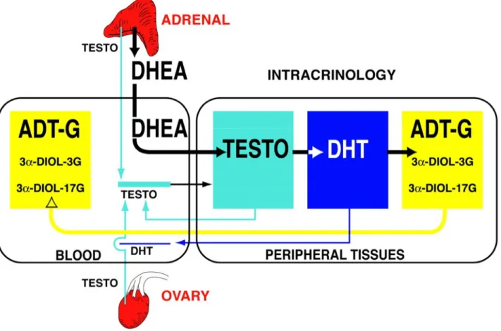
Transformation of the adrenal precursor steroids DHEA-S and DHEA into androgens and/or estrogens in peripheral target tissues depends upon the level of expression of the various steroidogenic and metabolizing enzymes in each cell of these tissues. This sector of endocrinology that focuses on the intracellular hormone formation and action has been called intracrinology (8) (Fig.2). This situation of a high secretion rate of adrenal precursor sex steroids in men and women is thus completely different from all animal models used in the laboratory, namely rats, mice, guinea pigs and all others (except monkeys), where the secretion of sex steroids takes place exclusively in the gonads (7). One explanation for the delayed progress in the field of formation of sex steroids in peripheral target tissues or intracrinology is the fact that the adrenals of the animal models usually used do not secrete significant amounts of adrenal precursor sex steroids, thus focusing all attention on the testes and ovaries as the exclusive sources of androgens and estrogens. The term intracrinology was thus coined (8) to describe the synthesis of active steroids in peripheral target tissues where the action is exerted in the same cells where synthesis takes place without release of the active steroids in the extracellular space and general circulation (8).
Fig1. Human steroidogenic and steroid-inactivating enzymes in peripheral intracrine tissues. 4-DIONE, androstenedione; A-4-DIONE, 5-alpha-androstane-3,17-dione; ADT, androsterone; epi-ADT, epiandrosterone; E1, estrone; E1-S, estrone sulfate; 5-DIOL-FA, androst-5-ene-3alpha,17beta-diol
fatty acid; 5-DIOL-S, androst-5-ene-3alpha,17beta-diol sulphate; HSD, hydroxysteroid dehydrogenase; TESTO, testosterone; RoDH-1, Ro dehydrogenase 1; ER, estrogen receptor; AR, androgen receptor; UGT2B28, uridine glucuronosyl transferase 2B28; Sult2B1, sulfotransferase 2B1; UGT1A1, uridine glucuronosyl transferase 1A1.
The almost exclusive focus on the role of ovarian estrogens in women’s reproductive physiology has removed attention from the dramatic 70% fall in circulating DHEA which already occurs between the ages of 20 to 30 and 40 to 50 years In fact, since DHEA is transformed to both androgens and estrogens in peripheral tissues, such a fall in serum DHEA and DHEA-S explains why women at menopause are not only lacking estrogens but are also likely to have been deprived of androgens for a few years, as illustrated by the 50–60% decrease in serum ADT-G (9) (Fig. 1). In a recent study nine androgens and their precursors and metabolites were measured by gas chromatography-mass spectrometry and liquid chromatography-tandem mass spectrometry in serum samples from 377 healthy postmenopausal women aged 55–65 years and 47 normally cycling 30- to 35-year-old premenopausal women. A decrease of 60% was then observed in the sum of ADT-G and 3α-diol-G while serum DHEA was decreased by 54% in postmenopausal compared with premenopausal women (8). Serum testosterone, on the other hand, did not decrease significantly from 0.18 ± 0.07 in premenopausal to 0.14 ± 0.07 ng/ml in postmenopausal women.
Since the serum levels of ADT-G and 3α-diol-G in women are 70% of those found in men of the same age while serum testosterone in women compared with men is only about 3% (0.15 ng/ml in women versus 4.5 ng/ml in men), it is clear that serum testosterone is not a valid marker of androgenicity in women. This situation is somewhat analogous to the situation in castrated men where castration causes a 90–95% reduction in the concentration of serum testosterone while the intra-prostatic concentration of DHT as well as of serum ADT-G and 3α-diol-G are only reduced by 50–70% (10).
Completion of the identification and characterization of all the human UDP-glucuronosyl transferases has made possible the use of the glucuronide derivatives of androgens as markers of androgenic activity. In fact, UGT2B7, UGT2B15 and UGT2B17 are the three enzymes responsible for the glucuronidation of all androgens and their metabolites in the human (11). The relatively simple inactivation mechanisms of androgens permits measurement of the sum of the metabolites of all androgens in the circulation, thus offering a precise assessment of the total androgenic activity in both women and men.
While the only means of determining androgenic activity in specific tissues is the direct measurement of the intra-tissular concentration of the active androgens, such measurements are not possible in the human except under exceptional circumstances such as in samples of cancer tissue obtained at surgery (7-9) However, while not permitting the assessment of androgenic activity in specific tissues, measurement by validated mass spectrometry techniques of the glucuronide
derivatives of ADT and 3α-diol permits an accurate assessment of total androgenic activity in the whole organism. In fact, since inactivation of the active androgens into ADT and 3α-diol and their subsequent glucuronidation into ADT-G and 3α-diol-G is the obligatory route of elimination of androgens (11-12) this approach appears to be the best means of evaluating total androgenic activity in individual subjects and patients. The clinician can then reliably correlate these values of androgenic activity with the other clinical findings.
As mentioned above, the level of transformation of the adrenal precursor steroid DHEA into androgens and/or estrogens in peripheral target tissues depends upon the level of expression of the various steroidogenic enzymes in each cell of each of these tissues (8). This situation of a high secretion rate of adrenal precursor sex steroids by the adrenals in men and women is thus completely different from all animal models used in the laboratory, namely rats, mice, guinea pigs and all others (except monkeys), where the secretion of sex steroids takes place exclusively in the gonads and the adrenals do not secrete significant amounts of DHEA (14).
The classical concept of androgen and estrogen secretion in women assumed that all sex steroids had to be transported by the general circulation following secretion by the ovaries before reaching the target tissues. According to this classical concept, it was erroneously believed that the active steroids could be measured directly in the circulation, thus providing a potentially valid measure of the general exposure of the whole body to sex steroids. In fact, this concept is valid only for animal species lower than primates but it does not apply to the human, especially in postmenopausal women where all estrogens and almost all androgens are made locally from DHEA in the peripheral tissues, which possess the enzymes required to synthesize active sex steroids. Such a local biosynthesis and action of androgens in target tissues eliminates the exposure of other tissues to androgens and thus minimizes the risks of undesirable masculinizing or other androgen-related side-effects. The same applies to estrogens, although we feel that a reliable parameter of total estrogen secretion (comparable with the glucuronides identified for androgens) has yet to be determined.
Fig. 2 Schematic representation of the very important contribution of the precursor DHEA of adrenal origin to total androgenic activity in postmenopausal women with a parallel minor contribution of testosterone (TESTO) of ovarian and adrenal origins. By intracrine mechanisms, DHEA is transformed into testosterone and DHT in peripheral tissues and then into the inactive metabolites ADT and 3α-diol before transformation into the water soluble glucuronide derivatives ADT-G, 3α-diol-3 G and 3α-diol-17 G by the UGTs 2B7, 2B15 and 2B17. These water-soluble metabolites are then released into the general circulation where they can be measured. A very small proportion of the testosterone and DHT made intracellularly by the steroidogenic enzymes of the intracrine pathway diffuse into the circulation. The height of the colored boxes is proportional to the concentration of each steroid.
2.1 Relative DHEA(S) Concentrations in Brain vs. Plasma vs. CSF in Humans
Higher concentrations of DHEA are found in the brain compared to plasma. In a study of ten postmortem human brains, DHEA concentrations were 29.4 nmol/kg in prefrontal lobe, 16.3 nmol/kg in parietal lobe, 13.1 nmol/kg in temporal cortex, 16.9 nmol/kg in cerebellum, and 18.7 nmol/kg in corpus callosum (15). These data were derived from nine women and one main (76–93 years old), and it is worth noting that large individual differences in DHEA brain concentrations were observed, with prefrontal lobe DHEA concentrations ranging from 9.8 to 470 nmol/kg (16). Mean DHEA concentrations were 1.83 nM in plasma of living human subjects of similar ages, which results in a brain-to-plasma ratio of ~6.5 (16). Although human brain concentrations of DHEA are higher than plasma concentrations, cerebrospinal fluid (CSF) concentrations of DHEA are lower than plasma concentrations. DHEA concentrations in CSF were ~5% of those found in the plasma of humans (16).
The validity of reported measurements of DHEAS and pregnenolone sulfate in the brain has recently been questioned (17,18). Many studies have relied on identification of parent compounds after separation of steroid sulfates from free steroids by organic:aqueous solvent extraction followed by a chemical reaction (solvolysis) to remove the sulfate. Analyses of sulfated steroids after extraction and solvolysis have found high concentrations of DHEAS and pregnenolone sulfate in rodent and human brains (19-20). Recent studies that measure intact sulfated compounds without deconjugation However, high DHEAS concentrations were found in two samples of human brain tissue using the new sample preparation method described above and gas chromatography-mass spectrometry (GC-MS) analysis (21). Hence, humans may indeed have high concentrations of brain DHEAS and older studies may turn out to be correct once verified using these newer analytic protocols (22,23). Studies relying solely on organic:aqueous extractions and solvolysis to measure DHEAS remain questionable and need to be reassessed.
2.2. Species Differences - Humans vs. Rodents
Humans and rodents (rats and mice) differ in the pathways through which sex steroids are synthesized. Whereas the Δ4 pathway predominates with rodents, the 17,20-lyase activity of the human P450c17 enzyme strongly prefers the Δ5 pathway (24). Subsequent conversion of DHEA into androstenedione by 3β-hydroxysteroid dehydrogenase (3βHSD) is the only pathway by which humans produce androstenedione (25). In rodents, conversion of cholesterol to androstenedione can occur through two pathways – the Δ5 pathway described above and the Δ4 pathway which involves the conversion of pregnenolone into progesterone (by 3βHSD) and progesterone conversion into androstenedione through the 17-OH-progesterone intermediary. Thus, humans make DHEA (Δ5 pathway) prior to downstream conversion into androstenedione and further metabolism into other sex steroids, whereas rodents go through the Δ4 pathway (predominantly) or Δ5 pathway. The species difference in predominant steroid pathways may partly explain species differences in peripheral circulating concentrations. Whereas DHEAS is the most abundant circulating steroid hormone in the human body (26), rats and mice (the species typically studied) have low circulating concentrations of DHEA(S) in the periphery (27). Unlike humans who secrete DHEA(S) from their adrenal glands and gonads, rats and mice can only synthesize and secrete DHEA(S) from their gonads, as their adrenal glands lack P450c17 (28).
Like humans, rats and mice have higher concentrations of DHEA in the brain compared to the plasma (4). For example, Sprague-Dawley rats had mean DHEA concentrations of 0.08 ng/ml (0.28 nM) in plasma, while brain concentrations of DHEA were 0.42 ng/g (1.46 nmol/kg) in anterior brain and 0.12 ng/g (0.42 nmol/kg) in posterior brain (4). These data are consistent with the hypothesis that in rodents, brain DHEA is derived mainly if not solely from local synthesis and not from peripheral synthesis. In human beings, brain DHEA may be derived from both local synthesis and peripheral synthesis. Thus, since DHEA is found in appreciable concentrations in brains of both human beings and rodents, rodents may indeed be a good model for studying the function of DHEA in the brain, but may not be an appropriate model for studying peripheral effects of these steroids.
3. DHEA(S) AS A NEUROSTEROID.
Important actions in the central nervous system (CNS) were initially inferred from observations that DHEA and DHEAS were synthesized de novo in brain, as brain concentrations were higher than plasma concentrations and brain concentrations remained high after adrenalectomy and gonadectomy of rats (4). Indeed, they have been termed “neurosteroids” for this reason (29-30). DHEA and DHEAS were among the first neurosteroids identified in rat brains. Cytochrome P450c17 was found in a subset of neurons of embryonic rodent brains (29). P450c17 expression was mainly neuronal; its expression was found as early as embryonic day 9.5, and persisted in the CNS during embryonic development. In one study, P450c17 was not detected in the CNS in adult rats and mice by immunocytochemistry, raising the possibility that this enzyme, and its neurosteroid products, function mainly during development (31). However, another study found P450c17 in adult male rat hippocampus by immunohistochemical staining (31). In the hippocampus, P450c17 was localized to pyramidal neurons in the CA1-CA3 region and to granule cells of the dentate gyrus. In these cells, P450c17 was localized in pre- and post-synaptic locations and in the endoplasmic reticulum by immunoelectron microscope analysis. While P450c17 protein was readily detected in the brain, the abundance of P450c17 mRNA transcripts in the embryonic mouse brain (31) or hippocampus of adult male rats was low, and was approximated to be 1/200th of the expression in the testis.
DHEA can be synthesized in vivo in rat and frog brains. Rat brains were capable of converting pregnenolone into DHEA and this may be activity-dependent (32). Basal P450c17 steroidogenic enzyme activity was low in the hippocampus, but could be enhanced by exposing neurons to N-methyl-D-aspartate (NMDA) (32). Similar findings have been reported for NMDA stimulation of pregnenolone synthesis from cholesterol in the hippocampus (33), suggesting that both P450scc and P450c17 are regulated by neurotransmitters. Frog brains also were found to synthesize DHEA from pregnenolone, and this enzymatic activity was reduced in a concentration-dependent manner by ketoconazole, an inhibitor of P450c17 (34). P450c17 enzymatic activity and protein expression were co-localized, further indicating that the enzymatic activity was due to P450c17.
P450c17 expression has also been found in adult rat spinal cord. Immunohistochemical studies localized P450c17 in both neurons and glial cells in the spinal cord. Slices of spinal cord tissue containing P450c17 protein converted (3H)pregnenolone into (3H)DHEA, and this conversion was reduced by ketoconazole. Thus, the spinal cord is one region in the CNS of rodents that expresses P450c17 and can synthesize DHEA endogenously from a precursor (35).
DHEAS may be synthesized in the brain from DHEA (36). Sulfation of DHEA has been observed in the brains of rhesus monkeys in vivo and in human fetal brain slices in vitro (36). Conversion of (3H)DHEA into (3H)DHEAS was also found in incubations of brain homogenates from pons, hypothalamus, olfactory bulb, cortex, and striatum/hippocampus of fetal and adult Sprague-Dawley rats and from thalamus, frontal cortex, basal ganglia, olfactory bulb, hippocampus, brainstem, midbrain, occipital cortex and cerebellum of adult Wistar rats . In addition to mammals, DHEAS synthesis from DHEA has been observed in brain homogenates from hypothalamus and telencephalon but not rhombencephalon of adult European green frogs.
Hydroxysteroid sulfotransferase (HST) or SULT2A1, also commonly referred to as DHEA sulfotransferase, is an enzyme that sulfonates DHEA (in addition to pregnenolone) (37). Western blotting and immunohistochemistry (with an antibody directed against partially purified rat liver HST) showed protein expression of an HST in adult Wistar rat brain (3). However, the characterization of this HST was not fully addressed, and hence its identity was uncertain. Other studies using different antibodies to purified or well-characterized proteins have confirmed the finding of HST in the brains of rats and frog. SULT2A1 mRNA expression has been shown in rat brains, thereby definitively demonstrating the presence of SULT2A1 in the brain. Future research on the activity and localization of newly discovered sulfotransferases, such as SULT2B and SULT4, may further our understanding of DHEA sulfonation in the brains of humans, rats and mice in the future (37).
It is unlikely that brain DHEAS comes from the periphery because sulfated steroids are hydrophilic and do not readily cross the blood-brain barrier, as evidenced by low recovery (0.03%) of radioactively labeled DHEAS in the brains of Sprague-Dawley rats following intracardiac injection (154). Although, one study has found increased pregnenolone sulfate in the brains of Sprague-Dawley rats after i.v. injection via the tail vein (325). What little steroid sulfates do enter the brain may occur through organic anion transporting peptides (OATP), which may work to transport DHEAS in both directions (13). However, steroid sulfates may egress from the brain more readily than they enter. The efflux clearance of (3H)DHEAS across the blood-brain barrier was determined to be tenfold greater than its influx (118 (µl/min-g efflux vs. 11.4 µl/min-g influx) (13). Hence, DHEAS is predominately transported out of the brain across the blood-brain barrier, further suggesting that DHEAS found in the brain is most likely due to local synthesis.
4. DHEA(S) MECHANISMS OF ACTION
Steroid hormones affect gene transcription by binding to specific cytoplasmic receptors, and then translocating into the nucleus, or binding to receptors that are resident in the nucleus, where they bind to steroid responsive elements on DNA. To date, no nuclear steroid receptor with high affinity for either DHEA or DHEAS has been found (38-40). The mechanisms by which DHEA(S) operate are not fully understood (38). DHEA(S) may mediate some of its actions through conversion into more potent sex steroids and activation of androgen or estrogen receptors in tissue (i.e. skin, liver, brain) (41). In addition to DHEA(S) having effects through its sex steroid metabolites (i.e. estradiol and testosterone), DHEA(S) may also have effects through its more immediate metabolites, such as 7α-hydroxy-DHEA (42). Although no unique DHEA or DHEAS nuclear steroid receptor has been found, DHEA and DHEAS have been found to affect receptors and to show affinity for some binding sites (38).
In the brain, DHEA(S) modulates actions of the γ-aminobutyric acid type A (GABAA) receptor, the NMDA receptor, and the sigma subtype 1 (σ1) receptor (43-48) among others (49-51). DHEA and DHEAS generally act as noncompetitive antagonists at the GABAA receptor, with DHEAS having more potent antagonistic effects than DHEA (52-53). DHEA(S) generally acts as a positive allosteric modulator of the NMDA receptor, although the binding of DHEA(S) with an interaction site on the NMDA receptor is not well documented (43,46). DHEA(S) can potentiate NMDA receptor function through its actions as a σ1 receptor agonist. However, in non-hippocampal brain regions DHEA(S) may inhibit glutamate neurotransmission through σ receptors, since σ receptor agonists were shown to reduce NMDA-induced dopamine release in the striatum (54). In an electrophysiological study with Sprague-Dawley rats, intravenous (i.v.) administration of DHEA (100–500 µg/kg) potentiated the NMDA neuronal response of CA3 rat hippocampus pyramidal neurons in a dose-dependent manner (32). The addition of σ receptor antagonist haloperidol or σ1 receptor antagonist N-dipropyl-2-(4-methoxy-3-(2-phenylethoxy)phenyl)-ethylamine monohydrochloride (NE-100), but not saline or spiperone (which has low affinity for σ receptors), inhibited the potentiating effect of DHEA, suggesting that DHEA can modulate the NMDA response through σ1 receptors (55). DHEAS potentiated the NMDA evoked release of (3H)norepinephrine from preloaded hippocampal slices, while the addition of σ receptor antagonists haloperidol or 1-(2-(3,4-dichlorophenyl)-ethyl)-4-methylpiperazine (BD1063) blocked the potentiating effect of DHEAS (56). Thus, DHEA(S) can modulate NMDA neurons and receptor activity by acting at the σ1 receptor (that is coupled to Gi/o proteins) in both in vivo and in vitro studies (55,56).
DHEAS, but not DHEA, augments cholinergic function in several animal models (57). Intraperitoneal (i.p.) administration of DHEAS (25–250 µmol/kg) increased acetylcholine (ACh) release from hippocampal neurons in rats (58). This effect has behavioral relevance in vivo, since DHEAS prevented (in a dose-dependent manner) the memory impairment induced by the ACh receptor antagonist scopolamine in mice (59). The σ1 receptor antagonist NE-100 blocked the ameliorating effects of DHEAS in this model, suggesting that the modulation of the cholinergic system by DHEAS involves interaction with σ1 receptors (59). Long term administration (15 days) of the STS inhibitor p-O-(sulfamoyl)-N-tetradecanoyl tyramine (DU-14) (which inhibits the conversion of DHEAS to DHEA) to rats increased plasma DHEAS concentrations, decreased DHEA concentrations, increased hippocampal ACh release, and blocked scopolamine-induced amnesia (60).
Additional intracellular sites where DHEA may act have also been described. DHEA may interact directly with certain cytoskeleton components or novel membrane receptors. DHEA was found to bind to microtubule-associated protein (MAP) 2C with strong affinity (61). MAP2C, which is expressed at early development stages, was found in adult retina and olfactory bulb, which are tissues in which neurogenesis persists in the adult (61). Intriguing leads are emerging for possible DHEA receptor sites in the periphery that may also exist in the central nervous system. A DHEA receptor was found on endothelial cell plasma membranes and it was coupled to endothelial nitric-oxide synthase (eNOS) activity through Gi/o proteins Gαi2 and Gαi3 (62). DHEA(S) may also have actions at other receptors, including the peroxisome proliferator-activated receptor α (PPARα), pregnane X receptor, constitutive androstanol receptor, and estrogen receptor β (63-65).
Mechanisms of action of DHEA and DHEAS in neurons. This cartoon summarizes many of the actions of DHEA and DHEAS described in detail in the text. DHEA and DHEAS have inhibitory effects (red blocking arrow) at the GABAA receptor (section 6 and 7.1). DHEA and DHEAS act as agonists (green arrow) at the σ1 receptor (section 6 and 7.1), which subsequently may activate the NMDA receptor. DHEA inhibits Ca2+ influx (red blocking arrow) into the mitochondria (section 7.1). DHEA influences embryonic neurite growth through stimulation (green arrow) of the NMDA receptor (section 7.2). DHEA increases (green arrow) kinase activity of Akt and decreases apoptosis, while DHEAS decreases (red blocking arrow) Akt and increases apoptosis (section 7.4). DHEAS increases (green arrows) TH mRNA and TH protein abundance (section 7.5) leading to increased catecholamine synthesis. DHEA and DHEAS stimulate (green arrows) actin depolymerization and submembrane actin filament disassembly and (green arrows), increasing secretion of catecholamines (“da” and “ne”) from secretory vesicles (section 7.5). DHEA and DHEAS inhibit (red blocking arrow) reactive oxygen species (ROS) activation of transcription mediated by NF-κB (section 7.6 and 7.7). DHEA inhibits (red blocking arrow) nuclear translocation of the glucocorticoid receptor (GR) (section 7.8). Mechanisms of action not pictured in this graph are: alterations of brain derived neurotrophic factor (BDNF) synthesis, inhibition of stress-activated protein kinase 3 (SAPK3) translocation, and inhibition of 11β-hydroxysteroid dehydrogenase type 1 (11β-HSDl) activity. Abbreviations: σ1, sigma 1 receptor; Akt, serine-threonine protein kinase Akt; Ca2+, calcium; da, dopamine; GABA
A, γ-aminobutyric acid type A receptor; GR, glucocorticoid receptor; ne, norepinephrine; NF-κB, nuclear factor kappa B; NMDA, N-methyl-D-aspartate receptor; ROS, reactive oxygen species; TH, tyrosine hydroxylase.
5. NEUROBIOGICAL ACTION OF DHEA WITH POTENTIAL RELEVANCE TO SEXUAL FUNCTION: EXPERIMENTAL FINDINGS
Neurobiogical action of DHEA involve directy DHEAS, DHEA and its more immediate metabolites (e.g., 7α-hydroxy-DHEA) in the brain but they are also due to conversion of DHEA(S) into sex steroids (estradiol, testosterone, DHT, 3alpha-DIOL, 3Beta-DIOL). Neurobiological actions of estradiol and testosterone are well established (66-69), less characterized are the function attributable directly to DHEA and DHEAS. These major biological actions of DHEA(S) involve neuroprotection, neurite growth, neurogenesis and neuronal survival, apoptosis, catecholamine synthesis and secretion, as well as anti-oxidant, anti-inflammatory and anti-glucocorticoid effects. In addition, DHEA affects neurosteroidogenis and endorphin synthesis/release
5.1 DHEA and catecholamine synthesis/secretion
DHEA(S) influence catecholamine synthesis and secretion (70). DHEAS was protective against the neurotoxin MPP+ (which inhibits catecholamine synthesis and triggers cell death) in rat cerebellar granule cell cultures. In an in vivo study of long-term DHEA treatment, obese and lean female Zucker rats were fed chow containing 0.6% DHEA for 28 days (71). Lean rats had higher norepinephrine (NE) in the lateral hypothalamus but lower NE in the paraventricular nucleus (PVN) of the hypothalamus compared to lean rats fed the control diet (71). There were no differences among obese Zucker rats in NE, serotonin (5HT), and serotonin metabolite 5-hydroxyindole-3-acetic acid (5-HIAA) in the hypothalamus (71). In another study, obese female Zucker rats treated with a large, acute i.p. injection of DHEA (200 mg/kg) had increased concentrations of dopamine (DA), 5HT, and 5-HIAA, and decreased concentrations of NE and epinephrine (EPI) in the paraventricular nucleus compared to controls that received oil vehicle (72). DHEA treatment had no effect on neurotransmitter concentrations in the lateral hypothalamus or ventromedial hypothalamus (72). These in vivo studies suggest that the duration and/or dose of DHEA treatment may be important and that DHEA may have different effects on different parts of the hypothalamus. In vitro, DHEAS (10−8 M and 10−6 M) has been found to stimulate dopamine release from rat hypothalamic cells in primary cultures (73).
DHEA(S) affects proliferation of catecholamine-producing adrenomedullary chromaffin cells. Although DHEA and DHEAS do not induce proliferation by themselves, they may modulate proliferation induced by growth factors and do so in an age-dependent manner. In bovine chromaffin cells from young animals, DHEA decreased cell proliferation induced by insulin-like growth factor-II (IGF-II), but had no effect on proliferation induced by basic fibroblast growth
factor (bFGF) (74). In another study, DHEA decreased cell proliferation induced by leukemia inhibiting factor (LIF) in bovine chromaffin cells from young animals (75). In bovine chromaffin cells from adult animals, DHEA decreased cell proliferation induced by epidermal growth factor (EGF) (75). DHEAS had no effect on LIF-induced proliferation of cells from young animals, but high micromolar concentrations of DHEAS enhanced EGF-induced proliferation of cells from adults (75). The effects of DHEA and DHEAS were not due to downstream metabolism into sex steroids since neither the estrogen receptor antagonist ICI 182,780 nor the androgen receptor antagonist flutamide affected chromaffin cell proliferation (75). Thus, local production of DHEA and DHEAS in the adrenal cortex can influence proliferation of chromaffin cells, and may have similar effects on catecholamine-producing neurons in the brain.
DHEA and DHEAS stimulate secretion of catecholamines from rat pheochromocytoma PC12 cells and are involved in inhibition of neuronal proliferation and promotion of differentiation of adrenal medullary cells to a more neuroendocrine phenotype. Administration of nerve growth factor (NGF) induced PC12 cells to differentiate into a neuronal phenotype, while administration of DHEA alone had no effect (76). PC12 cells incubated with both NGF and DHEA had lower survival and less neurite outgrowth than cells incubated with NGF alone in serum-free medium (76). NGF dose-dependently induced phosphorylation of extracellular signal-regulated kinases (ERK)1/2, which distinguished proliferation from differentiation processes, and this ERK1/2 phosphorylation was inhibited by DHEA (76-77). Furthermore, DHEA stimulated dopamine release from NGF-treated cells, while neither NGF nor DHEA alone had an effect on dopamine release (77). Another study compared the stimulatory effects of DHEA and DHEAS on catecholamine synthesis. The effect of DHEA was faster than that of DHEAS; whereas the effect of DHEA peaked at 10 min, the effect of DHEAS peaked at 30 min (58). In addition to stimulating secretion, DHEAS (but not DHEA) also stimulated catecholamine production. DHEAS increased tyrosine hydroxylase (TH) protein abundance in PC12 cells after four hours of stimulation, and also increased TH mRNA abundance in PC12 cells after only two hours of stimulation (58). These data suggest that DHEA and DHEAS function differently, and that DHEAS may directly affect TH gene transcription. Experiments to determine if DHEA increases TH gene transcription directly have not yet been reported.
DHEA and DHEAS have non-transcriptional effects on catecholamine secretion. DHEA and DHEAS have been found to stimulate actin depolymerization and submembrane actin filament disassembly, a fast-response cellular system regulating trafficking of catecholamine vesicles (78). An actin meshwork inhibits catecholamine secretory vesicles from reaching exocytosis sites. By decreasing this actin meshwork, DHEA and DHEAS increase the ability of catecholamines to be
secreted from secretory vesicles. Addition of DHEA and DHEAS to PC12 cells induced actin depolymerization, as measured by the ratio of G-monomeric to total cellular actin, an established marker of actin cytoskeleton dynamics (78). When PC12 cells were exposed to phallacidin, an actin filament stabilizer, the stimulatory effect of DHEA and DHEAS on both dopamine and norepinephrine secretion was prevented (78). These studies show that DHEA and DHEAS exert a direct effect on PC-12 cells (a model of chromaffin cells), and thus, provide in vitro evidence of how the zona reticularis and the adrenal medulla may be interacting in vivo. These findings also raise the possibility that DHEA and DHEAS could increase catecholamine production and release in the brain.
5.2 DHEA allopregnanolone and beta-endorphin
Allopregnanolone is a 3-α, 5-α reduced metabolite of Progesterone (79), and its major sources are the gonads and adrenal cortex, and, to a lesser extent, the CNS (79). Allopregnanolone acts as an agonist of GABAA receptor, modulating stress, mood, and behavior. Gonadal steroids may modulate allopregnanolone levels, as suggested by several experimental studies on animal models. In fact, female rats show significantly higher hippocampal allopregnanolone concentrations on the morning and afternoon of proestrous than at diestrous or estrous, reaching the lowest levels at estrous. Moreover, ovariectomy determines an increased adrenal allopregnanolone content and a reduction in allopregnanolone levels in brain and serum; this may be due to an estrogen-mediated enzymatic induction in the synthesis of allopregnanolone (79-81).
β-endorphin is the most important and biologically active endogenous opioid peptide; it has behavioral, analgesic, thermoregulatory, and neuroendocrine properties. A decrease in central and peripheral β-EP levels in ovariectomized rats and in circulating β-EP levels in postmenopausal women has been shown (82)
Dehydroepiandrosterone administration induces an increase in β-EP content in anterior and neurointermediate pituitary and hippocampus in a dose-dependent manner. High-dose DHEA administration induces an increase in β-EP circulating levels to those observed in fertile animals and an even greater increase in the hypothalamus. Comparing these results with those assessed administering E2 in ovx rats, we can observe several similarities. In all the examined organs, the effect of DHEA on β-EP levels closely reflects that of E2 valerate, with the exception of the neurointermediate pituitary, where DHEA induces a lower increase in β-EP.
Dehydroepiandrosterone administration induces an increase in allopregnanolone content in the hypothalamus, anterior pituitary, serum, and hippocampus, where DHEA administration restores allopregnanolone levels to those observed in fertile rats. Similar data have been observed at the central level when administering E2 valerate in ovx rats. However, DHEA does not determine a decrease in allopregnanolone adrenal content, as observed with E2. Moreover, the serum allopregnanolone levels reached in response to DHEA therapy are lower than those obtained with E2 administration (83)
The mechanisms of action of DHEA are not clear. Part of the effects of DHEA depends on its conversion to estrogens and androgens and on the recruitment of their receptors. In fact, at the vascular level, a reduction of atherosclerotic lesions by DHEA in rabbits was reported, partially through conversion to estrogens. However, there is evidence that DHEA may have a specific receptor. This has been demonstrated in blood vessels, where DHEA binds with high affinity to nonhuman endothelial cell membranes without being displaced by structurally related steroids. The present data seem to indicate that the effect of DHEA administration may not be entirely ascribed to the conversion in estrogenic metabolites. In fact, the E2 levels reached in DHEA-treated ovx animals were not significantly different from those observed in untreated ovx animals and significantly lower than those obtained in ovx animals in response to E2 valerate administration. Similar levels of β-EP and allopregnanolone, obtained in various organs, were observed in response to the two different therapies. Therefore, DHEA may act directly or through its local metabolites on neurosteroidogenesis and the opioidergic pathway. (83)
6. EFFECT OF DHEA THERAPY ON SEXUAL FUNCTION USING AN IN-VIVO MODEL The field of behavioural pharmacology is showing an increasing interest in female rat sexual behaviour as a model for evaluating drug actions and steroid interference. Endogenous sex steroids modify female sexual behaviour in rats. Fertile animals during the estrous phase show normal level of sexual receptivity, which is abolished by ovariectomy and it can be partially restored by the subcutaneous injections of Estradiol Benzoate (EB) and Progesterone (P) (84). Sex steroids influence the neurobiology of sexual function, acting directly on their receptors at nuclear and membrane level or indirectly throughout their effects on neuropeptides (oxytocin, beta-endorphin, ecc.), neurotransmitters (dopamine, serotonin) and neurosteroid metabolism (mainly allopregnanolone) (85-86).
Sexual behaviour in the female rat is characterized by both receptive and proceptive behaviours (87). Receptive behaviour consists in a reflexive posture, called lordosis, which represents the female readiness to allow copulation (87). Proceptive behaviours, including hops, darts and ear-wigglins, are exhibited by a sexually receptive female to arouse male sexual interest (88). Consequently they are considered appetitive behaviours, while lordosis represents the consummatory aspect of female sexual behavior (89). Sexual motivation of female rats can be also assessed by evaluating their preference for a sexually active male versus a sexually receptive female, during the partner preference test (90,91). In addition, if the mating occurs in a laboratory condition which allows the female to enter and exit from the male’s compartment, the estrous female will approach and withdraw from the male, controlling hence the number and the timing of sexual contacts (i.e. mount, intromission and ejaculation). This pattern, known as paced mating behaviour is thought to be influenced by sexual motivation besides to sensory discrimination between the different types of sexual stimulation (mount, intromission and ejaculation) (92).
In the present study, we evaluated the influence of DHEA administration on receptive and proceptive components of female rat sexual behaviour and whether the co-administration of estrogens might enhance sexual response in a model of ovariectomized rats.
6.1 Methods Animals
Adult female Sprague-Dawley rats weighing 200-250 g were obtained from Charles River Laboratories (Calco, LC, Italy). They were housed two per cage and maintained in standard conditions at 22±1°C and 55-60% humidity. A 12-h reversed light/dark cycle was employed to facilitate behavioural testing during the normally active (dark) phase of the cycle. Commercial rat
pellets and water were freely available. After a week of adaptation, all rats were bilaterally ovariectomized under ketamine hydrochloride (Ketavet 100®, Farmaceutici Gellini Spa, Italy) plus xylazine hydrochloride (Rompun®, Bayer, Germany) anesthesia, using standard surgical procedures. Sexually experienced Sprague Dawley male rats, weighing approximately 350-400 g, were used as stimulus animals in the behavioural tests.
Animal care, maintenance and surgery were conducted in accordance with the Italian law (D.L. n. 116/1992) and European legislation (EEC n. 86/609). The experimental design and procedures received the approval of the Bioethical Committee of the Italian Institute of Health.
Treatments
Treatments were started approximately 3 weeks after ovariectomy to allow a complete recovery from surgery. Animals were divided in 6 groups and submitted to the following treatments:
1) Estradiol Benzoate (Estradiol benzoate, Sigma-Aldrich, Milan, Italy) (EB) 10 µg/rat subcutaneously (s.c.) injected 48 h before the behavioural test;
2) Estradiol Benzoate (EB) 3 µg/rat s.c. injected 48 h prior to behavioural test;
3) DHEA (dehydroepiandrosterone, Sigma-Aldrich, Milan, Italy) 0.5 mg/kg by oral gavage (p.os) daily for 6 weeks;
4) DHEA 5 mg/kg p.os daily for 6 weeks;
5) EB 3 µg /rat s.c. 48 h prior to behavioural test plus DHEA dosed at 0.5 mg/kg p.os daily for 6 weeks;
6) EB 3 µg/rat 48 h prior to behavioral test plus DHEA dosed at 5 mg/kg p.os daily for 6 weeks. All the females were s.c. injected with 500 µg of progesterone (Prontogest, AMSA) 4 h before the tests. Hormones were delivered in a peanut oil vehicle. DHEA was solubilised in Tween 80 (10%) and water and administered in the volume of 5 ml/kg.
Sexual behaviour testing 1) Receptivity test
The test was conducted in a clear Plexiglas arena (70×35×40 cm high), where a male rat was placed for a 5-min period and was allowed two intromissions with a stimulus non-experimental female to
ensure his sexual vigor. Thereafter, an experimental female rat was placed in the arena with the male. The test ended when the female received 10 mounts with or without intromission. The lordosis response was scored on a four-point scale (0-3) as described (93). For each rat a lordosis quotient (LQ) was calculated as the number of lordosis responses (scores of 2 or 3) divided by the total number of mounts multiplied by 100. During the test the number of proceptive behaviours (hops-darts, ear-wigglings, crawlings, approach and withdrawal from the male and sniffings) and rejective behaviours exhibited per minute were recorded. The behavioural parameters were scored by observers unaware of the pharmacological treatments.
2) Partner preference test
After 5 weeks of treatment the partner preference test, performed according to Ågmo et al. (94), was used to evaluate sexual motivation in a no-contact condition. The apparatus consisted of an open field arena (100×50×40 cm high) with two round cages made of wire meshing (16 cm diameter, 40 cm high) diagonally positioned at the opposite corners of the arena. In the two cages a sexually active male and a female rat were individually placed as stimulus animals. A virtual area near to each animal cage was defined as the incentive zone: the sexual incentive area near to the male and the social incentive area near to the female. In these conditions it was allowed the transmission of visual, olfactory and auditory cues but it was avoided mating. Experimental females were individually placed in the centre of arena for a 5-min adaptation period at the presence of the stimulus animals and thereafter tested for 10 min. The number of visits to the male and the female as well as the time spent near each stimulus animal were recorded. The measure of sexual motivation is expressed by a preference score that is the ratio between the time spent in the sexual incentive area versus the total time spent in the two incentive areas (94).
3) Paced mating behaviour
After 6 weeks of treatment the experimental female rats were tested for paced mating behaviour according to the procedure described by Clark et al. (95). The apparatus consisted in a plexiglas arena (80×49×40 cm high) divided into three equal compartments by two clear plexiglas partitions (48.5×39.5 cm high) with holes (5 cm in diameter, placed 1.25 cm from the side and bottom edges) in the bottom corners. The small diameter of the holes enabled the female rat to enter and exit from the male compartment, while not allowing the access of male rat to the female’s compartment. Two opaque partitions (48.5×39.5 cm high), covering the holes in the plexiglas ones, were used to avoid the female rat and the males to see one another, when they were not being tested.
The mating test started by placing the experimental female in the central compartment and a stimulus male rat in each of the two side chambers. The female rat was allowed to access to a single male rat through the hole of the clear plexiglas partition, after the removal of the opaque divider. The mating test ended when the female received 10 mounts with intromission. If ejaculation occurred, she was allowed to leave the male rat’s compartment and then return. Thereafter the female was confined in the central compartment replacing the opaque partition. To complete the test the female rat was permitted to access to the second male rat. Paced mating test was terminated: (i) if the female returned to the stimulus male rat, following the 10th intromission; (ii) if the female received no intromission within the first 15 min from the beginning of the test; (iii) when the interval between two intromissions was longer than 30 min. During the test the following parameters of sexual behaviour were registered or calculated: a) the contact-return latency, that is the time between the female’s exit from male’s compartment following each type of sexual stimulation (mount, intromission, ejaculation) and her return; b) the percentage of exits, that is the number of times the female rat left the male’s compartment, following each type of stimulation.
Biochemical assays
Following the conclusion of all behavioural experiments, specifically 24 h after the paced mating test, female rats were sacrificed by decapitation and trunk blood collected. Brains and adrenal glands were removed for the subsequent biochemical analysis. From the brains the following areas were isolated: hypothalamus, hippocampus, anterior pituitary, neurointermediate lobe, frontal and parietal cortex.
All hormonal determinations were carried out during the same assay. Plasma DHEA, Progesterone (P), Estradiol (E2) Testosterone (T) concentrations were determined using commercially available radioimmunoassays kits (Radim, Pomezia, Rome, Italy). The intra-assay and inter-assay coefficients of variation (CV) and the sensitivity of the assay were: 7.8 and 8.3% and 0.02 ng/ml for DHEA, 8,9% and 0,03 ng/ml for T, 11.7% and 0.12 ng/ml for P, 8.5% and 4.7 pg/ml for E2, respectively. Serum corticosterone (ICN Biomedicals Inc., Irvine, CA, USA) was assayed by radioimmunoassay using trade kits. For corticosterone the assay sensitivity was 25 pg/ml and the intra- and inter-assay coefficients of variation were 5.8% and 7.5% respectively.
The supernatant of tissue homogenates and serum was passed through a C-18 Sep-Pak cartridge, previously equilibrated with homogenizing buffer. The cartridge was sequentially washed with homogenizing buffer, 50% aqueous methanol, and the unconjugated steroid fraction was eluted with absolute methanol and brought to dryness under nitrogen. Analytical grade solvents were purchased from Merck (Darmstadt, Germany); C-18 Sep-Pak cartridge were obtained form Waters Corporation (Milford, USA). Allopregnanolone contents were measured by a radioimmunoassay method, using an antiserum kindly provided by Dr. RH Purdy (San Diego, CA, USA). The sensitivity of this assay was 20 pg/mL, the recovery after extraction and chromatography was 86.51±2.7% (mean ± SEM), and the intra- and inter-assay coefficient of variation was 7% and 9%, respectively. Allopregnanolone levels were expressed in pg/mg of tissue in each tissue and in pg/ml in serum.
β-Endorphin assay
The supernatant of tissue homogenates and plasma were passed through a C-18 Sep-Pak cartridge, previously equilibrated with 50% aqueous methanol, and the unconjugated fraction was eluted with absolute methanol and brought to dryness under vacuum. β-endorphin levels were measured by a previously described specific radioimmunoassay (83), using camel β-endorphin as standard (Sigma Chemicals, St. Louis, MO, USA). The antiserum was used at the final dilution of 1:130.000. Analytical grade solvents were purchased from Merck (Darmstadt, Germany); C-18 Sep-Pak cartridges were obtained from Waters Corporation (Milford, USA). The sensitivity of this assay was 10 pg/ml, the recovery following acetic acid extraction and chromatography corresponded to 85±11% of the total amount, and the intra- and inter-assay coefficients of variation were 6% and 8%, respectively. β-endorphin levels were expressed in ng/organ (hypothalamus, hippocampus and neurointermediate lobe) or ng/mg tissue (frontal and parietal cortex) and in ng/ml in plasm
Statistical Analysis
Behavioral and hormonal data, reported as mean ± SEM, were analyzed using GraphPad Prism Software, version 5.0 for Windows (GraphPad Software, San Diego, CA, USA). One-way analysis of variance (ANOVA) with Newman–Keuls test for post-hoc comparisons was performed between individual treatment groups. Values of P < 0.05 indicate significant differences among groups.
6.2 Results
Hormonal evaluation
-Effects of Estradiol Benzoate (EB 3-10 µg/rat), of DHEA (0,5-5 mg/kg) and co-administration of DHEA (0,5-5 mg/kg) plus EB (3 µg/rat) on Allopregnanolone (Allo) concentration in selective brain areas and in adrenal gland of ovariectomized rats.
In all brain areas analyzed, with the only exception of the frontal lobe, the administration of EB at 10 µg/rat in ovariectomized rats increased Allo content in comparison with EB 3 µg/rat. The administration of DHEA at the lower dose (0,5 mg/kg) did not evidence any significant difference in comparison with EB 3 µg/rat in all brain areas. On the contrary, DHEA 5 mg/kg increased Allo levels in comparison with EB at 3 µg/rat in all CNS areas evaluated. The co-administration of DHEA 0,5 mg with EB did not affect Allo level in all brain areas, in comparison with EB alone and with DHEA at 0,5 mg/kg. On the contrary, the co-administration of EB with the highest dose of DHEA (5 mg/kg) increased Allo levels respect to EB 3 alone in all brain areas and also in comparison with DHEA alone, at the dose of 5 mg/kg, in the parietal cortex, in the hypothalamus and in the anterior pituitary. In the adrenal gland, only the co-administration of DHEA (0,5 mg/kg and 5 mg/kg) with EB 3 increased Allo content in comparison with EB 3 alone, whereas all other hormonal treatments did not affect its intra-glandular concentration.
-Effects of Estradiol Benzoate (EB 3-10 µg/rat), of DHEA (0,5-5 mg/kg) and co-administration of DHEA (0,5-5 mg/kg) plus EB (3µg/rat) on Beta-endorphin (β-end) concentration in selective brain areas.
In all brain areas analyzed, with the only exception of the frontal lobe, EB, when administered at the dose of 10 µg/rat increased β-end content respect to EB at 3 µg/rat. The administration of DHEA alone at the lower dose (0,5 mg/kg) did not evidence any significant differences in comparison with EB 3 µg/rat in all brain areas. DHEA treatment at the dose of 5 mg/kg increased β-end levels in comparison with DHEA 0,5 mg/kg and in comparison with EB 3 µg/rat in hippocampus, in the hypothalamus and in the neurointermediate lobe. The co-administration of DHEA 0,5 mg with EB did not modify β-end level in all brain areas analyzed. On the contrary, the co-administration of EB with the highest dose of DHEA (5 mg/kg) increased β-end respect to EB 3 µg/rat alone in all brain areas and also in comparison with DHEA alone at the dose of 5 mg/kg in the hypothalamus.
-Effects of Estradiol Benzoate (EB 3-10 µg/rat), of DHEA (0,5- 5 mg/kg) and co-administration of DHEA (0,5-5 mg/kg) plus EB (3µg/rat) on circulating hormonal levels.
Female rats receiving EB 10 µg/rat, DHEA 5 mg/kg and the co-administration of DHEA + EB (5 mg/kg + 3 µg/rat) showed an increased levels of circulating estradiol in comparison with EB 3 µg/rat. Administration of DHEA at 0,5 mg/kg alone showed reduced levels of estradiol compared to EB 3, whereas no modifications on estrogen levels were found following the co-administration of EB + DHEA 0,5 in comparison with EB 3.
Plasma level of DHEA was found increased following the administration of both doses of DHEA alone or in co- administration with EB, evidencing a dose-related increase, although the presence of EB did not modify plasma level of DHEA.
Only animals receiving DHEA alone or in combination with EB showed increased level of testosterone in comparison with EB 3 or EB 10. In these animals, T level were not affected by the dose of EB, while the concentration of T were higher in rats treated with DHEA 5 in comparison with those receiving DHEA at the dose of 0,5 mg/kg.
Progesterone and corticosterone circulating levels were not modified following the administration of any dose of EB, DHEA or their co-administration.
Allo levels resulted significantly increased following treatments with EB 10 and with the co-administration of DHEA 5 + EB 3 in comparison with EB 3 alone. DHEA therapy alone, at 0,5 mg/kg and 5 mg/kg, did not affect the concentration of this steroid compared to EB 3.
Beta-endorphin circulating levels were increased only following the administration of the EB 10, DHEA 5 mg and the combination of DHEA 5 + EB 3 in comparison with EB 3 alone.
Behavioural tests
1) Receptivity test
The oral administration of DHEA alone at both dosages failed to induce receptive behaviour in female rats, even if they were treated with progesterone; therefore their LQ values were always undetectable over the 4 weeks (data not shown). Rats treated with EB dosed at 10 µg/rat exhibited high level of sexual receptivity (LQ= 68.6 after 1 week of treatment, 93.8 after 2 weeks, 97.5 after 3 weeks and 90.0 after 4 weeks) (Fig.1). Rats treated with EB dosed at 3 µg/rat showed a lower level
(LQ= 71.4 after 1 week of treatment, 77.5 after 2 weeks, 73.8 after 3 weeks, 81.4 after 4 weeks) (Fig.1). Female rats treated with EB 3 µg/rat and DHEA 0.5 mg/kg displayed LQ values which were not statistically different from those of EB treated rats at the same dosage (3 µg/rat). The administration of DHEA dosed at 5 mg/kg in EB treated rats induced a significant increase in LQ value in comparison with EB alone treated rats, only after 1 week of treatment (p<0.05) (Fig.1). It must be underlined that LQ value was significantly higher than that of EB 10 treated rats (p<0.05) (Fig.1). In the following tests performed after 2, 3 and 4 weeks the difference in LQ values between EB + DHEA and EB alone treated rats was not statistically different (Fig.1). As concerns proceptive behaviours detected during the test, female rats treated with EB and DHEA at both dosages exhibited a marked increase in comparison with EB (3 µg/rat) treated rats already after the first week of treatment (p<0.05) (Fig. 2). Starting from the second week the difference between EB+DHEA and EB alone treated rats was statistically significant only when the highest dose of DHEA was administered (p<0.05 after 2 weeks, p<0.001 after 3 and 4 weeks) (Fig.2). It must be underlined that the number of proceptive behaviours exhibited by DHEA 5 mg/kg + EB 3 µg/rat was higher than that of EB 10 µg treated rats, at any tested time (Fig.2). No difference was detected in the number of rejective behaviours among the different experimental groups (data not shown). 2) Partner preference test
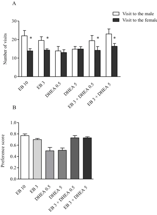
As expected, rats submitted to EB treatment at both dosages showed a marked preference to visit the male rat rather than the female (p<0.05) (Fig.3 panel A). Consequently the preference score of EB treated rats was 0.77 following the injection of 10 µg/rat and 0.70 following the injection of the lower dose (3 µg/rat) (Fig.3 panel B). Female rats treated with DHEA alone failed to show a preference for the sexual or the social stimulus (Fig.3 panel A): therefore the preference score of both treated groups was approximately 0.50 (Fig.3 panel B). Rats administered with DHEA at both dosages and EB (3 µg/rat) exhibited a significant preference for the male rat (Fig.3 panel A): the preference score was found to be 0.73 in both treated groups (Fig.3 panel B).
3) Paced mating behaviour
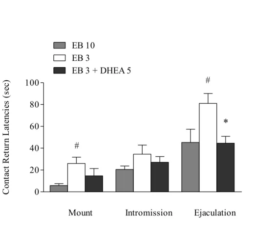
Paced mating test was performed only in 3 groups of rats treated as follows: 1) EB 10 µg/rat; 2) EB 3 µg/rat; 3) EB 3 µg/rat + DHEA 5 mg/kg.
As shown in Fig.4, there was a significant main effect of type of sexual stimulation on contact-return latencies (mount<intromission<ejaculation). It must be stressed that the mount, intromission and ejaculation contact return latencies of EB 3 + DHEA 5 mg/kg treated females were lower than that of EB (3 µg/rat) treated rats: the difference was found statistically significant as regards the
latency to return to male’s compartment after ejaculation (p<0.05) (Fig.4). For percentages of exits, there was a significant main effect of type of sexual stimulation but not a main effect of treatment (data not shown).
7.3 Conclusions
Sex steroids directly modify sexual function, evidenced as changes in sexual behaviour and in variations in the neurobiology of neuropetides and neurosteroids involved in sexual function. It’s well established by previous studies that ovariectomy reduces female brain content of allo and beta-endorphin in the hypothalamus and in sovra-hypothalamic areas and that two-weeks treatment with estrogens or DHEAS were able to increase the level of endogenous opioid and neurosteroidogenesis in a dose-related response. (83)
The present study confirms that chronic treatment (six weeks) of ovariectomized rats with DHEA only at the dose of 5 mg/kg increases allopregnanolone and beta-endophin content in different brain areas and in plasma when compared with available reference values of ovariectomized rats (16). Similarly, circulating hormonal changes after treatment with both doses of DHEA results in physiological increase of plasma estradiol, within the available range values of fertile animals (16) and no significant change of adrenal function (corticosterone level) were observed after treatment. These results, together with the available previous data, confirm that these animals received doses of DHEA inducing a biological response in a physiological range.
A single injection of EB at the higher dose (10 µg/rat) increased allopregnanolone and beta-endorphin brain and circulating content, supporting the concept that both systems are positively and directly influenced by estrogens in a dose-related way. Interestingly, co-administration of EB and DHEA showed a synergic effect on brain neurosteroidogenesis and opioid content compared to single treatments, although EB and DHEA therapies had a different time-schedule. In particular, this effect was shown over different brain areas and it was evidenced comparing animals receiving EB (3 µg/rat) and DHEA (5 mg/kg) as single therapy with rats receiving the combination of both. Opioid receptor agonists infused into the lateral ventricles can inhibit (through mu receptors) or facilitate (through delta receptors) the lordosis behavior of ovariectomized (OVX) rats treated with estrogen and a low dose of progesterone.
The sexual effects of beta-endorphin are reputed to occur mainly through their action on the pre-optic area and the amigdala (101). Beta-endorphin infused into the medial pre-pre-optic area inhibits mounting and intromission; infusion of this peptide into the medial amygdala inhibits the initial