UNIVERSITY OF CATANIA
INTERNATIONAL PH.D. COURSE IN NEUROBIOLOGY
24
rdCYCLE
Effect of growth factors, steroids, alfa-lipoic acid and Astroglial
Conditioned Media on the expression of some biomarkers of astroglial cell
proliferation and differentiation in primary culture
Dr. Sonia Grasso
Experimental Thesis
TUTOR: Prof. Roberto Avola
COORDINATOR: Prof. Roberto Avola
_____________________________________________________________
CHAPTER 1
INTRODUCTION
The nervous system of vertebrates (CNS) is mainly formed by neurons and glia, two heterogeneous classes of cells. CNS plays very delicate and complex functions. During the process of
neurogenesis, the proliferation and differentiation of neuroepithelial cells and the consequent formation in different types of neural cells play a key role in the development of the nervous system.
In the interactive dialogue between neurons and glia, the growth factors, coordinate some complex processes, such as the development and maturation of astrocytes and neurons.
The interactions between neurons and astrocytes play a crucial role during the process of
development and in the adult brain. During development, in fact, glial cells promote the growth of the precursors.
Growth factors influence the activity of the target cells by interaction with specific receptors located on the plasma membrane.
Several studies shown that the growth factors involved on astroglia, and also promote the accretion of neuritis (Engele J. and Bohn MC 1991, Petroski RE et al. 1991). Furthermore, it is known in the literature that the astroglia proliferation is closely related to cell growth (LC Wang et al. 1995). In addition, steroid hormones play a key role on the function and development of the brain in mammals. In fact, they adjust the neuroendocrine function with type mechanisms feedback to the hypothalamic-pituitary level (Kalra SP, et al. 1983) and they are also implicated in the control of cognitive and motor function (C. Beyer 1999), in the control of mental status through the regulation of neurotransmission aminergic and peptidergic (G. Fink et al. 1996). In particular, it has been observed that estrogens act as protective factors in neurodegenerative diseases, such as Parkinson's disease and Alzheimer's diseases (Xu H. et al. 1998). In contrast to this regulatory function and protective, estrogens play a different role during neuronal development. Alterations in the levels of
estrogen in the central nervous system (CNS) in development, affect critical aspects of cell differentiation including the extension of neurites, synapse formation, myelination, the expression of neurotransmitters and neuropeptides, death and survival cell (RC Miranda et al. 1994). Estrogens are crucial hormones for differentiation of the CNS. The first two weeks after birth, estrogen organize specific brain circuits male permanently and irreversibly (AP Arnold et al. 1985). In addition, estrogens affect the astroglial compartment and can promote the effects of neurotrophins at the level of the genome through interactions with the regulatory pathways of neurotrophic factors (Toran-Allerand CD 1996).
GROWTH FACTORS
The growth factors (GFs) are protein molecules with low molecular weight that play an important role in cellular communication, through interaction with specific receptors located in the plasma membrane of target cells. The growth factor interaction greatly resembles that of some hormones. At the level of the S.N.C. astrocytes express receptors for various GFs, neurotransmitters and neuromodulators. Nerve cells can respond to growth factors of astrocytic origin and can control the functions astrocyte through different signaling molecules and pathways of intracellular signal transduction. The growth factors, neurotransmitters, and peptides represent the principal agents of cell signaling (Sendtner M. et al. 1995).
The GFs include both the components that stimulate proliferation, as mitogens, and the components that depress it (for example, TGF and interferons).
However, these types of response are attributable to distinct GFs, as a single factor may stimulate or inhibit the proliferation in different cell populations, or in the same cell type, but in different conditions (concentration and time of application of the factor growth, functional status of the target cells, etc.). The stimulation of the cells with GFs induce a proliferative effect after about 15-20 hours after the receipt of the stimulus. Mitogenic activity exercised by growth factors on quiescent
cells induces G0 - G1 transition phase of the cell cycle, resulting a progression versus the S phase of
DNA replication.
This process is characterized at least by two subphases: an initial "competence" phase, which in the case of quiescent cells coincides with their transition from G0 to G1 phase, followed by a
"progression" phase in which the cell synthesizes components necessary for DNA synthesis, such as DNA polymerase, the "Proliferating cell nuclear antigen" (PCNA), ribonucleotide reductase,
thymidine kinase.
In some cases, the stimulation of proliferation requires the presence of the growth factor during all the time that elapses between the receipt of the stimulus and the stage of progression. In other cases, instead, it is sufficient that the growth factor is present only during the phase of competence.
The epidermal growth factor (EGF), basic fibroblast growth factor (bFGF), the insulin-like growth factor I (IGF-I), and insulin (INS) are neurotrophic agents inducing neuronal and astroglial cells proliferation and differentiation in culture (Avola R. et al. 1988a, Avola R. et al. 1988b).
Stiles C.D. and coll. (1979) have classified the growth factors such as factors of "competence" or
"progression". These two classes of growth factors cooperate for a full mitogenic response of the
cell.
The factors of "competence" are not able to initiate DNA synthesis, but are able to induce the "competence" to respond to other growth factors ("progression" factors) that stimulate the "progression" through the cell cycle. The competent cells do not respond to the factors of
"progression" and stop growth. The factors of "competence" include: the platelet-derived growth factor (PDGF) and fibroblast growth factor (FGF), while the factors of "progression" include the epidermal growth factor (EGF), and the insulin growth factor family (IGFs).
In addition to growth factors mentioned above, there is a wide range of molecules that participate in various development processes and neuronal growth. Among the most important are the class of neurotrophins that includes the "nerve growth factor" (NGF), the "brain derived neurotrophic factor" (BDNF) and neurotrophin 3 and 5.
The biological action of NGF is mediated by its binding to two classes of receptors localized on the plasma membrane: p75, known as low-affinity receptor, which binds also to other neurotrophins, and Trk-A, a glycoprotein of 140 kDa with activity of tyrosine kinases. NGF has multiple functions and in the near future could be used in the treatment of certain diseases such as neurotrophic corneal ulcer, Parkinson's disease, Alzheimer's disease, multiple sclerosis and some peripheral neuropathies.
Fibroblast growth factors (FGFs)
The family of fibroblast growth factors (FGFs) includes the aFGF and bFGF, two mitogenic proteins that were originally purified basing on their ability to bind to heparin, the FGF-5, FGF-6, the factor growth of keratinocytes, and the products of oncogenes int-2 and hst (F. Hefti et al., 1994).
The fibroblast growth factors are able to elicit strong morphological effects on the astroblasto, characterized by a narrowing of its cell body, and by an increase of the extension of cellular processes.
The FGFs are present in many peripheral tissues and are potent mitogens agents for various types of cells (A. Baird et al. 1986). The brain and the pituitary gland are particularly rich in bFGF. The basic fibroblast growth factor (bFGF) is a protein from the PM of about 17,000 D, can stimulate the proliferation of endothelial, mesenchymal and neuronal cells.
The bFGF promotes the survival and differentiation of cholinergic neurons in rat mesencephalic culture, of mesencephalic dopaminergic neurons (Mayer E. et al. 1993) new-striatal GABAergic neurons (Zhou and D. Daughter of M. 1991) and immortalized hypothalamic neurons (Gallo F. et al. 1996).
Epidermal growth factor (EGF)
The epidermal growth factor (EGF) is an acidic protein, that belongs to a large family of proteins which includes some viral proteins (Hefti F. 1994).
It is a "progression" polypeptide factor (molecular weight of about 6000 Da), and it is known in the literature its stimulatory effect on the proliferation of the epidermis, its inhibitory effect on gastric secretion (G. Carpenter and Wahl MI 1990), as well as the proliferation and differentiation of various cell types (Avola R. et al. 1988 b).
A lot of studies concerning EGF were addressed both to the role as a mitogen, both to its ability to affect the differentiation of not-neuronal cells (Carpenter G. and Cohen S. 1979), such as astroglial cells where the EGF stimulates both the synthesis of DNA and RNA, that the activity of
fosfoinositil 3-kinase (Avola R. et al. 1988a and 1988b). The EGF is able, moreover, to modify the morphology and to induce astrocyte "up-regulation" in the levels of glutamine synthetase and S-100 (Avola R. et al. 1988a and 1988b).
In recent years it has been demonstrated that EGF induces numerous biological responses: • increased or decreased cell adhesion
• morphological changes
• induction of ornithine decarboxylase • phosphorylation reactions.
INSULIN
Insulin and insulin like growth factors of type I and II are a family of proteins involved in the regulation of metabolism and cell growth of various tissues.
Insulin is a peptide hormone secreted by the cells of the pancreatic island of Langerhans , has a PM of 5807 Da and consists of two polypeptide chains of 21 (A-chain) and 30 (B-chain) amino acids.
Insulin is able to promote some metabolic pathways, and in particular it induces some key enzymes of glycolysis and causes a lowering of the levels of activity of fructose 1,6-bisphosphatase and pyruvic carboxykinase in the liver, causing a slowdown in gluconeogenesis.
The central nervous system has been regarded, for a long time, as a court Insulin-dependent, because the insulin produced by the pancreas crosses the blood-brain barrier to a limited extent. Currently there is increasing evidence that insulin found in the brain is synthesized on site probably by astrocytes and that insulin can function in nerve cells, more like that neurotransmitter neuromodulator (Wei L. et al . 1990).
Insulin, synthesized and released by nerve cells, has also been associated with the growth of the brain. In fact, it was demonstrated (Kappy MS et al. 1984) that the extent of protein synthesis in the brain is positively correlated with the number of insulin receptors since it promotes an active uptake of amino acids through specific transporters and that insulin stimulates the synthesis of macromolecules in mixed cultures brain (Raizada MK et al. 1980) and in cultured astrocytes from neonatal rat (Avola R. et al. 1988a and 1988b).
This action may be specific for astrocytes, which regulate the local availability of glucose in the central nervous system. In this way the Insulin could affect the availability of extra glucose in case of ischemic events, due to its ability to promote the "uptake" of glucose through its specific carrier (GLUT4).
Insulin like growth factors (IGF-I ED IGF-II)
Insulin like growth factors (IGFs) (IGF-I and IGF-II) are two peptides (MW about 7500) that promote cell growth and are structurally related to insulin.
The IGF-I is a potent mitogen and acts through interaction with its specific receptor type I, and it belongs to the family of tyrosine kinase receptors. This receptor for IGF-I has a higher affinity than IGF-II and insulin. The receptors for IGF-I are widely distributed in the central nervous system of mammals. E 'likely that, through the link with its high affinity receptor, the IGF-I is able to increase the synthesis of RNA in neurons and to stimulate DNA synthesis in embryonic brain cells in vitro. In the brain, IGF-I and IGF-II expression is remarkable during early development, but their expression decreases in the adult brain (Bondy CA 1991).
The IGF-I stimulates glial and neuronal growth and in the CNS increases the production of myelin stimulates DNA synthesis and induces a phenotype catecholaminergic in neural cells of chicken crest (V. Nataf and S. Monier, 1992).
In astrocytes, l 'IGF-I stimulates cell proliferation and glucose "uptake" (Shemer JM et al. 1987). Insulin, IGF-I and IGF-II promotes the survival and stimulate neurite outgrowth in central neurons and peripheral culture, including mesencephalic dopaminergic neurons (Engele J. and Bohn MC 1991). The fact that IGF-I is expressed in neurons during synaptogenesis in the projection, made me think for a functional role of IGF-I in the synaptic formation or stabilization (F. Hefti 1994). L 'IGF-I has also an important role in programmed cell death mechanisms by exerting a antiapoptotic role (Le Roith D. et al. 1998).
ASTROGLIAL CONDITIONED MEDIA (ACM)
Astroglial-conditioned media (ACM) influence the development and maturation of cultured nerve cells (Aloisi et al., 1987; Sensenbrenner et al., 1980) and may modulate biochemical, functional and morphological events linked to neuron-glia interactions.
ACM collected from epidermal growth factor (EGF) and insulin-like growth factor-I (IGF-I)-treated astrocytes are enriched of plasminogen activator (PA) (Toshnival et al., 1987). This factor interferes with cell proliferation and migration during brain development. ACM impair growth of meninges through a soluble inhibitory factor probably corresponding to PA in an in vitro model of glial-limiting membrane (Struckhoff et al., 1995). This protein is synthesized and released by astrocytes when stimulated with basic fibroblast growth factor (bFGF) (Rogister et al., 1988).
EGF, insulin (INS), IGF-I, and bFGF are released by astrocytes. Other factors interfering with astrocytes are nerve growth factor (NGF) and transforming growth factor b (TGF-beta). INS and IGF-I are powerful mitogenic and trophic factors for both neurons and astrocytes (Morrison et al., 1988; Casper et al., 1994). EGF stimulates proliferation and differentiation of cultured rat primary astrocytes (Avola et al., 1988a; Spina-Purrello et al., 2002) and acts synergically with IGF-I (Han et al., 1992).
Growth factors elicit their biological effects by binding to plasmalemmal tyrosine kinase receptors (Fantl et al., 1993). Binding activates the extra cellular signal-regulated kinase (ERK) cascade, followed by induction of some immediate early genes (IEGs) involved in the control of cell cycle progression (for review see Thomson et al., 1999). Moreover, growth factors induce cellular differentiation in neuronal and astrocyte cell lines (Knusel et al., 1990; Hajihosseini et al., 1999). Astroglial cell differentiation is characterized by changes in cell morphology and in cytoskeletal markers. Glial fibrillary acidic protein (GFAP) is the main cytoskeletal protein of differentiated astrocytes (Bignami et al., 1972). Developing astrocytes express first higher vimentin than GFAP levels. Subsequently, they become immunoreactive for both proteins. Vimentin immunoreactivity decreases in the course of development and in adult brain astrocytes remain positive for
GFAP only (Dahl et al., 1981). Astrocyte differentiation and switching in the intermediate filament cytoskeletal protein expression is mainly mediated by cyclic AMP-dependent mechanisms (Shafit-Zagardo et al., 1988; Le Prince et al., 1991), although some growth factor-triggered signaling molecules (the PI-3K/c-Akt binome and the NFkB transcription factor) are involved as well (Zelenaia et al., 2000).
Treatment of the immortalized hypothalamic neuronal line (GT-1-1 sub clone) with ACM from cultures at various stages of differentiation, promotes changes in neuronal proliferation,
morphology and release of LH-RH (Gallo et al., 1995). This phenomenon is probably mediated by TGF-beta present in the medium (Galbiati et al., 1996). The present work has assessed the effect of ACM collected from 15, 30, 60 or 90 days in vitro (DIV) on developing (15 or 30 DIV) cultured astrocytes pre-treated with growth factors (EGF, bFGF, IGF-I or INS). The study was specifically designed to assess up and down modulation by exogenous growth factors during interactive crosstalk with endogenous growth factors, released in ACM harvested from different stages of maturation of astrocyte cultures.
STEROID HORMONES
The organization of neural circuits is controlled by a broad spectrum of neuroendocrine responses. In particular, the cognitive and behavioral functions in adult mammals are constantly affected by specific sex hormones and the different exposure of the central nervous system (CNS) to steroid hormones produced by the gonads, especially estrogens and androgens.
The biosynthesis of steroid hormones is made from cholesterol, which is the precursor of the five most important classes of steroids, hormones, such as progestins and in particular progesterone, androgens, including testosterone, estrogens, which include estrone and estradiol (E2),
glucocorticoids, such as cortisol and mineralocorticoids such as aldosterone. The most important sites of synthesis of these classes of hormones are: progestins in the corpus luteum, estrogen in the ovaries, androgens in the testes, glucocorticoids and mineralocorticoids in the adrenal cortex.
Steroid hormones once in the bloodstream, bind in large part to carrier proteins albumin and globulins.
As part of the nervous system estrogen, together with glucocorticoids and androgen hormones are key to many brain activity.
The presence of estrogen appears to be crucial during brain development for perinatal sexual differentiation, masculinization and development of structures and functions of the CNS (AP Arnold and RA Gorski 1984). Numerous studies have confirmed that perinatal exposure to estrogen in the CNS changes permanently glial and neuronal morphology. Estrogens also modulate
functional and neuromorphological properties such as cell size, the formation of synapses, axonal growth and dendritic arborization and are involved in the control of neuronal development and, in particular, in the formation of brain sex-specific circuits (Arnold AP and Gorski RA 1984).
The formation of brain estrogens is catalyzed by the enzyme aromatase (also known by the name of estrogen synthase or P450arom. Or EC 1.14.14.1). The enzyme aromatase is known for its ability to catalyze the conversion of androgens such as testosterone and androstenedione into estrogens such as 17- E2 and estrone.
Many studies, in which were used monolayer cell cultures of rodent brain, have shown that the expression of aromatase and its activity are mainly localized in neurons with very low levels in glial cells (C. Beyer and Hutchison JB 1997) . Zwain I.H. and coll. (1997), have reported the production of E2 and the expression of aromatase in cultured astrocytes derived from the cerebral cortex of
neonatal rats, suggesting that under specific culture conditions, aromatase can be expressed by astrocytes. Astrocytes are potentially able to express the aromatase in response to injury and the distribution of aromatase immunoreactive astrocytes after a lesion and the distribution of reactive astrocytes after brain damage (Garcia-Estrada J. et al. 1993). Estrogens that are formed in astrocytes would be issued and would act as a trophic factor for damaged neurons in the processes of cell growth (Garcia-Segura LM et al. 1999). Glial cells do not express the aromatase under normal conditions, the induction of the enzyme may be part of the program of glial activation and may
cooperate with the new conditions in which it comes to finding the nerve tissue after damage. The brain aromatase activity appears to be mainly regulated by the steroid hormones.
The receptors for steroid hormones are also phosphoproteins and their functions are regulated by phosphorylation. This post-translational modification may play a role in the nuclear translocation, in DNA binding, and in interactions with other proteins in trans-activation (CS Hill and R.
Treisman, 1995). The primary site of phosphorylation is induced dall'E2 serine 118 (Ser118). The action of estrogens on neurite outgrowth includes the stimulation of the release of Ca2 + from
intracellular stores and the consequent activation of the trasductional cascade cAMP / PKA / pCREB (Beyer C. and H. Raab, 1998). The mobilization of cytosolic calcium and the subsequent activation of intracellular Ca2 + cascades seems to be a prerequisite for neuronal survival and dendritic growth (JD Kocsis et al. 1993).
It has been shown that estrogen inducing an increase of Ca2 + a adenilate cyclase stimulate, through the activation of a kinase Ca2+ / Calmodulin dependent (CaMK) (DMF Cooper et al. 1995). The rapid increase of Ca2+ ions is specific for 17-E2, in fact, testosterone and E2 at high
concentrations do not seem to affect the levels of intracellular Ca2+.
In the nerve cells have been described interactions between the signaling pathways dell'E2 and growth factors (Toran-Allerand CD et al. 1988; Singh M. e coll 1999). In particular, has been demonstrated co-localization of estrogen receptor with the ligand of neurotrophins and their receptor systems (p75 and the receptor tyrosine kinase) in neurons of the CNS in development (Toran-Allerand CD et al. 1992). The IGF-I is a growth factor with prominent neurotrophic effects, which stimulates the differentiation and survival of specific neuronal populations.
Estrogens may have a neuroprotective effect by preventing programmed cell death, the effect of many stress agents, reducing the risk and improving the symptoms of neurodegenerative diseases. One of the mechanisms by which estrogen may exert their neuroprotective effects, involving molecules important to apoptosis. The family of proteins related to the Bcl-2 is involved in the regulation of cell death of many types of cells, including neurons. Some members of this family
such as Bcl-2 and Bcl-XL are negative regulators of apoptosis, other as Bax, Bad and Bid, act as positive regulators of apoptosis (Martinou JC et al. 1994).
Depletion of estrogen after menopause is thought to increase the susceptibility of women to Alzheimer's. In fact, in women there is an increased prevalence of Alzheimer's disease (DL Bachman et al. 1992) than men. The estrogen replacement therapy may have therapeutic effects against Alzheimer's disease and improve cognitive function and stopping the progression of the disease (Fillit H. et al. 1986). However, the mechanism by which estrogens induce neuroprotection is unclear. As well as in neurodegeneration is apoptosis involved, in neuroprotection by estrogen seems involved the modulation of apoptosis.
ALPHA LIPOIC ACID
Alpha-lipoic acid (ALA) also known as thioctic acid, was first isolated from bovine liver in 1950 (Reed LJ et al., 2001). Lipoic acid contains two thiol groups, which may be oxidized or reduced. As with the thiol antioxidant glutathione, ALA is part of a redox pair, being the oxidized partner of the reduced form dihydrolipoic acid (DHLA). Unlike glutathione, for which only the reduced form is an antioxidant, both the oxidized and reduced forms of lipoic acid are antioxidants.
ALA is reduced in vivo to its dithiol form, DHLA, which also possesses biological activity. DHLA is a potent reducing agent with the capacity to reduce the oxidized forms of several important antioxidants, including vitamin C and glutathione (Jones et al., 2002). Although reduced glutathione has twice the chemical reactivity in its thiol group, DHLA is superior to glutathione in regenerating vitamin C (Suh et al., 2004). DHLA may also reduce the oxidized form of alpha-tocopherol (the alpha-tocopheryl radical) directly or indirectly, by reducing the oxidized form of vitamin C (dehydroascorbate), which is able to reduce the alpha-tocopheryl radical.
ALA is a naturally occurring compound that is synthesized in small amounts by plants and animals, including humans (Smith et al., 2004). Endogenously synthesized ALA is covalently bound to specific proteins, which function as cofactors for mitochondrial dehydrogenase enzyme complexes. In addition, to the physiological functions of protein-bound ALA, there is an increasing scientific and medical interest in potential therapeutic uses of pharmacological doses of free ALA (Kramer et al., 2001). Considering its role in biochemical processes, lipoic acid was initially included in the vitamin B complex. However, at present, ALA is not considered to be a vitamin.
ALA is synthesized de novo from an 8-carbon fatty acid (octanoic acid) and cysteine (as a sulphur source) in liver. Its catabolism also takes place in liver. Due to an asymmetric carbon having four different attached groups, ALA exists as two enantiomers: the R-enantiomer and the S enantiomer. Naturally occurring lipoic acid is the form, but synthetic lipoic acid is a racemic mixture of form and S-form. Both forms seem to have different potencies; it was previously shown that the R-form is more potent than the S-R-form in its ability to stimulate glucose uptake in L6 myotubes, (Estrada et al., 1996) as well as to increase insulin-stimulated glucose uptake in obese Zucker rats (Khanna et al., 1999) On the other hand, the S-form exerts a slightly increased affinity for
glutathione reductase (Pick et al., 1995), thus the two forms of ALA differ in the potency in which they exert the various biological activities of this compound.
ALA is unique among natural antioxidants in its ability to fulfil all of these requirements, making it a potentially highly effective therapeutic agent for a number of conditions in which oxidative damage has been implicated.
ALA’s antioxidant properties consist of the following:
1) its capacity to directly scavenge reactive oxygen species (ROS);
2) its ability to regenerate endogenous antioxidants, such as glutathione and, vitamins E and C; 3) its metal chelating activity, resulting in reduced ROS production
Due largely to its antioxidant properties, ALA has recently been reported to afford protection against oxidative injury in various disease processes, including neurodegenerative disorders (Evans et al., 2000). Although the ability of ALA to directly scavenge ROS appears to be responsible, at least partially, for its neuroprotective effects, it remains unknown whether the neuroprotecitve effects of ALA may also occur through other mechanisms, such as induction of the endogenous antioxidants and phase 2 enzymes in neuronal cells.
ALA might also be able to induce endogenous antioxidants and phase 2 enzymes in neuronal cells, and the increasing endogenous defences might afford protection against oxidative/electrophilic neuronal cell injury.
ALA has neuroprotective effects in neuronal cells. One possible mechanism for the antioxidant effect of ALA is its metal chelating activity (Ou et al., 1995).
In a further study, Müller and Krieglstein have tested whether pretreatment with ALA can protect cultured neurons against injury caused by cyanide, glutamate, or iron ions. Neuroprotective effects were only significant when the pretreatment with ALA occurred for >24h.
The authors conclude that neuroprotection occurs only after prolonged pretreatment with ALA and is probably due to the radical scavenger properties of endogenously formed DHLA (Muller et al., 1995).
Alpha lipoic acid is also a potent promoter of glutathione (another important intracellular antioxidant) increasing its availability.
Some experiments carried out on mice under physical activity showed that a supplemented diet with lipoic acid:
• prevents the decrease of glutathione transferase induced by exercise. • protects the muscle from oxidative damage.
• counteracts the lipid peroxidation of biological membranes.
Therefore, lipoic acid would also useful in sports field thanks to the action antioxidant so far described and the metabolic energy (favors conversion of pyruvate to acetyl-Co A).
Concerning its antioxidant activity, lipoic acid is often recommended in the cataract treatment, glaucoma, chronic fatigue syndrome, AIDS, lung cancer, liver disease, heart problems, and in some neurodegenerative diseases.
However, it is hypothesized effects mostly from studies in laboratory animals and therefore still waiting for further confirmation.
GLUTATHIONE
It is a tripeptide (Glutamilcisteinilglicina) produced in the liver and composed by three amino acids: cysteine, glutamic acid and glycine.
It performs many biological functions such as training, with selenium, glutathione peroxidase, an enzyme with antioxidant within the cell membrane.
The most important action of L-Glutathione is the antioxidant function. When oxidative stress in the body produces peroxides by this reaction:
2 GSH + ROOH ----> GSSG + ROH + H2O
2 GSH represent two molecules of L-glutathione and peroxide ROOH.
The reaction produces water, alcohol (ROH) and a molecule of glutathione disulfide (GSSG). If the peroxide in question is represented by hydrogen peroxide, the reaction will produce:
2 GSH + H2O2 ----> GSSG + 2 H2O
that is the formation of two molecules of water and of a glutathione disulfide.
Both reactions are catalyzed by the glutathione peroxidase enzyme, and the conversion of the oxidized form is reduced uninterruptedly. This regeneration is catalyzed by the enzyme glutathione reductase.
Into the body by metabolic needs continually occur conversion reactions GSH-GSSG and vice versa.
Given the great importance of oxidative stress in a variety of degenerative diseases and aging, it is easy to understand that the reduced glutathione is indispensable for the removal of body free radicals.
Protective activity toward the CNS
Many researches were performed on the role of glutathione in neurodegenerative pathologies such as Parkinson's disease and Alzheimer's. In effect, the brain is particularly sensitive to damage induced by free radicals because it already normally produces for its activities many more free radicals than any other tissue. The glutathione proves to be the most potent antioxidant brain. In Parkinson’s affecting patients there is actually a low level of glutathione in the substantia nigra of the brain, which is the seat of elective most important modifications induced by the disease. It was also found that the Parkinson's disease occurs a decrease in the level of dopamine produced by the neurons of the substantia nigra and a greater oxidation of the same.
Oxidation of dopamine originate the "reactive oxygen species" or ROS, highly toxic molecules to living tissues, among which include the same free radicals superoxide, peroxide and hydroxyl. ROS are also inevitably produced by normal physiological processes and the healthy organism is able to neutralize them. However, their excessive production is able to cause substantial damage. In fact the increase in ROS concentration in the substantia nigra in Parkinson’s patients determines substantial damage of its neurons.
Finally, in Parkinson's disease occurs a vicious circle oxidation-dopamine neuronal damage-reduction of dopamine, according to the above scheme shown.
α-GPC
Changes in cholinergic function are implicated in the pathogenesis of learning and memory
alterations occurring in adult-onset cholinergic dysfunction including dementia disorders (Davies et al., 1976; Gottfries et al., 1994). The cholinergic system is not the only neurotransmitter system affected in cognitive dysfunction common of Alzheimer’s disease or vascular dementia, but
analysis of its involvement in cognitive functions has shown that central cholinergic receptors might be involved in learning and memory through complex mechanisms (Davies et al., 1976; Gottfries et al., 1994).
Cholinergic strategies were therefore developed for restoring deficient cholinergic
neurotransmission which occurs primarily in basal forebrain (Terry et al., 2003). Cholinergic precursors have represented an old approach to treat cholinergic dysfunction and cognitive decline in adult-onset dementia disorders (Amenta et al., 2001; PArnetti et al., 2007). Many of these precursors were early leaved because their efficacy was not clearly demonstrated. This is not true for choline alphoscerate, a cholinergic precursor available in the pharmaceutical market of several countries, which has been studied both in preclinical paradigms and in clinical trials.
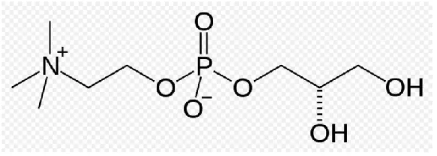
Choline alphoscerate or alpha-glycerylphosphorylcholine (ATC code N07AX02) (GPC) (Figure A) is a semi-synthetic derivative of lecithin. Following oral administration, it is converted to
phosphorylcholine, a metabolically active form of choline able to reach cholinergic nerve terminals where it increases acetylcholine synthesis, levels and release (Amenta et al., 2008).
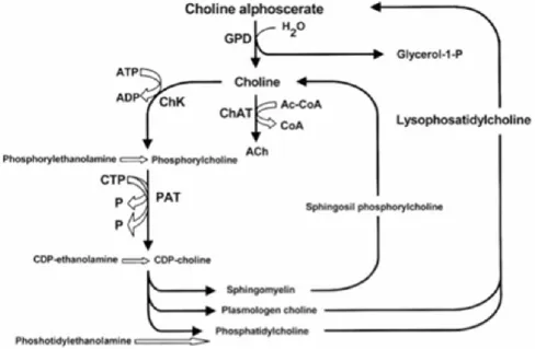
Figure B summarizes acetylcholine anabolic pathways (Amenta et al., 2008).
As shown, the enzyme phosphorylcholine diesterase transforms alpha glyceryl-phosphorylcholine into a molecule of choline and another of glycerol-1-phosphate.
Figure B: Acetylcholine synthetic pathways . Interference of choline-containing compounds.
This figure shows the steps in which choline alphoscerate can influence neurotransmitter biosynthesis. Cythidin diphosphate (CDP); Cythidin triphosphate (CTP); Glyceryl-phosphorylcholine diesterase (GPD); Choline acetyltransferase (ChAT); Choline kinase (ChK); Phosphocholine cytidyl transferase (PCT).
Mechanisms of action of choline alphoscerate are mainly two. In fact the compound interferes with brain phospholipid metabolism and increases brain choline and acetylcholine levels and release (Amenta et al., 2008; Amenta et al., 2001).
Pharmacodynamic studies on choline alphoscerate during phases of development of the compound were focused primarily on its role in potentiating brain cholinergic neurotransmission and in
interfering with brain phospholipid metabolism. Pre-clinical studies have demonstrated that choline alphoscerate increases the release of acetylcholine in rat hippocampus, facilitates learning and memory in experimental animals (Sigala et al., 1992), improves brain transduction mechanisms (Lopez et al., 1991; Schettini et al., 1990) and decreases the age-dependent structural changes occurring in the rat frontal cortex and hippocampus (Amenta et al., 1993). Moreover, the compound
contributes to anabolic processes responsible for membrane phospholipid and glycerolipid synthesis, positively influencing membrane fluidity (Aleppo et al., 1994). In several animal paradigms of impaired cognitive function, choline alphoscerate was demonstrated to improve cognitive deficit in experimental models of aging brain (Canonico et al., 1990; Drago et al., 1990) and to reverse mnemonic deficits induced by scopolamine administration (Parnetti et al., 2007; Sigala et al., 1992). Based on the above evidence, the central parasympathomimetic activity of choline alphoscerate was defined, suggesting its clinical use in patients affected by cognitive decline. Consistently with the activity profile, choline alphoscerate was classified as a centrally acting parasympathomimetic drugs both in international pharmacopoeias (Reynolds et al., 1996) and in the Chemical Therapeutical Anatomical Classification.
A restorative role of choline alphoscerate on central cholinergic system was also documented by studies performed in old rodents. In these investigations the compound was able to counter age-related changes in brain acetylcholine synthesizing (choline acetyltransferase) and degrading (acetylcholinesterase) enzymes (Amenta et al., 1994) and some subtypes of muscarinic cholinergic receptors (Amenta et al., 1994; Muccioli et al., 1996).
Neuroprotective effects of choline alphoscerate were also documented in a rodent model of altered cholinergic neurotransmission caused by lesioning of the Nucleus Basalis Magnocellularis which represents the main source of cholinergic innervation of cerebral neocortex (Amenta et al., 1995; Bronzetti et al., 1993). A neuroprotective effect of treatment with choline alphoscerate on
hippocampus microanatomy and glial reaction which represents an early sign of brain damage was documented in spontaneously hypertensive rats, used as an animal model of brain vascular injury (Tomassoni et al., 2006). A model used to mimic to some extent neuropathological changes occurring in vascular dementia (Tomassoni et al., 2006). Among cholinergic precursors tested, choline alphoscerate elicited the most relevant stimulation on vesicular acetylcholine transporter, and choline transporter in the same model of brain vascular injury suggesting that it represents a strong enhancer of central cholinergic neurotransmission (Tayebati et al., 2011).
Effects of choline alphoscerate were limited not only in rodent models of aging of lesioning of brain cholinergic nuclei, but also in Rhesus monkeys. In this species the compound revealed general facilitatory properties on retinal neurotransmission as well as specific spatial frequency tuning effects on retinal information processing (Antal et al., 1999).
Another series of more recent studies has shown that association of choline alphoscerate with (acetyl)cholinesterase inhibitors potentiates effects on cholinergic neurotransmission. In fact, administration of choline alphoscerate plus the acetylcholinesterase inhibitor rivastigmine induced an increase of brain acetylcholine levels and of high affinity choline uptake binding sites more pronounced than single drugs (Amenta et al., 2006). This investigation has suggested that
combination of a suitable precursor of brain acetylcholine such as choline alphoscerate and of an acetylcholinesterase inhibitor may represent an association worthwhile of being further investigated as a cholinergic replacement therapy in pathologies characterized by impaired cholinergic
neurotransmission (Amenta et al., 2006). This working hypothesis was supported by the demonstration of a more sustained neuroprotective action by choline alphoscerate plus the
acetylcholinesterase inhibitor galantamine than the two drugs administered alone (Tayebati et al., 2009).
Intermediate filaments cytosckeletal proteins
:
One of the main hallmarks of developmental neurobiology is to understand the molecular
mechanisms by which such cellular diversity is generated. Such diversification occurs at an early stage of development, especially by activation of sets of cell type-specific genes, which gives cells distinctive functions and morphological characteristics. Some of these cell-specific genes are the intermediate filaments (IF) protein genes, which are regulated during cell development. The two major IF proteins of astrocytes are vimentin and GFAP. In the course of astrocyte development, a transition in the expression of IF protein genes is observed. Early during development, radial glia and immature astrocytes express mainly vimentin (Bignami et al., 1995). Towards the end of gestation, a switch occurs whereby vimentin is progressively replaced by GFAP in differentiated astroglial cells (Wofchuk et al., 1995; Pixley et al., 1984). At present, there is no consensus on the functional role of these IF proteins. The application of molecular genetic approaches to IF function has been providing some significant insights as well as raising new questions about the functional role of individual IF proteins.
Vimentin
Vimentin IFs are the only IF type found in a variety of cells including astrocytes, fibroblasts, endothelial cells, macrophages, neutrophils and lymphocytes (Evans et al., 14998). Functional analysis of the vimentin gene promoter has already been carried out and several negative and positive elements were identified within this region (Gomes et al., 1999). Data obtained from vimentin knockout mice (-/-) demonstrated that those animals developed and reproduced without presenting an obvious new phenotype, thus heavily calling into question the biological function of vimentin (Colucci-Guyon et al., 1994). Several data, however, argue in favor of a relevant function for vimentin. Using the same vimentin (-/-) lineage as Colucci-Guyon and collaborators found that GFAP filaments were also absent in certain glial cells that normally coexpress vimentin and GFAP
such as the Bergmann glia and an astrocyte subpopulation of the corpus callosum (Colucci-Guyon et al., 1994). This was not due to the inability to express GFAP. Transfection of cultured vimentin -/- astrocytes with a vimentin cDNA restores the vimentin-GFAP filament network, suggesting that in these cells vimentin might be required for coassembly with GFAP filaments (Galou et al., 1996). Reactive gliosis is a prominent result of many types of insult to the central nervous system (CNS) and leads to the formation of glial scar that impedes the regeneration of axons. The intermediate filament protein vimentin is found in pathology of the CNS, mainly in the vicinity of injuries to the CNS. In the present study some authors investigated the role of vimentin in the formation of glial scars in vitro and in vivo by using immunohistochemistry, Western blot analysis, and in situ hybridization. In vitro experiments showed that the intensity of immunofluorescent labeling for vimentin and glial fibrillary acidic protein (GFAP) was consistently decreased in astrocytes after transfection with a retrovirus carrying antisense complementary DNA (cDNA) for vimentin. Transfection also inhibited the growth of astrocytes and decreased the expression of vimentin mRNA. In vivo studies demonstrated that transfection with the retrovirus carrying the antisense cDNA vimentin inhibited the upregulation of vimentin and GFAP in stab wounds in rat cerebrum. These results suggest that vimentin may play a key role in the formation of glial scars in the CNS. Moreover, vimentin appears to accompany the formation of glial scars. Vimentin may stabilize the formation of GFAP-type IF in some reactive astrocytes, and its expression may be required for the formation of GFAP in these cells (Galou et al., 1996). In normal adult CNS, vimentin is not
expressed in astrocytes, but only in some specialized glial cells such as those of Bergmann glia and radial glia, and ependymal cells. These findings suggest that vimentin may take part in the
formation of glial scar, and that there may be a relationship between the expression of GFAP and that of vimentin. During the formation of GFAP networks in some reactive astrocytes, vimentin may act as a cytoskeleton associated protein (Fuchs et al., 1998). At early stages of CNS
development, IF in radial glia and immature astrocytes are composed of vimentin (Bignami et al., 1982). Subsequently, at about the time of birth, a transition from vimentin to GFAP takes place;
vimentin disappears and is progressively replaced by GFAP in differentiated astroglial cells, which transiently coexpress these two proteins (Galou et al., 1996). The transient expression of vimentin observed in the present study has also been observed immunocytochemically in most models of gliosis (Stringer et al., 1996; Norenberg et al., 1994; Schiffer et al., 1986). In the normal adult rodent brain, vimentin expression is restricted to specialized glia such as ependymal cells,
Bergmann glia of the cerebellum, and Schwann cells, which has led to the suggestion that vimentin may be a more specific marker of gliosis than is GFAP (Norenberg et al., 1994; Lenze t al., 1997). Galou founds that the astrocytes in the immediate vicinity of stab wounds expressed considerable GFAP (Galou et al., 1996). However, these cells did not express GFAP after the vimentin gene had been knocked out, whereas in wild-type mice the cells not only expressed vimentin but also
expressed GFAP. These results suggest that the expression of GFAP in these astrocytes depends on the expression of vimentin, and that changes in the expression of vimentin affect the expression of GFAP. In summary the authors Lin and Kai found that the expression of vimentin and GFAP increased markedly after injury to CNS, and that restricting vimentin decreased the expression both of vimentin and GFAP, as well as formation of glial scar. In addition the authors therefore believe that vimentin may play an important role in reactive gliosis and the formation of glial scar.
Accordingly, we suggest that manipulating the expression of vimentin may control reactive gliosis and provide an environment that favours the regeneration of injured axons.
Glial fibrillary acidic protein (GFAP)
Initially isolated from multiple sclerosis plaques, GFAP has been widely recognized as an astrocyte differentiation marker, constituting the major IF protein of mature astrocyte (Eng et al., 1971). The developmental schedule of GFAP expression is not known in detail. In the mouse CNS, GFAP expression has been first detected at the end of gestation (Pixley et al., 1984).Transcriptional studies demonstrated that GFAP mRNA increases between birth and day 15 and then decreases until day 55 (Riol et al., 1992). After reaching a plateau lasting into the second year of adult life, GFAP mRNA and protein levels tend to increase again in some regions such as the hippocampus, striatum and cortex (Laping et al., 1994). This usually corresponds to the increase of reactive astrocytes. Such increase during senescence is one of the most generalized markers for brain aging.
Insights into the role of GFAP have only recently emerged with reports on subtle abnormalities in GFAP-deficient-mice (Gomi et al., 1995; Liedtke et al., 1996).
Recently, Pekny and collaborators have shown that primary cultures of GFAP-/- astrocytes
exhibited an increased final cell saturation density (Pekny et al., 1998). Those results led the authors to speculate that the loss of GFAP expression observed focally in a proportion of human malignant gliomas might reflect tumour progression to a more rapidly growing and malignant phenotype. Some data indicate a novel concept that GFAP might play an important role in the control of neurological disease. Liedtke and collaborators analyzed the astroglial response in GFAP-/- mice with experimental autoimmune encephalomyelitis, a model for multiple sclerosis. GFAP-/- astrocytes presented a disorganized cytoarchitecture due to irregular spacing and a decreased number of hemidesmosomes (Liedtke et al., 1998).
Although there is still some controversy about GFAP function in brain physiology and pathology, a large amount of evidence has been accumulating in the past few years in favour of an active and relevant role for this structural intermediate filament protein in brain development.
GFAP expression during neuron-glia cross-talk
Although some studies have postulated a requirement of GFAP for the formation of astrocytic processes, there is still some controversy about such mechanism. Pekny and collaborators reported that GFAP-deficient astrocytes in the primary cerebellar cultures could form normal processes in response to neurons (Pekny et al., 1998). Thus GFAP might not be a sole prerequisite for the formation of astrocytic processes.
Another remarkable example of the relevance of GFAP expression in neuron-glia interaction regards regeneration of axonal growth after CNS lesions. The GFAP-glial scar formed after NS lesion is considered to be a barrier to regeneration of axon growth.
GFAP synthesis inhibition relieved the blockage of neurite outgrowth that normally is observed after a lesion.
In contrast to this study, Wang and collaborators did not find a correlation between the absence of GFAP and axonal outgrowth (Wang et al., 1997). They reported no increase in axon sprouting or long distance regeneration in the cortical spinal tract fibers of GFAP-/- mice.
Given the relevant role of GFAP during CNS development, as well as a factor in the reactive response to injury, understanding the mechanism of its modulation should be useful to elucidate some steps of NS physiology and pathology.
Several lines of evidence indicate that glia influence the growth, migration and differentiation of neurons but the effect of neuronal cells on astrocytes is far from being well understood. Increasing evidence has been accumulated indicating that neurons are modulators of astrocyte gene expression and differentiation (Gomes et al., 1999; Swanson et al., 1997).
Recently, the authors demonstrated that neurons secrete brain region-specific soluble factors which induce GFAP gene promoter (Gomes et al., 1999).
Furthermore, GFAP is differently modulated by distinct brain regions. It is conceivable that differences in growth factor binding ability of the GFAP gene promoter from different regions could account for such diverse modulation.
Taken together, those results emphasize the complexity of neuron-glia interaction during CNS development and suggest that neurons may modulate the GFAP gene promoter and induce the astrocytic differentiation program. These data argue in favour of the possibility that modulation of intermediate filaments such as GFAP in astrocytes by growth factors might be implicated in cell differentiation as well as in cell-cell interactions during CNS development.
Nestin
Nestin is a class VI intermediate filament protein that was first identified in the progenitor cells that are found during the early developmental stages of the central and peripheral nervous systems (Lendahl et al., 1990). Subsequently, nestin was detected in progenitor cells of non-neuronal tissues, such as skeletal and cardiac muscle (Vaittinen et al., 2001), tooth (Terling et al., 1995), testis (Fröjdman et al., 1997), hair follicle cells (Li et al., 2003), pancreas (Delacour et al., 2004), oval cells in the liver (Koenig et al., 2006) and mammary gland (Li et al., 2007). Owing to its characteristic expression pattern, nestin generally is considered to be a marker of stem or progenitor cells.
In normal rat brain, Nestin occurs in few astrocytes of the brain stem, whereas in reactive astrocytes it has been observed everywhere: in hippocampus by lesions with kainic acid, in hemispheres in experimental ischemia (Duggal et al., 1997; Schmidt-Kastner et al., 1997) and in trauma where Nestin expression increases in time with GFAP (Sahin Kaya et al., 1999). Nestin is expressed in neuroepithelial stem cells, radial glia cells and progenitor cells. It is down-regulated at the onset of GFAP expression during terminal differentiation to mature astrocytes. There are today numerous contributions showing that Nestin is reexpressed in reactive astrocytes where, as an embryonal feature of neuroepithelial cells, it would play a role in their plasticity. This Nestin re-expression seems today well documented by its demonstration following experimental hippocampal lesions (Abdel-Rahman et al., 2004), spinal cord injuries (Frise´n et al., 1995), global cerebral ischemia (Li
et al., 1999), mechanical cortical injury (Krum et al., 2002) and also in human pathology, such as in multiple sclerosis (Holley et al., 2003) or around brain tumours (Almqvist et al., 2002).
The prevailing distribution of GFAP and Vimentin in the thin and long processes in comparison with Nestin is less evident in younger than in older reactive astrocytes, whereas the expression of Nestin in the cytoplasm is highly variable, probably as a consequence of their different durations. It is worth emphasizing that, in line with the above mentioned interpretation, reactive astrocytes with large bodies surrounding tumours can be either intensely positive for Nestin or almost negative, whereas their GFAP and Vimentin expression is more stable.
Further studies will be necessary to characterize fully the proliferating cells in primary cultures of glial cells, but the results obtained in a recent paper reveal a major contribution of the
nestin+/GFAP– cells to the increase in the number of astrocytes, even though nestin+/GFAP+ cells proliferate also. These observations suggested a role of nestin+/GFAP+ cells as NSC or a
contribution of these cells to the formation of glial scar. Interestingly, Ernst and Christie (Rohl et al., 2003) recently showed that the expression of nestin did not predict an active state of
proliferation. Indeed, nestin+/GFAP+ cells present in the unlesioned neocortex are not actively engaged in mitotic activity, although reactive astrocytes that express nestin in response to injury are mitotically active.
Cyclin D-1
Cyclin D is a member of the cyclin protein family that is involved in regulating cell cycle
progression. The synthesis of cyclin D is initiated during G1 and drives the G1/S phase transition.
Once the cells reach a critical cell size (and if no mating partner is present in yeast) and if growth factors and mitogens (for multicellular organism) or nutrients (for unicellular organism) are present, cells enter the cell cycle. In general, all stages of the cell cycle are chronologically separated in humans and are triggered by cyclin-Cdk complexes which are periodically expressed and partially redundant in function. Cyclins are eukaryotic proteins that form holoenzymes with
cyclin-dependent protein kinases (Cdk), which they activate. The abundance of cyclins is generally regulated by protein synthesis and degradation through an APC/c dependent pathway.
Cyclin D is one of the major cyclins produced in terms of its functional importance. It interacts with four Cdks: Cdk2, 4, 5, and 6. In proliferating cells, cyclin D-Cdk4/6 complex accumulation is of great importance for cell cycle progression. Namely, cyclin D-Cdk4/6 complex partially
phosphorylates Rb, which is able to induce expression of some genes (for example: cyclin E) important for S phase progression.
Growth factors stimulate the Ras/Raf/ERK that induce cyclin D production. One of the members of the pathways, MAPK activates a transcription factor Myc, which alters transcription of genes important in cell cycle, among which is cyclin D. In this way, cyclin D is synthesized as long as the growth factor is present.
Even though cyclin D levels in proliferating cells are sustained as long as the growth factors are present, a key player for G1/S transition is active cyclin D-Cdk4/6 complexes. Despite this, cyclin D
has no effect on G1/S transition unless it forms a complex with Cdk 4 or 6.
One of the best known substrates of cyclin D/Cdk4 and -6 is the retinoblastoma tumor suppressor protein (Rb). Rb is an important regulator of genes responsible for progression through the cell cycle, in particular through G1/S phase.
In its un-phosphorylated form, Rb binds a member of E2F family of transcription factors which
controls expression of several genes involved in cell cycle progression (example, cyclin E). Rb acts as a repressor, so in complex with E2F it prevents expression of E2F regulates genes, and this
inhibits cells from progressing through G1. Active cyclin D/Cdk4 and -6 inhibit Rb by partial
phosphorylation, reducing its binding to E2F and thereby allowing E2F-mediated activation of the
transcription of the cyclin E gene and the cell progresses towards S-phase. Subsequently, cyclin E fully phosphorylates Rb and completes its inactivation.
Cyclin D is regulated by the downstream pathway of mitogen receptors via the Ras/MAP kinase and the β-catenin-Tcf/LEF pathways and PI3K. The MAP kinase ERK activates the downstream
transcription factors Myc and AP-1 which in turn activate the transcription of the Cdk4, Cdk6 and Cyclin D genes, and increase ribosome biogenesis. Rho family GTPases and Focal Adhesion Kinase (FAK) activate Cyclin D gene in response to integrin.
Ornithine decarboxylase
The Ornithine decarboxylase (ODC) is an enzyme involved in polyamines metabolism: by
decarboxylation of ornithine, reaction catalyzed by the enzyme ornithine decarboxylase (ODC), is formed putrescine.
The expression of this enzyme is regulated by different stages, by transcriptional and post-transductional levels.
The first reaction consists in the production of putrescine by the ODC enzyme. It requires pyridoxal phosphate as a cofactor and also reducing agents containing thiol groups.
The polyamines are divalent regulators of cell function, promote the growth or cell death depending on the environmental signals and recently it has been shown that the polyamines are also involved in cell cycle regulation.
Recent "in vivo" studies shown that during the cell cycle also occur changes in the activity of ODC and in the concentration of polyamines.
Supporting the correlation between polyamines and cell growth, some reports indicate that high levels of polyamines, resulting in an increase of their synthesis, are present in the cells that make up many solid tumors, where the ODC results to be overexpressed, as well as in different precancerous manifestations and consequently exposure to chemical carcinogens.
Mitogen-activated protein (MAP) kinases
Mitogen-activated protein (MAP) kinases are serine/threonine-specific protein kinases belonging to the CMGC (CDK/MAPK/GSK3/CLK) kinase group. The closest relatives of MAPKs are
the cyclin-dependent kinases (CDKs). MAPKs are involved in directing cellular responses to a diverse array of stimuli, such as mitogens, osmotic stress, heat shock and proinflammatory cytokines. They regulate proliferation, gene expression, differentiation, mitosis, cell survival, and apoptosis - among many others. The first mitogen-activated protein kinase to be discovered was ERK1 (MAPK3) in mammals. Since ERK1 and its close relative ERK2 (MAPK1) are both
involved in growth factor signaling, the family was termed "mitogen-activated".
MAP kinases are found in eukaryotes only, but they are fairly diverse and encountered in all animals, fungi and plants, and even in an array of unicellular eukaryotes. Most MAPKs have a number of shared characteristics, such as the activation dependent on two phosphorylation events, a three-tiered pathway architecture and similar substrate recognition sites. These are the "classical" MAP kinases. But there are also some ancient outliers from the group as sketched above, that do not have dual phosphorylation sites, only form two-tiered pathways, and lack the features required by other MAPKs for substrate binding. These are usually referred to as "atypical" MAPKs. It is yet unclear if the atypical MAPKs form a single group as opposed to the classical ones.
Mitogen-activated protein kinases are catalytically inactive in their base form. In order to become active, they require (potentially multiple) phosphorylation events in their activation loops. This is conducted by specialized enzymes of the STE protein kinase group.
In the case of classical MAP kinases, the activation loop contains a characteristic TxY (threonine-x-tyrosine) motif (TEY in mammalian ERK1 and ERK2, TDY in ERK5, TPY in JNKs, TGY in p38 kinases) that needs to be phosphorylated on both the threonine and the tyrosine residues in order to lock the kinase domain in a catalytically competent conformation. In vivo and in vitro,
phosphorylation of tyrosine precedes phosphorylation of threonine, although phosphorylation of either residue can occur in the absence of the other.
The ERK1/2 pathway of mammals is probably the best characterized MAPK system. The most important upstream activators of this pathway are the Raf proteins (A-Raf, B-Raf or c-Raf), the key mediators of response to growth factors (EGF, FGF, PDGF, etc.); but other MAP3 kinases such as c-Mos and Tpl2/Cot can also play the same role. All these enzymes phosphorylate and thus activate MKK1 and/or MKK2 kinases, that are highly specific for ERK1 and ERK2. The latter
phosphorylate a number of substrates important for cell proliferation and cell cycle progression (RSK kinases, Elk-1 transcription factor, etc.)
CHAPTER 2
AIMS OF INVESTIGATION
1. The first session of the study was specifically designed to assess up and down modulation by exogenous growth factors during interactive crosstalk with endogenous growth factors, released in ACM harvested from different stages of maturation of astrocyte cultures. In particolar, we assessed the effect of of ACM collected from 15, 30, 60 or 90 days in vitro (DIV) on
developing (15 or 30 DIV) cultured astrocytes pre-treated with growth factors (EGF, bFGF, IGF-I or INS).
To clarify mechanisms of astroglial cell proliferation/differentiation in culture, incorporation of [methyl-3H]-thymidine or [5,6-3H]-uridine in cultured astrocytes was assessed. In addition, ERK immunoreactivity in 30 DIV astroglial cell coltures was performed.
2. Because α-Lipoic acid (ALA or thioctic acid ) plays a pivotal role as antioxidant and metabolic component of some enzymatic complexes involved in glucose metabolism of different cell types, and its main function is to increase production of glutathione, the second session of the study was focused to evaluate the effect of ALA R(+)enantiomer or raceme (+/-) on the expression of some proliferation and differentiation biomarkers of astroglial cells in primary culture. In particular, we assessed the expression of GFAP, vimentin, nestin, cyclin D1 as well as MAP-kinase, a signalling transduction pathway biomarker, in 15 DIV astrocyte cultures pretreated or unpretreated with 0.5 mM glutamate for 24h and than maintained under chronic or acute treatment with 50 M R(+)enantiomer or raceme ALA.
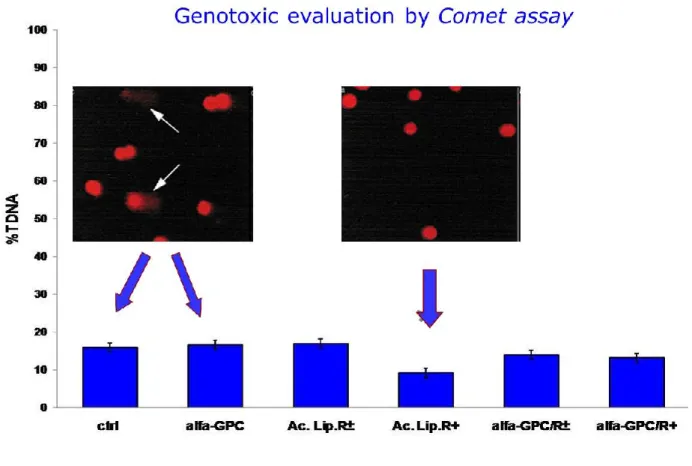
3. The final session of the study was aimed to evaluated the expression of some proliferation and differentiation markers in 15 or 21 DIV astrocyte cultures treated with 50 µM (+)thioctic acid or (+/-)thioctic acid and /or 10 mM GPC for 24h. In particular, it has been evaluated by Western blot analysis the expression of GFAP, vimentin, cyclin D1 Ornithyne decarboxilase and MAP-kinase, a signalling transduction pathway biomarker, in 15 or 21 DIV astrocyte cultures. In addition, it was evaluated the possible genoprotective effect by analysis of DNA status detected by Alkaline Comet assay.
CHAPTER 3
EXPERIMENTAL PROCEDURES
Experimental procedures for Astroglial Conditioned Medium
Materials
Culture medium (Dulbecco’s Modified Eagle’s Medium; DMEM), fetal calf serum (FCS) and the antibiotics penicillin and streptomycin were purchased from GIBCO-BRL. Plastic dishes were products Falcon (Oxford, USA), whereas radioactive precursors and an enhanced
chemiluminescence kit (ECL-plus) were obtained from Amersham International (Amersham, Buckinghamshire, UK). Glutamine, bovine serum albumin (BSA) and growth factors were
purchased by Sigma (St. Louis, USA). Rabbit anti-GFAP immunoglobulins were a product of Dako (Glostrup, Denmark), rhodamine-labeled (TRITC) affinity-purified goat antibody against rabbit IgGs were obtained from Kierkegard and Perry Laboratories Inc. (Faithersburg, ML, USA), antiphospho-ERK1 antibody was a product of Upstate Biotechnology (Lake Placid, NY, USA). Horse radish peroxidase (HRP)-labeled donkey antirabbit IgGs were purchased by Amersham. Other chemicals were of analytical grade and obtained from commercial sources.
Astroglial cell cultures
Primary astroglial cultures were prepared from newborn albino rat brains (1–2 day-old Wistar strain) as previously described (Bramanti et al., 2008). In particular, cerebral tissues, after dissection and careful removal of the meninges, were mechanically dissociated through sterile meshes of 82 mm pore size (Nitex). Isolated cells were suspended in Dulbecco’s Modified Eagle’s Medium (DMEM), supplemented with 20% (v/v) heat-inactivated foetal bovine serum (FBS), 2 mM glutamine, streptomycin (50 mg/ml) and penicillin (50 U/ml), and plated at a density of 3 9 106 cells/100-mm dish and 0.5 9 105 cells. The low initial plating density of dissociated cells was meant to favour the growth of astrocytes and only a very little oligodendroglial and microglial
contamination. Cells were maintained at 37 C° in a 5% CO2/95% air humidified atmosphere for 2
weeks. The first change of culture medium was made after six days and, thereafter, every three days. Approximately 96% of cultured cells displayed GFAP immunoreactivity confirming their astrocyte nature.Astroglial cells were characterized at 15 DIV, i.e. when confluent, by
immunofluorescent staining with the glial marker, GFAP, as previously reported. All efforts were made to minimize both the suffering and number of animals used. All experiments conformed to guidelines of the Ethical Committee of University of Catania, Italy.
Astroglial Conditioned Media
ACM used were collected from previous experiments.
Material included 15, 30, 60 or 90 DIV 24 h serumdeprived astrocyte cultures stored at –80_C until used. ACM were first centrifuged and then filtered with a sterile 0.2 lmpore to remove
contaminating cells or cell debris. A 8 ml amount of 24 hACMwas added per dish.
Growth factor-treated dishes were previously harvested for 24 h (starvation) before mitogen addition to ensure an optimal baseline starting condition.
Growth factors necessary for culture pre-treatment were added at the following concentrations: bFGF, 5 ng/ml; EGF, 10 ng/ml; IGF-I, 10 ng/ml; INS, 10 lg/ml. Treatments were performed as indicated below.
15 DIV cultures
24 h harvesting (DMEM+BSA), subsequent pre-treatment for 12 h with growth factor (EGF or bFGF or IGF-I or INS) and treatment for 24 h with ACM collected from 30, 60 or 90 DIV astrocyte cultures;
30 DIV cultures
24 h harvesting (DMEM+BSA), subsequent pre-treatment for 12 h with growth factor (EGF or bFGF or IGF-I or INS) and treatment for 24 h with ACM collected from developing 15 DIV, or mature 30, 60 or 90 DIV astrocyte cultures.
DNA and RNA labeling assay
[methyl-3H] thymidine (2Ci/ml culture medium) and [5,6-3H] uridine (10 Ci/ml culture
medium) incorporation into DNA and RNA, respectively, was assayed from growth factor-treated and untreated (control) astroglial cell cultures. Incubation was made for 12 h at 37_C to avoid possible artifacts induced from transient changes in the rate of labeled precursor transport and to ensure an adequate cellular uptake of precursors. After medium removal, dishes were rinsed three times with an ice-cold 0.9% NaCl solution (pH 7.4), and cells were extracted with 1 N perchloric acid for 30 min at 4_C. Acid insoluble material was washed three times with 0.5 N perchloric acid, once with ethanol, and solubilized in 0.3 N NaOH at 37_C for 30 min. Nucleic acids were then extracted as previously described and aliquots were taken for radioactivity measurements.
Radioactivity was expressed as dpm/mg of proteins. Proteins were determined by the method of Lowry and co-workers using bovine serum albumin as standard.
ERK1 immunoblotting
After growth factor treatment, cells were rinsed two times with ice-cold phosphate-buffered saline (PBS) with addition of orthovanadate to inhibit phosphatases.
Cells were then solubilized in a buffer containing 2% SDS, 10% glycerol, 50 mM dithiotreitol and 0.1% bromophenol blue. Samples were put on a SDS/12% polyacrylamide gel and electro blotted onto a 0.2 lm pore-size nitrocellulose film (Hybond C) for Western blotting. Filters were incubated for 2 h at 4_C in PBS with 0.5% Tween-20 (PBS-T) and 5% non-fat dried milk containing
antiphospho-ERK1 antibody diluted 1:10,000. Membranes were washed three times in PBST (3 · 10 min) at room temperature and then exposed to donkey anti-rabbit HRP-labeled IgGs diluted 1:10,000. Blots were washed three times in PBS-T and immunoprecipitate was visualized on a film by an ECL kit. The product of phospho-ERK1 immune reaction was quantified by computerized densitometry (ImageJ software; ImageJ Corporation, USA).
Statistics
Values in the text are expressed as the means ± standard error (SEM) of data obtained from five different dishes. The results of experiments of both DNA and RNA labeling were analyzed
statistically by analysis of variance (ANOVA) followed by Duncan’s multiple range test. Statistical significance was expressed at P values of <0.05, <0.01 and <0.001.
Experimental procedures for thioctic acid experiments
Drug Treatment for thioctic acid chronic or acute treatment
Astrocyte cultures at 15 DIV were mantained under following experimental conditions:
Control untreated astrocyte cultures unpretreated or pretreated with 0.5 mM glutamate for 24h
Chronic treatment with 50 M ALA raceme or R (+)enantiomer -treated astrocyte cultures unpretreated with 0,5 mM glutamate for 24h
Acute treatment with 50 M ALA R(+)enantiomer or raceme -treated astrocyte cultures pretreated or unpretreated with 0.5 mM glutamate for 24h
ALA is a potent antioxidant that is in clinical trials for diabetic neuropathy. Many reagents, when dissolved in growth media, release hydrogen peroxide. This can mystify the study of their
protective effects in cell cultures by producing a pro-oxidant effect. Since catalase breaks down hydrogen peroxide, the freshly dissolved ALA was treated with catalase to remove hydrogen peroxide before applying it to the cultures as follows.
ALA was dissolved in 0.5 M DMSO and then diluted in culture medium at the concentration 1 mM. Catalase (1000 U/ml) was added and the solution was incubated at 37_C for 30 min. This solution was diluted in the culture medium in order to obtained 50 M ALA final concentration and 1:10 in the culture medium in order to obtained 100 M ALA final concentration.
Drug Treatment for thioctic acid and alpha-glyceryl phosphoryl choline
Astrocyte cultures at 15 DIV were mantained under following experimental conditions:
thioctic acid was dissolved in 0.5M DMSO and than diluted in culture medium at the concentration 1mM. Catalase (1000 U/ml) was added and the solution was incubated at 37°C for 30 minutes. This solution was diluted 1:10 in the culture medium in order to obtained 50 µM thioctic acid