U
NIVERSITÀ DEGLI
S
TUDI DI
S
ASSARI
DEPARTMENT OF
BIOMEDICAL
SCIENCES
P
H
D C
OURSE IN
L
IFE
S
CIENCES AND
B
IOTECHNOLOGIES
XXIX C
YCLE
Coordinator: Prof. L
EONARDO
A. S
ECHI
S
TUDIES ON THE
M
ERISTEMATIC AND
E
2
F-D
EPENDENT
G
ENE
E
XPRESSION
IN
Arabidopsis thaliana
P
LANTS
Tutor:
Prof. D
IEGOA
LBANIPhD candidate:
TABLE OF CONTENTS
ABSTRACT...4
LIST OF ACRONYMS AND ABBREVIATIONS ...5
CHAPTER 1. Studies on DHFR/TS promoters in Arabidopsis thaliana ...6
1. INTRODUCTION ...6
1.1 Folates and C1-metabolism in plants ...6
1.2 THF structure and biosynthesis ...6
1.3 Roles of DHFR and TS enzymes in the synthesis of nucleotides ...9
1.4 DRTS genes in higher plants ...10
1.5 DRTS genes in Arabidopsis thaliana ...11
2. STATE OF THE ART AND AIMS OF RESEARCH ...12
3. MATERIALS AND METHODS ...13
3.1 Plant material and plant transformation ...13
3.2 Generation of promoter constructs ...14
3.3 Nucleic acids extraction and qPCR analyses ...16
3.4 GUS assays ...17
3.5 Treatments with cell cycle inhibitors ...18
4. RESULTS ...19
4.1 Molecular characterization of the AtDRTS genes ...19
4.2 Patterns of activity of the three AtDRTS promoters ...24
4.3 The AtDRTS genes are differentially expressed in both meristematic and differentiated tissues ...26
4.4 Intragenic regions regulate the activity of AtDRTS1 in root meristems ...30
4.5 In silico analyses of the AtDRTS promoters reveal distinctive promoter architectures ...33
4.6 The meristematic activity of AtDRTS1 in germinating seeds is cell cycle-regulated...43
4.7 E2F transcription factors are able to repress the activity of the AtDRTS2 and AtDRTS3 promoters ...45
CHAPTER 2. Visualization of the E2F-dependent transcriptional activation in planta ...60
1. INTRODUCTION ...60
1.1 Plant development...60
1.2 The plant cell cycle and its regulators ...61
1.3 Cell cycle and E2F/RB pathway ...63
1.4 The Retinoblastoma protein...63
1.5 The E2F transcription factors in plants ...64
1.6 Synthetic promoters in plant biology...67
2. STATE OF THE ART AND AIMS OF RESEARCH ...69
3. MATERIALS AND METHODS ...70
3.1 Plant material and plant transformation ...70
3.2 Generation of EM35S construct ...71
3.3 GUS assays ...71
3.4 Treatments with inhibitors of: DNA methylation, HDACs, phosphatases and kinases ...72
3.5 Treatments with cell cycle inhibitors ...72
3.6 qRT-PCR analyses on seedlings treated with Trichostatin A ...72
4. RESULTS ...74
4.1 The EM35S promoter shows two distinct patterns of activity ...74
4.2 Role of epigenetic mechanisms on the control of EM35S promoter activity...78
4.3 Phosphorylation/dephosphorylation events affect differently the EM35S promoter activity in the two types of lines ...82
4.4 The constitutive expression of an exogenous activating E2F reveals that only type A lines are E2F-responsive ...85
4.5 The E2F-dependent activation of the EM35S promoter in proliferating cells of type A lines occurs mainly during the G1/S transition...89
5. DISCUSSION ...90
ABSTRACT
This thesis focuses on two studies concerning some aspects of cell proliferation and development in plants. The first part describes additional results completing the characterization of the three AtDRTS genes of Arabidopsis thaliana, which code for bifunctional dihydrofolate reductase/thymidylate synthase enzymes whose activity is fundamental in proliferating cells. These analyses allowed the identification of different
AtDRTS isoforms, some of which are expected to encode monofunctional dihydrofolate
reductases, and revealed common and distinctive patterns of expressions that suggest redundant as well as specific roles of the three genes. The characterization of the AtDRTS promoters revealed distinctive features and an E2F-dependent repression of both AtDRTS2 and AtDRTS3. Moreover, evidence has been obtained that the first intron of AtDRTS2 and the intragenic region containing the second intron of AtDRTS1 play crucial roles in the control of promoter activity in the root meristems. Moreover, analyses conducted within this thesis revealed that the first intron of AtDRTS2 is able to confer strong activity in root apical meristem to a non-meristematic plant promoter. The second part of this thesis describes studies conducted to evaluate the regulation in planta of a synthetic promoter, named E2F-Minimal-35S (EM35S), that is expected to be specifically activated by E2F factors, important regulators of cell cycle progression in both plants and animals. Transgenic Arabidopsis plants harboring a construct in which the synthetic promoter drives the expression of the GUS reporter allowed the detection of the E2F-dependent transcriptional activation in different tissues. Moreover, these plants have been used to investigate the cell cycle-dependent regulation of the EM35S promoter activity as well as the effects of epigenetic mechanisms and phosphorylation/dephosphorylation events.
LIST OF ACRONYMS AND ABBREVIATIONS APH, Aphidicolin
BAM3, β-amylase 3
CDKs, Cyclin-Dependent Kinases CENH3, Centromeric Histone H3 COL, Colchicine
CYCs, Cyclins
DHFR/TS, Dihydrofolate Reductase/ Thymidylate Synthase (DRTS) DPs, Dimerisation-Partner proteins
EM35S, E2F-Minimal-35S eqFP611, Red Fluorescent Protein GEN, Genistein
GUS, β-glucuronidase HDACs, Histone Deacetylases
IME, Intron-Mediated Enhancement OKAD, Okadaic acid
pRB, Retinoblastoma protein
PP1/2A, Protein Phosphatase 1 and 2A RAM, Root Apical Meristem
RBR, Retinoblastoma-Related protein SAM, Shoot Apical Meristem
SFH, Sec14-like gene family STAU, Staurosporine
THF, Tetrahydrofolate TSA, Trichostatin A
CHAPTER 1.
Studies on the DRTS genes of Arabidopsis thaliana
1. INTRODUCTION
1.1 Folates and C1-metabolism in plants
Reactions involving the addition or removal of one-carbon units (C1-metabolism), are essential to all organisms, including plants. The majority of the C1 transfer reactions are mediated by tetrahydrofolate (THF) and its derivatives, commonly named folates or vitamin B9. Folates act as coenzymes in several cellular pathways, including the synthesis of purines and thymidylate, amino acid metabolism, pantothenate synthesis and the synthesis of methionine (Met). Furthermore, because methionine is the direct precursor of S-adenosyl-Met (Ado-Met), folates are indirectly required for the synthesis of molecules such as choline, chlorophyll or lignin, as well as ethylene and polyamines [Cossins, 2000; Hanson and Roje 2001]. THF derivatives are also key compunds necessary to support the massive photorespiratory fluxes that occur in green leaves of C3 plants. Photorespiration, in fact, relies on two THF-dependent enzymes present in leaf mitochondria, the glycine decarboxylase complex (GDC) and serine hydroxymethyltransferase (SHMT) [Oliver, 1994; Douce et al., 2001]. These enzymes use up to the 30% of the folate pool for the conversion of glycine to serine [Gambonnet et al., 2001]. Moreover, folates are also involved in the synthesis of pantothenate (vitamin B5) because the first enzyme of this pathway, ketopantoate hydroxymethyltransferase, uses 5,10-methylene tetrahydrofolate as a cofactor [Smith et al., 2007].
1.2 THF structure and biosynthesis
THF (figure 1) is a tripartite molecule, composed of pterin, p-aminobenzoate (pABA) and glutamate moieties. The one-carbon units at various oxidation levels are attached at N5 of the pteridine ring, N10 of the PABA unit, or bridged between the two nitrogens. Folates are synthesized de novo by plants, fungi, most bacteria and protozoa, whereas in animals folate
Fig. 1 Chemical structures of
tetrahydrofolate and its C1-substituted derivatives [From: Hanson and Gregory, 2002].
In all organisms folates occur predominantly as polyglutamylated molecules, with a short γ-linked chain of glutamyl residues attached to the first glutamate. These polyglutamylated folates are the preferred substrates for most folate-dependent enzymes [Cossins, 2000; Scott et al., 2000]. In plants, the biosynthesis of THF depends on the activity of enzymes that are localized in the cytosol, plastids and mitochondria (figure 2) [Neuburger et al., 1996; Rebeillé et al., 1997].
Fig. 2 Folate synthesis pathway in
plant cells, enzymes are:1, GTP cyclohydrolase I; 2, dihydrofolate aldolase;3, ADC synthase; 4/5, HPPK/DHPS; 6, DHFS; 7, dihydrofolate
reductase/thymidylate synthase; 8, folylpolyglutamate synthetase [From: Hanson and Gregory, 2002].
Cytosolic steps: synthesis of the pterin branch.
The initial reactions of the THF de novo synthesis in plants occur in the cytosol, where dihydropterin (or hydroxymethyldihydropterin, HMDHP) is synthesized from GTP in three steps. In the first reaction, GTP-cyclohydrolase I (GTPCHI) catalyzes the formation of dihydroneopterin triphosphate (DHNTP). Then the triphosphate side chain of DHNTP is removed to produce dihydroneopterin (DHN) in two reactions. First, the pyrophosphate group is detached by a specific nudix hydrolase [Klaus et al., 2005], then a non-specific phosphatase [Suzuki and Brown, 1974] cleaves the remaining phosphate. In the last step, dihydroneopterin aldolase (DHNA) removes the lateral three-carbon side chain of dihydroneopterin to release dihydropterin.
Plastidic steps: synthesis of the pABA branch.
The synthesis of pABA occurs in the plastids and requires the conversion of chorismate to aminodeoxychorismate, mediated by the aminodeoxychorismate (ADC) synthase. ADC is subsequently aromatized to pABA by ADC lyase [Basset et al., 2004].
Mitochondrial steps: synthesis of THF from pterin, pABA, and glutamate moieties.
The final reactions necessary for THF synthesis occur in mitochondria, which contain all the required enzymes. In mitochondria, dihydropterin is firstly activated into its pyrophosphorilated form which is then combined with pABA, resulting in dihydropteroate. These two reactions are catalysed in plants by a single mitochondrial bifunctional enzyme, which possesses the two activity necessary for these two steps: HPPK (hydroxymethyldihydropterin pyrophospho-kinase) and DHPS (dihydropteroate synthase) [Neuburger et al., 1996; Rebeillé et al., 1997]. In plants, DHPS is feedback inhibited by its own product, dihydropteroate [Prabhu et al., 1997]. The next step for THF synthesis is the ATP-dependent glutamylation of dihydropteroate to form dihydrofolate (DHFR) in a reaction catalyzed by dihydrofolate synthase (DHFS), which allows the attachment of the first glutamate to the carboxyl moiety of pABA. The presence of HPPK/DHPS and DHFS enzymes in plant mitochondria, but not in mammal cells, allow plants to be autotrophic for
the folates and appears to be necessary for a correct compartmentation within the cell [Appling, 1991].
1.3 Roles of DHFR and TS enzymes in the synthesis of nucleotides
In all organisms, the enzymes thymidylate synthase (TS) and dihydrofolate reductase (DHFR) are crucial for the synthesis of DNA precursors. In fact, the synthesis of thymidylate (figure 3), catalyzed by thymidylate synthase, requires N5,N10-methylene tetrahydrofolate to methylate and reduce deoxyuridine monophosphate (dUMP) to dTMP, yielding 7,8-dihydrofolate (DHF) as a secondary product. To enable efficient recycling of the resulting DHF, the activity of TS must be linked to the activity of dihydrofolate reductase (DHFR), the last enzyme of the biosynthetic pathway. The role of the DHFR and TS enzymes in the synthesis of DNA precursors highlights the importance of a coordinated regulation of both activities.
Fig. 3 Biosynthesis of thymidylate and role of the bifunctional DHFR/TS enzyme.
TS and DHFR have been described as distinct monofunctional proteins in animals, fungi, metazoa and bacteria, but plants and protozoa possess a bifunctional DHFR/TS enzyme (DRTS). The DHFR domain of the bifunctional enzyme maintains the tetrahydrofolate level by reducting the DHF originating from either the de novo synthesis pathway (monoglutamate form) or the oxidation of THF by the TS activity (polyglutamate form). It is not still clear whether bifunctionality arose independently during plant evolution or derived from a common ancestor shared with the protozoa [Philippe et al., 2000]. Nevertheless, also monofunctional DHFR enzymes have been identified in plants [Toth et al., 1987].
1.4 DRTS genes in higher plants
Plant DRTS genes have been described in Arabidopsis, carrot, soybean and maize [Lazar et al., 1993; Luo et al., 1993; Wang et al., 1995; Cox et al., 1999] but additional DRTS sequences of other species are available through genomic and EST databases, including sequences from several primitive plants and algal species. All the DRTS proteins possess a conserved N-terminal DHFR region separated from the conserved C-N-terminal TS domain by a junctional region of variable sequence which, according to studies in Plasmodium falciparum, has been shown to be essential for TS activity and domain-domain interaction of the bifunctional enzyme [Chaianantakul et al., 2013]. Furthermore, plant DHFR/TS bifunctional enzymes share conserved regions which specifically bind compounds such as metotrexate, dUMP and folates.
Studies on the different species, revealed peculiarities of the DRTS plant genes. Analyses of the 5'-ends of the carrot gene have demonstrated the presence of DRTS isoforms which are expected to have a specific subcellular localization (Luo et al., 1997). These isoforms are encoded from two distinct transcript species with differing lengths. Because the DHFR and TS activity are essential for the biosynthesis of nucleotides, analyses have focused on their importance in proliferating tissues or in tissues that are characterized by endoreduplication events. In situ hybridization analyses carried out in Daucus carota revealed that DcDRTS transcripts are particularly abundant in dividing cells of somatic embryos. In addition, Northern blot hybridization experiments revealed a stronger accumulation of DcDRTS
was found in the root elongation zone and leaves [Cox et al., 1999]. Also a recent investigation of the expression of the four ZmDRTS genes found in the maize genome revealed that all of them are maximally expressed at the beginning of kernel formation [Lian et al., 2015]
1.5 DRTS genes in Arabidopsis thaliana
All the information on the DRTS genes of Arabidopsis thaliana is reported in the TAIR (The
Arabidopsis Information Resource) database at the website https://www.Arabidopsis.org/. Arabidopsis possesses three DRTS genes, called AtDRTS1, AtDRTS2 and AtDRTS3 (or, alternatively THY1, THY2 and THY3), that show a similar genomic organization and are located downstream of three members of the sec14-like (SFH) gene family, which suggests their origin from evolutionary genome duplications [Jiao et al., 2012]. The SFHs are proteins with distinct subcellular localizations and varied physiological functions related to lipid metabolism, phosphoinositide mediated signalling and membrane trafficking.
The AtDRTS1 and AtDRTS2 genomic sequences have been described previously [Lazar et al., 1993] and a gene model has been proposed for AtDRTS3, but information concerning the expression and the regulation of the AtDRTS genes has not been reported so far. The AtDRTS1 gene, annotated as At2g16370 in the TAIR database, is located on the minus strand of chromosome 2. According to the proposed gene model, the gene extends 2774 bp, from position 7088865 to 7091639, and is divided into 10 exons that give rise to a transcript of 1924 bp. The predicted ATG start codon is located in the second exon and the resulting coding region translates into a protein of 519 aa with a MW of 58.1 KDa. The AtDRTS2 gene, annotated as At4g34570, based on the gene model spans 3310 bp on the minus strand of chromosome 4, from position 16511006 to 16514316 and contains 12 exons resulting in a 1926 bp transcript. The ATG start codon is found at the end of the second exon and the predicted coding region translates into a protein of 565 aa with a MW of 63.2 KDa. Finally, the AtDRTS3 gene, with annotation At2g21550, is located on the plus strand of chromosome 2 and the proposed gene model is divided into 10 exons and extends 2980 bp, from the ATG triplet at position 9234289 to the TAA stop codon at 9237269. The predicted transcript includes an open reading frame of 1476 bp that is expected to code for a protein of 492 aa with a MW of 55,3 KDa.
2. STATE OF THE ART AND AIMS OF RESEARCH
This part of my thesis describes additional data concerning the molecular characterization of the three DRTS genes of Arabidopsis thaliana, named AtDRTS1, AtDRTS2 and AtDRTS3. These genes play a major role in the biosynthesis of DNA precursors and, as a consequence, are expressed in dividing cells. Previous analyses conducted by our research group [Ghisaura, 2010; Marche, 2013] have demonstrated that the three DRTS promoters present differential profiles of activity. The results obtained, in fact, showed that the AtDRTS1 promoter was highly active in vascular tissues but, unexpectedly, not in root meristems. As for the AtDRTS2 promoter, a strong activity in both root and shoot apical meristems has been observed, whereas the AtDRTS3 promoter presented a meristematic expression in the shoot apex but not in the root, were strong activity has been detected only in the columella and in the central cylinder and not in the apical meristem. Furthermore, in silico analyses, allowed to identify several regulatory cis elements which have been reported to be involved in gene regulation in proliferating cells. Studies had been focusing in particular on the E2F sites contained in the
AtDRTS2 and AtDRTS3 promoters and on the HEXAMER site of the AtDRTS2 promoter.
Mutation of the E2F cis-elements increased considerably the activity of both promoters, suggesting that the E2F transcription factors act as repressors of AtDRTS2 and AtDRTS3, whereas mutation of the HEXAMER site determined a decrease of the AtDRTS2 promoter activity, suggesting an activating role of this cis element. Moreover, other analyses revealed also the importance of the first intron of AtDRTS2 for the expression in proliferating cells and it was shown that this intron is able to confer activity in root meristems not only to AtDRTS2, but also to the AtDRTS1 promoter [Marche, 2013].
In this respect, one of the aims of this part of my research project was to expand the molecular characterization of the AtDRTS genes, analysing the presence of different isoforms and extending the analysis in silico of the promoters. Experiments explaining contradictory aspects concerning the meristematic activity of the AtDRTS1 promoter were also carried out. Moreover, the suggested E2F-dependent repression of the AtDRTS2 and AtDRTS3 promoters was further investigated. Finally, additional studies concerning the first intron of AtDRTS2
3. MATERIALS AND METHODS
3.1 Plant material and plant transformationFor germination and growth in aseptic conditions, wild type or transgenic Arabidopsis
thaliana ecotype Columbia seeds were surface sterilized for 8/10 hours in 2% v/v PPM®
(Plant Preservative Mixture, Plant Cell Technology) supplemented with 50 mg/L magnesium salts (MgSO4). Seeds were imbibed for 2 days in 0,1 % agarose at 4°C in the dark and then germinated on petri plates containing MS salts (Duchefa Biochemie), supplemented with Sucrose (10g/l) and Phyto agar (8g/l) (Duchefa Biochemie) and incubated in a growth cabinet at 22°C under long day conditions of 16 h of light and 8 h of dark .
The transgenic Arabidopsis lines used in this study were generated by the floral dip method [Clough and Bent, 1998] using Agrobacterium tumefaciens EHA105 strain [Koncz and Schell, 1986].
For transformation, a colony of Agrobacterium containing the recombinant plasmid has been picked up to inoculate 4 ml of YEP medium(Bactotryptone 10 g/L; Yeast extract 10 g/L; NaCl 10 g/L adjusted at pH 7 with NaOH) containing the selection agents Kanamycin 40 mg/l and Rifampicin 50 mg/l , which are specific for the plasmid and for the Agrobacterium strain respectively. The culture was incubated O/N at 28°C with gentle shaking and then used to inoculate 400 ml YEP medium. After a further incubation at 28°C O/N, the culture was ready to transform plants. Each culture was transferred into 50 ml conical tubes, centrifuged at 4000 rcf for 7 minutes, at 4°C and the liquid poured away leaving a pellet. Infiltration media was prepared as following 50 g/l sucrose, 400 μl/l silwet L-77, and kept cold. A small amount of infiltration media was first added to the tubes to resuspend the Agrobacterium cells and then the remaining was added up to 500 ml to perform the floral dipping. Plants were dipped into infiltration media for 45 secs, placed on their side in a plastic bucket for 24 hours and left at RT. The transformed plants have then been transferred in a growth chamber to grow to maturity.
Transformed T1 and progeny plants were selected on MS plates containing the resistance antibiotic (Hygromycin, 10 mg/l or kanamycyn, 40 mg/l). At two weeks of age, the resistant plants were transferred to recovery plates and grown for one more week in aseptic conditions without the selection agent.
3.2 Generation of promoter constructs
All the primer sequences used for the production of the recombinant constructs are detailed in Table 1.
Table 1 List of the primers used for vector construction. The restriction sites are highlighted in red.
NAME SEQUENCE 5'-3' RESTR. SITE
F16F4 GTCTCTAGAGGTTTAGACTTTTGATGAAAC XbaI F16F5 GGCGGATCCAATGCTTCCCTACACAAAT BamHI T4L20 GTGGGATCCAGTCGCCGTCGTCTCCCGCC BamHI T4L21 AAATCTAGACCATGGTCAGAGTGAATCTACGCA XbaI; NcoI 221.9 Rev TGGACTAGTAGATCTCCCCCGTGTTCTCTCCA SpeI; BglII M13RV GGAAACAGCTATGACCATG
BAM3PRH AAAAAGCTTGCAGCATTCAGGCAGTCCA HindIII BAM3PRB GAAGGATCCTTTGTGTTTGAGAGAAAGA BamHI
SFH7/DRTS1i2 construct
For the production of the SFH7/DRTS1i2 dual reporter construct the region spanning from the start ATG codon of the AtSFH7 gene to the beginning of the third exon of the AtDRTS1gene was amplified by PCR from Arabidopsis thaliana genome, using high fidelity Pfx Taq polymerase (Invitrogen). This amplification was performed using the primers F16F4 (which anneals next to the AtSFH7 start codon, at position -1420 relative to AtDRTS1 transcription start, and creates a terminal XbaI site) and F16F5 (which anneals at the beginning of the
AtDRTS1 third exon, at position +760 relative to AtDRTS1 transcription start, and creates a BamHI site). The resulting DNA fragment was XbaI/BamHI digested and cloned into
pBlueScript-KS plasmid (also digested with XbaI/BamHI ), giving rise to the F16F45 plasmid, which has been sequenced to verify the fidelity. The F16F45 plasmid was then digested with XbaI/BamHI and the resulting fragment cloned into the SFH7/DRTS1 plasmid (digested with XbaI/BglII) [Ghisaura, 2010], replacing the fragment which comprises the intergenic region spanning from the start codon of the AtSFH7 gene to the ATG codon of
AtDRTS1 (located at the beginning of the second AtDRTS1 exon). In this dual reporter
BAM3/DRTS2i1 construct
The β-amylase 3 (BAM3) promoter (TAIR accession number: At4g17090) was isolated by PCR from A. thaliana genome with high fidelity Pfx Taq polymerase (Invitrogen), using the BAM3PRH primer, which includes a HindIII restriction site and pairs at position -794 respective to the BAM3 transcriptional start, together with the BAM3PRB primer, which anneals at position +9 respective to the BAM3 transcription start and contains a BamHI site. The resulting fragment was HindIII/ BamHI digested and cloned into the HindIII and BamHI sites of the TL2021 plasmid, positioning the BAM3 promoter upstream of the AtDRTS2 5'-UTR, giving rise to the B3TL2021 plasmid. The BAM3 promoter/AtDRTS2 5'-UTR region was then isolated digesting the B3TL2021 plasmid with HindIII and NcoI and cloned upstream of the GUS reporter gene into the pBI221.9 plasmid (cut HindIII/NcoI) producing the pBI221.9/B3TL2021. Finally, the pBI221.9/B3TL2021 was digested with HindIII and
EcoRI to isolate the BAM3 promoter/AtDRTS2 5'-UTR/GUS fragment which was cloned into
the HindIII and EcoRI sites of pBI121 binary vector, thus producing the BAM3/DRTS2i1 reporter construct.
BAM3 construct
For the production of the BAM3 reporter construct, firstly the B3TL2021 plasmid was digested with HindIII and BamHI and the resulting HindIII/BamHI fragment was cloned into the polylinker of a pCambia 1301 binary vector cut HindIII/BamHI, producing the pC13/BAM3 construct. The pC13/BAM3 plasmid was then digested with HindIII and NcoI to obtain a BAM3 promoter fragment suitable for cloning into the HindIII and NcoI sites of the pBI221.9 vector, upstream of the GUS reporter gene, to give rise to the pBI221.9/B3 plasmid. Finally, the BAM3 promoter/GUS region of pBI221.9/B3 was isolated as a HindIII/SacI fragment and cloned into the HindIII/SacI sites of the pBI121 binary vector, thus giving rise to the BAM3 reporter construct.
DRTS2i1/M35S construct
For the production of the DRTS2i1/M35S reporter construct the plasmid pGemT4L20 [Ghisaura, 2010] was digested with PstI and BamHI and the resulting fragment was cloned into the PstI/BamHI sites of the pBI221.9 vector, inserting the 5’-UTR containing the first intron of AtDRTS2 upstream of the -60 CaMV35S minimal promoter/GUS/ Nos PolyA region, thus creating the pBI221.9-DRTS2i1/M35S plasmid. This vector was then digested
with HindIII and EcoRI and the resulting fragment cloned into the pBI121 binary vector (digested Hind III/ Eco RI), suitable for Agrobacterium-mediated plant transformation, giving rise to the DRTS2i1/M35S reporter construct
M35S/DRTS2i1 construct
For the production of the M35S/DRTS2i1 reporter construct the 5'-UTR region of AtDRTS2, containing the first intron, was amplified by PCR (using high fidelity Pfx Taq polymerase-Invitrogen) from Arabidopsis genome using the T4L20 primer, which anneals at the beginning of the AtDRTS2 5'-UTR (at position +5 relative to the transcription start) and introduces a
BamHI restriction site, together with the T4L21 primer, which pairs at the beginning of the
first non-coding exon of AtDRTS2 (at position +222 relative to the transcription start) and contains a NcoI site overlapping a XbaI site. The resulting fragment was digested with
BamHI/XbaI and cloned into pBlueScript-KS plasmid (also cut with BamHI/XbaI), giving rise
to the TL2021 plasmid. Subsequently, the minimal -60 CaMV35S promoter was amplified from the pBI221.9/E2F plasmid [Albani et al., 2000], using the M13-REV universal primer and the 221.9 REV primer (which introduces a SpeI site and a BglII site). The fragment obtained was HindIII/BglII digested and cloned into the TL2021 plasmid (cut with HindIII and BamHI), upstream of the AtDRTS2 5'-UTR. The resulting plasmid, named M35S/TL2021, was then cut with HindIII/NcoI to isolate the minimal -60 CaMV35S/AtDRTS2 5'-UTR fragment, which was cloned into the pBI221.9 vector digested with HindIII/NcoI, upstream of the GUS reporter gene, to give rise to the pBI221.9-M35S/TL2021 construct. Finally, the minimal -60 CaMV35S/ AtDRTS2 5'-UTR/ GUS fragment was isolated by digestion with
XbaI/SacI and cloned into the XbaI and SacI sites of pBI121 binary vector, suitable for Agrobacterium-mediated plant transformation, thus producing the M35S/DRTS2i1 reporter
construct.
3.3 Nucleic acids extraction and qPCR analyses
Total RNA extractions were performed using the Qiagen RNeasy mini-kit. The RNA samples were digested with DNase I during the extraction using the Qiagen RNase-free DNase set, to
SuperScript® III Reverse Transcriptase with a combination of hexamers and oligo dT primers. Quantitative real-time PCR was performed on the BioRad iCycler iQ ™, using the Qiagen QuantiTect SYBR® Green PCR Kit. Triplicate PCR reactions have been performed, following the manufacturer's recommended amplification conditions. For all the analyses the amplification 18S RNA has been used as a reference for normalization. Quantification was calculated following the ΔΔCt method. The PCR primers were designed using the Primer3 online software (http://primer3.ut.ee/) and all their sequences are detailed in Table 2.
Table 2 List of the primers used for qRT-PCR analyses.
NAME SEQUENCE 5'-3' RT-DRTS1-F AAGTGTCGCCATTGAAATCC RT-DRTS1-R GCGAGTTTTCTGGAGAGGTG RT-DRTS2-F GAACAAGATCGCAGACGTGA RT-DRTS2-R ATGCCACATGTTTGCACAGT RT-DRTS3-F CACATGGCACGCTTATATCG RT-DRTS3-F TCTAGCTGCCACAACATTGC RT-18S-F CCTGCGGCTTAATTTGACTC RT-18S-R TTAGCAGGCTGAGGTCTCGT 3.4 GUS assays
Histochemical detection of GUS activity was performed on transgenic plants using 5-bromo-4-chloro-3-indolyl- β-D-glucuronide (X-Gluc) [Jefferson et al., 1987]. Plants at different developmental stages were incubated overnight at 37 °C in the GUS solution (50 mM pH 7 phosphate buffer, 1 mg/mL X-Gluc, 1 mM potassium ferricyanide). After staining, chlorophyll interference was removed treating the samples in 70% ethanol.
For quantitative analyses, the level of GUS activity was detected fluorimetrically using the fluorogenic substrate MUG (4-methyl umbelliferil–glucuronide). Seedlings of the same developmental stage were ground in GUS extraction buffer (50 mM NaPO4 pH 7, 10 mM EDTA, 0.1% Triton, 0.1% Sodium Lauryl Sarcosine, 10 mM β-Mercaptoethanol). An aliquot of 44 μl of the extracts was added to 396 μl of assay buffer (50 mM NaPO4 pH 7, 10 mM EDTA, 0.1% Triton, 0.1% Sodium Lauryl Sarcosine, 10 mM b-Mercaptoethanol, 1mM MUG) and the reactions were incubated at 37 °C. At four different time points, 100 μl of the reaction mix were added to 900 μl of stop buffer (0.2 M Na2CO3) and the amount of 4MU produced was measured using a fluorimeter (BioRad). The protein concentration of each extract was assayed using the Bradford method [Bradford, 1976] to allow calculation of the specific GUS activities.
3.5 Treatments with cell cycle inhibitors
To perform the treatments with cell cycle inhibitors, 30 seeds of selected homozygous transgenic lines harbouring the SFH7/DRTS1i2 construct were imbibed in sterile water alone (as control) or in water containing 5 µg/ml aphidicolin (Fisher Scientific) or 5 mg/ml colchicine (Apollo Scientific). After 72 h of imbibition in growth chamber at 22 °C under a regimen of 16 h of light at and 8 h of dark, proteins were extracted and fluorimetric assays of GUS activity were performed.
4. RESULTS
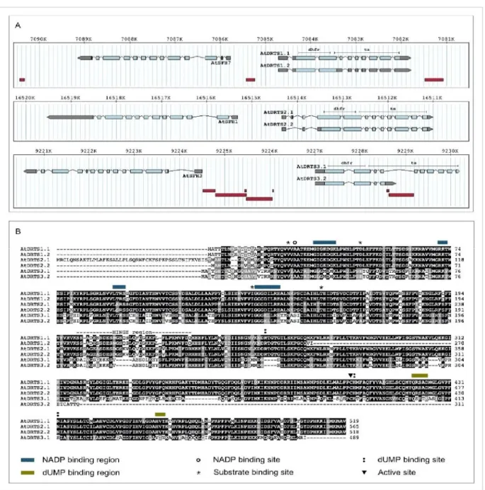
4.1 Molecular characterization of the AtDRTS genes
Database sequences and experimental analyses of the transcripts revealed the existence of at least two isoforms of each AtDRTS gene, some of which are potentially coding for truncated proteins lacking most of the TS domain (Fig. 4A and B). In this respect, although the genomic structure of AtDRTS1 and AtDRTS2 was supported by cDNA sequences, cDNA clones confirming entirely the predicted gene model of AtDRTS3 have not been reported. The only
AtDRTS3 cDNA sequence available in databases (accession number BX820604) confirms the
predicted position of the first three exons but extends the fourth exon into part of the following intron, that contains a transposon-like element in which an in frame stop codon interrupts the coding sequence (Fig. 4A). Thus, it appears that the presence of the transposon-like element in the fourth intron of AtDRTS3 can cause premature termination of the primary transcripts and yields a mRNA that retains part of the fourth intron and codes for a protein of 311 aa, with a predicted mass of 35 kDa, that is expected to possess only DHFR activity. However, although AtDRTS3 cDNA sequences including all the TS coding region have not been reported, microarray analyses (ATH1 Probe Set 263546_at) suggested the expression of transcripts spanning over the 3' end of the putative AtDRTS3 gene model. To verify whether full length AtDRTS3 transcripts corresponding the proposed gene model can be actually produced, RT-PCR reactions were performed using a forward primer that overlaps the ATG start codon in the first exon and a reverse primer that overlaps the predicted TAA terminating triplet, which is located in the tenth exon of the gene. These RT-PCR reactions were performed with high fidelity Taq polymerase, using retrotranscribed RNA isolated from
Arabidopsis seedlings, and allowed the amplification of a cDNA containing the entire
predicted coding region of the AtDRTS3 gene model. Although the resulting sequence did not show any nucleotide change compared to the exonic sequences reported in the TAIR database, the 5' splicing site of the sixth intron appears to occur 9 bp upstream of the predicted one and yields a mRNA that is coding for an AtDRTS3 protein of 489 aa, with a predicted MW of 54.9 kDa that is slightly smaller than the protein proposed by the gene model. Thus, in spite of the transposon element in the fourth intron, it appears that the AtDRTS3 gene can give rise to a full length transcript encoding a bifunctional DHFR/TS protein (Fig. 4B). Based on these results, the large isoform, corresponding nearly exactly to the gene model, has been called
intron and coding for a protein that lacks most of the C-terminal TS domain. Interestingly, a similarly truncated protein appears to be encoded also by an alternatively spliced transcript of
AtDRTS1 corresponding to the cDNA clone reported in databases with the accession number
BX820156. This isoform, called AtDRTS1.2, retains the third intron, containing an in frame stop codon, and the interrupted open reading frame is predicted to code for a protein of 270 aa, with a predicted MW of 30 kDa. Compared to the 519 aa long AtDRTS1.1 protein of 58,1 kDa, the AtDRTS1.2 isoform lacks most of the TS domain and, similarly to AtDRTS3.2, is expected to display only DHFR activity (Fig. 4B). Also concerning the AtDRTS2 gene two isoforms have been detected but both are coding for bifunctional DHFR/TS proteins (Fig. 4A). In this respect, 5’RACE analyses previously performed in our laboratory have revealed the existence of alternatively spliced AtDRTS2 transcripts lacking the second exon that contains the proposed ATG start codon of the gene. Its absence in the alternative transcripts results in the translation of a smaller isoform, named AtDRTS2.2, that begins from the in-frame ATG codon located in the fourth exon originally proposed as a start codon by Lazar et al. (1993).
As described in table 3, a comparison of the larger isoforms of the AtDRTS proteins reveal a close homology between AtDRTS1 and AtDRTS2, showing over 86 % amino acid identity, whereas AtDRTS3 appears to have partially diverged, with 56,8 and 57,4 % identity to
AtDRTS1 and AtDRTS2, respectively. This divergence is further highlighted by comparison of
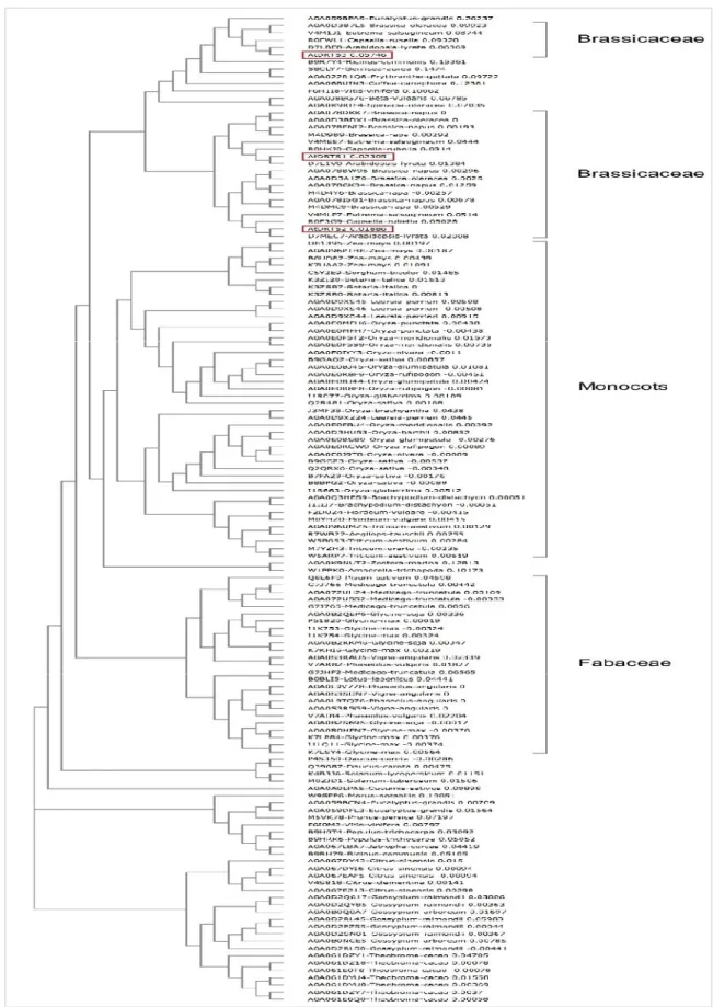
the highly variable hinge region separating the two functional domains, that shows as much as 64,7 % identity between AtDRTS1 and AtDRTS2 whereas for AtDRTS3 shows only 33,3 and 38,7 % identity compared to AtDRTS1 and AtDRTS2. Remarkably, as shown in figure 5, compared with the DRTSs of other angiosperms described in literature and databases,
AtDRTS3 groups together with a subset of the plant DRTS sequences. Moreover, although the
cysteine corresponding to the active site in the TS domain is conserved in all three AtDRTS large isoforms, nearly all the substrate binding sites are perfectly conserved between AtDRTS1 and AtDRTS2 but, as shown in figure 4B, several amino acid substitutions characterize most of the substrate binding sites of AtDRTS3 and could reflect functional peculiarities of this protein.
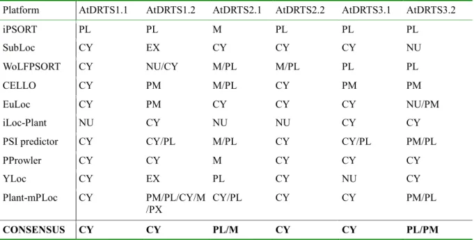
available online. As shown in Table 4, AtDRTS1.1 was predicted to be cytosolic by 8 of the platforms while the truncated AtDRTS1.2 isoform was predicted to be cytosolic by only 5 of the softwares and additional predictions, including cell membrane, chloroplast, and extracellular locations, were proposed by some of the programs. Interestingly, the localization of AtDRTS2.1, which possesses a N-terminal extension that is absent in the other AtDRTSs, was predicted mostly as plastidial and/or mitochondrial whereas the smaller AtDRTS2,2 isoform was largely predicted as cytosolic. For AtDRTS3.1 a prevalence of cytosolic over plastidial localization was reported while the truncated AtDRTS3.2 isoform was predicted more as plastidial or membrane bound rather than cytosolic. Thus, different subcellular localizations of the AtDRTS proteins and of some of their isoforms are likely to occur.
Table 3 Percent Identity Matrix of the AtDRTS large isoforms and of their Hinge region (H).
AtDRTS1 AtDRTS2 AtDRTS3 AtDRTS1/H AtDRTS2/H AtDRTS3/H AtDRTS1 100.00 AtDRTS2 86.85 100.00 AtDRTS3 56.82 57.41 100.00 AtDRTS1/H - - - 100.00 AtDRTS2/H - - - 64.71 100.00 AtDRTS3/H - - - 33.33 38.71 100.00
Table 4 Predicted subcellular localization of the AtDRTS isoforms. The consensus indicates the most
common predicted localization(s).
Platform AtDRTS1.1 AtDRTS1.2 AtDRTS2.1 AtDRTS2.2 AtDRTS3.1 AtDRTS3.2
iPSORT PL PL M PL PL PL SubLoc CY EX CY CY CY NU WoLFPSORT CY NU/CY M/PL M/PL PL PL CELLO CY PM M/PL CY PM PM EuLoc CY PM CY CY CY NU/PM iLoc-Plant NU CY NU NU CY CY
PSI predictor CY CY/PL M/PL CY CY/PL PM/PL
PProwler CY CY M CY CY CY
YLoc CY EX PL CY NU CY
Plant-mPLoc CY PM/PL/CY/M
/PX CY/PL CY CY PM/PL
CONSENSUS CY CY PL/M CY CY PL/PM
Legend: CY = cytosol PL = plastid M = mitochondrion NU = nucleus PX = peroxisome PM = plasma membrane EX = extracellular
Fig. 4 AtDRTSs gene structure and protein isoforms. (A) Genomic organization of the AtDRTS gene
paralogs and of the upstream AtSFH members. The exons are indicated as boxes with the UTR regions shown in gray and the coding portions in light blue. The portions corresponding to the DHFR and TS domains are indicated above the structure of the longest isoforms. The position of transposable elements is shown as dark red boxes below the gene structures. (B) Amino acid sequence comparison of the AtDRTS isoforms. The functional sites are indicated as described in the legend.
The Matrix was created by Clustal2.1 and the sequence alignments were performed using the T-Coffee program (http://www.ebi.ac.uk/Tools/msa/tcoffee/).
Fig. 5 Evolutionary relationships of angiosperm DRTS proteins. The Phylogenetic Tree was created
aligning the aminoacid sequences with Clustal Omega (http://www.ebi.ac.uk/Tools/msa/clustalo/). The branches including monocots, fabaceae and brassicaceae are pointed out. The AtDRTSs are indicated with red boxes.
4.2 Patterns of activity of the three AtDRTS promoters
Previous studies, conducted by our research group, were carried out to define the patterns of expression of the AtDRTS genes by analysing by the activity of their promoters in transgenic
Arabidopsis plants. Considering that the intergenic region upstream of the AtDRTSs contains
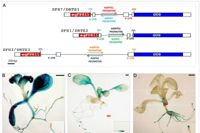
also the promoter of the divergent AtSFH genes, dual reporter constructs had been assembled in which the DRTS promoter was controlling the expression of the GUS reporter gene while a gene coding for the red fluorescent protein eqFP611 [Wiedenmann et al., 2002] was placed under the control of the SFH promoter (figure 6A). The genomic fragments of each intergenic region, spanning from the start codon of the AtSFH gene to the ATG codon of each AtDRTS gene, were amplified by PCR using high fidelity Taq polymerase and used for the production of the dual reporter constructs. All these dual promoter constructs, called SFH7/DRTS1, SFH1/DRTS2 and SFH3/DRTS3, contained the 5’ untranslated region of the genes, which in several cases has been shown to be important for the correct activity of the promoters. Concerning the histochemical GUS analyses of the transgenic lines, the most consistent patterns of GUS staining observed with each construct revealed remarkable differences in the activity of the three AtDRTS promoters (figures 6, B to D).
Interestingly, the AtDRTS1 promoter activity was very strong in both leaf and root vascular tissues but was not detectable in any of the root tips (fig. 6B). As for the AtDRTS2 promoter, it showed a strong activity in the proliferating cells of both root and shoot apical meristems, whereas its activity in differentiated tissues was practically undetectable (fig. 6C). Finally, concerning the AtDRTS3 promoter, it showed activity in the shoot apical meristem but not in the root apical meristems, and its activity in the root was confined to the root columella and the central cylinder (fig. 6D) [Ghisaura, 2010].
Fig. 6 (A) Schematic representation of the dual reporter constructs used to test the activity of the
divergent AtDRTS and AtSFH promoters; The gene coding for the GUS protein is under the control of the AtDRTS promoters whereas the gene encoding the red fluorescent protein eqFP611 is controlled by the AtSFH promoters. Patterns of activity of the (B) AtDRTS1, (C) AtDRTS2 and (D) AtDRTS3 promoter, in two weeks-old seedlings. In the insets is highlighted the pattern of GUS expression in the root apices. Scale bars: 1 mm.
4.3 The AtDRTS genes are differentially expressed in both meristematic and differentiated tissues
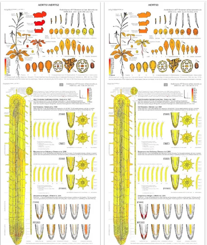
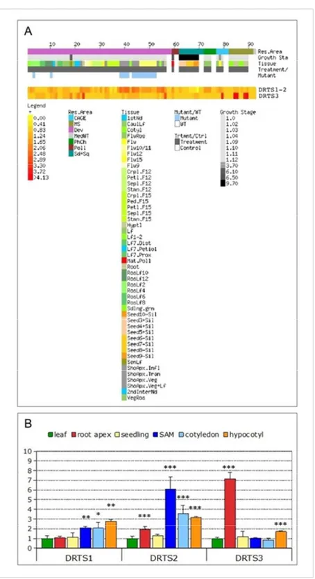
The surprising result that emerged from the previous analyses was the absence of activity of the AtDRTS1 promoter in the root apices. This feature could not be verified analysing microarray data which are reported at the Arabidopsis eFP browser of the Bio-Array Resource (BAR) website (http://www.bar.utoronto.ca/efp/cgi-bin/efpWeb.cgi; Winter et al., 2007) and at the Genevestigator V3 web tool (https://www.genevestigator.ethz.ch/gv/index.jsp; Hruz et al., 2008). These expression data, in fact, are related to experiments performed using the Affimetrix ATH1 array in which AtDRTS3 is represented by a specific probe set (263546_at) whereas AtDRTS1 and AtDRTS2 transcripts are hybridizing to a unique probe set (263601_s_at) and their individual patterns of expression is therefore not distinguishable. Nevertheless, as described in figure 7 and 8A, the expression of the AtDRTS1/AtDRTS2 couple and of the AtDRTS3 gene appear to be very distinctive. More specifically, the strongest signal for the AtDRTS1/AtDRTS2 probe set was detected in the shoot apex and in seeds 24 hours after imbibition, whereas the AtDRTS3 expression level is reported to be very strong in columella and lateral root cap (figure 7), confirming the results obtained from analyses on the transgenic plants, carrying the AtDRTS3 promoter reporter constructs. To further investigate the expression of the AtDRTS genes and to verify whether, in addition to AtDRTS3, also
AtDRTS1 and AtDRTS2 can show distinctive patterns of expression, qRT-PCR analyses were
performed on Arabidopsis seedlings and organs, using pairs of primers which specifically amplify the three AtDRTS cDNAs, as confirmed by sequencing the PCR fragments obtained. To discern the expression of the AtDRTSs in meristematic versus differentiated cells, the analyses were conducted with RNA isolated from root and shoot apexes, as well as leaves, hypocotyls and cotyledons. The relative level of expression of the AtDRTSs in the various organs compared to the leaves was calculated by the ΔΔCtmethod and is reported in figure 8B. These results reveal a remarkably higher expression of AtDRTS3 in the root apex compared to the other organs, which agrees with the high level of expression detected in root caps by microarray analyses. A slight upregulation of AtDRTS3 occurs also in hypocotyls, whereas similar levels of expression compared to the leaves are detected in seedlings, cotyledons and
hypocotils, as well as in the root apices to a lower extent. Conversely, AtDRTS1 shows the strongest expression in hypocotyls and is clearly upregulated also in shoot apices and cotyledons but shows similar levels of expression in the root apices and in leaves. The expression of all the AtDRTS genes in the shoot apex is likely to correlate to different extents with cell proliferation and, at least for AtDRTS1 and AtDRTS2, this correlation probably occurs in the root apex as well. The strong and variable expression of all three AtDRTS genes observed in differentiated tissues could be linked in part to cellular endoreduplication, but is likely to reflect also the involvement of the AtDRTS proteins in additional cellular processes.
Fig. 7 Spatial patterns of accumulation of the AtDRTS1/2 and AtDRTS3 transcripts according to
Microarray analyses.
Fig. 8 Analysis of the expression of the AtDRTS genes. (A) E-Northern analysis of the expression of
the AtDRTS transcripts revealed by microarray data. Heat maps showing the expression levels of the
AtDRTS1/AtDRTS2 common gene set and of AtDRTS3 across different samples were generated using
the Expression Browser tool of the Botany Array Resource (BAR) (http://bar.utoronto.ca/). (B) qRT-PCR analysis of the relative expression levels of the AtDRTSs in representative organs compared to leaves. The qRT-PCR analyses were repeated three times using independent biological replicates and quantification was normalized to 18S RNA levels. The bars show standard errors. *p<0.05, **p<0.01, ***p<0.001.
4.4 Intragenic regions regulate the activity of AtDRTS1 in root meristems
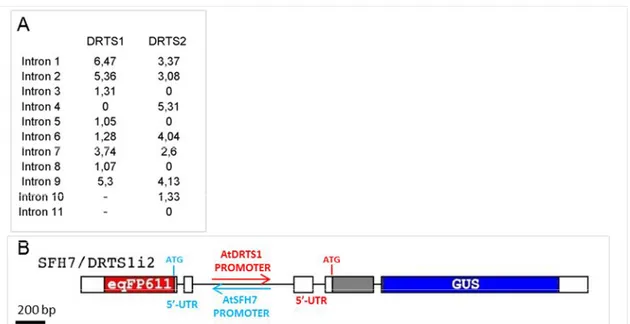
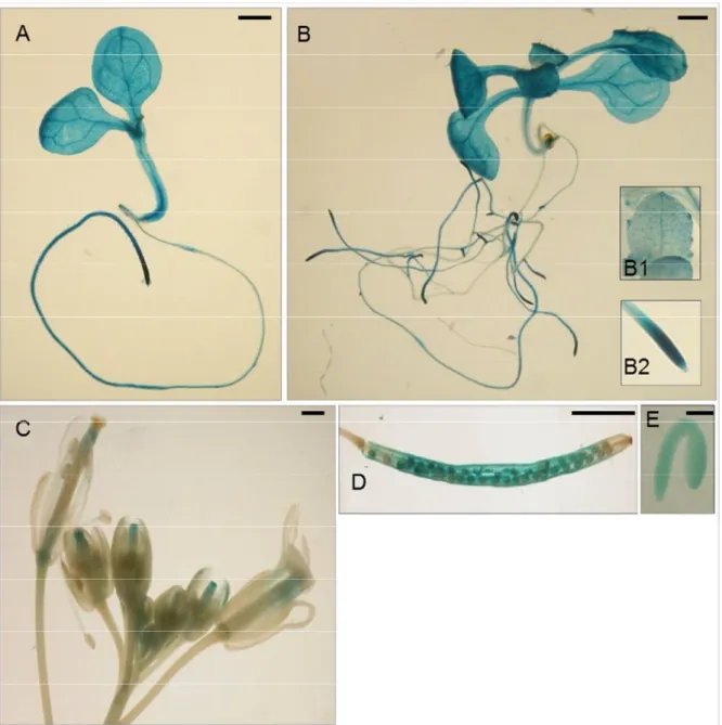
The qRT-PCR analyses suggested that regions required for the correct AtDRTS1 promoter activity in the root apex could be missing in the SFH7/DRTS1 construct. Because all the intergenic region upstream AtDRTS1 was included in the first promoter construct, experiments have been performed as part of this thesis to verify whether intragenic regions could be involved in the regulation of the AtDRTS1 promoter. Several studies have previously reported that some of the introns of various genes are able to strongly influence their expression, an effect known as intron mediated enhancement (IME) [Rose, 2008]. This IME is usually associated to the first intron that could be located in the 5’-untranslated region of the gene, close to the transcription start, but examples of the influence of additional introns, located in the coding regions, have also been described. A software programme, called IMEter (http://korflab.ucdavis.edu/cgi-bin/IMEter_2014/web-imeter2.1.pl), has been used. This software scores the probability of introns to act as IME elements [Parra et al., 2011]. As described in figure 9A, according to the IMEter analysis of AtDRTS1 both the first intron of the gene, located in the 5’-UTR and included in the SFH7/DRTS1 construct, as well as the second intron, located 420 bp downstream of the ATG codon and past the middle of the DHFR coding region, show remarkably high scores. Considering the lack of GUS activity in root apices of the SFH7/DRTS1 transformants and the high IMEter score of the second intron of AtDRTS1, an additional promoter construct was prepared that extends to the beginning of the third exon of the gene. In this construct, called SFH7/DRTS1i2, the GUS coding sequence is fused in frame with a large portion of the amino-terminal DHFR domain of the AtDRTS1 protein (figure 9B). Remarkably, the transgenic plants transformed with this construct revealed a strong GUS activity in the root apices indicating that the AtDRTS1 promoter can drive expression in the root apical meristem only when the intragenic region that includes the second intron of the gene is present downstream of the promoter (Fig. 10A to 10E). Interestingly a similar situation has been described for the CENH3 gene of Arabidopsis, whose expression in root meristems, but not in other meristematic tissues, requires the intragenic region containing the second intron of the gene [Heckmann et al., 2011].
Fig. 9 (A) IMEter analysis of the introns of the AtDRTS1 and AtDRTS2 genes. (B) Schematic
representation of the new SFH7/DRTS1i2 dual reporter construct.
Thus, with the inclusion of the intragenic region, the AtDRTS1 promoter becomes able to drive GUS expression in both apical meristems but is also broadly active in differentiated tissues of the roots, hypocotyls and cotyledons, which show particularly strong GUS staining of the vascular tissues (Fig. 10, D and E). The meristematic activity is already detectable in lateral root primordia. For some of the lines, AtDRTS1 promoter activity can be detected also in trichomes and in hydathodes (Fig. 10, inset B1). In mature flowers, the GUS staining can be detected in the style and ovary as well as in the vascular tissues of stamen filaments, whereas in developing flowers the promoter appears to be strongly active also in the stigmas (Fig. 10C). Moreover, GUS activity is clearly detected also in maturing seeds and in embryos (Fig. 10 D and E). Thus, the AtDRTS1 gene appears to be highly expressed in meristematic tissues but is also very active in various differentiated tissues, in agreement with the pattern of expression detected by qRT-PCR.
Fig. 10 Localization of GUS activity in lines carrying the SFH7/DRTS1i2 construct in: one-week-old
(A) and two-week-old (B) seedlings, which show a strong activity in hydatodes (inset B1) and RAM (inset B2); inflorescence; siliques (D); mature embryos (E). Scale bars: 1 mm in A, B and C; 3 mm in
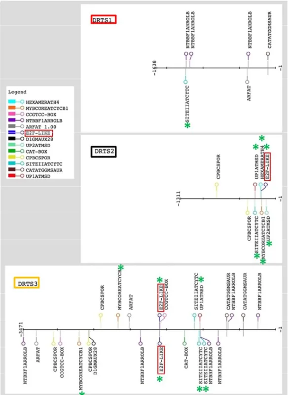
4.5 In silico analyses of the AtDRTS promoters reveal distinctive promoter architectures
The AtDRTS genes, although with variable strength, appear to be all expressed in the shoot apical meristem and common regulatory circuits could be involved in their control in this specific context. However, the different patterns of expression observed in root apical meristems and in other plant organs clearly suggest a distinctive regulation of the AtDRTS promoters. To verify the presence of common as well as specific regulatory elements in the
AtDRTS promoters, in silico analyses were performed searching against the PLACE
(http://www.dna.affrc.go.jp/PLACE/) and PlantPAN (http://plantpan2.itps.ncku.edu.tw/) databases, as well as using the RSAT (Regulatory Sequence Analysis Tools) (http://rsat.ulb.ac.be/rsat/) and JASPAR (http://jaspar.genereg.net/) web platforms. Because the 5’UTR of many genes have been shown to contain functional cis-elements, the analyses were carried out including all the DNA sequences upstream of the ATG start codons. Moreover, the intergenic region upstream of the AtDRTS genes contains also the promoters of
AtSFH genes. Only 1311 bp separate the coding regions of AtSFH1 and AtDRTS2, whereas
the intergenic region upstream of the AtDRTS1 ATG start codon is 1638 bp long and the
AtSFH3 and AtDRTS3 ATGs are separated by 3471 bp. Considering the presence of the two
promoters in the intergenic region, it is not possible to exclude that distant cis elements that are involved in the regulation of the AtSFH genes could be influencing also the activity of the
AtDRTS promoters. Thus, the promoter analyses were performed on the entire intergenic
regions separating the AtSFH and AtDRTS ATG start codons, although it is plausible that putative cis elements that are closer to the AtDRTS genes are more likely to regulate specifically their expression. The outputs of these analyses revealed a distinctive organization of the three AtDRTS promoters, with differences concerning the presence and the distribution of several putative cis elements. Overall, 93 different putative regulatory elements, varying in number and location, could be identified in at least one of the three intergenic regions (Table 5). Interestingly, only 17 of these 93 cis elements are found in all three intergenic regions and only 9 of them are also invariably located, in one or more copies, at positions that are suggesting their possible involvement in the regulation of the AtDRTS genes. 32 of the remaining putative regulatory elements are shared by only two of the intergenic regions: 18 are found in the intergenic regions upstream of both AtDRTS1 and AtDRTS3 and 13 are shared by the intergenic regions upstream of AtDRTS2 and AtDRTS3, whereas only one cis elements is found specifically in the AtDRTS1 and AtDRTS2 upstream regions and in both cases is located closer to the AtDRTS genes. Interestingly, almost half of the putative regulatory
elements (44) are found, in single or multiple copies, in only one of the three intergenic regions. In this respect, 16 cis elements are found specifically in the region upstream of
AtDRTS1, with 11 of them closer to AtDRTS1, and 5 cis elements are found only in the AtSFH1/AtDRTS2 intergenic region, with 3 of them closer to AtDRTS2, whereas 23 putative
elements are found specifically in the AtSFH3/AtDRTS3 intergenic region, 9 of which are also located in positions that are closer to AtDRTS2 (Table 5). Moreover, considering that an intragenic region containing the second intron of AtDRTS1 appears to be required for full activity of the AtDRTS1 promoter in the root apical meristem, additional in silico analyses were performed to verify whether the proposed intron mediated enhancement could be linked to the presence of particular cis elements in the second intron of the gene. 11 different putative regulatory sites are found in this intron, three of which are not found in the intergenic region upstream of the AtDRTS1 coding region (Table 6). Overall, the requirement of intragenic regions for full activity of the AtDRTS1 promoter in root apical meristems and the diversity of the various putative regulatory elements found closer to the AtDRTS genes in the upstream intergenic regions suggest very different architectures of the three AtDRTS promoters, which are likely to be controlled by distinctive transcriptional circuits.
Because all the AtDRTS promoters are able to drive expression in meristematic tissues, I focused my attention on the presence of promoter elements that have been reported to be involved in the control of gene expression in proliferating cells. Moreover, the balance of auxins and cytokinins plays important roles in the control of cell proliferation and cis elements linked to auxin and cytokinin regulation of gene expression were also taken in consideration. The presence and the location of these putative regulatory sites in the intergenic regions upstream of the AtDRTSs is described in figure 15. Because the E2F transcription factors have been reported to regulate genes involved in DNA synthesis and cell proliferation in both plants and animals [Berckmans and De Veylder, 2009], the presence of putative E2F binding sites was investigated in detail. The E2F factors are known to bind specifically a consensus sequence TTTSSCGSS (where S can be C or G) and an E2FAT cis element (TYTCCCGCC) has been reported in the promoters of many potential plant E2F target genes [Ramirez-Parra et al., 2003]. One copy of this element is actually found 199 nucleotides
regions upstream of the AtDRTSs revealed the presence of a putative E2F binding site also upstream of AtDRTS3 but not in the promoter of AtDRTS1. Remarkably, also using less stringent criteria to detect E2F-like elements (TSSCGSS) no additional putative E2F sites could be found in any of the intergenic regions upstream of the AtDRTS genes. Interestingly, the E2F-like cis element of AtDRTS3 is located 1591 nucleotides upstream of the ATG start codon, in the middle of a transposon-like element, and a recent study has reported that E2F sites are relatively common in plant transposable elements [Hénaff et al., 2014].
Other cis elements linked to cell proliferation that are found upstream of some of the AtDRTS genes include UP1ATMSD (GGCCCAWWW), which corresponds to the UP1 motif shown to be over-represented in the promoter of several genes that are up-regulated after main stem decapitation in Arabidopsis [Tatematsu et al., 2005]. This site contains the SORLIP2AT motif (GGGCC), an element over-represented in light-induced promoters of Arabidopsis, and is also overlapping with the SITEIIATCYTC element (TGGGCY), a site involved in the regulation of the Arabidopsis Cytc-1 promoter that is strongly active in root and shoot meristems [Welchen and Gonzalez, 2005]. Remarkably, combined UP1ATMSD/SITEIIATCYTC cis elements are located upstream of both AtDRTS2 and AtDRTS3, 283 bp upstream of the
AtDRTS2 ATG and 1040 bp upstream of the AtDRTS3 coding region. As in the case of the
E2F-like elements, these putative regulatory sites are much closer to the AtDRTS sequences than to the AtSFH genes and could be involved in the regulation of AtDRTS2 and AtDRTS3. Moreover, two additional SITEIIATCYTC elements are found 1054 and 1116 bp upstream of the AtDRTS3 coding region and could also regulate its expression. Interestingly, the intergenic region upstream of AtDRTS1 contains only one SITEIIATCYTC element that is very close to the AtSFH7 coding region (position -1238 bp) and is less likely to be involved in the control of AtDRTS1 expression. Therefore, the activity of the AtDRTS1 promoter in apical meristems is likely to be regulated differently than AtDRTS2 and AtDRTS3. This is also stressed by the fact that the intragenic sequence including the second intron of AtDRTS1 and required for promoter activity in root apical meristems does not contain any of the putative cis elements reported to be involved in gene regulation in proliferating cells. Moreover, specific regulatory circuits could control AtDRTS2 because additional cis elements that are known to be particularly relevant for the control of genes expressed in proliferating cells are found only in its upstream region. One of these cis elements corresponds to UP2ATMSD (AAACCCTA), another motif found in several genes up-regulated after main stem decapitation in Arabidopsis [Tatematsu et al., 2005], that is located at position -130 with respect to the ATG codon and
next to the splice donor site in the first intron of AtDRTS2. The second site is HEXAMERATH4 (CCGTCG), the hexamer motif of Arabidopsis histone H4 promoter [Chaubet et al., 1996], that is located 208 nucleotides upstream of the AtDRTS2 coding region. Finally, a MYBCOREATCYCB1 site (AACGG), known to control the M-phase-specific expression of the Arabidopsis cyclin B1:1 gene [Planchais et al., 2002], is found at position -196 in the AtDRTS2 gene. This putative cis element is also seen twice, although closer to the AtSFH3 gene, in the intergenic region upstream of AtDRTS3 but is not detectable upstream of AtDRTS1 (figure 15).
Concerning the distribution of putative cis elements linked to auxin and cytokinin control of gene expression, sequences of four elements associated to auxin-dependent promoter regulation and of one element associated to cytokinin-dependent expression are found in the intergenic regions upstream of some of the AtDRTS genes. Interestingly, auxin-related cis elements can be detected upstream of AtDRTS1 and AtDRTS3 but none of them is found upstream of AtDRTS2. In particular, upstream and close to AtDRTS1 coding region there is one NTBBF1ARROLB site (ACTTTA), at position -377, one ARFAT site (TGTCTC), at position -392, and one CATATGGMSAUR site (CATATG), 108 bp upstream of the ATG codon. Upstream of AtDRTS3, four of the five NTBBF1ARROLB sites and two of the three CATATGGMSAUR sites are close to the AtDRTS gene and could be influencing its expression whereas a D1GMAUX28 site is close to the AtSFH3 gene and is less likely to be involved in AtDRTS3 regulation. Moreover, the cis element CPBCSPOR (TATTAG), corresponding to a sequence critical for the cytokinin-dependent binding of a nuclear protein to the CsPOR promoter of cucumber [Fusada et al., 2005], is found twice upstream of
AtDRTS2 and three times upstream of AtDRTS3 but is absent upstream of AtDRTS1.
Nevertheless, the three CPBCSPOR sites in the AtSFH3/AtDRTS3 intergenic region are very close to the AtSFH3 coding region and are less likely to be involved in AtDRTS3 regulation whereas one of the two sites found upstream of AtDRTS2 is very close to the AtDRTS coding region, at position -388, and could be involved in the control of its expression.
Fig. 11 Map of the most relevant cis elements identified in the intergenic regions separating the
diverging AtDRTS and AtSFH coding sequences. The E2F sites found upstream of AtDRTS2 and
AtDRTS3 are indicated with red boxes. The map was created using the drawing tool of the RSAT
(Regulatory Sequence Analysis Tools) platform (http://www.rsat.eu/). The green asterisks show
Table 5 Presence and location of cis elements in the SFH/DRTS intergenic region. The distance from
DRTSs ATG codon is reported.
Sites which are closer to AtDRTS than to AtSFH coding regions are shown in red.
Sites associated to cell proliferation, endosperm expression or hormonal responses are highlighted in different colours as reported in the legend.
CIS element Sequence DRTS1 DRTS2 DRTS3
OSE1ROOTNODULE AAAGAT -1607, -1497, -1046, -959, -520, -216 -1244, -1141 -2395,-89-384, MYBST1 GGATA -1061, -974, -718, -465, -454, -122 -543 -2891, -912 REALPHALGLHCB2 1 AACCAA -1372, -925, -837, -800, -730 -633, -592 -2985, -1022, -634, -570, -20 PYRIMIDINEBOXO SRAMY1A CCTTTT -903,-163-314, -313 -3350, -2961, -2879, -2819, -2801, -645 DPBFCOREDCDC3 ACACNNG -1554, -1342,-92 -372 -2709, -865, -666, -270 MYBCORE CNGTTR -810, -773 -293 -3442, -2562, -2358, -2096, -1006, -121, -55 RAV1AAT CAACA -559 -1254, -784 -3442, -2059, -1771, -1556, -1002, -52 SURECOREATSULT R11 GAGAC -393, -75 -204, -143 -3287, -2010,-1232 CCA1ATLHCB1 AAMAATCT -1594, -631 -1075, -979 -1320, -1290,-512 SITEIIATCYTC TGGGCY -1238 -286 -1116, -1054,-1043 CCA1-B AGATAYR -1302 -492 -2392, -238 MYCATERD1 CATGTG -1343, -688 -524, -372 -667 -300CORE TGTAAAG -128, -179 -1246 -386 PREATPRODH ACTCAT -1524 -1300, -1175 -823 ACGTATERD1 ACGT -389 -156 -799, -213 SEF1MOTIF ATATTTAWW -591 -835, -625 -1384, -1302 SEF3MOTIFGM AACCCA -257 -747 -3204 LTRE1HVBLT49 CCGAAA -900, --663713, -301 -868, -582, -3327, -2857,-2524,
SP8BFIBSP8BIB TACTATT -625, -603 -2566, -2452, -1242, -840 GT1CORE GGTTAA -802 -3466, -3413, -1791, -1720, -1134, -632 CIACADIANLELHC CAANNNNATC -126 -3223, -1408, -1025, -396 PYRIMIDINEBOXH VEPB1 TTTTTTCC -1544 -2803,-831-878, AACACOREOSGLUB 1 AACAAAC -1368 -2336, -1865,-1268 LTRECOREATCOR1 5 CCGAC -430 -2182, -1984,-347 ARFAT TGTCTC -392 -3286, -2010 GAREAT TAACAAR -1428 -3192, -1864 CATATGGMSAUR CATATG -108 -673, -446 LECPLEACS2 TAAAATAT -1162 -2588, -1640 IBOX GATAAG -1417, -332 -1910 WBOXNTCHN48 CTGACY -189, -99 -718 CANBNNAPA CNAACAC -89 -543 S1FBOXSORPS1L2 1 ATGGTA -1493 -2622 SV40COREENHAN GTGGWWHG -464 -435 MYBPLANT MACCWAMC -728 -3110 CBFHV RYCGAC -774,-69-268, -1984, -959, -891, -347 CPBCSPOR TATTAG -811, -388 -3050, -2569,-2403 MYB2CONSENSUSA T YAACKG -293 -2562, -2096, -121, -55 SORLIP2AT GGGCC -310, -287 -1116, -1055,-1044 NAPINMOTIFBN TACACAT -672, -373 -665 RBCSCONSENSUS AATCCAA -915 -3385, -2854, -805, -27 MYBPZM CCWACC -1263 -3152, -3112,-1590 TBOXATGAPB ACTTTG -330 -2212, -1737,-1684 BOXLCOREDCPAL ACCWWCC -1263 -3111, -333 MYBCOREATCYCB1 AACGG -196 -2700, -2165 -10PEHVPSBD TATTCT -345 -1873
UP1ATMSD GGCCCAWWW -283 -1040 E2F-LIKE NNTSSCGSS -199 -1591 TATCCAOSAMY TATCCA -1061, -974,-465, -122 ASF1MOTIFCAMV TGACG -1262, -1108 EMHVCHORD TGTAAAGT -1228, -1179 BOXIINTPATPB ATAGAA -1274, -410 SREATMSD TTATCC -1060, -973 AMMORESIIUDCRN IA1 GGWAGGGT -1504 SURE1STPAT21 AATAGAAAA -1272
MYC2 ELEMENT TCACATG -688
TATCCACHVAL21 TATCCAC -465 REBETALGLHCB21 CGGATA -454 CAREOSREP1 CAACTC -308 ABRE-LIKE BACGTGKM -388 GADOWNAT ACGTGTC -389 BS1EGCCR AGCGGG -336 NRRBNEXTA TAGTGGAT -466 2SSEEDPROTBANA PA CAAACAC -89 BOXIINTPATPB ATAGAA -937 MYB1LEPR GTTAGTT -852 GCN4OSGLUB1 TGAGTCA -218 HEXAMERATH4 CCGTCG -208 UP2ATMSD TELOBOXATEEF1A A1 AAACCCTA -130 CCA1-A AATATCY -2889, -2684,-1478 SEBFCONSSTPR10 A YTGTCWC -3438, -2010 AMYBOX1 TAACARA -3192, -1864 CTRMCAMV35S TCTCTCTCT -2906, -2845
P1BS GNATATNC -3135, -2997 QELEMENTZMZM13 AGGTCA -2973, -1774 PALBOXAPC CCGTCC -2957, -1585 CGCGBOXAT VCGCGB -2711, -1715 HDZIP2ATATHB2 TAATMATTA -2238, -845 ANAERO2CONSENS US AGCAGC -943, -760 WUSATAg TTAATGG -2823 WBBOXPCWRKY1 TTTGACY -2794 ABRERATCAL MACGYGB -2710 D1GMAUX28 ACAGTTACTA -2561 L1BOXATPDF1 TAAATGYA -2364 SORLIP1AT GCCAC -1252 SORLIP5AT GAGTGAG -825 RHERPATEXPA7 KCACGW -797 RYREPEATLEGUMI NBOX CATGCAY -669 AUXIN CYTOCHININ GIBBERELLIN JASMONATE ABA ETHYLENE ENDOSPERM CELL PROLIFERATION
Table 6 Presence and location of cis elements in the intragenic 5' region of AtDRTS1. The distance
from DRTSs ATG codon is reported.
Position from ATG is the bp distance downstream from the ATG codon. Sites located within the second intron of the AtDRTS1 gene are shown in red.
CIS element Sequence Position from ATG DPBFCOREDCDC3 ACACNNG 34 TBOXATGAPB ACTTTG 160 ELRECOREPCRP1 TTGACC 163 GT1GMSCAM4 GAAAAA 186 LTRE1HVBLT49 CCGAAA 248 LTRECOREATCOR15 CCGAC 254 ACGTATERD1 ACGT 263 SEF3MOTIFGM AACCCA 285 GAREAT TAACAAR 348, 391 SREATMSD TTATCC 444 MYBST1 GGATA 445 REALPHALGLHCB21 AACCAA 475, 492, 496 MYBPLANT MACCWAMC 494 BOXLCOREDCPAL ACCWWCC 494