UNIVERSITÀ POLITECNICA DELLE MARCHE FACOLTÀ DI MEDICINA E CHIRURGIA
Corso di Dottorato di Ricerca in
SALUTE DELL’UOMO
Aged rats with different performances at environmental
enrichment onset display different modulation of
habituation and aversive memory
Tesi di Dottorato di
Relatore
Pugliese Arianna
Chiarissimo
Prof. Conti Fiorenzo
Index
1. Introduction
1.1 Aging 1
1.2 Environmental enrichment 4
1.3 Habituation, spatial and aversive memory 6
2. Aim of the study 9
3. Materials and Methods 3.1 Animals and housing conditions 10
3.2 OF 12 3.3 MWM 12 3.4 STPA 14 3.5 Statistical analysis 14 4. Results 15 4.1 Body weight 16 4.2 Habituation 16 4.3 Spatial memory 21 4.4 Aversive Memory 22 5. Discussion 24 5.1 Body weight 25 5.2 Habituation 25 5.3 Spatial memory 26 5.4 Aversive Memory 27 6. Conclusions 28 Acknowledgements 28 References 28
1
1. Introduction
1.1 Aging
Population aging is a phenomenon occurring throughout the world, driven by the decline of natality and the rise of life expectancy. The number of people aged over 60 was estimated to be 962 million in 2017, comprising 13 percent of the global population, and is expected to more than double by 2050 and more than triple by 2100. An even more marked trend involves the population aged 80 or over, that accounted for more than 2 percent of global population in 2017 and is projected to triple by 2050 and to increase nearly seven times by 2100. In Europe, the percentage of persons aged more than 60 reaches 25%, the highest among all other regions (Africa, Northern America, Asia, Latin America and the Caribbean and Oceania) (World Population Prospects: The 2017 Revision, Key Findings and Advance Tables, 2017).
From a biological point of view, the aging process is characterized by a progressive, time-dependent loss of physiological integrity, resulting from alterations occurring at the cellular level and leading to impaired function and increased vulnerability to death. In a recent review, López-Otín and colleagues (2013) proposed nine cellular and molecular disfunctions that can be considered hallmarks of aging, as they contribute to the aging process and together determine the aging phenotype of the organism: genomic instability, telomere attrition, epigenetic alterations, loss of proteostasis, deregulated nutrient sensing, mitochondrial dysfunction, cellular senescence, stem cells exhaustion and altered intracellular communication.
The aging brain
While we age, we experience numerous changes in sensory, motor and cognitive functions which depend largely on brain aging. Brain aging is characterized by several modifications of morphologic and functional brain characteristics. First, it is important to point out that physiological aging is not characterized, as previously thought, by a remarkable neuron death in hippocampus and neocortex of humans, non-human primates and rodents, with some exceptions restricted to specific areas (Burke and Barnes, 2006). On the other hand, a reduction of neurogenesis during aging has been reported across several species including humans (Kempermann et al, 2002; Amrein et al, 2011; Encinas and Sierra, 2012; Spalding et al, 2013; but see Kempermann et al, 2018). From a morphological point of view, regional changes in dendritic branching and spine density have been observed in humans, non-human primates and rodents (Duan et al, 2003; Burke and Barnes, 2006). Furthermore, both anatomical and electrophysiological data showed a decrease in synapse number in the aged rat hippocampus (Burke and Barnes, 2006). At the neuronal level, aging is associated to an increase in Ca2+
2 conductance and a disruption of Ca2+ homeostasis (Landfield and Pitler, 1984; Thibault and Landfield, 1996; Thibault et al, 2001; Toescu et al, 2004) that eventually contribute to plasticity deficits. In fact, impairments in long-term potentiation (LTP) induction/maintenance and an increased susceptibility to long-term depression (LTD) and to the reversal of LTP have been well documented in the hippocampus of aged rats (Barnes, 2003; Burke and Barnes, 2006; Arias-Cavieres et al, 2017). Moreover, alterations in neurotrophic factors (such as brain-derived neurotrophic factor (BDNF), nerve growth factor (NGF) and glial cell-derived neurotrophic factor (GDNF)) levels have also been consistently reported, leading to consequences on neuronal and synaptic plasticity processes (Budni et al, 2015).
These neurobiological alterations likely contribute to generate the physiological cognitive decline experienced, at different extents, in elderly humans and detectable in some aged animals too. Cognitive functions relying on the hippocampus and the prefrontal cortex are especially compromised, being these two regions particularly vulnerable to the aging process. To start, the age-related decline of spatial memory (i.e., memory for spatial configurations and ability to navigate in an environment) across species is very well known (Foster et al, 2012; Lester et al, 2017). Namely, aged humans show deficits in retrieving the contextual details of episodic memories (McIntyre and Craik, 1987; Spencer and Raz, 1995); also, they show impairments in performing spatial navigation tasks (Moffat, 2009; Techentin et al, 2014) as well as non-human primates (Lai et al, 1995; Rapp et al, 1997; Lacreuse et al, 2014), dogs (Head et al, 1995), rats (Barnes, 1979; Markowska et al, 1989) and mice (Bach et al, 1999). Another well-experienced age-related cognitive alteration is recognition memory impairment. If we distinguish two components in this process, i.e., recollection (recalling specific details of an experience, such as where and when it happened), and familiarity (having a sense of experience but lacking any specific details), general recollection deficits but unimpaired familiarity can be observed in both aged humans and aged rats. A generic alteration of object recognition ability was reported also in aged monkeys (Leal and Yassa, 2015). Also, discrimination ability, defined as the ability to disambiguate similar experiences to avoid interference, undergoes alterations in various domains during aging, as showed in humans, monkeys and rats (Leal and Yassa, 2015). Partly deriving from spatial memory impairment is the worsening of habituation (a very simple form of learning that consists in the reduction of the response to a novel stimulus as it progressively becomes familiar) occurring in aging rats (Vila-Luna et al, 2012) and mice (Fraley and Springer, 1981). Such impairments in spatial and recognition memory and discrimination ability all contribute to the well-known decline of episodic memory. Less evident, but still documented, cognitive alterations involve associative learning and executive functions. Associative learning
3 generically implies the association between two previously unrelated stimuli and was reported to be compromised by aging in humans, mice, rats and rabbits (Burke and Barnes, 2006); also
aversive memory, a form of associative memory which determines avoidance of a location that
has been associated with an aversive stimulus, is affected in old rats (Lovatel et al, 2013; Leffa et al, 2014) and mice (Flood and Roberts, 1988; Reddy and Kulkarni, 1998). Finally, both aged humans and monkeys are less performing in tasks designed to test executive functions (Burke and Barnes, 2006).
Cognitive reserve, cognitive training and physical exercise
The process of age-associated cognitive decline is extremely heterogenous; this means that while some aged individuals behave quite similarly to healthy adults, others experience cognitive impairments that can vary much in terms of nature and degree (Gallagher et al., 2006; Dixon and de Frias, 2014). Such differences derive from a combination of intrinsic (i.e., genetic) and external (i.e., deriving from the environment) factors, that together shape the aging process. In this context, Cognitive Reserve (CR) plays a key role. The concept of CR has been proposed to explain why individuals characterized by the same extent of brain damage show different clinical manifestations, and posits that the greater is the CR, the higher is the capability to sustain damage before demonstrating functional deficits. CR involves an active attempt of the brain to cope with damage by implementing compensatory processes and recruiting alternative brain circuits; its development has been linked to genetic factors and early societal environment, but also to education, occupational attainment, engagement in leisure activities and physical health (Scarmeas and Stern, 2003; Stern, 2009).
Importantly, engagement in physical and cognitive activities during aging still produces beneficial effects; in fact, interventions based on cognitive and/or physical stimulation applied in aged people were proven to be effective in preventing/contrasting physiological and pathological cognitive impairment (Bamidis et al., 2014). Cognitive interventions can consist in procedures designed to provide general cognitive stimulation (such as structured group activities and discussions) or in standardized sets of exercises that target specific cognitive functions (i.e., cognitive training); similarly, physical interventions can involve activities that require a generic body movement (such as games or dance) or planned and specific exercises. Both type of interventions can include a social component which plays itself an important role in the observed outcome. For example, it was shown that playing strategy videogames (Basak et al, 2008), acting (Noice et al, 2004; Noice and Noice, 2008), and attending a computer course (Klusmann et al, 2010) were effective in improving cognitive performance in healthy elderlies.
4 Moreover, numerous controlled trials proved that also more specific cognitive trainings can exert an amelioration of performances both in the targeted and in other domains (Gates and Valenzuela, 2010). As far as physical activity is concerned, Kattenstroth and colleagues (2013) tested the effects of participating for 6 months in a dance class, and showed that not only motor performance, reaction times and subjective well-being, but also cognitive performance were positively affected. Similarly, designed resistance and aerobic trainings were indeed sufficient to ameliorate cognitive function (Hindin and Zelinski, 2012; Bamidis et al., 2014). Interestingly, some studies reported that the combination of the two strategies (i.e., physical and cognitive stimulation) yielded a greater effect than each strategy alone (Fabre et al., 2002; Oswald et al., 2006).
1.2 Environmental Enrichment
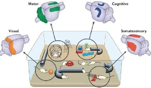
Environmental enrichment (EE) refers to a housing condition that facilitates enhanced sensory, motor, cognitive and social stimulation in laboratory animals, with the effect of boosting exploration, curiosity and attentional processes. For rodents, it is based on the interaction with stimulating objects (e.g. toys, tunnels, wooden sticks) that are frequently changed, voluntary physical exercise (on running wheels and stairs), and increased sociality (animals are reared in large groups) (Figure 1).
Figure 1. Brain stimulation deriving from environmental enrichment. Enhanced sensory stimulation, including
increased somatosensory and visual input, activates the somatosensory (red) and visual (orange) cortices. Increased cognitive stimulation — for example, the encoding of information relating to spatial maps, object recognition, novelty and modulation of attention — is likely to activate the hippocampus (blue) and other cortical areas. In addition, enhanced motor activity, such as naturalistic exploratory movements (including fine motor skills that differ radically from wheel running alone), stimulates areas such as the motor cortex and cerebellum (green). From Nithianantharajah and Hannan, 2006.
5 EE as an intervention was first proposed in the late 1940s by Donald Hebb, who anecdotally observed that rats that he had took home as pets showed behavioural improvements over their littermates housed in the laboratory (Hebb, 1947). In the early 1960s, Rosenzweig and colleagues introduced for the first time EE as an experimental paradigm (Rosenzweig et al, 1962; Rosenzweig, 1966; Rosenzweig and Bennett, 1996). Since then, tens of studies have been showing the positive effects of EE on neurogenesis, dendrite branching, synaptic plasticity and behaviour in both physiological and pathological conditions (van Praag et al., 2000; Nithianantharajah and Hannan, 2006; Patel, 2012; Sale et al, 2014).
Application of EE in laboratory animals is extremely useful to reproduce and understand the effects of physical exercise and complex mental activity in humans (Kramer et al, 2006; Nithianantharajah and Hannan, 2009; Petrosini et al, 2009). Focusing on aging, EE administration from youth or adulthood can be used to mimic the protective effects of cognitive reserve potentiation, while EE starting in old animals can serve as a model for interventions based on physical and cognitive stimulation in aged humans (Sale et al, 2014). To date, a large amount of evidence has been provided on the beneficial effects of EE in preventing or attenuating the age-associated cognitive decline (Mora, 2013; Sale et al, 2014). For example, life-long EE training was able to improve spatial learning and memory (Harati et al, 2011) and recognition memory (Leal-Galicia et al, 2008), prevent the degradation of attention performances (Harati et al, 2011) and decrease anxiety-like behaviours (Leal-Galicia et al, 2008) in aged rats. Interestingly, similar positive effects were obtained also when EE was started from adulthood or even during aging: 10-12 weeks of continuous EE were proven sufficient to reduce the age-related spatial memory decline in aged mice (Bennett et al, 2006) and rats (Kumar et al, 2012), and similar results were obtained with 6 weeks of EE in 21 months old mice (Harburger et al. 2007) and with only 23 days of EE in 27 months old mice (Frick and Fernandez, 2003).
Such effects have been related to EE positive action on hippocampal neurogenesis (Leal-Galicia et al, 2008, Kempermann et al, 2002), neurotrophic factors (BDNF) (Obiang et al, 2011), dendritic length (Kolb et al, 2003), synaptic plasticity (Kumar et al, 2012), and neurotransmitters systems (Frick and Fernandez, 2003; Segovia et al, 2006; Leal-Galicia et al, 2008).
6 1.3 Habituation, Spatial and Aversive Learning and Memory
Habituation
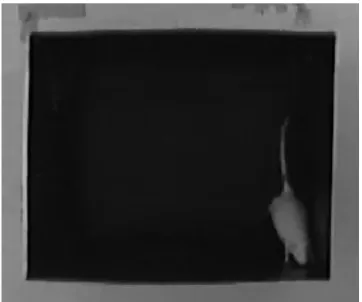
When an animal is repeatedly exposed to a novel stimulus without any biologically relevant consequence, its response to the stimulus progressively decreases. This behaviour is based on a simple form of non-associative learning present in animals of all phyla, known as habituation (Harris, 1943). In rodents, habituation is commonly studied monitoring the reduction of exploration during continued or repeated exposure to a novel environment. An Open Field (OF, i.e., an empty arena delimited by walls) is frequently used as the testing environment (Figure 2).
Figure 2. Rat in an open field. The picture, taken through the camera installed in our laboratory, shows a rat doing
a rearing, i.e., raising on its rear legs.
Initially, exploration is triggered by the necessity to gather information about a number of survival-related factors (e.g., availability of mates, sources of food and nesting, presence of predators and potential escape routes) and also restrained by the innate fear of novelty. As the environment becomes more and more familiar, exploratory behaviour progressively decreases. In these experiments, exploration is commonly measured as distance travelled, line crosses (on the floor of the OF), number of rearings (Leussis and Bolivar, 2006).
Two aspects of habituation can be distinguished and studied: intrasession and intersession habituation. In the former, the progressive decrease in exploration within a single exposition to the new environment is considered; in the latter, the response of the animal when re-exposed (after a variable time interval) to the environment is evaluated. Thus, while intrasession habituation involves only a learning component, intersession habituation requires also memory retention (Leussis and Bolivar, 2006). Consistently with the dependence of habituation in an
7 OF from the formation of a spatial representation in the brain, it has been shown that anatomical or pharmacological disruption of hippocampal functionality impairs habituation (Save et al., 1992; Vianna et al., 2000).
Spatial learning and memory
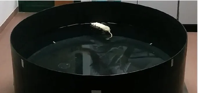
Spatial navigation refers to the process through which animals and humans learn about the spatial configuration of the environment and localize themselves with respect to it. Navigation is based on the use of cues, that can be of the type of dynamic self-motion cues and/or static environmental cues, the former deriving from the perception of self-motion, the latter consisting in stable objects localized in the space and extended boundaries (Lester et al, 2017). Employing such cues, the brain of mammalians assembles a variety of spatial representations or maps (O’Keefe and Nadel, 1978), thanks to the activation of different groups of spatially modulated cell types that fire in response to spatial information (i.e., hippocampal place cells, enthorinal grid cells, head direction cells in several cortical and subcortical structures, enthorinal borders cells, enthorinal speed cells). Such representations can be then transformed into persisting long-term memories to be retrieved when necessary. Spatial learning and memory rely primarily on the hippocampus, as shown by the first lesion studies (Morris et al, 1982), but other brain regions have been shown to be involved (D’Hooge and Deyn, 2001; Chersi and Burges, 2015). In laboratory animals, spatial navigation and spatial memory can be tested through a variety of experimental paradigms. Among them, the most used and known for its high reliability, validity and relative simplicity is for sure the Morris Water Maze Test (MWM). The protocol was first described by Morris in 1981 (Morris, 1981), who showed for the first time that rats are able to learn to locate an invisible platform in a pool exploiting only room distal cues. Since then, the MWM has been applied in a huge variety of experiments and adapted to other species (mice and even humans through virtual mazes (Kallai et al., 2005)), to study the neurobiology of spatial learning and memory but also to validate rodent models for neurocognitive disorders and to study the effect of a variety of pharmacological interventions. The MWM consists in a circular pool filled with water in which a small escape platform that the animal can’t see, hear or smell, is placed in a fixed position. The animal is repeatedly released from various start positions across the days and gradually learns to navigate to the platform relying on distal cues in the testing room; possibly, spatial reference (short or long term) memory can be assessed by evaluating the preference of the animal for the platform area when the platform is absent (Figure 3) (Vorhees and Williams, 2006).
8
Figure 3. MWM apparatus from our laboratory. The rat has been just released from the north point in the black
tank. The transparent submerged platform in the southwest quadrant (bottom left) is barely visible. Aversive learning and memory
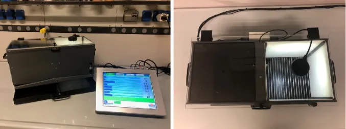
Aversive learning is a form of conditioned learning that takes place when an animal avoids a location where it previously received an aversive stimulus. It is strongly based on emotionality, notably on fear, and includes, for example, the process by which we learn not to touch a hot pot. Aversive learning can be tested in laboratory animals using avoidance tasks, distinguished in active avoidance (when the animal needs to move away from the feared location) and passive or inhibitory avoidance (PA, when the animal learns not to enter the feared location); between them, PA is the most studied. Two variants of PA are applied in rats and mice, giving overlapping results: step-down PA and step-through PA (STPA). In the former, an apparatus consisting of a platform placed upon an electrified grid is used. Initially, the animal is placed on the platform and is allowed to explore the entire apparatus. After one or more habituation trials, the electrified grid is activated and delivers a foot-shock to the animal as soon as it steps down from the platform (training phase). STPA uses, instead, a box divided in one illuminated and one dark compartment, with a metallic grid floor (Figure 4). At first, the animal is placed in the illuminated compartment and, following its preference for the dark, tends to move to the dark one. After one or more habituation trials, during the training session a foot-shock is given once the animal enters the dark compartment. The memory performance is then evaluated after a variable time interval from the training session; in both cases, the ability of the animal to avoid the location where the shock occurred is measured as time passed before stepping down or through. Numerous brain regions are involved and interact in the formation of an aversive memory trace, including hippocampus, baso-lateral amygdala, medial septum, entorhinal,
9 perirhinal, anterior cingulate, retrosplenial, prefrontal and posterior parietal corteces (Izquierdo et al., 2016).
Figure 4. Step-through passive avoidance apparatus from our laboratory. It consists of a shuttle box divided in
two compartments, one illuminated by a lamp and the other one dark for the presence of a black cover. The box is connected to a controller through which lighting condition, door opening and shock characteristics can be set.
2. Aim of the study
A wide agreement exists on the benefits deriving from EE on age-related cognitive impairment. Nonetheless, lack of EE standardization remains an open question as differences in type and duration as well as in the animals’ age at EE onset can significantly influence the results (Simpson and Kelly, 2011). Among all the possible variables, one has not been explored enough, i.e., cognitive performances at EE beginning. A possible, different effect of EE on animals displaying different age-related deficits was firstly proved by Fuchs and colleagues (2016) who evidenced that late enrichment was able to preserve spatial memory only in aged rats that were unimpaired when middle-aged.
To further examine the issue and delve deeper whether animals displaying different cognitive performances might react differently to EE, we applied a 12-week EE protocol to late adult rats pretested for habituation (OF), spatial memory (MWM) and aversive memory (STPA). Age of EE onset was prior to the median life span. Indeed, evidence has been recently provided in mice that the application of EE after the median life span failed to prevent age-related deficits (Bouet et al., 2011), while EE induced positive effects when started before the median life span (Freret et al., 2012). The finding that EE induces different effects on the basis of rats’ behaviour at EE onset unveils the opportunity to pre-define a performance threshold that can allow to optimize the positive consequences and minimize the negative ones.
10
3. Materials and methods
3.1 Animals and housing conditions
Male Sprague Dawley rats of the INRCA (Ancona) colony were used. All animals were bred, for the entire experimental period, under standard and strictly controlled environmental conditions (constant room temperature of 20-22 °C, relative humidity of 50-55 % and artificial light-dark cycle of 12 h) with ad libitum access to food and tap water.
Experimental rats (Exp) were screened by OF, MWM and STPA (basal/baseline) and the day after, at the age of 17 months, semi-randomly assigned -to balance their performances- to continue standard housing (SH) or begin EE. Age-matched naïve animals (N), used as controls to differentiate the effect of housing condition and retesting, were randomly allocated to SH or EE with no pre-screening (Fuchs et al., 2016). EE cages (79x52x140 cm) had three floors, two balconies and four flights; they contained several objects that were changed or moved twice per week to maintain the novelty and encourage foraging behavior (Figure 5). The objects used for the enrichment were carefully selected to respect two mandatory requirements: non-toxic and no possibility of causing wounds or suffocation. Housing density was 8/10 animals per cage for EE and 2 animals per cage for SH.
11
Figure 5. Environmental Enrichment (EE) cage. EE cages had three floors, two balconies and four flights. Plastic
and wooden toys, tunnels, wooden sticks to gnaw, balls, paper nesting material, a running wheel and several feeding boxes and water bottles were disseminated in the whole cage.
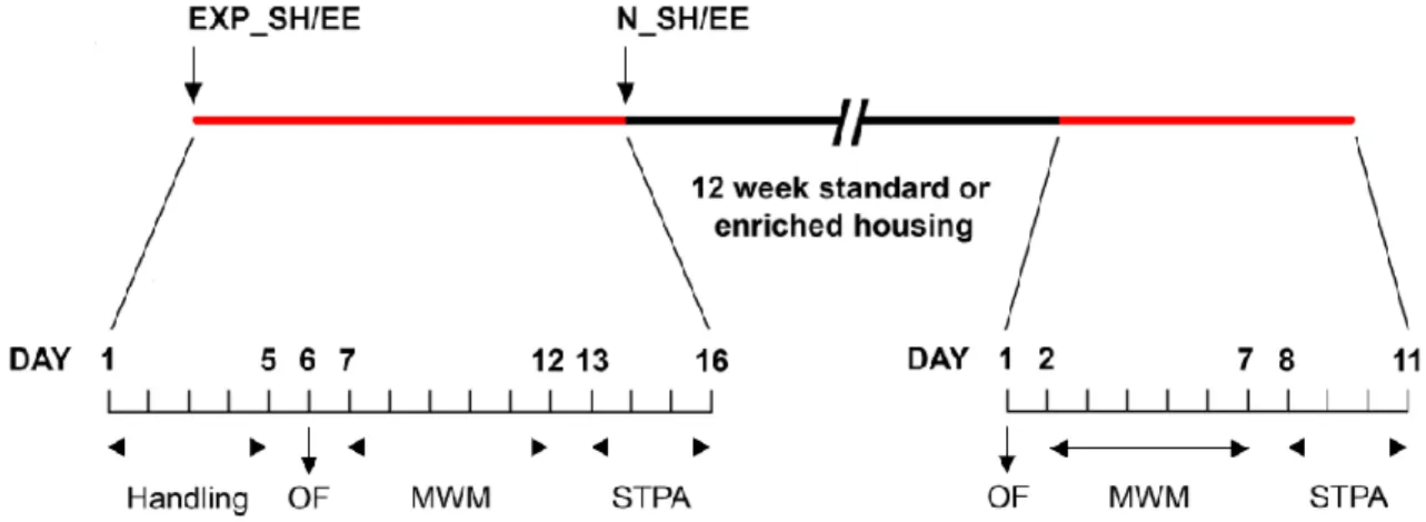
After 12 weeks, the surviving animals (23 Exp_SH (i.e., experimental animals maintained in SH), 24 Exp_EE (i.e., experimental animals kept in EE), 24 N_SH (i.e., naïve animals maintained in SH), 21 N_EE (i.e., naïve animals kept in EE)) were immediately retested (Exp) or tested for the first time (N) by OF, MWM and STPA (post housing, PH). Figure 6 schematizes the timetable of the protocol.
Figure 6. Study design. Experimental rats (Exp_SH/EE) were evaluated by Open Field (OF), Morris Water Maze
(MWM) and Step-trough Passive Avoidance (STPA) tests. The day after, at the age of 17 months, they were assigned, together with non-pretested, age-matched animals (N_SH/EE), to continue the standard housing (SH) or begin the environmental enrichment (EE). Immediately after the 12 weeks, when 20 months old, all rats underwent the battery of behavioral tests.
Before behavioral tasks, rats were handled and habituated to the test room 5 minutes per day for five consecutive days. Animals’ health status was constantly monitored and rats displaying non-adequate general conditions were immediately excluded (n=7). Body weight was measured daily during the behavioral tasks and twice a week during the 12-week-SH/EE housing. Cages were cleaned twice a week to ensure animals’ welfare. All procedures were conducted in accordance with the European Union legislation (Directive 2010/63/EU) and approved by the Italian Ministry of Health (code 336/2016-PR). Behavioral tasks were performed from 8.00 to 12.00 am. To guarantee the reproducibility of the protocols, several separate cohorts of animals were tested for each experimental group. EthoVision XT 10 (Noldus, The Netherlands) was used to record animals’ performances in the OF and in the MWM.
12 3.2 OF
A rectangular arena (60 cm wide, 70 cm long and 35 cm high) digitally divided into 2 regions, i.e. center and border, was used. The test room was dimly, diffusely and bottom-up illuminated by light sources allocated at the room corners (4-6 lux throughout the arena). Each rat was released at the same corner and allowed to freely explore for 10 minutes. The distance moved (DM) per minute was recorded and the exploration change (((ƩDMfrom 1st to 5th minute-ƩDMfrom 6th
to 10th minute)/ƩDMfrom 1st to 5th minute)*100)) was used to evaluate habituation (Hughes and Collins,
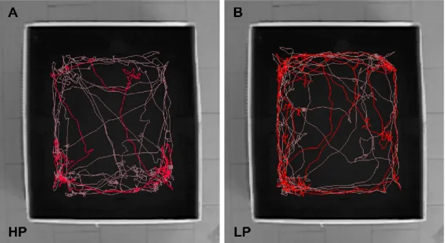
2010). Rats with an exploration change <50% or ≥50% were classified as low (LP; 13 Exp_SH and 11 Exp_EE) or high (HP; 10 Exp_SH and 13 Exp_EE) performers, respectively (Figure 7). Mean velocity, number of rearings (with and without a wall support), time spent at center, and number of fecal boli were also measured.
Figure 7. Examples of locomotion tracks in OF of a high performer rat (HP) (A) and a low performer rat (LP) (B).
Pink track derives from the first five minutes, while red track derives from the last five minutes. HP rat explores much during the first five minutes and markedly decreases its activity during the last five; on the contrary, LP rat shows a high exploratory activity during the whole task.
3.3 MWM
The MWM set-up consisted of a circular black pool (150 cm diameter; Ugo Basile Srl, Italy) filled with water (24±1 °C). The pool was virtually divided into four quadrants and a transparent, invisible platform was placed in the middle of the south-west (SW) quadrant 2 cm below the water surface. Visual distal cues were placed on the walls surrounding the maze, and an additional cue was represented by the operator who stood in a fixed position near the SW quadrant. The test room was dimly, diffusely and bottom-up illuminated by light sources
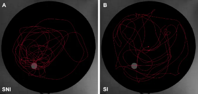
13 allocated at the room corners. The training phase consisted of 4 trials per day for 5 consecutive days. In each trial, rats were released in the pool facing the wall from one of four possible start positions (east, north, south-east, north-west); start positions were always different for the 4 trials in the same day, and their sequence varied across the days. In each trial, rats were given 120 seconds to find the platform; the trial was considered to be completed when the rat found the platform, climbed on it and remained on it for at least 5 seconds. If the rat failed to find the platform, it was gently guided to the platform by hand. In any case, animals were kept on the platform 20 seconds before returning to the cage; the inter-trial time was 1 minute. 24 hours after the last training trial, a 120 seconds probe trial without platform was administered using a new release point, i.e., north-east. During the training trials, latency to reach the platform, swimming path length, time spent at the border and velocity were recorded; for the analysis, swimming path length was preferred to latency to reach the platform to minimize the possible confounding effect of differences in swim velocity (Vorhees and Williams, 2006). During the probe trial, mean distance to the platform (proximity) and time spent in the target quadrant were recorded. At baseline, rats were classified as impaired for spatial memory (SI) if % time spent in the target quadrant was ≤ 25% (9 Exp_SH and 7 Exp_EE) and as not-impaired (SNI) if % time spent in the target quadrant was > 25% (14 Exp_SH and 17 Exp_EE). To compare reference memory results, proximity was preferred to percent time spent in the target quadrant as it allows a better graduation of performances also for SI animals (Figure 8).
Figure 8. Examples of swimming path tracks at probe day of one animal not impaired for spatial memory (SNI)
(A) and one impaired for spatial memory (SI) (B). SNI animals tend to swim closer to the platform zone (in grey) and spend more time in the target than in other quadrants, while SI animals randomly swim in the whole pool.
14 3.4 STPA
The task was performed in accordance to Platano et al. (2008). A shuttle box (57 cm long, 27 cm wide and 30 cm high) divided into two equally sized compartments separated by a sliding door (Ugo Basile Srl, Italy) was used. One compartment was illuminated and the other one was dark. The first day rats were allowed to freely explore the entire apparatus for 5 minutes, while the second day they were kept for 3 minutes in the dark compartment, once they had spontaneously entered it. The third day, rats were released in the illuminated chamber, after 10 seconds the sliding door was opened and when the animal entered the dark compartment with all four paws an electric foot shock was delivered (0.6 mA, 2 seconds). Rats that failed to vocalize or jump were excluded (n=2) (Wilmott and Thompson, 2013). After 24 hours, rats were put in the illuminated compartment, after 10 seconds the sliding door was opened and the time passed before the animal entered the dark chamber (latency) was recorded; if the animal remained in the illuminated compartment, the test was considered over after 300 seconds. Rats with a latency lower than 100 seconds or higher than 200 seconds were classified as impaired (AI; 16 Exp_SH and 18 Exp_EE) or not impaired (ANI; 7 Exp_SH and 6 Exp_EE) for aversive memory, respectively. These two cut-off values were chosen because nearly all rats dichotomized below or above these levels; we excluded animals with intermediate performances to avoid a possible classification bias (n=3).
3.5 Statistical analysis
Results were expressed as mean±standard error of the mean (SEM) (continuous variables) or as percentage (categorical variable). t-Student test was applied to compare two groups at one time-point and paired t-Student test was used to compare two time-points of one subgroup. Pearson’s (parametric analysis) or Spearman’s (nonparametric analysis) coefficients were used to assess correlation between variables. Linear regression was used to analyze body weight trend. Two-way ANOVA for repeated measures was applied to compare two groups at two or more time points, using, as interacting factors: i) housing condition and day to compare body weight of Exp_SH and Exp_EE; ii) housing condition and retesting to compare total DM by Exp_SH and Exp_EE, exploration change of Exp_SH and Exp_EE, mean proximity of Exp_SH and Exp_EE and latency at STPA first day of Exp_SH and Exp_EE; iii) minute and retesting to compare DM per minute at baseline and PH; iv) minute and housing condition to compare DM per minute of N_SH and N_EE; v) day and housing condition to compare swimming path length of Exp_SH and Exp_EE and swimming path length of N_SH and N_EE; vi) day and retesting to compare swimming path length of Exp_SH and Exp_EE between
15 baseline and PH. The categorical variable (percentage) was analyzed by binary logic regression for repeated measures using housing condition and retest as interacting factors; χ square test was used to compare two groups and McNemar’s test was applied to repeated measures. Significance was accepted for p<0.05.
4. Results
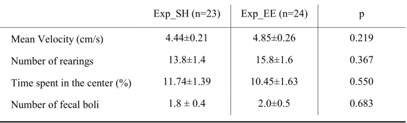
Table 1 reports the OF parameters used to evaluate basal locomotor activity and anxiety of experimental animals at baseline (Komsuoglu Celikyurt et al., 2014; Alves et al., 2018). No statistical differences were found between Exp_SH and Exp_EE rats, thus excluding any possible bias due to the semi-random assignment to the housing condition.
Table 1. Parameters of locomotor activity and anxiety of experimental rats before the 12-week protocol (baseline).
Statistical comparison was performed by t-Student test. Exp_SH, rats that continued standard housing; Exp_EE, rats that started enriched housing.
Correlation analysis between performance levels in the different tests at baseline demonstrated no significant correlation for both Exp_SH and Exp_EE animals, thus showing that impairments in the three cognitive domains were independent (Table 2).
Table 2. Correlation coefficient (r) and p for each couple of tests at baseline.
Exp_SH (n=23) Exp_EE (n=24) p
Mean Velocity (cm/s) 4.44±0.21 4.85±0.26 0.219
Number of rearings 13.8±1.4 15.8±1.6 0.367
Time spent in the center (%) 11.74±1.39 10.45±1.63 0.550
Number of fecal boli 1.8 ± 0.4 2.0±0.5 0.683
OF vs MWM OF vs PA MWM vs PA
r p r p r p
Exp_SH -0.086 0.696 -0.336 0.136 -0.173 0.454
16
Pearson’s correlation analysis was used for variables with normal distribution, otherwise Spearman’s coefficient was calculated. OF, Open Field test; MWM, Morris Water Maze test; PA, Step-Through Passive Avoidance test; Exp_SH, rats that continued standard housing; Exp_EE, rats that started enriched housing.
4.1 Body weight
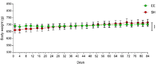
Figure 9 reports the body weight trend of SH and EE animals (experimental and naïve rats were grouped) during the 12-week protocol: while SH animals evidenced a significant body weight increase during the housing protocol (p<0.001), EE rats showed a steady trend. Despite the body weight gain of SH animals, no significant difference was found at each time point between the two groups.
Figure 9. Body weight trend during the 12-week protocol. No statistical differences were found comparing rats
under standard (SH) or enriched (EE) housing at each time point, but while body weight remained stable in EE rats, it significantly increased in SH animals. Experimental and naïve animals were grouped (SH, n=47; EE, n=45). Animals were 17 months old at the beginning and 20 months old at the end of the protocol. *** significant body weight gain in SH rats, p<0.001.
4.2 Habituation
Comparing baseline and PH, the total DM significantly decreased in both Exp_SH (2599.86±125.84 vs. 1555.92±133.33 cm, p<0.0001) and Exp_EE (2854.40±153.25 vs. 2088.43±72.91 cm, p<0.0001) animals (significant effects of both housing condition
(F(1,45)=7.1, p=0.011) and retesting (F(1,45)=87.5, p<0.0001)). At PH, total distance moved was
higher in Exp_EE than in Exp_SH animals (2088.43±72.91 cm vs 1555.92±133.33 cm, p=0.007) (Figure 10).
17
Figure 10. Total distance moved (DM) at baseline and after 12 weeks (PH) of standard housing (SH) or
environmental enrichment (EE). Total DM decreased at PH in both SH (Exp_SH) and EE rats (Exp_EE). At PH, total DM was significantly lower in Exp_SH than in Exp_EE animals. ** p<0.01; **** p<0.0001.
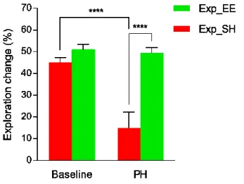
While in the SH group also the exploration change significantly decreased (p<0.0001), in the EE group it remained unchanged, so that Exp_EE animals attained a significantly higher exploration change than Exp_SH rats at PH (p<0.0001); significant effect was found for the interaction between housing condition and retesting (F(1,45)=12.6, p=0.0009) (Figure 11).
Figure 11. Exploration change at baseline and after 12 weeks (PH) of standard housing (SH) or environmental
enrichment (EE). Exploration change of SH animals (Exp_SH) significantly decreased at PH, while exploration change of EE animals (Exp_EE) remained unchanged. In fact, at PH exploration change of Exp_EE rats was significantly higher than exploration change of Exp_SH rats. **** p<0.0001.
Indeed, the exploration pattern of Exp_EE and Exp_SH animals at PH was strikingly different. Exp_EE animals, despite a significant reduction of the distance moved at 1st, 4th, 5th, 8th and 9th
18 minute with respect to baseline (p<0.0001, p=0.005, p=0.0005, p=0.04 and p=0.02, respectively) explored more during the first minutes of the task and progressively decreased their activity (significant effect of time and retesting interaction, F(9,207)=3.6, p=0.0003) (Figure 12A, 12C). On the contrary, Exp_SH rats showed a flat pattern, with a significant drop of the DM per minute from the 1st to the 8th minute with respect to baseline (p<0.0001, p<0.0001, p<0.0001, p=0.007, p=0.001, p=0.004, p=0.03, p=0.01, respectively; interaction between time and retesting had significant effect (F(9,198)=28.7, p<0.0001)) (Figure 12B, 12D).
Figure 12. Distance moved (DM) per minute at baseline and post housing (PH) of animals kept in standard housing
(Exp_SH) and animals housed in environmental enrichment (Exp_EE), and PH track examples. (A) Exp_EE rats maintained a decreasing exploration pattern also at PH, with a significant reduction of DM per minute at 1st, 4th,
5th, 8th and 9th minutes. (B) On the contrary, at PH Exp_SH rats had a flat explorative pattern instead of the typical
habituation profile; the DM per minute was significantly lower from the 1st to the 8th minute. (C, D) Examples of
locomotion tracks of one Exp_EE (C) and one Exp_SH (D) animal at PH; while Exp_EE animals kept exploring the arena, Exp_SH rats spent most of the time resting. *, p<0.05; **, p<0.01; ***, p<0.001; ****, p<0.0001.
19 In Exp_EE animals, a significant negative correlation was found between the basal exploration change and its variation at PH (∆, that is the exploration change value at PH minus the exploration change value at baseline) (p<0.001) (Pearson’s correlation) (Figure 13). This correlation indicates that animals with a lower exploration change value at baseline tended to increase it at PH, while animals with a higher exploration change value at baseline tended to decrease it, thus suggesting a possible different response to EE on the basis of basal behavior. Unlike Exp_EE animals, Exp_SH rats displayed no significant correlation between the basal exploration change and its variation at PH (r=-0.773).
Figure 13. Significant negative correlation between basal exploration change and its variation after the 12-week
protocol (delta) in Exp_EE rats. Almost all animals with a basal exploration change <50% had a higher exploration change at PH (i.e. their delta was positive), while almost all animals with a basal exploration change ≥50% had a lower exploration change at PH (i.e. their delta was negative). The Pearson’s correlation coefficient (r) indicated a strong association.
To clarify the phenomenon, Exp rats were divided in two subgroups: low performers, with basal exploration change <50%, and high performers, with basal exploration change ≥50%. The analysis of the exploration change at baseline and PH in the two subgroups showed that EE low performer rats significantly increased (p=0.003) and EE high performer rats significantly decreased (p=0.001) the exploration change (Figure 12A). On the contrary, consistently with the lack of correlation between basal exploration change and Δ in Exp_SH, both SH low (p=0.03) and high (p=0.004) performer rats significantly decreased the parameter (Figure 14B).
20
Figure 14. Exploration change at baseline and post housing (PH) of low (LP) and high (HP) performer rats
maintained in environmental enrichment (Exp_EE) or standard housing (Exp_SH). (A) Exploration change significantly increased in Exp_EE low performers, while it significantly decreased in Exp_EE high performers. (B) For animals maintained in standard housing, both low and high performers showed a significant decrease of exploration change. * p<0.05; ** p<0.01.
Finally, regarding the naïve groups, the total DM of N_EE rats was significantly higher than the total DM of N_SH animals (2.638.12±115.57 vs. 2317.97±101.37, p=0.047 cm). Moreover, N_EE showed a significantly higher exploration change than N_SH (p=0.01) (Figure 15A) due to the significantly higher DM at the first minute (p<0.0001) after the release in the arena (interaction between housing condition and time had significant effect (F(9,387)=4.3, p<0.0001)) (Figure 15B); no differences were found in the number of fecal boli (2.3±0.5 vs. 1.9±0.5) and in time spent in the center of the OF arena (10.33±1.43 % vs. 9.31±0.95 %).
Figure 15. (A) Exploration change of naïve animals housed in environmental enrichment (N_EE) and standard
21
trend of naïve animals. N_EE showed a significantly higher DM than N_SH during the first minute of exploration. * p<0.05; **** p<0.0001.
4.3 Spatial memory
Comparing the learning curves of Exp_EE and Exp_SH animals, no differences were found at baseline and PH. For both Exp_EE and Exp_SH animals the swimming path length was significantly lower at PH at days 1, 2 and 3, suggesting that animals retained memory from the previous training (for Exp_SH: significant effect of interaction between day and retesting
(F(4,88)=3.4, p=0.01); post-hoc comparison evidenced a significative difference at day 1
(p<0.0001), day 2 (p<0.0001) and day 3 (p<0.007); for Exp_EE: significant effect of day (F(4, 92)=39.0, p<0.0001) and retest (F(1,23)=21.8, p=0.0001); post-hoc comparison evidenced a significative difference at day 1 (p=0.0001), day 2 (p=0.001) and day 3 (p=0.04)) (Figure 16).
Figure 16. Learning curves of experimental animals maintained in standard housing (Exp_SH) and experimental
animals maintained in environmental enrichment (Exp_EE) at baseline and post-housing (PH). Learning curves of Exp_EE and Exp_SH animals overlapped both at baseline and at PH. At days 1, 2 and 3 of PH, both groups showed a significant reduction of the swimming path length. # significant difference.
Consistently, at probe day the mean proximity was lower at PH than at baseline in both Exp_SH (p=0.0001) and Exp_EE animals (p=0.02) (main effect of retest (F(1,45)=25.5, p<0.0001)), while it was comparable between the two groups both before and after the housing protocol (Figure 17).
22
Figure 17. Mean proximity to platform of experimental animals maintained in standard housing (Exp_SH) and
environmental enrichment (Exp_EE). Mean proximity decreased at PH in both Exp_SH and Exp_EE. * p<0.05; *** p<0.001.
No effect of EE on spatial learning and memory was detectable in naïve animals, except for a reduction of swimming path length at day 3 (p=0.02) (Figure 18).
Figure 18. (A) Learning curves of naïve animals kept in standard housing (N_SH) and naïve animals kept in
environmental enrichment (N_EE). A significative difference was found only at day 3. (B) Mean proximity at probe day of N_SH and N_EE animals. No difference was found between the two groups. * p<0.05.
4.4 Aversive memory
To verify if animals retained memory of the baseline aversive event, thus creating the prerequisite for a possible extinction phenomenon due to PH habituation sessions, baseline and PH latencies at first day were compared: both Exp_SH (12.72±2.01 vs. 8.27±2.06 seconds) and Exp_EE (9.38±1.24 vs. 6.96±1.19 seconds) animals showed no significant differences.
23 Comparing baseline and PH, the percentage of AI animals significantly decreased in Exp_SH group (p=0.04; OR=0.267 and 95% confidence interval=0.076-0.930 for housing condition) while it did not change in Exp_EE rats, so that the EE group gained a significantly higher percentage of AI rats in comparison to the SH group at PH (p=0.04; OR=0.286 and 95% confidence interval=0.085-0.963 for housing condition) (Figure 19).
Figure 19. Percentage of animals impaired for aversive memory (AI) before (baseline) and after (PH) the 12-week
protocol. At PH, the value significantly decreased for animals in standard housing (Exp_SH) whereas it did not change for those under environmental enrichment (Exp_EE), so much that at PH the EE group showed a significantly higher percentage of AI rats. At PH, the percentage of AI rats included animals that were AI at baseline and remained AI as well as animals that were ANI at baseline and converted to AI after the 12-week protocol. * p<0.05.
Focalizing the attention on basal performances, Exp_SH and Exp_EE AI animals showed a significant conversion to ANI (p=0.02 and p=0.02, respectively) (Figure 20A), Exp_SH ANI animals remained unchanged, and Exp_EE ANI animals had a significant conversion to AI (p=0.04) (Figure 20B).
24
Figure 20. (A) Baseline and post-housing (PH) performance of experimental animals classified at baseline as
impaired for aversive memory (AI) (the basal percentage was normalized at 100) and maintained under standard (Exp_SH) or enriched (Exp_EE) housing. A significant fraction of both Exp_SH and Exp_EE animals converted to not impaired (ANI). (B) Baseline and PH performance of Exp_SH and Exp_EE animals classified at baseline as ANI (the basal percentage was normalized at 100). Exp_SH totally remained ANI while a significant percentage of Exp_EE became AI. * p<0.05.
Comparing naïve rats, N_EE had significantly higher percentage of AI vs. N_SH (p<0.001) (Figure 21).
Figure 21. Percentage of naïve animals maintained in standard housing (N_SH) or in environmental enrichment
(N_EE) classified as impaired for aversive memory (AI). The EE group showed a significantly higher level in comparison to the SH group. *** p<0.001.
The results included in the paragraphs “Body weight”, “Habituation” and “Aversive Memory” are part of a paper submitted for publication to the journal Neurobiology of Learning and Memory (Balietti, M., Pugliese, A., Fabbietti, P., Di Rosa, M., Conti, F. Aged Rats with Different Performances at Environmental Enrichment Onset Display Different Modulation of Habituation and Aversive Memory. Neurobiology of Learning & Memory, submitted).
5. Discussion
The present study showed that aging rats displaying different learning and memory performances at EE onset react differently to the stimulating protocol. Indeed, habituation improved in LP but did not change in HP animals and aversive memory was modulated by the enriched housing condition only in ANI rats while EE AI animals, as SH rats, responded only to retesting.
25 5.1 Body weight
During the 12-week protocol, Exp_SH rats significantly increased their body weight, whereas Exp_EE animals did not change it, probably as a consequence of the opportunity to perform voluntary exercise (i.e. the presence of a running wheel but also larger spaces organized in several levels connected by stairs). However, SH and EE rats showed no differences at each time point, thus the contribution of a possible homeostatic control on the body weight to EE effect might be marginal, even if cannot be ruled out.
5.2 Habituation
Habituation can be considered the simplest form of learning: when the animal is exposed to a constant environment (intra-task analysis) or is re-exposed to familiar settings (inter-task analysis), an effective information processing induces a progressive decrease in exploration (Leussis and Bolivar, 2006). Both Exp_SH and Exp_EE rats evidenced intertask habituation -rats significantly reduced the total DM when retested in the OF-, but the housing condition differently influenced their intra-task behavior -in comparison to baseline, at PH Exp_SH had a significant reduction of the exploration change while Exp_EE rats showed no differences. Indeed, at PH, Exp_SH animals had a constant locomotion during the entire 10-minute registration, while Exp_EE animals showed a continue drop of activity from the first to the last minute. Therefore, while retention and recall of OF arena memory determined a minimum, random locomotion after SH (i.e. on the basis of the baseline exploration, animals remembered that the arena contained no “rewards”), it coexisted with a finalized activity after EE (i.e. an initial active exploration of a potentially “rewarding” environment followed by a progressive reduction in searching in what revealed itself to be an empty arena). This difference could partly derive from the fact that, while SH cages have always the same organization, the EE cage, better “mimicking” the natural habitat, undergoes periodic changes, included food and water position. Thus, EE rats could be more motivated to actively explore the environment, even when familiar, in search of potential, novel stimuli, in particular in terms of foraging. Furthermore, evidence exists that the multiple stimuli provided by EE may increase the exploration attitude (Sampedro-Piquero et al., 2014), without compromising habituation capability (Kempermann et al, 2002); accordingly, it is reasonable that in Exp_EE animals the higher propensity to an active interaction with the surroundings initially overwhelmed the re-exposure to a familiar setting, without impairing the successive environmental analysis and adaptation. A plausible support to this interpretation can be gathered from naïve controls. N_EE animals displayed a significantly higher DM per minute than N_SH rats immediately after the release in the
26 apparatus, while successively the locomotor patterns of the two groups overlapped. Rats, despite an innate aversion to novel, potentially dangerous spaces, are naturally pushed to explore in search of food, escape sites or mating partners (Sousa et al., 2006); our naïve animals’ behavior confirmed that EE can significantly enhance the latter instinct triggering a stronger explorative behavior (i.e. the higher DM of N_EE animals in comparison to N_SH rats during the first minute of OF) and a faster information extraction (i.e. the rapid overlapping of DM/minute decreasing trend of the two naïve groups). Moreover, the similar number of fecal boli and percent time spent in the center of the OF arena found in N_EE and N_SH rats seems to exclude, in our experimental model, anxiety/fearfulness modulation and to strictly link the DM differences to curiosity and exploration. EE capability to promote habituation when applied to young (Varty et al., 2000; Zimmermann et al., 2001; Elliot and Grunberg, 2005; Del Arco et al., 2007; Ronzoni et al., 2016) or early-adult (Hughes and Collins, 2010) rats has been previously described. The data obtained in our naïve groups (i.e. significantly higher exploration change of N_EE in comparison to N_SH) extended the responsiveness to a later age. However, aged rats, unlike younger animals (Zimmermann et al., 2001; Elliot and Grunberg, 2005; Del Arco et al., 2007; Hughes and Collins, 2010; Ronzoni et al., 2016), displayed an increased general activity due to EE, thus suggesting that during senescence the learning process lies on a different strategy. Interestingly, we also firstly proved that EE does not work homogenously on aging animals as its effect was critically influenced by the initial cognitive condition. Indeed, the housing stimulation ameliorated habituation capability of animals with less efficient data processing (LP) but did not further improve information extraction of HP rats. Despite the starting impairment, LP animals revealed a significant residual plasticity that guaranteed, after the housing stimulation, a considerable enhancement of environmental analysis. Regarding HP animals, their basal performances likely represented the maximum achievable level for that age and even if EE did not boost that level, it did support its maintenance during aging.
5.3 Spatial memory
Analysis of learning and memory performances at MWM test evidenced an amelioration, due to the retesting protocol, in both enriched and non-enriched animals so that, at the end of the protocol, both groups reached the same performances. In fact, it has been shown for aged rats that a pre-experience of MWM task up to 12 months before is facilitating for the subsequent execution of the same task (Pitsikas et al, 1991). Thus, in our experimental settings, the effect of the previous exposition was overwhelming and prevented us from studying any consequence
27 of EE on subgroups. Anyway, no effect of EE was detectable in naïve animals too, sustaining that, in our conditions, EE had no influence on spatial memory performances.
5.4 Aversive memory
Aversive memory involves the suppression of an innate instinct (in STPA the preference for darkness) when associated with a negative stimulus. It primarily relies upon dorsal hippocampus and basolateral amygdala (Paris et al., 2011) and deeply depends on emotionality. Both Exp_SH and Exp_EE AI rats took advantage from task re-exposure, as a significant percentage became ANI regardless the housing condition. On the contrary, Exp_SH and Exp_EE ANI animals showed a different response to the housing condition. Indeed, while a positive effect of retesting was found in Exp_SH ANI animals that maintained their performance at PH, the Exp_EE ANI group apparently showed a memory impairment after the 12 weeks of enriched housing. However, the performances of naïve animals seem to suggest a different interpretation. N_EE rats not only had a significantly higher percentage of AI animals than N_SH animals, but were almost all AI. This result is in line with Garrido and colleagues (2013) who found a significantly lower retention of the inhibitory response in EE young rats, probably due to the behavioral amelioration under stress. The hypothalamic-pituitary-adrenal (HPA) axis plays a pivotal role in the correct handling of stressors. During aging, this axis undergoes several alterations that contribute to learning and memory deficits, as hyperactivation and hypersecretion of glucorticoids (Pedersen et al., 2001). EE seems to reduce anxiety and fearfulness acting exactly on the HPA axis (Fox et al., 2006), maybe creating a continuous, mild stressful stimulus able to produce a “hormetic” effect. Indeed, the constant exposition to high social interactions, voluntary exercise as well as novel objects might operate as a stress inoculator that leaves EE animals more resilient to successive stressful events (Crofton et al., 2015), as the electric foot shock of the STPA test. Surprisingly, we showed, for the first time to our knowledge, that this “anxiolytic” effect is not uniform: animals with a basal intact aversive memory learned to better cope with a familiar stressful situation while animals with a basal impaired aversive memory, when re-exposed to the task, did not efficiently modulated their anxiety (i.e., similarly to the Exp_SH AI subgroup, a significant part enhanced its response to the negative stimulus converting to the ANI category while the remaining fraction, plausibly, simply maintained its age-related deficit).
28
6. Conclusions
These results showed that EE modulates habituation and aversive memory in aging rats enhancing their exploration attitude and lowering their anxiety level, but also that not all animals are able to benefit from this stimulating protocol. Indeed, only rats with basal lower performances improved their habituation and only animals initially unimpaired for aversive memory were able to manage their fear when re-exposed to a stressful condition. The pre-classification of animals in subgroups on the basis of their performances (i.e. LP and HP as well as AI and ANI) allowed to evidence differences that could be missed in experimental approaches that analyze age-matched animals as a uniform cohort. Considering the heterogeneous nature of senescence, especially regarding cognition, the pre-evaluation of aging animals might be crucial to better define EE mechanisms and applicability. The issue needs further analyses but certainly opens the way for the opportunity to design individually tailored approaches to optimize their efficacy and minimize possible unwanted consequences.
Acknowledgements
This project was conducted together with Dr Marta Balietti, who I want to deeply thank for her constant scientific and human support. I also thank Paolo Fabbietti and Mirko Di Rosa for their help with statistical analysis of aversive memory results.
References
Alves, C.D.S., Frias, H.V., Kirsten, T.B., Cordeiro, F., Bernardi, M.M., Suffredini, I.B. (2018). Luffa operculata fruit aqueous extract induces motor impairments, anxiety-like behavior, and testis damage in rats. J. Ethnopharmacol. 222, 52-60. doi: 10.1016/J.jep.2018.04.044.
Amrein, I., Isler, K., Lipp, H.P. (2011). Comparing adult hippocampal neurogenesis in mammalian species and orders: influence of chronological age and life history stage. Eur. J. Neurosci. 34, 978–987. doi: 10.1111/j.1460-9568.2011.07804.x.
Arias-Cavieres, A., Adasme, T., Sánchez, G., Muñoz, P., Hidalgo, C. (2017). Aging impairs hippocampal-dependent recognition memory and LTP and prevents the associated RyR up-regulation. Front. Aging Neurosci. 9, 111. doi: 10.3389/fnagi.2017.00111.
Bach, M., Barad, M., Son, H., Zhuo, M., Lu, Y.-F., Shih, R., Mansuy, I., Hawkins, R., Kandel, E. (1999). Age-related defects in spatial memory are correlated with defects in the late phase of hippocampal long-term potentiation in vitro and are attenuated by drugs that enhance the cAMP signaling pathway. PNAS. 96, 5280–5285. doi: 10.1073/pnas.96.9.5280.
29 Bamidis, P. D., Vivas, A. B., Styliadis, C., Frantzidis, C., Klados, M., Schlee, W., Siountas, A., Papageorgiou, S.G. (2014). A review of physical and cognitive interventions in aging. Neurosci. Biobehav. Rev. 44, 206–220. doi: 10.1016/j.neubiorev.2014.03.019.
Barnes, C.A. (1979). Memory deficits associated with senescence: a neurophysiological and behavioral study in the rat. J. Comp. Physiol. Psychol. 93, 74–104. doi: 10.1037/h0077579. Barnes, C.A. (2003). Long-term potentiation and the ageing brain. Phil. Trans. R. Soc. Lond. B. 358, 765-772. doi: 10.1098/rstb.2002.1244.
Basak, C., Boot, W., Voss, M., Kramer, A. (2008). Can training in a real-time strategy video game attenuate cognitive decline in older adults? Psychology and aging 23(4), 765-777. doi: 10.1037/a0013494.
Bennett, J.C., McRae, P.A., Levy, L.J., Frick, K.M. (2006). Long-term continuous, but not daily, environmental enrichment reduces spatial memory decline in aged male mice. Neurobiol. Learn. Mem. 85, 139-152. doi: 10.1016/j.nlm.2005.09.003.
Bouet, V., Freret, T., Dutar, P., Billard, J.M., Boulouard, M. (2011). Continuous enriched environment improves learning and memory in adult NMRI mice through theta burst-related-LTP independent mechanisms but is not efficient in advanced aged animals. Mech. Ageing Dev. 132, 240-248. doi: 10.1016/j.mad.2011.04.006.
Budni, J., Bellettini-Santos, T., Mina, F., Garcez, M., and Zugno, A. (2015). The involvement of BDNF, NGF and GDNF in aging and Alzheimer’s disease. Aging Dis. 6, 331. doi: 10.14336/ad.2015.0825.
Burke, S.N., Barnes, C.A. (2006). Neural plasticity in the ageing brain. Nature Rev. Neurosci. 7, 30-40. doi: 10.1038/nrn1809.
Chersi, F., Burgess, N. (2015). The cognitive architecture of spatial navigation: hippocampal and striatal contributions. Neuron 88(1), 64-77. doi: 10.1016/j.neuron.2015.09.021.
Crofton, E.J., Zhang, Y., Green, T.A. (2015). Inoculation stress hypothesis of environmental enrichment. Neurosci. Biobehav. Rev. 49, 19-31. doi: 10.1016/j.neubiorev.2014.11.017. D’Hooge, Deyn, D. (2001). Applications of the Morris water maze in the study of learning and memory. Brain Res Brain Res Rev 36, 60–90. doi: 10.1016/S0165-0173(01)00067-4.
Del Arco, A., Segovia, G., Garrido, P., de Blas M., Mora, F. (2007). Stress, prefrontal cortex and environmental enrichment: studies on dopamine and acetylcholine release and working memory performance in rats. Behav. Brain Res. 176, 267-273. doi: 10.1016/j.bbr.2006.10.006. Dixon, R.A., de Frias, C.M. (2014). Cognitively elite, cognitively normal, and cognitively impaired aging: neurocognitive status and stability moderate memory performance. J. Clin. Exp. Neuropsychol. 36, 418-430. doi: 10.1080/13803395.2014.903901.
Duan, H., Wearne, S.L., Rocher, A.B., Macedo, A., Morrison, J.H., Hof, P.R. (2003). Age-related dendritic and spine changes in corticocortically projecting neurons in macaque monkeys. Cereb. Cortex 13(9), 950–961. doi: 10.1093/cercor/13.9.950.
30 Elliott, B.M., Grunberg, N.E. (2005). Effects of social and physical enrichment on open field activity differ in male and female Sprague-Dawley rats. Behav. Brain Res. 165, 187-196. doi: 10.1016/j.bbr.2005.06.025.
Encinas, J.M., Sierra, A. (2012). Neural stem cell deforestation as the main force driving the age-related decline in adult hippocampal neurogenesis. Behav. Brain Res. 227, 433-439. doi: 10.1016/j.bbr.2011.10.010.
Fabre, C., Chamari, K., Mucci, P., Massé-Biron, J., Préfaut, C. (2002). Improvement of cognitive function by mental and/or individualized aerobic training in healthy elderly subjects. Int. J. Sports Med. 23, 415–21. doi: 10.1055/s-2002-33735.
Flood, J., Roberts, E. (1988). Dehydroepiandrosterone sulfate improves memory in aging mice. Brain Res. 448, 178–181. doi: 10.1016/0006-8993(88)91116-X.
Foster, T.C., DeFazio, R.A., Bizon, J.L. (2012). Characterizing cognitive aging of spatial and contextual memory in animal models. Front. Aging Neurosci. 4, 12. doi: 10.3389/fnagi.2012.00012.
Fox, C., Merali, Z., Harrison, C. (2006). Therapeutic and protective effect of environmental enrichment against psychogenic and neurogenic stress. Behav. Brain Res. 175, 1-8. doi: 10.1016/j.bbr.2006.08.016.
Fraley, S.M., Springer, A.D. (1981). Memory of simple learning in young, middle-aged, and aged C57/BL6 mice. Behav. Neural Biol. 31, 1-7. doi: 10.1016/S0163-1047(81)90986-9. Freret, T., Billard, J.M., Schumann-Bard, P., Dutar, P., Dauphin, F., Boulouard, M., Bouet, V. (2012). Rescue of cognitive aging by long-lasting environmental enrichment exposure initiated
before median lifespan. Neurobiol. Aging. 33, 1005.e1-1005.e10.
doi: 10.1016/j.neurobiolaging.2011.09.028.
Frick, K.M., Fernandez, S.M. (2003). Enrichment enhances spatial memory and increases synaptophysin levels in aged female mice. Neurobiol. Aging. 24, 615-626. doi: 10.1016/S0197-4580(02)00138-0.
Fuchs, F., Herbeaux, K., Aufrere, N., Kelche, C., Mathis, C., Barbelivien, A., Majchrzak, M. (2016). Late enrichment maintains accurate recent and remote spatial memory only in aged rats that were unimpaired when middle aged. Learn. Mem. 23, 303-312. doi: 10.1101/lm.041236.115.
Gallagher, M., Colantuoni, C., Eichenbaum, H., Haberman, R.P., Rapp, P.R., Tanila, H., Wilson, I.A. (2006). Individual differences in neurocognitive aging of the medial temporal lobe. Age (Dordr). 28, 221-233. doi: 10.1007/s11357-006-9017-5.
Garrido, P., De Blas, M., Ronzoni, G., Cordero, I., Antón, M., Giné, E., Santos, A., Del Arco, A., Segovia, G., Mora, F. (2013). Differential effects of environmental enrichment and isolation housing on the hormonal and neurochemical responses to stress in the prefrontal cortex of the adult rat: relationship to working and emotional memories. J. Neural. Transm. (Vienna) 120, 829-843. doi: 10.1007/s00702-012-0935-3.
Gates, N., Valenzuela, M. (2010). Cognitive exercise and its role in cognitive function in older adults. Curr. Psychiat. Rep. 12, 20–27. doi:10.1007/s11920-009-0085-y.
31 Harati, H., Majchrzak, M., Cosquer, B., Galani, R., Kelche, C., Cassel, J.C., Barbelivien A. (2011). Attention and memory in aged rats: impact of lifelong environmental enrichment. Neurobiol. Aging. 32(4): 718-736. doi: 10.1016/j.neurobiolaging.2009.03.012.
Harburger, L.L., Labert, T.J., Frick. K.M. (2007). Age-dependent effects of environmental enrichment on spatial reference memory in male mice. Behav. Brain Res. 185, 43-48. doi: 10.1016/j.bbr.2007.07.009.
Harris, J.D. (1943). Habituatory response decrement in the intact organism. Psychol. Bull. 40, 385–422. doi: 10.1037/h0053918.
Head, E., Mehta, R., Hartley, J., Kameka, M., Cummings, B.J., Cotman, C.W., Ruehl, W.W., Milgram, N.W. (1995). Spatial learning and memory as a function of age in the dog. Behav. Neurosci. 109(5), 851-858. doi: 10.1037/0735-7044.109.5.851.
Hebb, D.O. (1947). The effects of early experience on problem-solving at maturity. Am. Psychol. 2, 306–307.
Hindin, S.B., Zelinski, E.M. (2012). Extended practice and aerobic exercise interventions benefit untrained cognitive outcomes in older adults: a meta‐analysis. J. Am. Geriatr. Soc. 60, 136–141. doi: 10.1111/j.1532-5415.2011.03761.x.
Hughes, R.N., Collins, M.A. (2010). Enhanced habituation and decreased anxiety by environmental enrichment and possible attenuation of these effects by chronic alpha-tocopherol (vitamin E) in aging male and female rats. Pharmacol. Biochem. Behav. 94, 534-542. doi: 10.1016/j.pbb.2009.11.008.
Izquierdo, I., Furini, C. R., Myskiw, J. C. (2016). Fear memory. Physiol. Rev. 96, 695–750. doi:10.1152/physrev.00018.2015.
Kallai, J., Makany, T., Karadi, K., Jacobs, W. J. (2005). Spatial orientation strategies in Morris-type virtual water task for humans. Behav. Brain Res. 159, 187–196. doi:10.1016/j.bbr.2004.10.015.
Kattenstroth, J.C., Kalisch, T., Holt, S., Tegenthoff, M., Dinse, H. (2013). Six months of dance intervention enhances postural, sensorimotor, and cognitive performance in elderly without affecting cardio-respiratory functions. Front. Aging Neurosci. 5, 5. doi: 10.3389/fnagi.2013.00005.
Kempermann, G., Gast, D., Cage, F.H. (2002). Neuroplasticity in old age: sustained five fold induction of hippocampal neurogenesis by long-term environmental enrichment. Ann. Neurol. 52, 135-143. doi: 10.1002/ana.10262.
Kempermann, G., Gage, F.H., Aigner, L., Song, H., Curtis, M.A., Thuret, S., Kuhn, H.G., Jessberger, S., Frankland, P.W., Cameron, H.A., Gould, E., Hen, R., Abrous, D.N., Toni, N., Schinder, A.F., Zhao, X., Lucassen, P.J., Frisé J. (2018). Human adult neurogenesis: evidence and remaining questions. Cell Stem Cell 23, 25-30. doi: 10.1016/j.stem.2018.04.004.
Klusmann, V., Evers, A., Schwarzer, R., Schlattmann, P., Reischies, F., Heuser, I., and Dimeo, F. (2010). Complex mental and physical activity in older women and cognitive performance: a 6-month randomized controlled trial. J. Gerontol. Ser 65A, 680–688. doi: 10.1093/gerona/glq053.