UNIVERSITA’ POLITECNICA DELLE MARCHE
Scuola Di Dottorato In Medicina E Chirurgia
Curriculum “Medicina Sperimentale”, XIV ciclo
Doctoral Thesis
SKIN AGIN: A NEW PERSPECTIVE FOR PLASTIC SURGEONS.
CLINICAL ASPECTS, BIOLOGICAL IMPLICATIONS AND MOLECOLAR
DIAGNOSTICS TO UNDERSTAND AGING AND SUPPORT
REJUVINATION FOR INCREASING PATIENTS SATISFACTION
.
TUTOR: Prof. Giovanni Di Benedetto
Co-tutor: Pasquapina Ciarmela PhD
DOTTORANDA: Dr.ssa Elisabetta Petrucci
ABSTRACT ENG
Upper blephafoplasty is one of the most common aesthetic procedures performed by plastic surgeons. This high demand is due to an increased interest in beauty and rejuvenation.
In order to understand the aging process of periorbital skin, in our study, we investigated clinical aspects and biological mechanisms performing statistical analysis.
Methods: This study is composed of 3 phases:RETROSPECTIVE PHASE:A questionnaire given to patients before and after bilateral upper blepharoplasty was used to evaluate differences between them correlated with aging and smoking habits.CLINICAL PHASE: in our clinic practice we notice parameters to investigate, as palbebral edema, healing timing and scar redness and effects of smoking in periorbital wrinkles. EXPERIMENTAL PHASE: we used immunohistochimical procedures and statistic analysis in order to investigate if there are biomolecular bases and statistic correlation to support our hypothesis highlighted in the clinical phase.
We enrolled 30 patients, divided into 4 main groups based on smoking habits (smoker/no-smokers) and aging(under55/over55y.o.).
Results: the immunohistochemical stain showed Activin A expression in the subdermis in the same area where macrophages (CD68 positive cells) were present. The linear regression analysis showed that the percentage of Activin A and CD68 positive cells decreases with aging, maybe related to palpebral edema seen in younger patients. In addition, the percentage of Activin A and CD68 positive cells correlates to the wound healing timing. This finding suggest that Activin A and CD68 modify the healing process, increasing the inflammatory phase, and maybe determining the persistent redness present in scars of younger patients.
We observed that all patients under 55 y.o. with an high level of periorbital skin laxity were smokers. In order to validate our results, we plan to extend our study to a larger number of patients.
ABSTRACT ITA
Introduzione:La blefaroplastica superiore è l’intervento di chirurgia plastica estetica più richiesto e permette di ridonare freschezza ad uno sguardo appesantito dalla lassità cutanea dettata dall’inarrestabile trascorrere degli anni.
In una società ossessionata dalla necessità di apparire sempre giovani, ci proponiamo di studiare i meccanismi biologici dell’invecchiamento cutaneo focalizzando la nostra attenzione sulla zona periorbitaria.
Metodo:Il nostro studio si compone di 3 parti:
Fase retrospettiva: i pazienti compilano un questionario pre e post operatorio, che permette di differenziare i pazienti in base alle loro caratteristiche fisiche, alla loro motivazione nel sottoporsi all’intervento chirurgico e alla loro eventuale abitudine tabagica.
Fase clinica: nella nostra pratica clinica abbiamo osservato che i pazienti più giovani che si presentano in consulenza per suddetto intervento estetico presentano edema palpebrale, non associato ad altri sintomi, più frequentemente rispetto ai più maturi. Inoltre i pazienti più giovani sottoposti a blefaroplastica superiore sembrano presentare cicatrici visibili, quindi arrossate, per un tempo più lungo. È comune infine osservare che i pazienti fumatori presentino con maggior frequenza una texture palpebrale di peggior qualità e più rughe cutanee perioculari.
Fase sperimentale: abbiamo utilizzato un’analisi immunoistochimica e statistica per verificare l’esistenza di meccanismi biomolecolari e correlazioni statistiche in grado di supportare le ipotesi formulate durante la fase clinica.
Nel nostro studio abbiamo reclutato 30 pazienti sani sottoposti a blefaroplastica superiore bilaterale per motivi estetici. Tali pazienti sono stati divisi in 4 gruppi: fumatori/non fumatori/ <55anni/>55anni.
Risultati: l’analisi immunoistochimica dei tessuti analizzati ha rivelato l’espressione dell’ Activina A nel derma nelle stesse zone in cui vi sono infiltrati macrofagici (cellule CD68).
L’analisi statistica, utilizzando il metodo di regressione lineare, ha mostrato che la percentuale di cellule che esprimono Activina A e CD68 diminuiscono con l’età ed è forse questo il meccanismo alla base dell’edema palpebrale nei giovani.
Inoltre, la frequenza di cellule positive per Activina A e CD68 risulta essere proporzionale al tempo di guarigione della ferita. Ciò suggerisce che tali fattori alterino il processo di guarigione delle ferite, aumentando la fase infiammatoria del processo di cicatrizzazione e potrebbero essere la causa della visibilità maggiore delle cicatrici nelle prime settimane postoperatorie nei pazienti più giovani.
L’analisi statistica dei dati ha inoltre evidenziato che le pazienti giovani (>55aa) con grado più marcato di lassità perioculare erano tutte fumatrici.
In futuro potrebbe essere interessante estendere il nostro studio ad un numero maggiore di pazienti per poter convalidare i risultati ottenuti.
SUMMARY
1. INTRODUCTION [A-B-C-D] ... 6
2. HISTORY OF BLEPHAROPLASTY[E] ... 7
3. SKIN ANATOMY [G)] ... 8
3.1 THE EPIDERMIS ... 9
3.2 THE DERMIS ... 10
3.3 THE DERMAL-EPIDERMAL JUNCTION[F)] ... 11
3.4 NEUROVASCULAR AND LYMPHATIC PATTERNING ... 12
4. DYNAMISM OF SKIN ... 13
5. EYELIDS ANATOMY[H)] ... 16
6. SKIN AGING[H)J)] ... 17
7. AGING OF PERIORBITAL REGION[K)] ... 18
8. MAIN AGING FACTORS ... 19
8.1 NUTRITION ... 19
8.2 ESTROGENS ... 22
8.3 PHOTOAGING AND OXIDATION ... 24
8.4 SMOKING HABITS ... 25
9. RECRUITMENT ... 26
10. AIM ... 27
11. MATERIALS AND METHODS ... 28
11.1 COLLECTION OF TISSUES AND IMMUNOHISTOCHEMICAL ANALYSIS ... 29
11.1.1 MASSON’S TRICHOME ... 29
11.1.2 IMMUNOHISTOCHEMISTRY ... 30
11.1.3 INTRODUCTION TO THE SIMPLE LINEAR REGRESSION MODEL140 ... 32
11.1.4 INTRODUCTION TO CORRELATION MATIX140 ... 33
12. RESULTS AND DISCUSSION ... 35
12.1 IMMUNO HISTOCHEMICAL RESULTS AND DISCUSSION ... 35
12.2 STATISTIC RESULTS AND DISCUSSION ... 38
12.2.1 NO-SMOKERS ... 41
12.2.2 UNDER 55 ... 43
12.2.3 OVER 55 ... 44
13. CONCLUSION AND OBSERVATION: ... 45
14. LIMITS AND PERSPECTIVES ... 46
15. BIBLIOGRAFY ... 46
16. ATTACHMENT ... 53
16.3 UNDER 55 ... 54 16.4 SMOKERS ... 54 16.5 NO SMOKERS ... 55
1.
INTRODUCTION
[A-B-C-D]Dermatochalasis is a common condition of skin redundancy of the upper eyelids hanging on or even beyond the eyelashes, mostly caused by aging. This condition can easily be treated by an upper blepharoplasty, one of the most commonly performed procedures by plastic surgeons.
In our understanding of aesthetics, everyones has their own exact idea of what is beautiful and what is not. Nowadays, the efforts that are being made for the preservation of youthful appearance demonstrate that youthfulness is the prerequisite for beauty. This is not just a phenomenon of modern society, however. A look back into the history of human civilization shows there has always been a fascination with overcoming aging and conserving eternal youth. From the ancient to the medieval ages, myths like the fountain of youth, where elderly people step into magic waters and a gradual process of rejuvenation takes place, were very popular. Skin mirrors the first signs of natural aging, and the maintenance and improvement of its quality have gained particular attention. As life expectancy increases and people are getting older, the public pressure to retain a youthful appearance has led to a rapid growth in rejuvenation procedures, nonmatter the cost. Understanding the mechanisms underlying skin aging is of great importance to use proper and safe intervention modalities.
Increasing life expectancy, the phenomenon of population aging, increased income levels, and increased interest in beauty are some of the factors that lead to the high demand for plastic surgery or wrinkle reduction and the increasing interest in antiaging therapies. This is a global trend that is expected to continue.
We, as plastic surgeons, perform surgery to correct the physical effects and signs of the aging process.
What we have not focused on nor have we been trained to perceive are the underlying physiological causes of aging.
2. HISTORY OF BLEPHAROPLASTY
[E]Surgical relief of full overhanging eyelid and of puffiness and wrinkling of the lower eye-lid was undertaken by Arabic surgeons of the tenth and eleventh centuries A.D. according to Sichel writing in the Annales d’Oculistique (1844). Sichel’s specific reference to Albucasis’work is of historical interest. Abul Qasim (936-1013), or Albucasis as he is more commonly known, was born in the Andalusian town of Zahra near Cordova and was the author of a great medical-surgical treatise called the Al-Tas’rif. The surgical part of the treatise survives in Channing’s Arabic Text and translation (Oxford,
Clarendon Press, 1778) and was translated again in 1861 by Dr. Lucien Leclerc. In volume II Albucasis wrote chapter 15 entitled ‘Cauterization for the relaxation of the eyelid’.
PRE SURGERY POST SYRGERY PRE SURGERY POST SYRGERY
PRE SURGERY POST SYRGERY PRE SURGERY POST SYRGERY
3. SKIN ANATOMY
[G)]Human skin is composed of three distinct layers; epidermis (Figure 1), dermis and hypodermis, with varying degrees of specialization within each layer (Figure 2)
There are between 155-174 different proteins in skin with the main constituents consisting of collagens (I, II, III, VI, XII and XIV), extracellular matrix proteins (Elastin, lumican, mimecan, prolargin, periostin, decorin), keratins ([type I cytoskeletal 9, 10, 13, 14, 15 and 16] and [type II cytoskeletal 1, 2, 5, and 75]) cellular proteins (vimentin, desmoplakin, actin, myosin, tubulin, laminin, histones, annexins and 14-3-3 protein. How the structural mechanics of the skin relates to the assembly these matrix components remains largely unknown.
Figure 2
3.1 THE EPIDERMIS
The epidermis is the most superficial and biologically active of these layers as the basal layer of the epithelium (stratum basale) is constantly renewing. Despite the high turnover, a stable landscape of fractal geometric shapes is seen on the skin surface. These shapes deform on movement, responding to the translation of forces through the underlying dermal fibrillar network and form topographical lines. Over 40 different names have been ascribed the different skin lines and folds1.Epithelial cells densely
pack the epidermis to a depth of between 75 and 150 µm (up to 600 µm thick on palms/soles). The superficial epidermis undergoes a process of cornification which is one of the adaptive processes to providing the body with a barrier to the elements2. The pattern of the epidermal ridges in humans
form at around the 10th week of pregnancy from undulations in the basal layer of the epidermis3.
The epidermal structure changes from anuclear cells of the stratum corneum superficially to distinct hexagonal shaped cells in the stratum basale4. Adherence between neighbouring keratinocytes is
maintained by tight junction complexes (e.g. Claudins, Zo-1, Occludins) which form an important intracellular barrier5 the hexagonal confirmation has been mathematically shown to be the most
efficient two-dimensional building block in nature6 as seen in other anatomical sites such as liver
lobules and retina. The interfollicular epidermis form further hexagonal clusters of around 30 cells with intervening furrows that act as a fulcrum for movement33. The corneocytes have dimensions of
30-40 μm in diameter and a thickness of 0.1-1.0 μm embedded in a multifaceted matrix of multi-lamellar organized lipids8. Their presence gives the epidermal layer their suppleness to flex and move
with the body it is covering. The high lipid content, from ceramides, cholesterols and fatty acids generated by the stratum granulosum, account for the firmness and partial lucidity of the epidermis9.
These sit on a complex assembly of collagen IV, laminin, nidogen, perlacan, heparin sulphate proteoglycans and junctional molecules known as the basement membrane10. This is an important part
of the dermo-epidermal junction11 which defines and also adheres the epidermis and dermis together,
providing a strong mechanical barrier against pathogens.
3.2 THE DERMIS
The dermis is usually less than 2 mm thick, but maybe up to 4 mm thick (e.g. adult back) and provides most of the mechanical strength to the skin. The dermis has two regionally distinct areas; the superficial papillary dermis and the deeper reticular dermis. The papillary dermis and reticular dermis are formed from distinct fibroblast lineages that may explain their differences in fibril architecture42.
The papillary dermis interacts closely with hair follicles13,14. Small diameter collagen fibres (mean
38000nm) interspersed with elastic fibres are found in the papillary dermis15. The reticular dermis is
made up of predominantly large diameter collagen fibres (mean 80000 nm), which is less densely packed, and organized into large interwoven fibre bundles of branching elastic fibres which form a superstructure around the collagen fibres15,16. These fibres consist of oxytalan (fine branch like
fibrillin-rich microfibrils), elaunin (arciform microfibrils with an elastin core) and elastic fibres (thick, fibrillin-rich microfibrils with elastin rich core) making up an elastic microfibrillar network17,18. The
predominant collagens found are type I (80-90%) and type III (10-20%)21. The collagen fibre
configuration of the dermis forms “retinaculum cutis” which gives rise to the troughs seen on the epidermal surface.
3.3 THE DERMAL-EPIDERMAL JUNCTION
[F)]The dermal–epidermal junction (DEJ) ensures the interface and cohesion between the two. The DEJ may be characterized by a convoluted appearance due to dermal papillae and epidermal projections, which increases nutrient and signalling exchanges between epidermal and dermal cells. The dermis includes two distinct regions, the superficial part (papillary dermis) and the deeper part (reticular dermis).
The Hypodermis
The hypodermis mainly consists of loose connective tissue which depending on site forms gliding layers or large pockets of adipose tissue that insulates and protects the skin. The tissue is particularly rich in proteoglycan and glycoaminoglycans, which attracts fluid into the tissue giving it mucous like properties22. The types of cells found in the hypodermis are fibroblasts, adipose cells, and
macrophages which have a particular role in adipocyte homeostasis in obesity23, possibly associated
with tissue remodeling23 and may stimulate thermogenesis of fat during cold exposure and exercise24, 25. Adipocytes are organized into lobules with the fibrous septa and rich blood and lymphatic supply in
between. The hypodermis has an important role in adipose homeostasis and is particularly rich in G protein coupled receptors, which regulate lipolysis, adiponectin and leptin secretion26. The deepest
extent of the hypodermis is largely devoid of fat and where the chaotic pattern of fibres is best appreciated. It can be seen to coalesce and separate to form new vacuoles with compression, expansion, shearing and stretch. The polyhedral shape of these vacuoles has been demonstrated to be the most efficient 3D structure to maintain space and fibre confirmation27 The “microvacuolar” tissue
that makes up the hypodermis22 acts as an active reservoir for interstitial fluid that can dynamically
alter the structural stiffness of the tissue28, 29. The hyaluronan, glycoaminoglycan and proteoglycan
are exceeded e.g. during inflammation. The tissue swelling is limited by the fibrous components of this tissue which has been shown to have an active role in either increasing or decreasing the interstitial compartment pressure through cytoskeletal tension that the cells exert on the collagen fibrils28. The
integrity of the microvacuolar tissue is therefore very important to fluid distribution homeostasis in the body.
3.4 NEUROVASCULAR AND LYMPHATIC PATTERNING
Regulation of fluid flow in skin requires carefully controlled vasomotor activity therefore, it is no coincidence that during development there is synergistic expression of transmembrane protein NRP1 which signals both neuronal (via SEMA3A) and vascular (via VEGF164) progenitor cell migration. Many somatosensory and autonomic neurons exist in the skin that and may be associated with Merkel cells (Aβ fibres), hair follicles (Aβ fibres), the epidermis (C fibres), the dermis (Aδ fibres) to provide sensory feedback30,31. The face has a high spatial acuity and is rich in both myelinated and
unmyelinated nerve fibres32. Patterning that exists to distinct regions of the body are largely perfused
by defined arterial domains known as “angiosomes”33 or perforating vessels known as
“perforasomes”34, which are frequently associated with nerves but do not necessarily overlap with
dermatomes. The terminal branches of the arterial tree give rise to capillary loops that form small dermal arterial units35. Large islands of skin can be perfused on a single perforating vessel34 which
indicate that the skin has significant horizontal flow. Each unit of skin supplied by these terminal capillary loops is between 0.77-1.88 mm236. Draining of the skin does appear to arise from a fine
dermal plexus of venous polygons at the subpapillary level with branches approximately 70 μm in diameter37 hence the territories for artery and vein are quite distinct. The superficial lymphatic plexus
is less easily visualized and situated below the subpapillary venous plexus. The deeper lymphatic plexus is found in the lower dermis and hypodermis and form a pattern that outlines the fat globules allowing for efficient interstitial fluid turnover. These neurovascular networks are cushioned by the microvacuolar system, which allows them to be incredibly mobile, extendible and compressible to accommodate the movement of skin.
4. DYNAMISM OF SKIN
The skin structure is designed to minimize stress across the tissue as it is deformed through movement or external forces. Gibson described how differing densities of collagen in the papillary dermis, reticular dermis and hypodermis ensure that the skin can extend in any direction when a force is applied. It also contracts in a plane at right angles with a progressive reduction in volume in the stretched specimen. The assembly of the collagen network is carefully intertwined so that pull in any direction is possible. However, there is a favoured “grain” or anisotropy, which more stretch can be applied before a mechanical plateau and end point is reached. This is an important characteristic to understand when trying to close skin wounds. Skin can adapt to these forces by mechanical relaxation of the collagen fibres and biological remodelling of the fibrous structure38. Manipulation of this
phenomenology is frequently used in tissue expansion for breast and burns reconstruction. The movement of skin relates to the large amounts of amorphous matrix, fibrillar collagen, sulphated proteoglycans, glycoproteins, glycosaminoglycans and hyaluronic acid.
The difference in fibrillar composition from superficial to deep becoming less dense is what allows a huge range of excursion of the deep tissues translating to very little movement on the surface. The interstitial fluid provides hydrostatic turgor to the fibrous components of the skin that are important in maintaining tissue volume and pliability.
Site differences
The characteristics of skin differ vastly in topology, pH, temperature, moisture and microbiology at different body sites. On an area such as the face, thickness can vary from 0.1 mm on the upper eyelid to 1 mm to on the upper lip. Studies have shown that certain parts of the body vary greatly in terms of laxity and extensibility. In addition, skin has different mechanical properties at different body sites due to skin ligament distribution.
Race differences
There are a variety of differences in skin structure depending on race despite the obvious differences in pigmentation associated with melanin types and distribution39. There are differences in stratum
corneum thickness and adherence. The stratum corneum in African skin is found to be thicker but lower in lipid and water content than Caucasian epidermis, and Asian skin found to be the thinnest and the highest in lipid and water content40. Caucasian and Asian skin had less fibrillar collagen but more
elastin than African skin, and African skin was thicker with significantly greater fibrillin-rich microfibrils in the dermis87. Asian and Caucasian facial skin have fewer pores and smoother pore
architecture than seen in Hispanic or African skin42 which is associated with a more youthful look43.
African skin also has a higher number of mast cells which has been postulated to provide the inflammatory driver for the higher incidence of hypertrophic and keloid scars seen in these populations44,45.
Sex differences
Gender differences in skin can partly be attributed to hormonal differences between sexes which regulate facial and body hair distribution, sebum production, sweating and skin pH46. Structurally skin
is thicker in men than women 47 and loss of estrogen in menopause causes the skin to thin even further
which can be reversed by oestrogen therapy48.
Age differences
Clinical hallmarks of aged skin include xerosis, melanocytic hyperplasia, telangiectasia and diminished elasticity49. With age there is rigidification of the stratumcorneum with loss of echogenicity and
weakening in the upper dermis that leads to wrinkles50. Other causes of wrinkle formation include loss
of retinaculum cutis and thinning of dermis, thickening of the stratum corneum and thinning of the stratum spongiosum51, thinning of the epidermis and loss of collagen IV and VII at dermoepidermal
junction at the base of the wrinkle52. Aging shows loss of elastin, degradation of collagen and changes in tissue interstitial fluid with age. Collagen becomes sparser and less soluble in intrinsically aged skin but more thickened and soluble in sun damaged skin with elastin deposited in the papillary dermis53
making the skin more mechanically fragile54. Studies have found a generalized loss in skin volume
from 30% at 50 years to 52% at 80 years55 so relatively it seems that elastic fibre density increases56.
clumping of elastic fibres in the papillary dermis is a hallmark sign of photoaged skin16. The
components of the elastic fibre network such as the fibrillinrich microfibrils may play a role in absorbing the damaging effects of ultraviolet radiation57.
Despite the wealth of studies in aged skin is remarkable that little is known about how ageing effects the mechanical integrity of the retaining structures which visibly degenerate over time.
The appreciation of the architectural continuum of skin provides us with many concepts that helps us better understand how aging, disease and injury effect the skin health and cosmesis. By studying the physical and temporal dynamism of skin we can further appreciate, simulate or engineer more realistic skin.
5. EYELIDS ANATOMY
[H)]The eyelids are bilamellar structures, with anterior and posterior lamellae58. The anterior lamella
consists of skin and the loosely adherent orbicularis oculi muscle, which can be divided into pretarsal, preseptal, and orbital segments. The pretarsal orbicularis is fixed to the tarsal plate of both upper and lower eyelids. The posterior lamella is composed of the tarsal plate and the conjunctiva. The tarsal plate provides structural support to the eyelid. The conjunctiva is a stratified squamous nonkeratinizing epthielial layer, which is the most posterior layer of the eyelid59.
6. SKIN AGING
[H)J)]As plastic surgeons, we are used to seeing the effects of aging on the face and body, but we now have an opportunity to understand and interact with the fundamental causes of aging as well.
The most important biologic processes involved in skin aging are alterations in DNA repair and stability, mitochondrial function, cell cycle and apoptosis, extracellular matrix, lipid synthesis, ubiquitin-induced proteolysis and cellular metabolism.
On the face, we are used to observing loss of skin tone due to decreased skin elasticity and collagen and elastin levels. Also noted on the face is a marked change in the overall shape and thickness of the skin due to loss of underlying facial fat, loss of muscle tone, and to some small degree, loss of bony and cartilaginous prominence and support.
The skin acts as an envelope to the body and is closely integrated to the underlying fascial endoskeleton through retinacular ligaments60,61, blood vessels62 nerves63 and lymphatics64. Skin can be
defined from the hypodermal fat and fascial endoskeleton by “dissection or surgical planes” which are created artificially through loose connective tissue regions that are key to the gliding of skin over muscle contraction. The fascial endoskeleton or retinacular system65 is important in determining the
limits of skin movement. When the retinacular system degenerates in ageing, obesity and disease, we see a changein form66.
The surgical treatments for the above changes are focused on rhytidectomy, blepharoplasty, laser resurfacing, chemical peels, fat grafting, muscle suspension, and tightening procedures.
Young skin was characterized by regular polygonal keratinocytes and thin reticulated collagenfibers. With aging, more irregularly shaped keratinocytes and areas with unevenly distributed pigmentation and increased compactness of collagen fibers were observed. In the elderly, thinning of the epidermis, marked keratinocyte alterations, and huddles of collagen and curled fibers, corresponding to elastosis, were present. A side-by-side correlation between confocal descriptors and histopathologic aspects has been provided in a few cases.
Skin aging is a complex biological phenomenon, but aging processes, whether intrinsic or extrinsic, have a definite qualitative and quantitative effect on both collagen and elastin in dermis.
7. AGING OF PERIORBITAL REGION
[K)]In the last 20 years there has been an increasing appreciation of the multi-factorial nature of changes in the aging face with respect to the periorbital region. In the past, soft-tissue changes were widely believed to be largely responsible for the characteristics of periorbital aging, such changes which include festoon formation, deepening of the nasojugal groove, and prolapse of sub-orbicularis oculi fat (SOOF)68 In recent years, studies of the facial skeleton have demonstrated age related changes in the
bony architecture of the face that are reflected in the overlying soft tissue and its attachments. Flowers has discussed changes in the relationship of the orbital rim and maxilla with the skull base as potential contributors to the tear trough deformity67 In a study using computer-assisted tomography (CT) scans
and three-dimensional soft-tissue reformatting, Pessa demonstrated progressive posterior inclination of the zygoma and maxillary wall, which contributed to the development of the nasojugal fold16 Berger
and Musch demonstrated that vertical laxity of the lower eyelid with secondary horizontal laxity was responsible for involutional entropion in elderly Caucasian subjects70; subsequently, van den Bosch71
and associates confirmed this observation, adding that aging caused shortening of the horizontal eyelid fissure as well as downward shift and laxity of the lower eyelid. It is becoming ever clearer that
Blepharochalasis is a condition characterized by atrophic, redundant skin of the eyelid, resulting by cutis laxa syndrome or facial aging.
In young patients Motegi et al. Demonstrated loss of elastic fibers an neutrophil infiltration within and around the vessels in the entire dermis. Neutrophils contain various proteinases as MMPs (MMP3-MMP9).
The degradation of elastic fibers is mediated by elastases, including MMP, also in aging people. Laxity skin and collagen loss may cause in older patients decreasing of visus due to the excess of skin above upper eyelashes.
Blepharoplasty is the surgical treatment primarily chosen for blepharochalasis, to correct functional visus problems and/or severe cosmetic disturbance.
8. MAIN AGING FACTORS
8.1 NUTRITION
Figure 4
The most important concept in understanding the relationship of sugars to aging is that the simple act of cross-linking two collagen fibers will render both of them incapable of being repaired through the
usual process of remodelling. Because the appearance of youth depends on maintenance of youthful, flexible, and repairable collagen fibers, cross-linking should be avoided whenever possible.
Exogenous glycation
Exogenous glycation occurs when foods that contain glycated proteins are consumed. At temperatures greater than 248°F, sugars readily bind to proteins to form advanced glycation end products, aptly abbreviated AGEs. This reaction is known as the Maillard reaction when it occurs in foods. Examples of glycated proteins consumed in the common diet include the grill marks on grilled meat, the brown crust on bread, the brown colour of malted barley, and the browning of toasted bread. The reaction that occurs in foods results when the reactive carbonyl group of the sugar reacts with the nucleophilic amino group of the amino acid to create molecules that impart flavour and odour to foods.
Food with high levels of AGEs include donuts, barbecued meats, fried food and dark-coloured soft drinks75.
Ultraviolet exposure also increases cross-linking in the skin,75 and cross-links of the proteins of
antioxidant enzymes further decrease our natural defences against free radicals.
AGEs are proinflammatory and accumulate on nucleic acids, proteins, and lipids.
Endogenous glycation
This process of glycation likely starts quite early in life and is well established by our late 20s. Glycated collagen accumulates at a rate of 3.7% yearlya percentage that will vary according to diet. It occurs mainly in the bloodstream when glucose, fructose, and galactose bind to structures, such as red blood cells.
Nerves, brain tissue, retina cells, and pancreatic beta cells, also are easily glycated. High serum glucose levels lead to increased glycation, resulting in tissue damage.
Advanced glycation proteins resulting from high serum glucose levels interfere with molecular and cellular functioning, and promote the release of damaging side products, such as hydrogen peroxide.
with advancing age, and weakens blood vessel collagen, leading to aneurysms causing cerebral hemorrhage.76. The effects of glycation are most pronounced in the eye, where the proteins deposit in
the lens, cornea, and retinal cells.
Advanced glycation end products AGEs are highly stable and almost impossible to eradicate from the body. Glycation cross-linked elastin and collagen cannot be repaired like normal elastin and collagen.8 It is said that glycation collagen accumulates at a rate of 3.7% annually. This rate is accelerated with ultraviolet exposure.
The increased glucose results in higher levels of nicotinamide adenine dinucleotide, which raises the proton gradient beyond which complex III prevents further increase by stopping the mitochondrial electron transport chain. This results in the production of reaction oxygen species by the mitochondria, damaging the DNA78.
Advanced glycation end products and aging
The body has difficulty excreting AGEs; however, some are removed in the urine as breakdown products of AGEs. There are several ways of dealing with the damaging effects of glycation: eating a low glycemic diet to reduce serum glucose levels, breaking AGE sugar/protein crosslinks, and inhibiting the formation of AGEs. Compounds that may prevent the negative effects of AGEs include resveratrol, a substance derived from fermented red grape skins abundant in red wine79 Resveratrol is
a potent antioxidant and has also been shown to have an additional antiaging effect by modulating sirtuins. Other substances that may inhibit AGE formation include aspirin, carnosine, carnitine, taurine, metformin, and alpha-lipoic acid cinnamon, cloves,
oregano, ginger, garlic, some flavonoids, and benfotiamine.79-89. Some antiaging cosmeceuticals are
incorporating carnosine and alpha-lipoic acid, and making appearance claims related to skin glycation. It is unclear whether topical application has an effect on existing skin glycation or a preventative effect on future skin glycation.
Glycation is an example of how diet can increase the tendency toward aging and accelerate demise of the body.
Lowering AGEs in the diet can help by reducing systemic oxidative stress and inflammation72.
Dietary advice needs to be part of our everyday discussion with our patients, whether medical, surgical or cosmetic.
Nutrition, oxidation, and aging skin
Oxidative damage is inevitable in the oxygen-rich environment required for human existence.
54yPhotons of ultraviolet radiation from the sun constantly bombard our bodies on a daily basis, leading to the production of reactive oxygen species. These reactive oxygen species must be quenched to prevent oxidative tissue damage, which requires the donation of a free electron from another source. The primary antioxidant in the skin is vitamin E, which donates an electron to the reactive oxygen species, but in the act is itself oxidized. To again function as an antioxidant, vitamin E must replenish its lost electron from vitamin C, which is considered a secondary antioxidant. Vitamin C also must be replenished either from new dietary sources or from a tertiary antioxidant, such as vitamin A. It is this transfer of electrons that extends the human lifespan, but it requires the ingestion of the vitamins for skin health.
8.2 ESTROGENS
As the population of postmenopausal women increases, interest in the effects of estrogens grows. The influence of estrogens on several body systems and especially reproductive tissues, nervous and cardiovascular systems, and skeleton has been well documented. However, a less explored area is the effect of estrogens on skin. Estrogens affect several skin functions as hair growth 84-86, pigmentation4–6,
vascularity89–91, elasticity92, and water-holding capacity93. Since its first use in the 1940s, systemic
estrogen therapy has been known to have obvious, visible effects on the skin. In particular, observations have been made regarding the ability of estrogen to improve wound healing in postmenopausal women and to combat the phenomenon of skin aging. However, despite the knowledge that estrogens have such important effects, the cellular and subcellular sites and
mechanisms of estrogen action in skin are still poorly understood. In this article, we review the effects of estrogen on skin and particularly its ability to prevent skin aging. We also report the recent discoveries regarding the molecular mechanisms of estrogen effect in skin particularly its ability to prevent skin aging.
The aging of skin occurs in all individuals at a variable rate that is influenced by genetic, environmental, and hormonal factors. This process can be divided into intrinsic and photoaging. Photoaging is the term given to the superposition of chronic sun damage on the intrinsic aging process94. It describes premature skin aging in chronically photo-exposed skin and is characterized by
severe wrinkling and pigmentary changes such as solar lentigo and mottled pigmentation. Intrinsic aging or chronologic aging describes skin damage due to the passage of time and is characterized by smooth, pale, finely wrinkled skin and dryness95. Estrogens prevent skin aging by influencing skin
thickness, skin wrinkling, and skin moisture96-101.
Collagen is a main constituent of the skin and provides the major support for skin resistance. The most abundant type, collagen I, predominates in the reticular dermis and type III collagen in the papillary dermis as well as at sites of new collagen deposition. It was first noticed in 1941 by Albright et al.25
that postmenopausal women with osteoporosis had skin that was noticeably atrophied. These observations were corroborated by Brincat et al26. when they demonstrated that there was a decrease
in skin thickness and skin collagen content. They found an average linear decline of 2.1% of skin collagen and 1.13% skin thickness per postmenopausal year in the initial 15–18 postmenopausal years. Varila et al105 showed not only that topical estrogen increases the collagen content, but also that
the estrogen causes an increase in collagen synthesis.
The ability of skin to hold water is related to statum corneum and estrogens may play a role in its barrier function having effect on skin moisture.
Wrinkles are modifications of the skin associated with cutaneous aging appearing preferentially on sun-exposed areas (actinic aging). Moreover, they can be increased by various intrinsic (heredity, ethnic, hormonal, and pathological) or extrinsic factors (irradiation, pollution, temperature, and
humidity). Histological studies of wrinkles have shown alterations of dermal component with atrophy of dermal collagen, alterations of elastic fibers and marked decrease in glycosaminoglycans107,108.
Estrogens cause an icrease in collagen85 and glycosaminoglycans106.
8.3 PHOTOAGING AND OXIDATION
Cutaneous aging is a complex biological phenomenon affecting the different constituents of the skin1.
There are two independent, clinically and biologically distinct, processes affecting the skin simultaneously. The first is the intrinsic aging, ‘the biologic clock’, that affects the skin in the same manner as it affects the internal organs, by slow, irreversible tissue degeneration. The second process is the extrinsic aging, ‘photoaging’, which is the result of exposure to out-door elements, primarily UV irradiation111. The photoaged skin usually shows a variety of clinical manifestations, including
coarseness, wrinkling, sallow discoloration, telangiectasia, irregular pigmentation, and a variety of benign, premalignant and malignant neoplasms112. Old photo-protected skin may have increased laxity
and fold accentuation but clearly does not develop the leathery, sagging appearance of actinically damaged teguments.
The histologic differences between the two states are dramatic with the intrinsic aging generally demonstrating overall loss of the extracellular matrix and photoaging demonstrating selective increases,such as in elastin113. Histologically, intrinsically aged skin is characterized by a flat
epidermal–dermal interface with loss of the dermal papillae. However, cellular polarity and normal epidermal differentiation appear to be maintained. Diminished collagen synthesis is also noted, perhaps correlating with dermal atrophy and decreased wound healing ability110. The elastic tissue is
lost primarily in the fine subepidermal network. However, within the reticular dermis, the elastic network is irregularly thickened, fragmented, and disorganized113.
The histologic hallmark of photo-aging is dermal elastosis which largely consists of thickened, tangled, and ultimately granular amorphous elastic structures114–116. This elastic material is postulated to result
from direct ultraviolet-mediated damage to the dermal fibroblasts which then produce abnormal elastin, or it may result from chronic low-grade enzymatic digestion of extracellular matrix by
proteases elicited by inflammatory mediators117. The accumulation of elastic material is accompanied
by degeneration of the surrounding collagenous meshwork118.
Quantitatively, there is gradual accumulation of elastin in sun-exposed skin and its loss and fragmentation in the protected skin. This is accompanied by a gradual reduction in the amount of collagen fibers in both sun-exposed and protected skin. A previous human in vivo study has demonstrated that even a single, relatively short exposure to ultraviolet radiation results in an increase in matrix metalloproteinases (MMPs) with significant degradation of type I collagen. Also, exposure to UV irradiation inhibits the synthesis of type I and type III procollagens122. Therefore, it
appears likely that UV irradiation inhibits the synthesis of collagen and induces its degradation118,122.
At the same time UV irradiation induces elastin gene transcriptional activity, possibly involving a mechanism mediated by free radicals generated by a xanthine/xanthine oxidase system119,123, and
resulting in increased deposition of elastin that replaces the degenerated collagen.
8.4 SMOKING HABITS
Smoking is a major cause of premature facial aging. Skin aging in general, often accompanied by wrinkling and furrowing, plays a significant role in the decision to undergo aesthetic surgery. This is the first study to describe an earlier need for upper eyelid correction in smokers. It is known that smoking, age, and sun exposure are independent risk factors that contribute to wrinkle formation9.
Sun exposure is one of the most studied factors leading to premature aging. However, cigarette smoke predisposes skin to aging more than sun exposure, and the daily use of 20 cigarettes is equivalent to almost 10 years of aging125. This was confirmed in a study of monozygotic twins that showed a
significant cumulative increase in apparent age relative to the years of smoking130. Smoking results in
blockage of the cellular responses of the fibroblasts, leading to decreased collagen synthesis, an induced matrix metalloproteinases expression, and abnormal accumulation of elastic fibers and proteoglycans131.
Cigarette smoke and chronic sun exposure are thought to be factors contributing to premature skin aging125–128.
9. RECRUITMENT
In our study we enrolled patients with dermatochalasis who were scheduled for bilateral upper Blepharoplasty.
Patients will be recruited between the ages of 36 and 77 years.
None of the patients received sedation or per os (PO)/intravenous (IV) pain medication prior to surgery.
Both the right and left eyelids were operated immediately.
In all patients, a volume of about 3 ml of anaesthetic was injected in each upper eyelid. If necessary because of insufficient pain relief, additional infiltration with the initially used anaesthetic was provided.
Written informed consent was obtained from all patients.
The exclusion criteria were:
previous surgery on the upper eyelids; neurologic diseases;
Grave’s disease and metabolic diseases (diabetes).
A questionnaire was given to patients before and after surgery.
The preoperative questionnaire was used to evaluate aesthetic complaints (as excess of skin of upper eyelid, upper eyelid folds and periorbital wrinkles), smoking habits (smokers, never-smokers and ex-smoker), ocular discomfort (none, visus limitation, bloodshot eyes, excessive tearing, dry eyes, itch), eyelid discomfort (erythema, edema) and ages.
The postoperative questionnaire was about aesthetic results and patient’s satisfaction, wound healing, ocular and eyelid discomfort.
10. AIM
The aim of our study is to investigate histological tissues changing in periorbital skin during aging and the effects of smoking habits on proinfiammatory cells. Collagen fibers, CD68 and Activin A were assessed in samples collected at the time of upper blepharoplasty
This study is composed of 3 phases:
1. RETROSPECTIVE PHASE to compared healthy patients underwent to a standard cosmetic procedure, defined upper blepharoplasty, and to evaluate differences between patients correlate with aging (including intrinsic and extrinsic aging factors) and smoking habits, using a questionnaire given them before and after surgery.
2. CLINICAL PHASE. In our clinic we observed that younger patients showed palpebral edema more frequently than the older ones probably due to macrophages infiltration in dermis. Then we noticed differences in wound healing, indeed 2 months after surgery older patients had an almost invisible scar, while in younger patients redness usually remained few weeks longer and that was maybe due to different fibroblast activation or modulation. We ascertain in our daily practice, as aesthetical surgeons, that smoking habits increase periorbital wrinkles in younger patients, maybe due to collagen fibers decreasing or proinfiammatory cells modulation.
3. EXPERIMENTAL PHASE. In our study we propose to document the changes in epidermal thickness as well as in dermal collagen through different decades in smokers and no-smokers patients using immunohistochimical procedures and to investigate if there are biomolecular bases for our hypothesis highlighted in the clinical fase.
11. MATERIALS AND METHODS
We enrolled 30 patients, aging range from 36 to 77 (average age 52 y.o.), 3 male, 27 female and we investigate: immunohistochemical parameters (Activin A and CD68+), smoking habits ( 18 smokers, 12 no-smokers), palpebral discomfort (edema), upper eyelids skin laxity (grade from 1 to 4) and wound healing (months).
We defined upper eyelid skin laxity into 4 grades, based on preoperative pictures, as follow:
Grade 1) skin laxity is showed only in upper lateral eyelids;
Grade 2) skin laxity and folds involve the entire upper eyelids, but is less evident when eyes are closed;
Grade 3) skin laxity involves the entire upper eyelids and skin folds are evident also when eyes are closed;
Grade 4) upper eyelids excess of skin compromise visus covering tarsus and eyelashes.
GRADE 1 POST SURGERY
GRADE 2 POST SURGERY
GRADE 3 POST SURGERY
The wound healing measurement was performed by using a qualitative approach, based on post-operative questionnaire. The patients specified how many month after surgery do not they notice more of their eyelids scars, than means that previous redness was mostly disappeared.
Postoperative satisfaction was declared by patients in the above-mentioned questionnaire as follow:
0)bad result. 1)good result.2)very good result.3)excellent result.
11.1 COLLECTION OF TISSUES AND IMMUNOHISTOCHEMICAL ANALYSIS
Skin tissues are collected at the time of upper blepharoplasty. After surgery, tissues are immersed-fixed for 24 h in buffered formalin and paraffin embedded for subsequent histological and immunohistochemical investigations. Paraffin sections (3 µm) were cut.
11.1.1 MASSON’S TRICHOME
Masson’s trichrome stain was used to study amount of extracellular matrix. The formalin-fixed and paraffin-embedded tissue sections were deparaffinised and rehydrated. The sections were washed in distilled water and were stained with the Masson Trichome Goldner (Bio-Optica, Milan, Italy) following the manufacturer instructions.
Quantitative morphometric analysis was performed as previously reported132.
Collagen expression was quantified by measuring the area of five fields of Masson's trichrome staining using Adobe Photoshop 7.0 and Image J 1.49n (National Institutes of Health, USA http://imagej.nih.gov/ij). Random images were captured from collagen stained slides using a Nikon Y-THS microscope (Japan). The area of collagen was calculated from the images by performing sequential commands in the Photoshop (open image>select>colour range>fix fuzziness less than 100>select blue-green stained colour from image>ok). For white areas (vessels lumen and broken parts of the tissue), fuzziness was set as less than 50. Selected parts were copied and pasted into a new page and saved in Tiff format. Tiff image were opened in Image J, and the scale set as distance in pixels: 317, known distance: 200, pixel aspect ratio: 1.0 and unit of length: µm. Tiff images were converted into a RGB stack and then the threshold adjusted. Area, area fraction and limit to threshold were
selected as parameters. Then, analyse>measure, the percentage of the area was visible in a new window, and saved as an Excel file. The percentage of white area was removed from the total area (100) and the percentage of collagen occupied area was recalculated. The final value was obtained by the averaging all fields in the slide (at least 6, up to 10).
11.1.2 IMMUNOHISTOCHEMISTRY
Paraffin sections were deparaffinized and rehydrated via xylene and a graded series of ethyl alcohol. To break the methylene bridges and expose the antigenic sites to allow the antibodies to bind, the section were immersed containing 1 mM EDTA buffer, pH 8, and kept in a water bath at 100 C for 20 minutes. To inhibit endogenous peroxidase activity, sections were incubated for 60 minutes with 3% hydrogen peroxide in methanol and washed in phosphate-buffered saline (PBS). To block nonspecific background, the sections were incubated, 20 minutes at room temperature (RT) with normal horse serum (NHS) for CD68, diluted 1:75. Sections were then incubated overnight at 4 C with CD68 monoclonal mouse Anti-Human antibody (DakoCytomation) diluted 1:400.After washing in PBS, the sections were incubated with peroxidase anti mouse IgG made in goat diluted 1:200 (Vector Laboratories) for 30 minutes. The peroxidase ABC method (Vector Laboratories) was performed for 1 hour at RT using 3,30 diaminobenzidine hydrochloride (DAB; Sigma) as chromogen. Sections were counterstained in Mayer's haematoxylin, dehydrated, and mounted with Eukitt solution(KindlerGmbH and Co.) 133-134.
Activin A (rabbit, anti-βA, 81-113)-NH2 anti-serum diluted 1:2500 was kindly donated by Dr Joan Vaughan and Dr Peter Gray (Salk Institute for Peptide Biology, La Jolla, CA) and were used as previously described135.
Positive cells were counted in ten different images (×400 magnification) carried out by Nikon Y-THS microscope. For accuracy, analyses were performed by two individuals in a blinded fashion.
The quantification of Collagen content, Activin A and CD68 positive cells are reported in the tabel below: number patientsZ≥X %Collagen Skin excess/upper eyelid fold Smokers Edema Wound Healing (months)
Age Satisfaction % positive cells activin A %positive cells CD68 Sex p01/2016 14,10 2 No Smoker 4 43 3 20,40 28,49 F p02/2016 25,61 2 Smoker 6 41 2 24,00 31,54 F p03/2016 20,90 4 Smoker 6 53 3 33,50 16,95 F p04/2016 15,11 4 No Smoker 6 77 3 22,27 24,10 F p05/2016 18,99 3 Smoker x 3 45 3 13,97 18,23 F p06/2016 23,35 1 Smoker 3 42 3 12,03 16,62 F p07/2016 18,11 2 Smoker 6 56 2 24,72 34,85 F p08/2016 20,31 2 Smoker 3 38 2 33,48 19,90 F p09/2016 18,52 3 Smoker x 6 44 3 27,25 14,80 F p10/2016 16,11 3 No Smoker 2 67 3 24,31 16,84 F p11/2016 36,82 2 No Smoker x 6 51 3 43,42 23,50 F p12/2016 35,47 3 Smoker x 6 51 3 20,15 27,89 M p13/2016 21,75 1 Smoker x 1 36 3 10,53 24,02 F p14/2016 24,96 4 Smoker 2 73 3 17,37 16,72 F p15/2016 14,77 3 Smoker 3 68 3 8,31 17,65 M p16/2016 12,89 3 Smoker x 3 57 3 27,78 29,69 M p17/2016 23,91 2 Smoker 1 41 3 14,49 25,57 F p18/2016 28,34 2 No Smoker 1 42 3 15,96 21,96 F p19/2016 34,09 2 Smoker x 1 59 3 13,61 24,93 F p20/2016 30,06 3 Smoker x 2 60 2 23,70 19,67 F p21/2016 30,66 2 No Smoker 2 42 3 41,75 44,83 F p22/2016 31,38 2 No Smoker 2 51 3 25,14 24,17 F p23/2016 9,67 1 No Smoker x 4 40 3 19,52 37,68 F p24/2016 22,85 1 Smoker 2 40 3 28,72 26,27 F p25/2016 16,24 2 Smoker x 3 57 2 22,59 25,31 F p26/2016 20,86 4 No Smoker 2 70 2 28,25 15,21 F p27/2016 15,90 2 No Smoker 4 54 3 11,64 9,18 F p28/2016 18,80 4 No Smoker 1 68 3 18,64 21,72 F p29/2016 20,60 1 No Smoker 2 37 3 13,99 18,39 F p30/2016 15,28 3 Smoker 2 54 3 30,35 11,90 F Table 1
In our study we divided patients in groups based on aging and smoking habits. The main 4 groups are:
smokers no-smokers under 55 y.o. over 55 y.o.
Figure 5
55 y.o. is the cut off commonly used to take in consideration the effect of menopausal state on biological paramethers. Our patients over 55 y.o. were in menopause.and were not under hormons therapy.
We evaluated the combination of these main groups with immunohistochemical factors (Activin A and CD68), percentage of collagen and upper eyelids skin excess
In order to evaluate the correlation between parameters, a linear regression model and correlation matrix were used
11.1.3 INTRODUCTION TO THE SIMPLE LINEAR REGRESSION MODEL140
The goal of simple (univariate) linear regression is to model the relationship between a single feature (explanatory variable x) and a continuous valued response (target variable y). The equation of a linear model with one explanatory variable is defined as follows:
y=w0 +w1x
Here, the weight w0 represents the y axis intercepts and w1 is the coefficient of the explanatory variable.
Linear regression can be understood as finding the best-fitting straight line through the sample points, as shown in the following figure:
Figure 6
This best-fitting line is also called the regression line, and the vertical lines from the regression line to the sample points are the so-called offsets or residuals—the errors of our prediction.
We used Ordinary Least Squares (OLS) method to estimate the parameters of the regression line that minimizes the sum of the squared vertical distances (residuals or errors) to the sample points.
11.1.4 INTRODUCTION TO CORRELATION MATIX140
Using this scatterplot matrix, we can now quickly eyeball how the data is distributed and whether it contains outliers.
To quantify the linear relationship between the features, we will now create a correlation matrix. The correlation matrix is a square matrix that contains the Pearson product-moment correlation
coefficients (often abbreviated as Pearson's ), which measure the linear dependence between pairs
of features. The correlation coefficients are bounded to the range -1 and 1. Two features have a perfect positive correlation if r =1, no correlation if r = 0 , and a perfect negative correlation if r = −1 , respectively. As mentioned previously, Pearson's correlation coefficient can simply be calculated as the covariance between two features x and y (numerator) divided by the product of their standard deviations (denominator):
Here, μ denotes the sample mean of the corresponding feature, σxy is the covariance between the
features x and y , and σx and σy are the features' standard deviations, respectively.
Plotting the correlation matrix array as a heat map
.
Figure 7
The correlation matrix provides us with another useful summary graphic that can help us to select features based on their respective linear correlations:
12. RESULTS AND DISCUSSION
12.1 IMMUNO HISTOCHEMICAL RESULTS AND DISCUSSION
By Masson’s trichrome stain it was possible to assess the presence of collagen in the tissues. As reported by the manufactures instructions, the stain showed the difference components of the tissue:
Nuclei: black Muscle fibers: red Collagen: green Erythrocytes: yellow
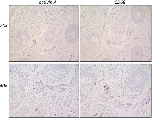
The presence of Activin A and CD68 was assessed by immunohistochemistry. The localization of activin A that we observed looks like the one previously reported137-138
.
In detail, activin A localizationwas confined
to basal epithelial cells (∆) and
in the subdermis (▼
).Figure 9
We observed that the localization of activin A in the subdermis often is in the same area where macrophages (CD68 positive cells) are present.
activin A
CD68
Figure 11
activin A
Activin A
CD68
CD68
40x
40x
20x
20x
Figure 12
12.2 STATISTIC RESULTS AND DISCUSSION
Analysing data shown in table 1 we obtained the following results. We performed regression analysis for the parameters below:
SKIN (skin laxity)/AGE; CD68/AGE;
SKIN/CD68;
MONTH (wound healing timing)/ACTIV (ActivinaA). To have a better view of graphics we standardized the parameters.
Figure 13
Linear regression analysis shows that skin laxity improves with aging (r=0,78), as we daily observe in our clinic practice. Figure 13
Figure 14
We found that CD68+ cells slightly decrease with aging (r=-0,29). CD68+ is macrophages, proinflammatory cells. Figure 14
In our clinic phase, we assumed that younger patients show more commonly edema than the older ones and it could be caused by macrophages infiltration in upper eyelids skin. Motegi et al demonstrated139 prominent infiltration of neutrophils in dermal vessels in a case study about a young
patient with history of swelling of bilateral upper eyelids. The graphic below Figure 15 show the relation to skin aging and CD68+ (r=-0,35) validate the previous result obtained.
Figure 15
Figure 16
We notice that wound healing timing increases while Activin A level growing (r=0,32). Figure 16Activin A is a proinflammatory cell, belonging to TGF-beta family able to stimulate fibrotic tissue. We investigate also the correlation between CD68+ and activina A (r=0,28) and it doesn’t depend on aging.Figure 17
Figure 17
During statistic analysis we noticed that smoking habits and aging are variables influencing correlation factors, therefore we decided to divide our sample into 4 groups:
SMOKERS; NO-SMOKERS; UNDER 55yo; OVER 55yo.
In the further paragraphs we have detailed the most significant correlations between clinical and immunohistochemical parameters for each group.
As far as smoker patients are concerning, we didn’t present any results because no significant correlations were found compared with other groups.
12.2.1 NO-SMOKERS
For NO-SMOKERS group we performed regression analysis for the parameters below: COLL(percentage of collagen)/ ACTIV (ActivinaA);
CD68/AGE;
CD68/ ACTIV (ActivinaA).
In smokers patients we obtained stronger correlations between percentage of collagen/Activin A (r=0,65 vs r=0,22)Figure 18, CD68+/aging (r=-0,44 vs r=-0,29) Figure 19 and CD68+/Activin A (r=0,48 vs r=0,28) Figure 20, than in smokers ones.
Figure 18
Figure 20
12.2.2 UNDER 55
For UNDER 55 group we performed regression analysis for the parameters below: COLL(percentage of collagen)/CD68.
Figure 21
Patients under 55 y.o.showed higher correlation between percentage of collagen and CD68+ than the value obtained considering all the patients (r=0,26 vs r=0,15).Figure 21
We build a histogram to show the influence of smoking habits on periorbital skin aging (LAXITY).
Figure 22
We found a very interesting result considering smoking habits in younger patients Figure 22: all patients with skin laxity 3 and 4 were smokers. We assumed that smoking enhances skin aging in younger patients.
12.2.3 OVER 55
For OVER 55 group we performed regression analysis for the parameters below: CD68/AGE;
MONTHS (wound healing timing)/CD68.
Graphic number Figure 23confirms results showed by graphic number 2, but in over 55 y.o. patients the correlation CD68+/aging is stronger (r=-0,64 vs r=-0,29). In clinical phase we assumed that postoperative scars redness stay longer in younger patients; maybe this result could be related to CD68+ higher level in youngers.
Figure 24
CD68+cells influence wound healing in patients over 55 y.o. more than analysis the entire sample(r=0,57 vs r=0,13) Figure 24and we notice that is decreasing with aging process Figure 14;we assumed that proiflammatory cells elongate healing timing, as we show about Activin A Figure 16.
13. CONCLUSION AND OBSERVATION:
The immunohistochemical stain showed Activin A expression in the subdermis in the same area where macrophages (CD68 positive cells) were present. The linear regression analysis showed that the percentage of Activin A and CD68 positive cells decreases with aging, maybe related to palpebral edema seen in younger patients. In addition, the percentage of Activin A and CD68 positive cells correlates to the wound healing timing. This finding suggest that Activin A and CD68 modify the healing process, increasing the inflammatory phase, and maybe determining the persistent redness present in scars of younger patients.
The analysis performed in our study could be useful also to support rejuvenation. It is fundamental to understand aging process and smoking effect in skin, in order to reduce such negative influence and increase the satisfaction in patients who underwent to cosmetic procedure
As stated in the paragraph above, we were forced to subdivide our sample into 4 groups to better investigate the effect of smoking and aging on clinical and imminohistochemical parameters.
In order to validate our results and to increase their consistency we suggest to involve more patients in future studies to make available more parameters for each group.
14. LIMITS AND PERSPECTIVES
In the future, we plan to extend our study to a larger number of patients, to validate our results, It could be interesting to investigate:
1. myofibroblats modulation including cellular markers as alfa-SMA and desmina;
2. neutrophils infiltration in dermal vessels in younger patients with bilateral upper eyelids edema;
3. effects of hormonal therapy in menopause patients (over 55).
4. How smoking habits could influence skin aging in younger patients (under 55).
15. BIBLIOGRAFY
A) Improvement of photoaged skin wrinkles with cultured human fibroblasts and adipose-derived stem cells: A comparative study Jae Hoon Jeong , Yingfang Fan , Ga Young You , Tae Hyun Choi, Sukwha Kim. Journal of Plastic, Reconstructive & Aesthetic Surgery (2015) 68, 372e381.
B) A randomised double-blinded crossover study comparing pain during anaesthetising the eyelids in upper blepharoplasty: First versus second eyelid and lidocaine versus prilocaine Shariselle M.W. Pool, Michel M.R.F. Struys, Berend van der Lei.Journal of Plastic, Reconstructive & Aesthetic Surgery (2015) 68, 1242e1247
C) Clinical aspects and molecular diagnostics of skin aging Christos C. Zouboulis, MD⁎, Evgenia Makrantonaki, MD.Clinics in Dermatology (2011) 29, 3–14
D) Anti-Aging Medicine and the Aesthetic Surgeon: A New Perspective for Our Specialty Vincent C. Giampapa,1 Antonio Fuente del Campo,2 and Oscar M. Ramirez3. Aesth. Plast. Surg. 27:493–501, 2004
E) The history of blepharoplasty to correct blepharochalasis. Kathryn L. Stephenson MD. Aesth Plast Surg 1:177-194,1977. Springer-Verlag New york inc.
F) The Caucasian and African skin types differ morphologically and functionally in their dermal componentSarah Girardeau, Sole`ne Mine, Herve´ Pageon and Daniel Asselineau
G) The Caucasian and African skin types differ morphologically and functionally in their dermal componentSarah Girardeau, Sole`ne Mine, Herve´ Pageon and Daniel Asselineau