D
EPARTMENT OF
P
HARMACY
SCHOOL OF MEDICINE AND SURGERY
Ph.D. Thesis
XXXI cycle
N
EW TARGET PROTEINS FOR DRUG DISCOVERY
IN COLON CANCER
E
STERP
AGANOT
UTOR:
P
ROF.
F
RANCESCAB
ORRELLIC
OORDINATOR:
P
ROF.
M
ARIAV.
D’A
URIAPh.D. program in Pharmaceutical Sciences
A.Y.
2015-2018
TABLE OF CONTENTS
ABSTRACT ... 6
ABBREVIATION LIST ... 8
INTRODUCTION ... 11
Chapter I. Colorectal cancer (CRC) ... 11
1.1 Physiopathology and statistics ... 11
1.2 Microenvironmental regulation of tumour angiogenesis ... 12
Chapter II. Monoacylglycerol lipase (MAGL) ... 19
1.1 Monoacylglycerol lipase (MAGL): biochemistry and physiopathology ... 19
1.2 2-arachydonoyl glycerol (2-AG) is a MAGL substrate ... 19
1.3 The role of MAGL and its substrate 2-AG in cancer ... 20
Chapter III. N-acylethanolamine acid amide hydrolase (NAAA) ... 24
1.1. N-acylethanolamine acid amide hydrolase (NAAA): biochemistry and physiopathology . 24 1.2 Palmitoylethanolamide (PEA) is a NAAA substrate ... 25
1.3 The role of NAAA and its substrate PEA in cancer ... 25
Chapter IV. G protein-coupled receptor 35 (GPR35) ... 31
1.1 Orphan G protein-coupled receptor 35 (GPR35) ... 31
1.2 GPR35: evidence supporting its involvement in cancer ... 32
1.3 Glucose metabolism in cancer ... 32
AIM ... 37
MATERIALS AND METHODS ... 38
1.1 Patients ... 38
1.2 Mice ... 38
1.3 Drugs ... 38
1.4 Cell culture... 39
1.4.1 Colorectal cancer cell lines ... 39
1.4.2 Healthy colonic epithelial cells (HCEC) ... 39
1.4.3 Endothelial cell lines ... 39
1.5 Primary cells isolation and culture ... 39
1.5.1 Murine bone marrow-derived macrophages (BMDM) ... 39
1.5.2 Isolation of Intestinal Crypts and 3D Organoid Culture ... 40
1.6 In vivo models of colorectal cancer ... 40
1.6.1 AOM DSS model of colitis associated to cancer (CAC) ... 40
1.6.3 APCmin model of sporadic colon cancer... 41
1.6.4 Xenograft model of colon cancer ... 41
1.6.4.1 Xenograft tumour secretome preparation ... 42
1.7 Cell viability (neutral red assay) ... 42
1.8 Proliferation assays ... 42
1.8.1 BrdU incorporation in colonic epithelial cells ... 42
1.8.2 EdU incorporation in colonic organoids ... 42
1.8.3 Proliferation assay in HUVEC ... 43
1.9 Migration assay ... 43
1.9.1 Scratch assay in human colon adenocarcinoma cell lines ... 43
1.9.2 Scratch assay in HUVEC ... 43
1.10 Angiogenesis analysis ... 44
1.10.1 Tube formation assay ... 44
1.10.2 CXCL1/KC detection ... 44
1.10.3 Aortic ring assay ... 44
1.10.4 Quantification of angiogenesis modulators in HUVEC by ELISA ... 44
1.11 Evaluation of metabolic profile ... 44
1.11.1 Glucose uptake ... 44
1.11.2 Seahorse extracellular flux analysis ... 45
1.11.3 Lactate measurements ... 45
1.12 BrdU incorporation and quantification ... 45
1.13 Identification and quantification of endocannabinoids, PEA and related molecules .... 45
1.14 Immunohistochemistry ... 46
1.15 Immunostaining for CD31... 46
1.16 Gene expression analysis by quantitative Real-Time (q)-PCR ... 47
1.17 Western blot analysis ... 47
1.18 Statistical analysis ... 48
RESULTS ... 51
Chapter I. Pharmacological inhibition of MAGL reduces experimental colon tumorigenesis ... 51
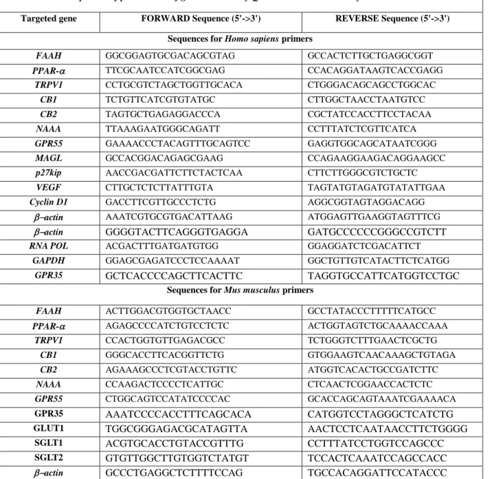
1.1 MAGL expression in human colorectal carcinoma (HCT 116) cells and xenograft tumour tissues ... 51
1.2 The MAGL inhibitor URB602 reduces tumour growth in the xenograft model of colon cancer ... 51
1.3 Effect of URB602 (a MAGL inhibitor) on 2-AG levels in healthy and tumour tissues from xenografted mice ... 51
1.4 The MAGL inhibitor URB602 drives in vivo angiogenesis output in xenograft tumour tissues ... 56
1.5 Direct antiangiogenic effect of the MAGL inhibitor URB602 in HUVEC ... 56
1.6. The MAGL inhibitor URB602 regulates Cyclin D1 in xenograft tumour tissues ... 56
1.7. URB602 exerts chemopreventive effects in the azoxymethane (AOM) model of colon carcinogenesis ... 57
Chapter II. NAAA is crucially involved in proliferation, migration and in vivo colon tumorigenesis ... 65
1.1 NAAA is down-regulated in human CRC tissues ... 65
1.2 The NAAA inhibitor AM9053 reduces tumour growth in the xenograft model of colon cancer ... 65
1.3 Effect of the NAAA inhibitor AM9053 on NAAA and PEA targets expression in xenograft colon cancer tumours ... 65
1.4 The incubation with xenograft tumour secretome reduces NAAA expression in colorectal cancer (CRC) cells ... 69
1.5 The NAAA inhibitor AM9053 and its substrate PEA reduce the number of tumours induced by azoxymethane (AOM) ... 69
1.6 AM9053 (NAAA inhibitor) and PEA inhibit the proliferation of colorectal cancer - but not of healthy colonic - cells ... 69
1.7 AM9053 and PEA treatments lessen the migration of colorectal cancer cells ... 69
Chapter III. GPR35 promotes glycolysis, proliferation and colon tumorigenesis ... 76
1.1 Gpr35-/- BMDM have reduced glucose uptake ... 76
1.2 The lack of GPR35 impairs the metabolic profile of macrophages ... 76
1.3 GPR35 influences angiogenesis via macrophages ... 80
1.4 GPR35 is involved in experimental colitis-associated cancer and sporadic colon cancer ... 83
1.5 GPR35 affects intestinal epithelial cell turn-over under homeostatic conditions ... 83
DISCUSSION ... 87
Part I. Pharmacological inhibition of MAGL reduces experimental colon tumorigenesis ... 87
1.1 Presence of MAGL and 2-AG in the xenograft tumour tissue ... 87
1.2 Effect of URB602 on 2-AG levels, tumour progression and angiogenesis ... 87
1.3 Effect of URB602 in the azoxymethane (AOM) model of colon carcinogenesis ... 89
1.4 Concluding remarks ... 90
Part II. NAAA is crucially involved in proliferation, migration and in vivo colon tumorigenesis ... 91
1.1 NAAA and PPAR− expression in human CRC ... 91
1.2 Effect of AM9053 on experimental carcinogenesis ... 91
1.3 Effect of AM9053 and exogenous PEA on proliferation and migration of colorectal cancer cells ... 93
1.4 Concluding remarks ... 94
Part III. GPR35 promotes glycolysis, proliferation and colon tumorigenesis ... 95
1.2 GPR35 affects angiogenesis ... 96
1.3 GPR35 has a role in colonic proliferation and tumorigenesis ... 96
1.4 Concluding remarks ... 97
GENERAL CONCLUSIONS ... 98
ABSTRACT
Background: Colorectal cancer (CRC) is one of the most common causes of death in Western countries. Because of the high heterogeneity and incidence of this disease, it is crucial to improve the knowledge on its biology – a fundamental step for drug discovery - and to develop clinically-relevant diagnostic and prognostic biomarkers.
Evidence suggests that MAGL [(a serine hydrolase that converts monoacylglycerols, such as the endocannabinoid 2-arachydonoyl glycerol (2-AG), in glycerol and fatty acid], NAAA [(a cysteine hydrolase responsible of the catabolism of palmitoylethanolamine (PEA)] and GPR35 (an orphan-G protein coupled receptor) affect biological events (e.g. proliferation, differentiation, survival) and regulates pathophysiological states (e.g. intestinal inflammation) which are suggestive of a possible involvement in CRC. Here, we explored the possible contribution of MAGL, NAAA and GPR35 to colon tumorigenesis.
Material and Methods: The role of MAGL, NAAA and GPR35 was assessed in vivo, via genetic or pharmacological blockade, in APCmin mice as well as in the azoxymethane (AOM),
AOM/dextran sodium sulfate (DSS) and xenograft models.
Cell proliferation was evaluated in CRC cells, healthy colonic epithelial cells and colonic organoids 3D culture by using the BrdU and EdU incorporation; migration was examined in CRC and endothelial cells by using the scratch assay. Angiogenesis was assessed in tumour tissues [by microvessel counting and by investigating the expression of vascular endothelial growth factor (VEGF) and fibroblast growth factor-2 (FGF-2) proteins] as well as in the aortic ring model and in endothelial cells by using the tube formation assay. Cell metabolism was measured by the quantification of extracellular acidification rate (ECAR), oxygen consumption rate (OCR), lactate production and glucose uptake in bone marrow derived macrophages (BMDM). MAGL, NAAA and GPR35 expression was evaluated by RT-PCR and immunohistochemistry; 2-AG and PEA levels were measured by liquid chromatography mass spectrometry.
Results: MAGL- The MAGL inhibitor URB602 reduced xenograft tumour growth, the effect being associated to regulation of VEGF and FGF-2, reduction in the number of vessels and down-regulation of cyclin D1. A direct antiangiogenic effect was observed in human endothelial cells, too. In experiments aiming at investigating the role of MAGL in chemoprevention, URB602 attenuated AOM-induced preneoplastic lesions and tumours in wild-type but not in MAGL-deficient mice. NAAA- NAAA expression was reduced in biopsies of clinically-diagnosed CRC patients as well as in CRC cells incubated with tumour secretome. The NAAA inhibitor AM9053 and its substrate PEA inhibited proliferation and migration in CRC cells. Increased PEA levels in
in the AOM model of colon carcinogenesis and reduced xenograft tumour growth. GPR35- GPR35 deletion resulted in a reduction of cell energetic demand and production in M0-, M1- and M2- BMDM. Also, depletion of GPR35 in M2-BMDM decreased their capability to produce the CXCL1 (pro-angiogenic chemokine) and to stimulate the tube formation of endothelial cells. Also, GPR35-/- mice showed a reduced intestinal turnover in physiological conditions and, importantly, a reduced colon tumorigenesis, as highlighted in two different experimental models of colon cancer. It is noteworthy that the protective role of GPR35 in APCmin mice was confirmed with the conditional deletion of Gpr35 (Villin-Cre) in the intestinal epithelium.
Conclusions: In summary, by elucidating the physiopathological role of MAGL, NAAA and GPR35 in experimental colon tumorigenesis and by ascertain their dysregulation in intestinal tumours, this PhD thesis put forth such proteins as possible innovative prognostic markers in clinically-diagnosed CRC and as new molecular targets to be explored in drug discovery.
ABBREVIATION LIST
15-LOX 15-lipoxygenase
2-AG 2-arachydonoyl glycerol
2DG 2-deoxy-D-glucose
2DG6P 2-deoxy-D-glucose-6-phosphate
5-FU 5-fluorouracil
AA arachidonic acid
ABHD12 α/β-hydrolase domain containing 12
ABHD6 α/β-hydrolase domain containing 6
ACF aberrant crypt foci
Adenosine-A3 adenosine A3 receptor
AEA N-arachydonoylethanolamide or anandamide
AOM azoxymethane
APC adenomatous polyposis coli
ATCC American type culture collection
BMDM bone marrow-derived macrophages cell line
BrdU 2'-deoxy-5-bromo-uridine
BSA bovine serum albumin
CAC colitis associated to cancer
Caco-2 human colorectal adenocarcinoma cell line
CAFs cancer-associated fibroblasts
CB1 cannabinoid receptor 1
CB2 cannabinoid receptor 2
CD Crohn’s disease
CD31 cluster of differentiation 31
cDNA complementary DNA
cGMP cyclic guanosine 3’,5’-monophosphate
COX-2 cyclooxygenase 2
CRC colorectal cancer
CXCL CXC-chemokine ligand
CXCL17 chemokine ligand 17
DAG diacylglycerol
DAGL diacylglycerol lipase
DMEM Dulbecco's modified eagle medium
DSS dextran sodium sulphate
ECAR extracellular acidification rate
ECM extracellular matrix
ECs endothelial cells
EDTA ethylenediaminetetraacetic acid
EdU edoxyudine
EGF epidermal growth factor
EGM-2 endothelial growth medium
ELISA enzyme-linked immunosorbent assay
EMT endocannabinoid membrane transporter
EVs extracellular vesicles
FAAH fatty acid amide hydrolase
FAP familial adenomatous polyposis
FBS fetal bovine serum
FITC fluorescein 5-isothiocyanate
FOLFIRI fluorouracil/leucovorin and irinotecan
FOLFOX fluorouracil/leucovorin and oxaliplatin
GABAA type A γ-aminobutyric acid receptor
GAPDH glyceraldehyde 3-phosphate dehydrogenase
GDP guanosine diphosphate
GPCR G protein-coupled receptor
GPR55 G protein-coupled receptor 55
GTP guanosine triphosphate
GWAS genome-wide association study
HCEC healty human colonic epithelial cells
HCT116 human colorectal adenocarcinoma cell line
HNPCC hereditary non-polyposis colorectal cancer
HPLC high performance liquid chromatography
HUVEC human umbilical vein endothelial cells
IBD inflammatory bowel disease
IECs intestinal epithelial cells
IFNα interferon-α
IFNγ interferon-γ
IL interleukin
IL-4 interleukin-4
ip intraperitoneal administration
K-Ras Kirsten RAt Sarcoma
LPS lipopolysaccharide
M1 pro-inflammatory phenotype of macrophages
M2 pro-resolving phenotype of macrophages
MAGL monoacylglycerol lipase
M-CSF macrophage colony-stymulating factor
MMP matrix metalloproteinase
MΦ macrophages
NAAA N-acylethanolamine acid amide hydrolase
NAEs N-acylethanolamines
NAPE-PLD N-acyl-phosphatidylethanolamine selective phospholipase D
NAT N-acyl-transferase
NPPE N-palmithoylphosphatidylethanolamine
NR neutral red
OCR oxygen consumption rate
OEA N-oleoylethanolamide
p53 tumour suppressor protein
PBS phosphate-buffered saline
PC prostate cancer
PCR polymerase chain reaction
PEA N-palmythoilethanolamide PGD2 prostaglandin D2 PGE2 prostaglandin E2 PGF2 prostaglandin F2 PGs prostaglandins PLC phospholipase C
PPAR-α peroxisome proliferator-activated receptor α
PPAR-γ peroxisome proliferator-activated receptor γ
PSC primary sclerosing cholangitis
RPMI 1640 Roswell Park Memorial Institute 1640 medium
SDS-PAGE sodium dodecyl sulfate-polyacrylamide gel electrophoresis
SNP single-nucleotide polymorphism
SVEC4-10 immortalized murine endothelial cells
TAMs tumour-associated macrophages
TASCs tumour-associated stromal cells
TBS Tris-buffered saline
TGFβ transforming growth factor β
TNF tumour necrosis factor
TNM classification of malignant tumours
TRPV1 transient receptor potential cation channel subfamily V type 1
UC ulcerative colitis
VEGF vascular endothelial growth factor
VEGFA vascular endothelial growth factor A
VEGF-R vascular endothelial growth factor receptor
CHEMICAL NAMES AM630 [6-iodo-2-methyl-1-(2-morpholin-4-ylethyl)indol-3-yl]-(4-methoxyphenyl)methanone AM9023 1-isothiocyanatopentadecane ARA-C 4-amino-1-[(2R,3S,4S,5R)-3,4-dihydroxy-5-(hydroxymethyl)oxolan-2-yl]pyrimidin-2-one ARN077 5-phenylpentyl-N-[(2S,3R)-2-methyl-4-oxo-oxetan-3-yl]carbamate ARN726 4-cyclohexylbutyl-N-[(S)-2-oxoazetidin-3-yl]carbamate DAB 4-(3,4-diaminophenyl)benzene-1,2-diamine
EDTA 2-[2-[bis(carboxymethyl)amino]ethyl-(carboxymethyl)amino]acetic acid
EdU 5-ethyl-1-[(2R,4S,5R)-4-hydroxy-5-(hydroxymethyl)oxolan-2-yl]pyrimidine-2,4-dione
FCCP 2-[[4-(trifluoromethoxy)-phenyl]hydrazinylidene]propanedinitrile
FITC 3',6'-dihydroxy-6-isothiocyanatospiro[2-benzofuran-3,9'-xanthene]-1-one
HEPES 4-(2-hydroxyethyl)-1-piperazineethanesulfonic acid
HU-210 11-hydroxy- Δ8- tetrahydrocannabinol
JZL184 (4-nitrophenyl) 4-[bis(1,3-benzodioxol-5-yl)-hydroxymethyl]piperidine-1-carboxylate MK886
3-[3-tert-butylsulfanyl-1-[(4-chlorophenyl)methyl]-5-propan-2-ylindol-2-yl]-2,2-dimethylpropanoic acid
S-OOPP N-[(3S)-2-oxooxetan-3-yl]-3-phenylpropanamide
INTRODUCTION
Chapter I. Colorectal cancer (CRC) 1.1 Physiopathology and statistics
Colorectal cancer (CRC) represents a common cause of cancer-related deaths. It has been estimated that, in 2018, it will be the third most common cause of deaths in USA, with an equal incidence in males and females (8%) and with a prediction of 140,250 new diagnosed cases (Siegel et al., 2018; Figure 1). Similarly, CRC has been placed at the second position in the oncologic death rank in European countries for 2018 (Malvezzi et al., 2018; Figure 2) and in Italy 53.000 new CRC cases has been estimated to be diagnosed in the same year (www.airtum.it).
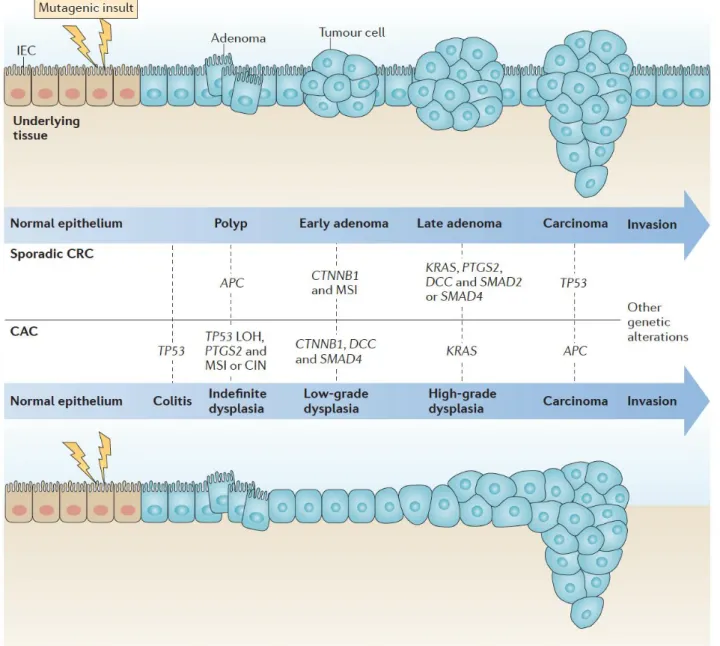
The majority of colorectal cancers are sporadic, arising from dysplastic adenomatous polyps. In this case, CRC develops as a consequence of the progressive accumulation of genetic and epigenetic alterations that cause transformation and progression of normal mucosa to adenoma and subsequently to carcinoma (Lao and Grady, 2011) (Figure 3). These alterations are the consequence of mutations in genes involved in cell growth regulation, such as tumour-suppressor genes (e.g. APC, Smad4 and p53) or oncogenes (e.g. K-Ras, c-myc, c-neu, c-src) (Calvert and Frucht H, 2002). This multi-step process spans 10 to 15 years, thereby providing an opportunity for prevention. It is also well established that CRC is largely influenced by lifestyle, genetic predisposition and longstanding intestinal inflammation. About 10% of CRCs develops in the setting of well-defined hereditary syndromes i.e. hereditary non-polyposis colorectal cancer (HNPCC) and familial adenomatous polyposis (FAP) (Lynch and de la Chapelle, 2003; Rustgi, 2007).
Chronic gut inflammation is one of the main causes of increased risk of developing CRC (Terzic et al., 2010; Ullman and Itzkowitz, 2011) (Figure 3). Patients affected by inflammatory bowel diseases (IBD), such as ulcerative colitis (UC) and Crohn’s disease (CD), are at increased risk of developing neoplasia (Gupta et al., 2007; Itzkowitz and Harpaz, 2004), with an extended incidence rate of 2.75 and 2.64 of CRC in patients with UC and CD, respectively (Bernstein et al., 2001). It is worth noting that colitis can promote tumorigenesis by altering microbial composition as well as the expansion of microorganisms with genotoxic capabilities (Richard et al., 2018; Dejea et al., 2018). Other risk factors include obesity, meat and fat-rich diet, smoking, alcohol consumption and hyperinsulinemia (Marley and Nan, 2016; Wynder et al., 1969).
CRC is largely asymptomatic until the latter stages, when the cancer has already metastasized, thus a huge emphasis is also placed on early detection. Standard clinical practice for CRC patients include primarily the surgical resection, followed by neoadjuvant radiotherapy (for patients with rectal cancer), and adjuvant chemotherapy (for patients with stage III/IV and high-risk stage II colon cancer) (Marley and Nan, 2016). 5-fluorouracil (5-FU) is used in several poly-chemotherapy
Introduction - Chapter I Colorectal cancer
regimens in CRC advanced patients, such as FOLFOX (Fluorouracil/Leucovorin and Oxaliplatin) and FOLFIRI [Fluorouracil/Leucovorin and Irinotecan (IR)] (Gustavsson et al., 2015). However, intrinsic or acquired resistance to 5-FU may occur (Ahn et al., 2015).
Despite the clear progresses raised in colon cancer diagnosis and therapy, the number of deaths still remain unacceptably high. Therefore, it is crucial to improve the knowledge on CRC pathogenesis, and, importantly, to identify new strategies, including chemoprevention and early diagnosis, in order to reduce the public health burden of this disease.
1.2 Microenvironmental regulation of tumour angiogenesis
Microenvironment plays a crucial role in the tumour growth and a considerable number of key transduction signals are involved in tumour microenvironment progression. Specifically, it is largely reported the importance of angiogenesis for the development of solid tumours, because the dissemination of tumours requires the growth of new blood vessels (Risau, 1997; Hanahan and Folkman, 1996).
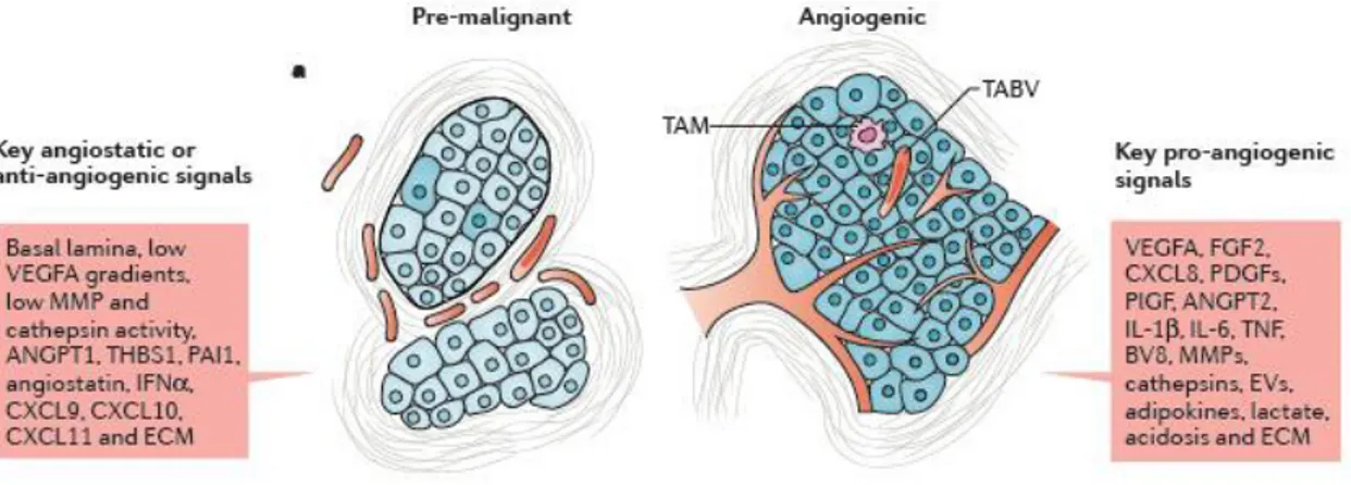
Hypoxic cancer cells secrete vascular endothelial growth factor A (VEGFA), which initiates tumour angiogenesis by engaging VEGF receptor 2 expressed on the endothelial cells of neighbouring blood vessels (Potente et al., 2011). In pre-malignant stages of epithelial tumours, a basal lamina precludes the interaction between early lesions and the vascularized peritumoral tissues (Bossi et al., 1995; Bluff et al., 2009) (Figure 4). Instead, in the malignant tumours, cancer cells acquire invasive behaviours and induce a stromal response involving the formation of new blood vessels (Bergers and Benjamin, 2003) (Figure 4). Tumour-associated blood vessels typically acquire an aberrant morphology, which indicates the impaired vascular maturation, poor vessel functionality and incoherent tumour perfusion (Potente et al., 2011; Bergers and Benjamin, 2003; Morikawa et al., 2002). This angiogiogenic switch, strictly correlated with cancer progression, shows substantial variances in the patterns of tumour vascularization, which reflects differences in tumour type, grade and stage, anatomical site and expression of pro-angiogenic and anti-angiogenic factors (Bluff et al., 2009; Fukumura et al., 1997; Jubb et al., 2011; Morrissey et al., 2008).
Concerning CRC, it is characterised by enhanced VEGF expression and high microvascular densities, - suggestive of increased angiogenic activity - which negatively affect patient survival (Des Guetz et al., 2006). Consistently, therapy with anti-VEGF antibodies improves CRC patient survival, thus emphasising VEGF as a major clinical angiogenic factor (Carmeliet, 2005; Ferrara et al., 2007; Hawinkels et al., 2008).
Although cancer cells play a pivotal role as source of VEGFA and other pro-angiogenic mediators, many signals are produced from various tumour-associated stromal cells (TASCs), including bone marrow-recruited cells (e.g. macrophages, lymphocytes and neutrophils) as well as tissue-resident
cells such as vascular cells (i.e. endothelial cells and pericytes) and fibroblasts (Fang and Salven, 2011) (Figure 5).
In mouse models of cancer, macrophages derive mainly from circulating monocytes that extravasate into tumours in response to various chemoattractant stimuli (Qian and Pollard, 2010; Lahmar et al., 2016). Thereafter, monocytes differentiate and mature into tumour-associated macrophages (TAMs), i.e. the major constituents of malignant tumours - under the influence of colony-stimulating factor 1 (M‑CSF) (Qian and Pollard, 2010; Lahmar et al., 2016).
Current therapeutic interventions often fail to prevent disease progression to metastatic dissemination. Hence, innovative therapeutic approaches include the targeting of specific molecular pathways such as proteins involved in the tumour angiogenesis and metastasis (Gelmon et al., 1999; Goetz et al., 2003).
Introduction - Chapter I Colorectal cancer
Figure 1. Ten Leading Cancer Types for the Estimated New Cancer Cases and Deaths by Sex, United States, 2018 (from Siegel, 2018).
Estimates are rounded to the nearest 10 and cases exclude basal cell and squamous cell skin cancers and in situ carcinoma except urinary bladder. Ranking is based on modeled projections and may differ from the most recent observed data.
Figure 2. Prediction of cancer mortality in the European Union (EU) for the year 2018 (from Malvezzi et al., 2018).
Bar-plots of age-standardized (world population) death rates per 100 000 persons for the year 2012 (blue) and predicted rates for 2018 (green) with 95% prediction intervals for total cancer and the eight major cancer sites in EU men and women.
Introduction - Chapter I Colorectal cancer
Figure 3. Adenoma to carcinoma sequence (from West et al., 2015)
Canonical mechanisms of sporadic colorectal cancer (CRC) and colitis-associated cancer (CAC) development are shown in the top and bottom panels, respectively. CRC and CAC share similarities in their developmental pathways, including microsatellite instability (MSI), activation of the oncogene KRAS, activation of cyclooxygenase 2 (COX2; encoded by PTGS2), and mutation and eventual loss of heterozygosity (LOH) of TP53, adenomatous polyposis coli (APC), deleted in colon cancer (DCC) and SMAD4. However, the frequency and sequence of these events differs between the cancers. For example, mutation in APC is one of the first events in CRC, whereas it occurs at later stages in CAC. By contrast, TP53 mutations usually occur early in CAC but at a later stage in the progression of CRC. Although CRC shows a clear progression of morphological changes, from polyp to carcinoma, CAC progression involves increasing histological grades of dysplasia that culminate in an invasive carcinoma. CTNNB1, gene encoding β-catenin; CIN, chromosomal instability; IEC, intestinal epithelial cell.
Figure 4. Angiogenesis during malignant progression (from De Palma et al., 2017)
Early-stage (pre-malignant) tumours typically display scant or no intratumoural vascularization, although a vascularized stroma surrounds the tumours and may adjoin parenchymal tumour domains (left panel). In malignant tumours, the cancer cells acquire invasive behaviours and induce a stromal response involving robust intratumoural angiogenesis, along with leukocyte infiltration, fibroblast proliferation and extracellular matrix (ECM) deposition (right panel). In pre-malignant lesions, a basal lamina separates the tumour from the surrounding tissues; this, together with angiostatic signals conveyed by some ECM components, and the relatively low levels of pro-angiogenic factors, prevents intratumoural vascularization or constrains it into a quiescent state. In malignant lesions, angiogenesis is largely controlled through the actions on vascular endothelial cells (ECs) of multiple pro-angiogenic mediators, which include growth factors, cytokines, various ECM proteins, ECM-remodelling enzymes, as well as extracellular vesicles (EVs) and by-products of deregulated tumour metabolism. ANGPT, angiopoietin; CXCL, CXC-chemokine ligand; EV, extracellular vesicle; FGF2, fibroblast growth factor 2; IFNα, interferon-α; IL, interleukin; MMP, matrix metalloproteinase; PAI1, plasminogen activator inhibitor 1; PDGF, platelet-derived growth factor; PlGF, placental growth factor; TABV, tumour-associated blood vessel; THBS1, thrombospondin 1; TNF, tumour necrosis factor; VEGFA, vascular endothelial growth factor A.
Introduction - Chapter I Colorectal cancer
Figure 5. Tumour microenvironment and role of the inflammation (from West et al., 2015) Production of inflammatory cytokines by several cell types including CD4+ T cells, innate lymphoid cells (ILCs) and tumour-associated macrophages (TAMs). Many of the inflammatory cytokines can act directly on transformed intestinal epithelial cells (IECs) to promote proliferation, inhibition of apoptosis, invasion, angiogenesis, epithelial to mesenchymal transition (EMT) and metastasis. More recently, it has been appreciated that these cytokines can activate cancer-associated fibroblasts (CAFs) to produce cytokines and growth factors that modulate both neoplastic cells and the tumour microenvironment.
Chapter II. Monoacylglycerol lipase (MAGL)
1.1 Monoacylglycerol lipase (MAGL): biochemistry and physiopathology
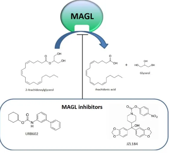
Monoacylglycerol lipase (MAGL) is a serine hydrolase that converts monoacylglicerols, such as the endocannabinoid 2-arachydonoyl glycerol (2-AG) to glycerol and fatty (arachidonic) acid. MAGL consists of two tissue specific isoforms with a molecular weights of 33 kD and 36 kD, which could reflect different splice variants (Karlsson et al., 2001; Long et al., 2009a). The enzyme associates with membranes and its active site resides within the cytosol.
MAGL is localized in different areas of brain and in peripheral tissues, including the gastrointestinal tract (Ahn et al., 2008; Duncan et al., 2008; Izzo and Camilleri, 2009). MAGL blockade shows tissue-specific differences in monoacylglycerol metabolism, with the brain showing the most dramatic elevations in 2-AG and peripheral tissues often showing greater changes in other monoacylglycerols, consistent with the lipolytic role of MAGL as the final step of triglyceride hydrolysis in peripheral tissues (Long et al., 2009b).
In addition to the role of MAGL in terminating 2-AG signaling, MAGL exerts a crucial control over brain prostaglandins production in both basal and neuroinflammatory states. (Nomura et al., 2011a). In fact, through the hydrolysis of 2-AG, MAGL releases arachidonic acid (AA), i.e. the major precursor for the synthesis of pro-inflammatory eicosanoids (Nomura et al., 2011a). MAGL blockade decreases AA levels in the brain, stoichiometrically to 2-AG elevation, which also results in a reduction of lipopolysaccharide (LPS)-induced pro-inflammatory levels of downstream COX-driven prostaglandins [i.e. prostaglandin E2 (PGE2), PGD2, PGF2], and thromboxane production (Nomura et al., 2011a). For this reason, MAGL is an important target for neuroinflammatory and neurodegenerative disorders.
In recent years, the understanding of MAGL role has greatly increased, due to the progress made in the knowledge of the structure of MAGL, the synthesis of highly potent and selective in
vivo efficacious inhibitors, e.g. JZL184 and URB02, as well as the development of MAGL-deficient
mice (Chanda et al., 2010; Long et al., 2009a; Sclosburg et al., 2010) (Figure 6). Consequently, an impressive number of studies have described the therapeutic potential of MAGL inhibitors in a variety of human diseases – including cancer - through the bidirectional manipulation of endocannabinoids, eicosanoids, and other lipid signaling pathways.
1.2 2-arachydonoyl glycerol (2-AG) is a MAGL substrate
MAGL plays a predominant role in catalyzing the hydrolysis of the endocannabinoid 2-arachydonoyl glycerol (2-AG) (Dihn et al., 2002; Dihn et al., 2004; Hohmann et al., 2005). 2-AG is generated “on demand” through stimulus-dependent cleavage of membrane phospholipid precursors and its levels are regulated by the balance between its production and degradation (Murataeva et al.,
Introduction - Chapter II Monoacylglycerol lipase
2014). 2-AG is thought to be produced through hydrolysis of phospholipids by phospholipase C (PLC) β or δ to release diacylglycerols (DAG), which are then degraded to 2-AG by diacylglycerol lipase (DAGL) α or β (Gao et al., 2010; Tanimura et al., 2010). (Figure 7).
Genetic or pharmacological inhibition of MAGL reduces 2-AG hydrolysis by >80% in most tissues, including the brain. The remaining amount of 2-AG is metabolized by the hydrolytic enzymes α/β-hydrolase domain 6 (ABHD6), ABDH12, fatty acid amide α/β-hydrolase (FAAH) (Blankman et al., 2007; Dihn et al., 2004; Marrs et al., 2010; Goparaju et al., 1998), 15-lipoxygenase (Kozak et al., 2002) and monoacylglycerol acyltransferases/kinases (Simpson et al., 1991). Although the other enzymes may have roles in 2-AG hydrolysis in certain settings, MAGL blockade lead to dramatic elevations, especially in the brain, in 2-AG levels, confirming that MAGL is the primary enzyme involved in degrading 2-AG in vivo (Long et al., 2009b; Nomura et al., 2011a; Schlosburg et al., 2010; Fowler, 2012).
2-AG is an endogenous lipid mediator involved in a variety of physiological and physiopathological processes. 2-AG behaves as full agonist for the cannabinoid (CB)1 and CB2 receptors (Sugiura et al.,
1999; Sugiura et al., 2000). The 2-AG homologues 2-linoleoylglycerol and 2-palmitoylglycerol, which are present in tissues and presumably released together with 2-AG, act as ‘entourage compounds’ to potentiate the effects of 2-AG at CB receptors without themselves having a direct action on CB receptors (Ben-Shabat et al., 1998). In addition, several studies report that 2-AG may also exert physiological effects independently from CB1 and CB2 receptors activation, for example by binding to and activating GABAA (Sigel et al., 2011), PPAR-γ (Bouaboula et al., 2005),
adenosine A3 (Lane et al., 2010), TRPV1 (Iwasaki et al., 2008), and GPR55 (Ryberg et al., 2007) (Figure 7). However, CB1 receptor is the major brain target through which 2-AG exerts its physiopathological effects, including regulation of food intake and metabolism, neuro-inflammation, addiction, anxiety and pain (Baggelaar et al., 2018).
1.3 The role of MAGL and its substrate 2-AG in cancer
MAGL is up-regulated in aggressive human ovarian, prostate, breast cancer cells and in primary tumours, where it promotes migration, invasion, survival, and in vivo tumour growth (Mulvihill and Nomura, 2013; Nomura et al., 2010; Nomura et al., 2011b). In cancer cells, MAGL plays a role in controlling global levels of free fatty acids, which serve as building blocks for the synthesis of pro‐tumorigenic signalling lipids, such as prostaglandin E2 and lysophosphatidic acid (Qin et al., 2014; Nomura et al., 2010). Recently, it has been reported that MAGL is upregulated in melanoma tissue and its expression in tumour cells is significantly associated with tumour aggressiveness, vascular invasion of the primary lesion and tumour progression (Baba et al., 2017).
Concerning 2-AG, i.e. the main substrate of MAGL, studies have shown that it exerts antiproliferative effects in a number of cancer cell lines (Melck et al., 2000; Costa et al., 2014; Orellana-Serradell et al., 2015), including colorectal cancer cells (Ligresti et al., 2003). For example, the increase of endogenous 2-AG levels as a consequence of MAGL inhibition was shown to reduce prostate cancer invasion in vitro (Nomura et al., 2011b). On the other hand, the reduction of physiological endogenous 2-AG levels has been reported to stimulate cell invasion (Nithipatikom et al., 2011).
Our understanding of how MAGL impacts colon cancer progression is so far hindered by under-reported and controversial data (Schicho and Storr, 2011; Izzo et al., 2015), since the enzyme has been associated to both cancerogenic (Sun et al., 2013) and anti-cancerogenic processes (Ye et al., 2011). Nevertheless, the effect and role of MAGL in relation to 2-AG production and angiogenesis have been not evaluated to date.
Introduction - Chapter II Monoacylglycerol lipase
Figure 6. Conversion of 2-Arachidonoylglycerol (2- AG) into arachidonic acid and glycerol by monoacylglycerol lipase (MAGL). MAGL inhibitors such as URB602 and JZL184 increase the endogenous levels of 2-AG.
Figure 7. Metabolic pathways and molecular targets of 2-AG (adapted from Iannotti and Piscitelli, 2018).
ABH4/6/12, αβ-hydrolase 4/6/12; CB1/2, cannabinoid receptor 1/2; COX2, cyclooxygenase 2;
DAG, diacylglycerol; EMT, 'endocannabinoid membrane transporter'; FAAH, fatty acid amide hydrolase; GDE1, glycerophosphodiester phosphodiesterase; GPR55, G-protein-coupled receptor 55; PLCβ, phospholipase Cβ; PLD, phospholipase D; PTPN22, protein tyrosine phosphatase, nonreceptor type 22; TRPV1, transient receptor potential, vanilloid subtype 1 receptor; GABAA, type-A γ-aminobutyric acid receptor; adenosine A3, adenosine A3 receptor; PPARγ, peroxisome proliferator-activated receptor gamma; 15-LOX, lipoxygenase-15; PMs, prostaglandin-ethanolamides/prostamides; PG-GEs, prostaglandin-glyceryl esters.
Introduction - Chapter III N-acylethanolamine acid amide hydrolase
Chapter III. N-acylethanolamine acid amide hydrolase (NAAA)
1.1. N-acylethanolamine acid amide hydrolase (NAAA): biochemistry and physiopathology N-acylethanolamine acid amide hydrolase (NAAA) is a N-terminal cysteine hydrolase of the choloylglycine hydrolase family. Being a lysosomal enzyme, NAAA is produced as an inactive pro-enzyme and autocatalytically activated by cleavage at acidic pH. Human and murine NAAA are similar with 76% sequence identity (Tsuboi et al. 2005). Glycosylation of the enzyme was firstly believed to be necessary to reach its full activity. However, more recent findings have demonstrated that glycosylation is most probably necessary for trafficking of the enzyme to the lysosomes and its maturation, with minimal effects on enzyme activity (Pavlopouloset al. 2018).
NAAA is responsible of the hydrolysis of N-acylethanolamines (NAEs), mainly PEA and to a very less extent OEA and AEA. Consistently, NAAA inhibitors increase preferentially PEA levels in
vitro and in vivo, making NAAA a promising target for drug discovery (Tsuboi et al. 2005; Ueda et
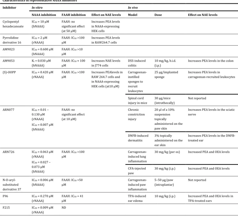
al., 2013; Alhouayek and Muccioli, 2014; Bottemanne et al., 2018) (Figure 8). With the recent molecular identification of NAAA, a number of selective and stable NAAA inhibitors have been synthesised and tested in a variety of disease models. While the first generation of inhibitors were irreversible and moderately potent, in more recent years new potent, reversible, stable and selective inhibitors have been synthesised (Bottemanne et al., 2018; Figure 8). Makriyannis’s research group developed a series of isothiocyanate-based NAAA reversible inhibitors such as AM9053 and AM9023 (West et al., 2012; Malamas et al., 2015) (Table 1). Other series of potent NAAA inhibitors were developed by Piomelli and co-workers around -lactone and -lactam moieties, such as -lactone (S)-OOPP, ARN077 and ARN726 (Solorzano et al., 2009; Duranti et al., 2012; Ponzano et al., 2013; Ribeiro et al., 2015; Nuzzi et al., 2016) (Table 1). These new inhibitors are precious tools to study the role of NAAA.
Because NAAA is highly expressed in bone marrow and immune system, especially in macrophages, its pathophysiological role was firstly evaluated in inflammatory processes (Solorzano et al., 2009; Sasso et al., 2013; Yang et al., 2015). Moreover, because NAAA is believed to primarily control PEA levels (Tsuboi et al. 2005; Tai et al., 2012), its inhibition is expected to mimic the anti-inflammatory, neuroprotective and analgesic properties of PEA (Alhouayek and Muccioli, 2014). NAAA is also largely expressed in human and murine small and large intestine (Borrelli et al., 2015; Alhouayek et al., 2015) and its pharmacological inhibition reduces colon inflammation by locally increase PEA levels (Alhouayek et al., 2015). However, it is important to note that NAE levels as well as the expression of NAAA may be altered differently depending on the tissue or the disease considered.
1.2 Palmitoylethanolamide (PEA) is a NAAA substrate
N-acylethanolamines (NAEs) are endogenous lipids consisting of an acyl chain linked by an amide bond to ethanolamine. Although sharing the same basic scaffold, NAEs can bind to several different receptors and exert a plethora of biological effects (Bottemanne et al., 2018). Besides the well characterized NAEs, such as the endocannabinoid arachidonoylethanolamine (AEA), N-palmitoylethanolamine (PEA) is one of the most-studied NAEs, being it clinically used for its anti-inflammatory, analgesic and neuroprotective properties mostly mediated by PPAR- (Alhouayek and Muccioli, 2014; Gugliandolo et al., 2018; Esposito and Cuzzocrea, 2013; Skaper et al., 2015; Iannotti et al., 2016). Despite PPAR is the main reported target (Lo Verme et al., 2005), additional and pharmacologically relevant targets of PEA have been reported (Petrosino and Di Marzo, 2017) (Figure 9). Thus, PEA has also been shown to be an agonist of orphan G-protein coupled receptor 55 (GPR55) (Ryberg et al., 2007), although this pharmacological implication remains to be clarified. In addition, unlike AEA, PEA does not bind to cannabinoid (CB) receptors, although it can indirectly activate them via the so-called “entourage” effect, i.e. through the inhibition of FAAH, the enzyme responsible for endocannabinoids degradation (Di Marzo et al., 2001; Petrosino et al., 2016). Finally, PEA may directly or indirectly modulate (via enhancement of AEA levels or via PPAR-α activation) transient receptor potential cation channel subfamily V (TRPV)1 channels (De Petrocellis et al., 2002; Ho et al., 2008; Ambrosino et al., 2013; Ambrosino et al., 2014) (Figure 9).
In addition to be an endogenous lipid molecule, PEA is also a food component, first discovered in the late 1950s, when it was shown that the anti-allergic and anti‑inflammatory activities exerted by peanut oil or soybean lecithin were due to a specific lipid fraction corresponding to PEA (Coburn et al., 1954; Ganley et al., 1958). PEA has been detected in a wide variety of food sources such as milk, tomato and corn (Venables et al.,2005; Kilaru et al., 2007; Gouveia-Figueira and Nording, 2014) (Table 2).
1.3 The role of NAAA and its substrate PEA in cancer
Several splice variants of NAAA are expressed in human cells. Among these, two code for catalytically inactive proteins which represent up to 20% of the expressed NAAA in certain cell lines, including the human prostate cancer cell line VCAP) (Sakura et al., 2016). The expression and the activity of NAAA were reported in a number of tumour types and the enzyme has been also proposed as a possible biomarker of prostate cancer (Wang et al., 2008). More recently, NAAA expression was found higher in non-aggressive prostate cancer, assuming the enzyme as a promising signature for tumour aggressiveness (Liu et al., 2014). Also, NAAA is highly expressed in aggressive mouse ovarian cancer (Du et al., 2016). Interestingly, it has been recently reported
Introduction - Chapter III N-acylethanolamine acid amide hydrolase
that pharmacological inhibition of NAAA decreased proliferation and migration and caused cell death in different bladder cancer cell lines (Vago et al., 2017). However, to date there is no information about the possible role of NAAA in colorectal cancer physiopathology.
Concerning PEA, there are some findings which are suggestive of a possible role in cancer. First, some of PEA targets (e.g. CB receptors, TRPV1 and PPAR-) are involved in the carcinogenesis mechanisms. Furthermore, PEA has been shown: i) to slow up melanoma cell survival (Hamtiaux et al., 2012); ii) to induce cell death in high grade astrocytomas/neuroblastoma cells (Stock et al., 2012); iii) to be dramatically decreased in human tumour brain tissues compared to healthy area (Maccarone et al., 2001) and iv) to potentiate the cytotoxic effect of anandamide in human breast cancer cells (De Petrocellis et al., 2002). A recent study on colorectal cancer cells has shown antiproliferative and antiangiogenetic effects of PEA, possibly through a selective PPAR− dependent inhibition of AKT/mTOR pathway (Sarnelli et al., 2016). Nevertheless, the knowledge of PEA role in CRC is still greatly fragmentary, lacking functional studies that corroborate its involvement and potential molecular mechanisms.
Figure 8. Conversion of palmitoylethanolamide (PEA) into palmitic acid and ethanolamide by acylethanolamine acid amide hydrolase (NAAA) (adapted from Bandiera et al., 2014).
Representative chemical structures of NAAA inhibitors are shown. Specific NAAA inhibitors and their pharmacological properties are reported in Table 1.
Introduction - Chapter III N-acylethanolamine acid amide hydrolase
TABLE 1Characteristics of representative NAAA inhibitors Characteristics of representative NAAA inhibitors
Inhibitor In vitro In vivo
NAAA inhibition FAAH inhibition Effect on NAE levels Model Dose Effect on NAE levels
Cyclopentyl hexadecanoate IC50 = 10 µM (hNAAA) FAAH: no significant effect (at 50 µM)
Increases PEA levels in NAAA-expressing HEK cells Pyrrolidine derivative 16 IC50 = 2 µM (rNAAA) FAAH: IC50 >100 µM
Increases PEA levels in RAW264.7 cells AM9023 IC50 = 0.600 µM (hNAAA) FAAH: IC50 >10 µM AM9053 Ki = 0.030 µM (hNAAA) FAAH: IC50 ≈ 100 µM
Increases NAE levels in J774 cells
DSS-induced colitis
10 mg/kg, b.i.d. (i.p.)
Increases PEA levels in the colon (S)-OOPP IC50 = 0.420 µM
(rNAAA)
FAAH: IC50 >100 µM
Increases PEAlevels in RAW 264.7 cells and in NAAA-expressing HEK cells (at10 µM)
Carrageenan-instilled sponges to recruit leukocytes 25 µg/implanted sponge
Increases PEA levels in carrageenan-recruited leukocytes Spinal cord injury in mice 30 µg/mice (intrathecally) Not reported ARN077 IC50 = 0.01 – 0.130 µM (rNAAA) IC50 = 0.007 µM (hNAAA) FAAH: no significant effect (at 10 µM) Chronic constriction injury 20 µl of a 10% suspension topically administered on the paw skin
Increases PEA levels in the sciatic nerve DNFB-induced dermatitis 3% topically administered on the ear skin
Increases PEA levels in the DNFB-treated ear ARN726 IC50 = 0.063 µM (rNAAA) IC50 = 0.027 – 0.073 µM (hNAAA) FAAH: IC50 >100 µM Carrageenan-induced lung inflammation
30 mg/kg (per os) Increased PEA and OEA levels
CFA-injected paw
30 mg/kg (i.p.) Increased PEA and OEA levels N-O-aryl- substituted derivative 37 IC50 = 0.006 µM (hNAAA) FAAH: IC50 >50 µM Carrageenan-induced paw inflammation 5–50 µg/paw (intraplantar) Not reported F96 IC50 = 0.270 µM (rNAAA) FAAH: IC50 = 41 µM TPA-induced ear edema
10 mg/kg (i.p.) Increased PEA and OEA levels in TPA-treated ears
F215 IC50 = 0.009 µM (rNAAA)
ND
Figure 9. Metabolic pathways and molecular targets of PEA (from Petrosino and Di Marzo, 2017).
(A) PEA is biosynthesized from a membrane phospholipid, N-palmitoylphosphatidylethanolamine (NPPE), via several routes, the most investigated of which is through the direct hydrolysis by NAPE-PLD. PEA can be then degraded to palmitic acid and ethanolamine by either FAAH or NAAA (Iannotti et al., 2016). NAT, N-acyl-transferase.
(B) PEA can directly activate PPAR-α (Lo Verme et al., 2005b) or, more controversially, GPR55 (Ryberg et al., 2007).
(C) PEA, for example through the inhibition of the expression of FAAH, may increase the endogenous levels of AEA and 2-AG, which directly activate CB2 (or CB1) receptors and TRPV1 channels (entourage effect) (Di Marzo et al., 2001; Petrosino et al., 2016).
(D) PEA, possibly through an allosteric modulation of TRPV1 channels, potentiates the activation and desensitization by AEA and 2-AG of TRPV1 channels (entourage effect) (De Petrocellis et al., 2001; Di Marzo et al., 2001; Ho et al., 2008; Petrosino et al., 2016).
(E) PEA may also activate TRPV1 channels via PPAR-α (Ambrosino et al., 2013, Ambrosino et al., 2014).
Introduction - Chapter III N-acylethanolamine acid amide hydrolase
(from Petrosino and Di Marzo, 2017)
TABLE 2. Food sources that contain PEA
Food source
Concentration of PEA
(ng·g-1 fresh weight)
Bovine milk 0.25
Elk milk 1.81
Human breast milk 8.98 ± 3.35 nmol·L-1
Human breast milk (110 ± 32.3 lactation days) 23.4 ± 7.2 nmol·L-1
Common bean (Phaseoulus vulgaris) 53.5
Garden pea (Pisum sativum) 100
Southern or blackeyed peas (Vigna unguiculata) 138
Tomato 100
Medicago sativa 1150
Corn 200
Soybean (Glycine max) 6700
Soy lecithin 950000
Chapter IV. G protein-coupled receptor 35 (GPR35) 1.1 Orphan G protein-coupled receptor 35 (GPR35)
The G protein-coupled receptors (GPCRs) superfamily of transmembrane-spanning proteins is composed of ∼1,000 members (Lagerstrom and Schioth, 2008) and comprises ∼3% of the human genome (Insel et al., 2007). Considering their ubiquity and central role in signal transduction, it is not surprising that GPCRs constitute the largest class of drug targets for a wide range of pathological conditions (Santos et al., 2017; Campbell and Smrcka, 2018). Although GPCRs-targeted therapies are hampered by undesirable side effects [due to a lack of receptor subtype selectivity (Wang and Lewis, 2013) and/or a pathological interference with physiological signaling (Kenakin, 2005)], GPCRs remain attractive targets for drug design. In last years, more than 140 GPCRs with unknown endogenous ligands have been discovered, including the so-called orphan
receptors (Levoye et al., 2006). These are involved in different physiological effects and may
provide access to signal transduction pathways currently unknown, allowing for new strategies in drug design. Importantly, orphan GPCRs-targeted therapies may be more selective than those currently known, resulting in a potential side effects reduction.
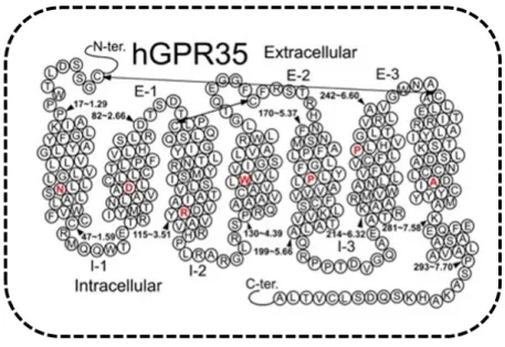
G protein-coupled receptor 35 (GPR35) is an orphan receptor discovered in 1988 (O’Dowd et al., 1998) (Figure 10). Despite being able to be activated by high concentration of endogenous molecules, foremost the tryptophan metabolite kynurenic acid (KYNA), 2-oleoyl lysophosphatidic acid (LPA), CXCL17 and cGMP (cyclic guanosine 3’5’ monophosphate), it has not been demonstrated that any of these agonists binds selectively to GPR35 (Oka et al. 2010; Wang et al. 2006; Maravillas-Montenero et al., 2015). For example, KYNA shows huge species-dependent differences in potency, which is lowest (EC50>10-3 M) for human GPR35 (Jenkins et al., 2011;
Milligan, 2011), and CXCL17 does not act as GPR35 agonist in some experimental system (Park et al., 2018).
A number of synthetic surrogate ligands for GPR35 have been proposed. One of the earliest and most useful GPR35 ligands is the cGMP phosphodiesterase inhibitor zaprinast (2-(2-propyloxyphenyl)-8-azapurin-6-one) (Taniguchi et al., 2006). Despite its wide application as reference GPR35 agonist (with reported potency between 2-8 μM at the human ortholog), zaprinast, similarly to kynurenic acid and many other synthetic agonists, display a marked animal (human vs rodent) species selectivity (Mackenzie et al., 2014; Jenkins et al. 2010, Jenkins et al. 2012).
GPR35 is mainly expressed in colon, spleen and immune cells (e.g. macrophages and dendritic cells) in both humans and mice (Maravillas-Montero et al. 2015; Divorty et al. 2015; Taniguchi et al., 2006; Wang et al., 2006. In the rat, high expression of GPR35 has been found in spleen, colon, dorsal root ganglion, and uterus (Taniguchi et al., 2006; Ohshiro et al., 2008).
Introduction - Chapter IV G protein-coupled receptor 35
It has been reported that GPR35 plays a role in a wide range of human diseases, but the signaling pathways have been not elucidated (Sun 2008, Okumura et al., 2004). In terms of classical GPCR signaling, GPR35 appears to couple to various Gα subunits depending on the cell type and/or background animal species (Divorty et al. 2015).
1.2 GPR35: evidence supporting its involvement in cancer
A genome-wide association study (GWAS) identified a single-nucleotide polymorphism (SNP) of GPR35 as novel risk locus for ulcerative colitis (UC) and primary sclerosing cholangitis (PSC) (Ellinghaus et al. 2013). The GPR35 rs3749171 SNP, leading to a threonine to methionine shift at Thr3.44, is located in the third transmembrane helix of the receptor (Ellinghaus et al. 2013). The third intracellular domain with its DRY motif is crucial for keeping the receptor in its inactive state. Only after ligand binding GPCRs change their structure so that the DRY motif allows the activation of downstream signaling. Remarkably, patients with UC and PSC have a higher risk to develop cancer (Krugliak Cleveland et al. 2017, Karlsen et al. 2017, Ullman and Itzkowitz, 2011).
GPR35 is highly expressed in human gastric cancer cells (Okumura et al., 2004). Moreover, GPR35 was identified as a potential receptor for CXCL17, a mucosal chemokine that promotes tumour growth and angiogenesis (Maravillas-Montero et al. 2015, Weinstein et al. 2006). Thus, CXCL17 levels are significantly increased in primary colonic cancers and clinical analyses suggest that CXCL17 could be an important biomarker of CRC poor prognosis (Ohlsson et al. 2016). Recently, it has been proposed a clinical significance of the CXCL17-CXCR8 (GPR35) axis in breast cancer, but the mechanistic basis of this effect remains poorly understood (Guo et al., 2017).
Recently, a significant increase of GPR35 expression, correlating with poor prognosis, has been reported in non-small-cell lung cancer (NSCLC) tissues (Wang et al., 2018). Notably, GPR35 was upregulated in chemoresistance cell model of NSCLC, and GPR35-mediated chemoresistance occurred partially via β-arrestin-2/Akt signalling (Wang et al., 2018).
Despite these findings, GPR35 function as tumour suppressor or inducer in cancers is still uncertain and may be related to cancer/cell types and/or specific microenvironment of the tumour.
1.3 Glucose metabolism in cancer
Cancer cells metabolize glucose in a different manner compared to normal cells (Warburg, 1956). Glucose metabolism and glycolysis is accelerated in cancer cells by preferential expression of transporters and enzyme isoforms that drive glucose flux. While in normoxia, healthy cells use the degradation of glucose to pyruvate and later the TCA (tricarboxylic acid) cycle to produce ATP, neoplastic cells prefer to use glycolysis to produce energy rather than oxidative phosphorylation (Warburg, 1956) (Figure 11). Indeed, tumour cells switch metabolism with high lactate production, even in aerobic conditions, and mitochondrial metabolism suppression. This metabolic adaptation is
called “aerobic glycolysis” or the “Warburg effect” (Warburg, 1956) (Figure 11). In solid tumours, the hypoxic environment supports glycolytic metabolism and provides resistance to therapy as well as an optimal niche for the maintenance of cancer stem cells (Trèdan et al., 2007; Persano et al., 2011; Fidoamore et al., 2016). Several genes (e.g. oncogenes) involved in the glycolytic pathway regulate the adjustment of cancer cells to the metabolic switch, suggesting them as possible target in cancer therapy.
Activation of macrophages or dendritic cells with a range of stimuli, including LPS (Krawczyk et al., 2010), the TLR3 ligand poly(I:C) (Pantel et al., 2014) and type I interferon (IFN) (Pantel et al., 2014), induces a metabolic switch from OXPHOS to glycolysis, in a phenomenon similar to the Warburg effect (Pantel et al., 2014; Kelly and O’Neill, 2015) (Figure 12). Recent immunometabolism studies have shown that alterations in the metabolic profile of macrophages shape their activation state and function (Kelly and O’Neill, 2015). Thus, metabolic reprogramming of macrophages could become a therapeutic approach to treat diseases, such as cancer, with a high macrophage involvement (Geeraerts et al., 2017).
Here, we took advantage of macrophages in order to investigate the metabolic role of GPR35 and the possible impact on experimental models of colorectal cancer.
Introduction - Chapter IV G protein-coupled receptor 35
Figure 10. Helix net representation of human GPR35 receptor structure (from Shore and Reggio, 2015) and simplified activation of GPR35 by endogenous or synthetic ligands.
The most highly conserved residue in each transmembrane helix (among Class A GPCRs) is shown in red. Possible disulfide bridges are indicated by double-headed arrows.
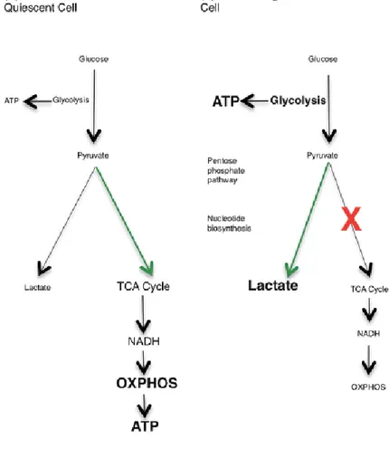
Figure 11. The Warburg effect (from Kelly and O’Neill, 2015)
(A) In resting cells, glucose is metabolized to pyruvate via glycolysis. Some pyruvate is converted to lactate, but most is directed to the TCA cycle via acetyl-CoA. The TCA cycle generates NADH, which donates electrons to the mitochondrial electron transport chain so that OXPHOS can progress.
(B) In highly proliferative or tumour cells, the metabolic profile switches from OXPHOS to aerobic glycolysis, known as the Warburg effect. Mature innate immune cells also rely on glycolysis, although they do not proliferate after activation. The majority of the pyruvate generated by glycolysis is converted to lactate, and glycolytic intermediates build up, meeting the high energy demand of the cell. Glycolysis is the source of ATP in these cells, and also provides glucose-6-phosphate for nucleotide biosynthesis in the PPP.
Introduction - Chapter IV G protein-coupled receptor 35
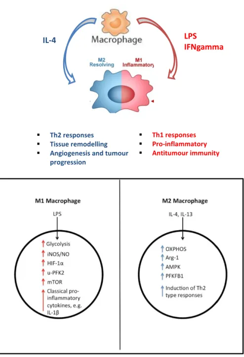
Figure 12. Physiological and metabolic differences between M1 and M2 phenotypes of macrophages (Adapted from Kelly and O’Neill, 2015)
M1 macrophages (pro-inflammatory phenotype) rely on glycolysis for ATP production and have increased levels of iNOS, HIF-1α and u-PFK2, while M2 macrophages (pro-resolving phenotype) are fueled by OXPHOS and have increased levels of Arg-1, AMPK and PFKFB1. M1 macrophages release pro-inflammatory cytokines such as IL-1β, while M2 macrophages are involved in the response to parasite infection, as well as in wound healing, and they release the anti-inflammatory cytokine IL-10. In fact, it is thought that a spectrum of macrophage activation exists, with different populations of macrophages exhibiting different inflammatory and metabolic phenotypes.
LPS
IFNgamma
IL-4
▪ Th2 responses
▪ Tissue remodelling
▪ Angiogenesis and tumour
progression
▪ Th1 responses
▪ Pro-inflammatory
AIM
The general aim of this work is to investigate the possible role of three specific targets, namely two enzymes [i.e. monoacylglycerol lipase (MAGL) and N-acylethanolamine acid amide hydrolase (NAAA)] and the G protein-coupled receptor (GPR) 35 in colorectal cancer (CRC).
To adequately pursue our objective, both pharmacological and genetic blockade of MAGL, NAAA and GPR35 were exploited in well-established models of sporadic colon cancer (i.e. AOM-induced preneoplastic lesions, polyps and tumours and APCmin mice), in a model of colitis associated to cancer (tumours induced by the AOM/DSS combination) and in xenograft CRC tumours.
More in depth studies were performed in colorectal cancer cell lines (in order to evaluate proliferation and migration), in endothelial cells (to evaluate the angiogenic process) and in bone marrow-derived macrophages (to evaluate the impact on metabolic profile).
Finally, we have measured the expression of these targets in human colonic biopsies of CRC patients, which is of obvious translational value.
In few words, the ultimate goal of my PhD thesis is to provide potential innovative therapeutic targets for CRC.
Materials and Methods
MATERIALS AND METHODS 1.1 Patients
Human studies were approved by Ethical Committee of the University of Naples Federico II, Department of Medicine (protocol number 245_2015). Patients with a well-established diagnosis of colorectal cancer (CRC, stage II-IV) were included. Human colon samples were obtained by surgical colorectal resection. Healthy tissues, obtained from the peritumoral or distal resection margins of tumour, were used as control. All patients provided written informed consent.
1.2 Mice
All procedures performed in studies involving animals were carried out in accordance with the Italian D.L. no. 116 of 27 January 1992 and associated guidelines in the European Communities Council Directive of 24 November 1986 (86/609/ECC and 2010/63/UE). Experiments were conducted in UK with the approval of the UK home office and in Italy with the approval of the Institutional Animal Ethics Committee for the use of experimental animals. Male ICR mice (weighting 25–30 g) and athymic nude female (4-weeks old) mice were purchased from Harlan Italy (S. Pietro al Natisone, UD, Italy) and fed ad libitum with standard food (Mucedola srl, Settimo Milanese, Italy). MAGL knock-out (Mgll-/-) mice were a kind gift of R. Zechner and R. Zimmermann, University of Graz, Austria. GPR35 knock-out (Gpr35-/-) mice were obtained from the KOMP repository and the intestinal epithelia-specific GPR35 were generated by outbreeding the FlpO and then crossing into mice carrying Villin-Cre. Ear-biopsy genomic DNA was used for routine genotyping of all mice. GPR35 mice were bred and maintained in specific pathogen-free conditions at the Central Biomedical Services (CBS) facility, University of Cambridge. Athymic female mice, fed ad libitum with sterile mouse food, were maintained under pathogen-free conditions at the Department of Pharmacy, University of Naples Federico II. All mice were used after 1 week-acclimation period (temperature 23±2 °C; humidity 60%, free access to water and food).
1.3 Drugs
Azoxymethane (AOM) was purchased from Sigma (Milan, Italy). URB602 and MatrigelTM were obtained from Cayman Chemical (Cabru SAS, Arcore, Italy) and BD Biosciences (Buccinasco, Milan, Italy), respectively. DSS was obtained from MP Biomedicals (Canada). PEA was a kind gift from Epitech Group. AM9053 was synthesized in the laboratory of A. Makriyannis and M. Malamas. All reagents for cell cultures were obtained from Sigma, Bio-Rad Laboratories (Milan, Italy) and Microtech Srl (Naples, Italy). The vehicles used for in vivo (10% ethanol, 10%
Tween-20, 80% saline, 2 ml/kg) and in vitro (0.1% DMSO or 0.1% ethanol) experiments had no effect on the responses under study.
1.4 Cell culture
1.4.1 Colorectal cancer cell lines
For in vitro experiments, two human colon adenocarcinoma cell lines (i.e. Caco-2 and HCT116, ATCC from LGC Standards, Milan, Italy), with a different genetic profile (APC gene and p53 mutated in Caco-2 cells, K-RAS mutated in HCT116 cells) have been used (Ahmed et al. 2013). These cell lines were cultured in Dulbecco’s modified Eagle’s medium (DMEM) containing 10% fetal bovine serum (FBS), 100 U/ml penicillin and 100 μg/ml streptomycin, 1% non-essential amino acids, 2 mM L-glutamine and 1 M HEPES, in conformity with the manufacturer’s protocols. Cell viability was evaluated by trypan blue exclusion.
1.4.2 Healthy colonic epithelial cells (HCEC)
The immortalized healthy human colonic epithelial cells (HCEC), derived from human colon biopsies, have been used as a comparison with tumoural cells. HCEC were a kind gift of Fondazione Callerio Onlus (Trieste, Italy). HCEC were cultured in Dulbecco's modified Eagle’s medium (DMEM) supplemented with 10% FBS, 100 Units/ml penicillin, 100 μg/ml streptomycin, 200 mM L-Glutamine, 100 mM Na-pyruvate and 1 M HEPES. Cell viability was evaluated by trypan blue exclusion.
1.4.3 Endothelial cell lines
The human umbilical vein endothelial cell line (HUVEC, Promocell, Heidelberg, Germany) and immortalized murine endothelial cells (SVEC4-10, ATCC from LGC Standards, UK) were used. Cells were routinely maintained in 75 cm2 polystyrene flasks, at 37 °C in a 5% CO2 atmosphere, in
Dulbecco’s modified Eagle’s medium (DMEM) for SVEC4-10 or in endothelial growth medium (EGM-2) for HUVEC. DMEM was supplemented with 10% foetal bovine serum (FBS), 100 U/ml penicillin, 100 μg/ml streptomycin, 1% non-essential amino acids and 2 mM L-glutamine. EGM-2, containing VEGF, R3-IGF-1, hEGF, hFGF, hydrocortisone, ascorbic acid, heparin and GA-1000 (Clonetics, Cambrex Bio Science Walkersville, USA) was supplemented with 10% FBS.
1.5 Primary cells isolation and culture
1.5.1 Murine bone marrow-derived macrophages (BMDM)
Bone marrow was flushed out of the murine femur and tibia with macrophages culture medium (RPMI 1640, supplemented with 1 mM HEPES pH 7.4, 50 U/ml PenStrep and 10 % FBS). Cells were then filtered through 70 µm cell strainer and incubated for 6 days in culture medium supplemented with 100 ng/ml (h)M-CSF in plates. After harvesting and seeding, BMDM were
Materials and Methods
polarized for 16 h toward M1 or M2 phenotype with LPS (20 ng/ml) plus IFN (50 ng/ml) or IL-4 (20 ng/ml), respectively.
1.5.2 Isolation of Intestinal Crypts and 3D Organoid Culture
The small and large intestine was flushed with cold PBS, cut longitudinally and into 5 mm pieces. To dissociate the crypts, pieces were placed in 25mM EDTA/PBS for 1h at 4°C and suspended in PBS. Isolated crypts were washed, counted and pelleted. A total of 200 crypts were mixed with 40 μl of Matrigel and plated in 24-well plates (Sato et al. 2009). After polymerization of Matrigel, 500 μl of IntestiCult Organoid Growth Medium (Stem Cell) containing Wnt-3a (50 ng) was added. Medium was changed every 3 days. For passage, organoids were removed from Matrigel and mechanically dissociated into single-crypt domains, and then transferred to fresh Matrigel. Passage was performed once per week with a 1:2 split ratio.
1.6 In vivo models of colorectal cancer
1.6.1 AOM DSS model of colitis associated to cancer (CAC)
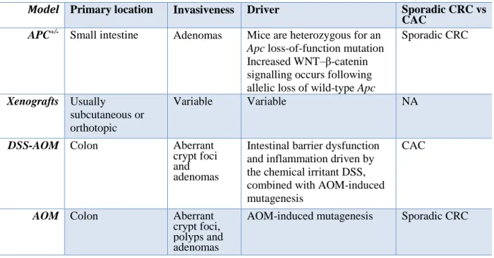
6-8-week-old wild type and Gpr35−/− mice were injected i.p. with azoxymethane (12.5 mg/kg, AOM). Colitis was induced by two cycles of 2.5% dextran sodium sulfate (DSS) in drinking water for 5 days, followed by a 16 day tap water period (Greten et al., 2004). The final DSS cycle (2%) was administered for 4 days, followed by a 10 day tap water period. The size and number of colonic tumours were evaluated at day 61 microscopically in longitudinally cut and formalin fixed specimen. The typical features of the AOM DSS model are depicted in Table 3.
1.6.2 AOM model of colorectal cancer
AOM (40 mg/kg in total, i.p.) was administered in mice at the single dose of 10 mg/kg at the beginning of the first, second, third and fourth week. The drugs were given i.p. three times a week starting one week before the first administration of AOM. All animals were euthanized by asphyxiation with CO2 three months after the first injection of AOM. Based on our laboratory
experience, this time (at the used dose of AOM) was associated with the occurrence of a significant number of aberrant crypt foci (ACF, which are considered pre-neoplastic lesions), polyps and tumours (Izzo et al. 2008) (see table 3 for the AOM model features).
For ACF, polyps and tumours determination, the colons were rapidly removed after sacrifice, washed with saline, opened longitudinally, laid flat on a polystyrene board and fixed with 10% buffered formaldehyde solution before staining with 0.2% methylene blue in saline. Colons were examined using a light microscope at 20X magnification (Leica Microsystems, Milan Italy). The detection and quantization of ACF, polyps and tumours on the colon were performed as previously reported (Izzo et al., 2008). Briefly, in comparison to normal crypts, aberrant crypts have greater size, larger and often elongated openings, thicker lining of epithelial cells, compression of adjacent