from inclusion-body myositis muscle
MyoD expression restores defective myogenic differentiation of human mesoangioblasts
Roncaglia, Sergio Rutella, Stefano Ferrari, Pietro Attilio Tonali, Enzo Ricci, and Giulio Cossu
Angelis, Enrico Tagliafico, Maurilio Sampaolesi, Teresa Gidaro, Manuela Papacci, Enrica
Roberta Morosetti, Massimiliano Mirabella, Carla Gliubizzi, Aldobrando Broccolini, Luciana De
doi:10.1073/pnas.0603386103
2006;103;16995-17000; originally published online Oct 31, 2006;
PNAS
This information is current as of February 2007.
& Services
Online Information
www.pnas.org/cgi/content/full/103/45/16995
etc., can be found at:
High-resolution figures, a citation map, links to PubMed and Google Scholar,
Supplementary Material
www.pnas.org/cgi/content/full/0603386103/DC1
Supplementary material can be found at:
References
www.pnas.org/cgi/content/full/103/45/16995#BIBL
This article cites 27 articles, 16 of which you can access for free at:
www.pnas.org/cgi/content/full/103/45/16995#otherarticles
This article has been cited by other articles:
E-mail Alerts
.
click here
at the top right corner of the article or
Receive free email alerts when new articles cite this article - sign up in the box
Rights & Permissions
www.pnas.org/misc/rightperm.shtml
To reproduce this article in part (figures, tables) or in entirety, see:
Reprints
www.pnas.org/misc/reprints.shtml
To order reprints, see:
MyoD expression restores defective myogenic
differentiation of human mesoangioblasts
from inclusion-body myositis muscle
Roberta Morosetti*†, Massimiliano Mirabella*‡§, Carla Gliubizzi*‡, Aldobrando Broccolini*, Luciana De Angelis¶, Enrico Tagliafico储, Maurilio Sampaolesi**, Teresa Gidaro*, Manuela Papacci*, Enrica Roncaglia储, Sergio Rutella††, Stefano Ferrari储, Pietro Attilio Tonali*‡, Enzo Ricci*‡, and Giulio Cossu**‡‡§§
*Department of Neurosciences and†Interdisciplinary Laboratory for Stem Cell Research and Cellular Therapy, Catholic University, Largo A. Gemelli 8, 00168
Rome, Italy;‡Fondazione Don Carlo Gnocchi, 00194 Rome, Italy;‡‡Institute of Cell Biology and Tissue Engineering, San Raffaele Biomedical Science Park,
00128 Rome, Italy;¶Department of Histology and Embriology, University ‘‘La Sapienza,’’ 00161 Rome, Italy;储Department of Biomedical Sciences, University
of Modena and Reggio Emilia, 41100 Modena, Italy; **Stem Cell Research Institute, San Raffaele Hospital, 20132 Milan, Italy;††Institute of Hematology,
Catholic University, 00168 Rome, Italy; and§§Department of Biology, University of Milan, 20133 Milan, Italy
Edited by Tullio Pozzan, University of Padua, Padua, Italy, and approved September 19, 2006 (received for review April 28, 2006)
Inflammatory myopathies (IM) are acquired diseases of skeletal muscle comprising dermatomyositis (DM), polymyositis (PM), and inclusion-body myositis (IBM). Immunosuppressive therapies, usu-ally beneficial for DM and PM, are poorly effective in IBM. We report the isolation and characterization of mesoangioblasts, ves-sel-associated stem cells, from diagnostic muscle biopsies of IM. The number of cells isolated, proliferation rate and lifespan, markers expression, and ability to differentiate into smooth muscle do not differ among normal and IM mesoangioblasts. At variance with normal, DM and PM mesoangioblasts, cells isolated from IBM, fail to differentiate into skeletal myotubes. These data correlate with lack in connective tissue of IBM muscle of alkaline phospha-tase (ALP)-positive cells, conversely dramatically increased in PM and DM. A myogenic inhibitory basic helix–loop– helix factor B3 is highly expressed in IBM mesoangioblasts. Indeed, silencing this gene or overexpressing MyoD rescues the myogenic defect of IBM mesoangioblasts, opening novel cell-based therapeutic strategies for this crippling disorder.
T
he idiopathic inflammatory myopathies (IM), characterized by mononuclear cells infiltration of skeletal muscle, are the largest group of acquired muscle diseases and encompass three major forms: dermatomyositis (DM), polymyositis (PM), and inclusion-body myositis (IBM) (1). Causes of DM, PM, and IBM are unknown, but an autoimmune pathogenesis is supported by marked up-regulation of cytokines and adhesion molecules, evidence of a T cell-mediated myocytotoxicity in PM and IBM and of a comple-ment-mediated microangiopathy in DM (2). Current immunother-apies are usually effective in DM and PM patients, whereas IBM, the most frequent myopathy in elderly patients, responds poorly or not at all to immunosuppressive therapies and its course steadily progresses to severe disability. In IBM muscle, the presence of degenerative features, such as vacuolated fibers containing amyloid and amyloid-related proteins (3), reflects a complex pathogenesis involving misfolded and unfolded proteins and increased oxidative stress in the context of a cellular ‘‘aged’’ milieu acting in concert with chronic inflammation (4). Regeneration and repair of muscle fibers are fundamental processes accounting for rebuilding muscle integrity and gradual recovery of muscle strength in IM after suppression of mononuclear cells infiltration. Satellite cell-dependent regeneration occurs also in IBM muscle wherein mul-tiple metabolic pathways normally involved in muscle development are activated (5, 6). However, in IBM, despite the activation of potentially repairing mechanisms, regeneration is inefficient.Mesoangioblasts are vessel-associated stem cells, firstly isolated from dorsal aorta of mouse embryos (7), able to differentiate into a variety of mesoderm tissues including skeletal, cardiac and smooth muscle (8, 9). When delivered intraarterially, mesoangioblasts
restore to a significant extent muscle morphology and function in a mouse model of muscular dystrophy (10).
Because mesoangioblasts express numerous receptors for in-flammatory cytokines, we assumed that the human counterpart of murine mesoangioblasts should be recruited in high numbers during muscle inflammation.
Here, we describe the isolation and functional characterization of pericyte-derived adult mesoangioblasts (herein simply called me-soangioblasts) from diagnostic muscle biopsies of IM patients and show that IBM mesoangioblasts fail to differentiate into skeletal muscle. This differentiation block can be corrected in vitro by transient expression of MyoD, making these cells potential attrac-tive candidates for cellular therapy of this disabling disease.
Results
Mesoangioblasts Are Efficiently Isolated from IM Muscle Biopsies.
After 10–15 days of organ culture from biopsies of three normal controls, three DM, three PM, and six IBM, we isolated a popu-lation of cells morphologically different from satellite cells. Ap-proximately 3–4⫻ 104cells could be obtained from each biopsy.
From the first passage on, cells were characterized by a triangular, adherent, refractive shape and by a floating兾loosely adherent round component, particularly abundant in DM (Fig. 1A). Peculiar cell morphology, phenotypic characteristics, and differentiation poten-tial indicated that our cells were human mesoangioblasts, as recently characterized (A. Dellavalle, M.S., R. Tonlorenzi, E.T., B. Sac-chetti, L. Perani, B. G. Galvez, G. Messina, R.M., S. Li, G. Peretti, J. S. Chamberlain, W. E. Wright, Y. Torrente, S.F., P. Bianco, and G.C., unpublished data).
Cells were kept in culture up to 25 population doublings (PD) (25 for control and DM, 19 for IBM, and 20 for PM) with a proliferation rate comparable for DM, IBM, and PM and independent from patients’ age. Doubling time from all biopsies was 33.5⫾ 2.38 h (Fig. 1B). At both early and late passages, cells kept a diploid kariotype (data not shown). There were no differences in the number of cells isolated from freshly dissected or fresh-frozen muscles at both early
Author contributions: R.M. and M.M. contributed equally to this work; R.M., M.M., and G.C. designed research; R.M., M.M., C.G., A.B., L.D.A., E.T., M.S., T.G., M.P., E. Roncaglia, and S.R. performed research; R.M., M.M., A.B., E.T., S.F., P.A.T., E. Ricci, and G.C. analyzed data; and R.M., M.M., and G.C. wrote the paper.
The authors declare no conflict of interest. This article is a PNAS direct submission.
Abbreviations: ALP, alkaline phosphatase; IM, idiopathic inflammatory myopathies; IBM, inclusion-body myositis; DM, dermatomyositis; PM, polymyositis; SDMC, satellite-derived myogenic cells; mdx, mouse muscular dystrophy; bHLH, basic helix–loop– helix; BHLHB3, bHLH domain containing class B3 transcription factor.
§To whom correspondence should be addressed. E-mail: [email protected].
© 2006 by The National Academy of Sciences of the USA
and late passages, neither phenotypic characteristics of the 12 IM studied were significantly different after 8 and 18 PD (Fig. 2A and Fig. 7, which is published as supporting information on the PNAS web site). Because we isolated on average of 3–4⫻ 104cells from
a single biopsy, the estimated final number of cells after 25 PD is 50–120⫻ 1010, and the real number that could be obtained before
the appearance of senescent cells in significant proportion is between 10 and 20⫻ 109cells. This number would be suitable for
intraarterial delivery to adult patients, based on a per kg compar-ison with the mouse model used before (10).
Clonogenic Potential, Cell Cycle, and Phenotypic Characteristics Do Not Differ Among IM Mesoangioblasts.We dissociated mesoangio-blasts to single cell suspension and cloned them by limiting dilution: clones appeared in 9.75⫾ 3.9, 8.87 ⫾ 3.1, and 10.5 ⫾ 4.0 wells for DM, PM, and IBM, respectively, all with the same double mor-phology of the original cells. By replating the clones at clonal density they were able to give rise to new clones.
The cell cycle distribution was similar for all mesoangioblasts of 12 IM in three separate experiments (each one conducted in duplicate) (G0兾G1, 65.5⫾ 6.4%; S, 23.7 ⫾ 4.3%; G2兾M, 10.8 ⫾
2.9%) regardless of the IM type. The pattern of distribution was significantly different (P ⱕ 0.01) from that observed in control proliferating satellite-derived myogenic cells (SDMC) (G0兾G1,
87.58⫾ 3.6%; S, 8.43 ⫾ 3.0%; G2兾M, 4 ⫾ 1.8%) (Fig. 1C). Results
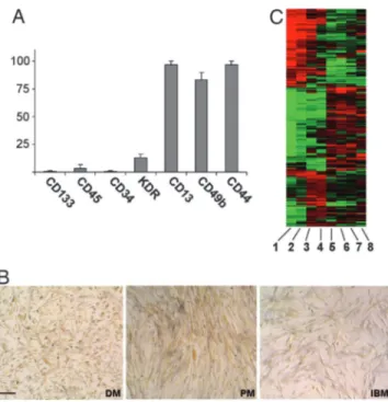
were always consistent throughout all experiments. Cells from all IM were strongly positive for CD44 and CD13, positive for CD49b, homogeneously negative for CD34, CD133, CD45 by FACS (Fig. 2 A), consistently with what observed in normal human mesoan-gioblasts (A. Dellavalle, M.S., R. Tonlorenzi, E.T., B. Sacchetti, L. Perani, B. G. Galvez, G. Messina, R.M., S. Li, G. Peretti, J. S. Chamberlain, W. E. Wright, Y. Torrente, S.F., P. Bianco, and G.C., unpublished data). By immunocytochemistry and Western blot, all of the cells were positive for vimentin, weakly positive for␣-SMA and desmin, and did not express glial fibrillar acidic protein (GFAP), nestin,III-tubulin, and MyoD (data not shown). Alka-line phosphatase (ALP) staining was positive in all IM mesoan-gioblasts, with the highest levels observed in PM and only a weak labeling in IBM (Fig. 2B). Together, these markers identify human adult mesoangioblasts as the in vitro progeny of pericytes.
The Ability to Differentiate into Smooth Muscle Cells (SMCs) and in Osteoblasts Are Similar Among All IM Mesoangioblasts. Murine mesoangioblasts differentiate into mature SMCs upon TGF treat-ment (11, 12). Therefore, we exposed mesoangioblasts from all patients to TGF. Approximately 80% of cells from all biopsies differentiated into strongly positive␣-SMA-positive SMC, with no significant difference between the various IM (Fig. 8 which is published as supporting information on the PNAS web site).
Similarly to murine mesoangioblasts (8), human cells responded to BMP2 with a rather low percentage (⬇5%) differentiating into strongly ALP-positive osteoblast-like cells expressing osteocalcin and osteopontin (data not shown). In contrast, both control and IM mesoangioblasts failed to differentiate into neurons or glia when grown in neural stem cell differentiation media (data not shown).
Genome-Wide Gene Expression in IM Mesoangioblasts.Proliferating mesoangioblasts from normal and IM muscle were analyzed for gene expression by Affimetrix gene array. As expected, gene expression profile was similar in all samples with only few genes differentially expressed. Clustering results are shown in Fig. 2C. Two main classes were defined: the first included mesoangioblasts from normal muscle, whereas the second consisted of mesoangio-blasts from DM and IBM. Interestingly, the clustering procedure paired together DM and IBM replicates.
A summary of the analysis is shown in Table 1, which is published as supporting information on the PNAS web site. In particular, mesoangioblasts from controls and IM (DM and IBM) did not express myogenic factors such as MyoD, or Pax3, Pax7, MEF2C, or MEF2D. As expected for mesoderm cells, mesoangioblasts did not
Fig. 2. FACS, immunophenotyping, ALP histochemistry, and gene expression profiling of IM mesoangioblasts. (A) More than 90% of cells from all samples were strongly positive for CD44 and CD13 with high percentage of cells CD49b-positive. None of the markers positive in murine mesoangioblasts were significantly expressed. Bars represent the mean⫾ SD of 36 samples from the 12 patients with IM (3 DM, 3 PM, and 6 IBM) (each performed in triplicate). (B) IM mesoangioblasts in vitro are all ALP-positive. After simultaneous staining in the same culture conditions, more intensely labeled cells can be observed in PM and to a lesser extent in DM, whereas IBM mesoangioblasts are only weakly positive. (Scale bar: 20m.) (C) Clustering results show two main classes: mesoangioblasts from normal controls (lanes 5– 8), mesoangioblasts from DM (lanes 1 and 2), and IBM (lanes 3 and 4). Clustering procedure pairs together DM and IBM replicates.
Fig. 1. Cell morphology, growth curve, and cell cycle. (A) From the organ culture on, refractive triangular, adherent, and round loosely adherent兾 floating cells were observed. (Scale bar: 40m.) (B) Cell growth was assessed after 24 h, 48 h, and 72 h. Viable cells were judged by trypan blue exclusion. Results are expressed as absolute counts. Bars represent mean⫾ SD of tripli-cate samples of one representative experiment of three. (C) Cell cycle distri-bution of proliferating normal human SDMC and mesoangioblasts from three controls, three DM, three PM, and six IBM (run in duplicate) after 24 h of culture was assessed by propidium iodide and FACS. For each sample, per-centage of cells in G0兾G1, S, or G2兾M phases of cell cycle is indicated. One
representative experiment of three is shown.
express cytocheratins or neurofilaments; at variance with their embryonic counterparts, human postnatal mesoangioblasts did not express endothelial markers but rather pericyte markers such as ALP, PDGF-receptor, or NG2 proteoglycan. Differently from controls, IM mesoangioblasts expressed numerous immune-related genes, such as IFN-induced proteins and, particularly in DM, high levels of integrins, that greatly facilitate diffusion into muscle interstitium. As recently shown, mesoangioblasts muscle homing can be increased by exposure to stromal cell-derived factor 1␣ (SDF-1␣) and subsequent transient expression of ␣-4 integrin or L-selectin (13). Of note, increased SDF-1␣ has been observed in IM muscle (14). These data suggest a possible advantage of IM mesoangioblasts over controls in migration through endothelium and into muscle fibers that can be more effective during inflam-mation and after intra-arterial delivery. Interestingly, only IBM mesoangioblasts expressed high levels of genes known to inhibit myogenesis such as TGF-1, SFRP-2 (secreted frizzled-related protein 2), and BHLHB3 (basic helix–loop–helix domain contain-ing class B3 transcription factor) (15–17).
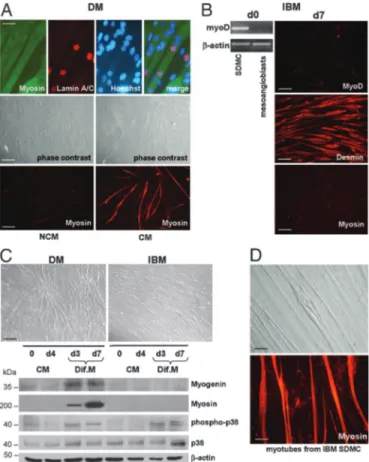
DM and PM Mesoangioblasts Efficiently Differentiate into Skeletal Muscle Under a Variety of Conditions.Upon coculture with C2C12 myoblasts, approximately 10% of myosin-positive myotubes con-tained nuclei expressing human lamin A兾C, thus confirming fusion of human and murine cells (Fig. 3A). To investigate whether mesoangioblasts from normal or IM muscle were capable of spontaneous myogenic differentiation in vitro, cells at 80% confluence were maintained in culture medium without growth factors (18). Control, DM and PM mesoangioblasts fused into multinucleated myosin-positive myotubes with a fusion
index (expressed as number of myonuclei兾number of total
nuclei) of 0.15⫾ 0.05 for three controls and 0.1 ⫾ 0.015 for three DM and three PM. Differences between controls and DM and PM did not reach statistically significance (P ⬎ 0.05). This feature was never observed in mouse mesoangioblasts, which differentiate only upon coculture (8, 9). Interestingly, DM, PM, and control mesoangioblasts, cultured for 4 days in growth medium conditioned by normal human SDMC, acquired homo-geneous myoblast-like morphology and, after exposure to dif-ferentiation medium for 7 days, fused into numerous multinu-cleated myosin-positive myotubes (fusion index of 0.7 ⫾ 0.1) (Fig. 3 A and C) with a marked up-regulation of myogenin and myosin by Western blot (Fig. 3C). This evidence indicates that factor兾s produced by satellite-derived cells is兾are necessary for the myogenic commitment of human mesoangioblasts.
IBM Mesoangioblasts Fail to Differentiate into Skeletal Muscle.In none of the above mentioned culture conditions, IBM mesoangio-blasts were able to differentiate into multinucleated myosin-positive myotubes. However, when previously exposed to SDMC-conditioned growth medium, they became oriented with elongated morphology displaying a strong desmin immunoreactivity. MyoD protein expression was not detectable after exposure to conditioned medium (Fig. 3B). By Western blot, IBM cells harvested at the same time points of DM did not express myosin or myogenin, but showed that activation of the p38 pathway, known to be involved in skeletal myogenesis, was not disrupted (19, 20) (Fig. 3C). Of note, satellite cells isolated from the same IBM biopsies (18) were able to normally differentiate in myotubes (Fig. 3D).
Connective Tissue of IM Biopsies Contains ALP-Positive Cells Recruited to Myogenic Fate.Results from in vitro studies have shown that ALP is a marker of adult mesoangioblasts. By investigating the distribu-tion of ALP-expressing cells, we found ALP reactivity only in small arteries in normal muscle, whereas in all DM and PM biopsies a very strong ALP staining was evident in perimysial and endomysial connective tissue (Fig. 4A), as described (21). On the contrary, IBM muscle showed no ALP connective tissue staining, although blood
vessels were normally represented. We asked whether at least part of the cells strongly ALP-positive in connective tissue of DM or PM muscle could be recruited to a myogenic fate by neighboring regenerating fibers. Indeed both PM and DM showed numerous strongly ALP-positive round cells in the endomysium with a MyoD-positive nucleus (Fig. 4B). In the same areas, satellite cells expressed Pax7, whereas ALP-positive cells in the interstitium were PAX7-negative. Because ALP is not expressed in satellite cells and myoblasts (22), the presence of cells expressing both ALP and myogenic markers suggests recruitment of pericytes-derived cells into the myogenic lineage. Neither ALP-positive nor double-positive cells were detected in the IBM sections analyzed. Because, at least early in the disease, both MyoD-positive satellite cells as well as pericytes are not significantly reduced, the lack of
MyoD-Fig. 3. Skeletal muscle differentiation. (A) (Top) Immunofluorescence for myosin and human lamin A兾C. Mesoangioblasts from DM efficiently fused with C2C12 murine myoblasts into mature myosin-positive myotubes, as indicated by the presence of nuclei expressing human lamin A兾C. (Scale bar: 10m.) (Middle and Bottom) DM mesoangioblasts exposed 4 days to SDMC-conditioned growth medium (CM) and subsequently cultured for 7 days in differentiation medium (Dif.M) spontaneously fuse into differentiated myotubes. (Scale bar: 40m.) Each experiment was performed in duplicate at least three times. (B) IBM mesoangio-blasts do not constitutively express MyoD (RT-PCR) and when exposed 4 days to CM and 7 days to Dif.M are negative for MyoD and myosin, but display a strong desmin immunoreactivity. (Scale bar: 20m.) (C) Mesoangioblasts from DM, but not from IBM, differentiate into multinucleated myotubes (phase contrast). (Scale bar: 40m.) For Western blot analysis, cells were harvested at day 0, after 4 days in CM and after 3 and 7 days in Dif.M. A marked up-regulation of myogenin and myosin is observed already at day 3 of differentiation for DM mesoangio-blasts; no up-regulation of myosin and myogenin is visible in IBM cells. For both DM and IBM mesoangioblasts, activation of p38 (phospho-p38) is observed. One representative experiment of six is shown. (D) SDMC from primary muscle cultures from all six IBM biopsies normally differentiate in multinucleated myotubes (phase contrast, Upper) and show strong myosin-immunoreactivity (Lower). (Scale bar: 20m.) Two representative cultures are shown. NCM, nonconditioned medium.
expressing pericytes-derived cells seems correlated with the specific defect in myogenic commitment of this cell population in IBM.
Transient Expression of MyoD Rescues the Myogenic Defect of IBM Mesoangioblasts.IBM mesoangioblasts did not give rise to termi-nally differentiated myotubes and did not constitutively express MyoD mRNA, and MyoD could not be induced by our differen-tiating conditions (Fig. 3 B and C); therefore, we transduced IBM mesoangioblasts with an adenoviral vector expressing full-length murine MyoD. Expression of MyoD caused myogenic differentia-tion in ⬇60% (fusion index 0.6 ⫾ 0.02) of cells, as defined by activation of muscle-specific genes and myotubes formation (Fig. 5A).
Myogenic potential of IM mesoangioblasts in vivo was tested by intramuscular transplantation in irradiated scid兾mdx immunodefi-cient mice (Table 2, which is published as supporting information on the PNAS web site). After two consecutive injections, the tibialis anterior (TA) of mice treated with mesoangioblasts from DM displayed, in the injected areas, numerous muscle fibers containing human nuclei and expressing human dystrophin (average percent-age of positive fibers was 60⫾ 15% and 57 ⫾ 10% in two animals transplanted with DM-derived mesoangioblasts, respectively; Fig. 5B Right). In contrast, none of such fibers could be detected in the TA of two mice injected with ‘‘wild-type’’ IBM mesoangioblasts. These muscles showed the presence of isolated (1–2%) human lamin A兾C-positive nuclei in the interstitium and within some muscle fibers, but dystrophin expression was negligible (Fig. 5B Left). However, after transplantation of adenoMyoD-transduced IBM mesoangioblasts, the TA of the two mice treated showed, in the injected region, the presence of large areas reconstituted with dystrophin-lamin A兾C-positive fibers (33 ⫾ 10% and 29 ⫾ 8%, respectively) (Fig. 5B Middle). Controls are shown in Fig. 9, which is published as supporting information on the PNAS web site).
siRNA Inhibition of BHLHB3 Expression Restores a Normal Myogenesis in IBM Mesoangioblasts. IBM mesoangioblasts had increased mRNA levels of BHLHB3 (Fig. 10, which is published as supporting
information on the PNAS web site), a group E subfamily member of bHLH factors, which negatively regulates myogenesis by mod-ulating the transcriptional activity of MyoD (17). Therefore, we examined the effect of siRNA-mediated suppression of BHLHB3 in mesoangioblasts from three IBM patients. Interestingly, siRNA-transfected cells were able to differentiate, giving rise to multinu-cleated myosin-positive myotubes after 7 days in differentiation medium (Fig. 6). To verify the siRNA specificity, we used a second siRNA for BHLHB3 and found that also this siRNA had a similar ability to rescue myogenesis (data not shown), unlike nonsilencing control siRNA that was ineffective.
Discussion
Our study demonstrates that human adult mesoangioblasts can be efficiently isolated from diagnostic muscle biopsies of patients with IM. Antigenic and molecular characterization of these cells indi-cated that mesoangioblasts represent a distinct type of mesoderm progenitor cells, different from mesenchymal stem cells (E.T., and S.F., unpublished observations). In all IM patients, these cells retain the same proliferation ability of cells isolated from normal muscle, and can be grown and expanded for as many as 25–30 passages, although not indefinitely. Here we have shown that exposure of DM
Fig. 4. ALP histochemistry in IM muscle biopsies. (A) In normal muscle only small arteries show ALP-positive staining. Intense ALP staining is present in PM and DM biopsies in perymisial and endomysial connective tissue, particularly strong in round cells associated to vessels’ walls. IM biopsies contain also a variable number of small-sized regenerating muscle fibers with slight punctuate ALP positivity. IBM muscle shows no ALP connective tissue staining. (Scale bar: 40m.) (B) Immunohistochemistry for MyoD or Pax7 with peroxidase-antiperoxidase (PAP) on the same unfixed frozen sections of biopsies from PM, DM, IBM, and normal muscle from which mesoangioblasts were isolated. Representative fields of a PM biopsy showing a round vessel-associated cell (open arrowhead) strongly ALP-positive containing a MyoD-ALP-positive nucleus. Several PAX7-ALP-positive cells (filled arrowheads), identified as activated satellite cells, are associated with muscle fibers showing regenerative aspects with slight ALP positivity, whereas the roundish ALP-positive cells in the interstitium are PAX7-negative (arrow). CTRL, control. (Scale bar: 10m.)
Fig. 5. MyoD transduction and induction of differentiation in IBM mesoangio-blasts. (A) As shown by immunofluorescence and Western blot, AdenoMyoD-transduced IBM mesoangioblasts after 7 days in differentiation medium, fused into multinucleated myosin-positive myotubes. Approximately 90% of cells were MyoD-positive. (Scale bar: 10m.) Efficiency of transduction was evaluated 48 h after the infection by Western blot showing a marked up-regulation of MyoD. -actin was used as loading control. A representative experiment of three, each performed in duplicate, is shown. (B) Double immunohistochemistry for human dystrophin and lamin A兾C. In vivo myogenicity of IM mesoangioblasts was tested by intramuscular transplantion in scid兾mdx mice. After two consecutive trans-plants, the TA of mice injected with IBM mesoangioblasts showed the presence of occasional human nuclei (blue with AMCA-labeled human lamin A兾C, arrow-heads) within muscle fibers and in the interstitium, but dystrophin expression was undetectable (Left). On the contrary, after IBM adenoMyoD-infected mesoan-gioblasts transplantation (Middle), large areas of injected muscle were reconsti-tuted with fibers containing human lamin A兾C-positive myonuclei (blue, arrow-heads) and expressing dystrophin along the sarcolemma (red with Texas red-labeled specific antibody). When DM mesoangioblasts were used (Right), the majority of muscle fibers in the injected areas expressed dystrophin and con-tained human nuclei (positive for lamin A兾C, arrowheads). (Scale bars: 10m.) SYTOX green staining on the same sections was used to identify both human and murine nuclei.
and PM mesoangioblasts to normal SDMC-conditioned medium is greatly effective in inducing skeletal muscle differentiation, outlin-ing the importance of muscle-secreted factors for myogenic mat-uration of these stem cells.
IBM mesoangioblasts show the same proliferation ability ob-served in normal muscle suggesting that the disease has not reduced their proliferation potency. Nevertheless, although they normally differentiate into SMCs, their differentiation into skeletal muscle seems markedly impaired, because no myotubes were observed under the same conditions that promote massive skeletal muscle differentiation of DM, PM and control mesoangioblasts. Interest-ingly, cultures of satellite cell-derived progenitors (MyoD-positive) obtained from the same IBM biopsies show normal myogenic differentiation. These data agree with previous studies showing that cultured IBM muscle cells proliferate and differentiate normally and can be properly innervated (23), although the possibility exists that satellite cells activation in vivo could be defective.
However, we show here that a progenitor cell, resident in a perivascular niche of IBM muscle, is defective in myogenic deter-mination and differentiation. No significant differences of age exist between our IBM and DM兾PM patients, excluding a consequence of muscle aging and strongly suggesting a causal correlation with the specific pathophysiology of IBM. In fact, IBM presents an inade-quate long-term regeneration despite a normal number of satellite cells, a complete lack in muscle connective tissue of ALP-positive cells, likely activated pericytes representing the cells from which mesoangioblasts are established in vitro, and a failure of mesoan-gioblasts to differentiate in vitro. This defect can be rescued by transient MyoD expression, a strategy that, independently from the underlying molecular mechanism, immediately offers a therapeutic opportunity. Skeletal myogenesis, a complex multistep process mainly regulated by bHLH myogenic factors, depends upon a finely controlled equilibrium with repressors bHLH such as Id (24). IBM mesoangioblasts highly express BHLHB3, which inhibits MyoD
function (17). Our data demonstrate that reestablishing a correct balance between positive and negative bHLH factors, by MyoD transient transduction or siRNA inhibition of BHLHB3 expression, restores a normal myogenesis in IBM mesoangioblasts. As BHLHB3 gene expression is regulated in a cell-type specific man-ner by multiple extracellular stimuli including cytokines and growth factors, it is likely that cytokines or other factors present in the IBM muscle ‘‘milieu’’ but not in other IM muscle induce BHLHB3 expression. Like most bHLH, BHLHB3 probably transactivates its own promoter, thus maintaining its inhibitory effect on myogenesis also in vitro, away from the microenvironment that initially induced its expression. Whereas several genes may control muscle differ-entiation of mesoangioblasts, we identified BHLHB3 as one of the important mediators of myogenic block in IBM. How much this defect impacts on the pathogenesis of IBM remains to be investi-gated; it is however intriguing that satellite cells are not directly affected and yet IBM muscle fails to regenerate.
Currently it is unknown to what extent other progenitors such as mesoangioblasts participate to regeneration in normal muscle after inflammatory damage and in degenerative myopathies, either directly or by feeding into the satellite cells pool, should this reservoir be exhausted. Postnatal mesoangioblasts in vivo express ALP, are located in perithelial position and can be considered as part of the pericyte population (A. Dellavalle, M.S., R. Tonlorenzi, E.T., B. Sacchetti, L. Perani, B. G. Galvez, G. Messina, R.M., S. Li, G. Peretti, J. S. Chamberlain, W. E. Wright, Y. Torrente, S.F., P. Bianco, and G.C., unpublished data). A characteristic histochem-ical feature of PM and DM is a strong ALP positivity in perimysial and endomysial connective tissue, as opposed to IBM as well as other myopathies with increased connective tissue, usually display-ing no or very little ALP-positivity (21). ALP staindisplay-ing correlates more with regenerative properties of muscle rather than with inflammatory changes and has been thought to reflect activated or proliferating fibroblasts (21). Our study indicates that human ALP-positive mesoangioblasts likely represent the cell population activated in muscle connective tissue of IM, originating from the perivascular niche and susceptible of myogenic determination in vivo, as indicated by MyoD expression.
Mesoangioblasts present a distinct advantage over satellite cells, i.e., the ability to cross the vessel wall which, together with easy isolation and good myogenic potential, make them an ideal candi-date for muscle reconstitution therapy by means of cell transplan-tation. Their safety as therapeutic tool is demonstrated by limited lifespan, absence of kariotype abnormalities or tumorigenesis in nude mice (A. Dellavalle, M.S., R. Tonlorenzi, E.T., B. Sacchetti, L. Perani, B. G. Galvez, G. Messina, R.M., S. Li, G. Peretti, J. S. Chamberlain, W. E. Wright, Y. Torrente, S.F., P. Bianco, and G.C., unpublished data). Moreover mesoangioblasts can be easily isolated with high efficiency even from frozen material and expanded in vitro to number of cells suitable for a potential in vivo treatment. The use of frozen material is particularly relevant in IBM, often misdiag-nosed as PM and suspected only after unsuccessful immune-suppressive therapy, because it makes possible to go back to stored muscle to expand mesoangioblasts. An intrinsic myogenic defect of IBM mesoangioblasts can be corrected with viral transduction of exogenous MyoD that overcomes BHLHB3 inhibition and, by act-ivating endogenous MyoD, irreversibly commit cells to myogenesis. Thus, in vitro manipulation of these autologous cells induced to effectively differentiate into skeletal muscle (without a stable viral transduction as necessary in muscular dystrophies) could be par-ticularly valuable to promote muscle repair and regeneration in a disease such as IBM where relentless muscle involvement occurs very selectively. The characteristic early and severe atrophy of quadriceps, responsible for sudden falls and disabling gait impair-ment, could be treated by infusing mesoangioblasts with selective catheter-mediated delivery into the iliac arteries. The use of autol-ogous cells eliminates the obligatory need of immune suppression, although more selective anti-dysimmune drugs may eventually be
Fig. 6. Induction of myogenic differentiation of IBM mesoangioblasts by BHLHB3 siRNA. (A) BHLHB3 siRNA (Hs 1) was transfected into IBM mesoan-gioblasts. Twenty-four hours after transfection, cells were harvested for RNA extraction and RT-PCR analysis. (B) MAPK siRNA was used as positive control; 24 h after transfection, cells were harvested for Western blot. (C) BHLHB3 siRNA-transfected cells were shifted to differentiation medium after 24 h. After 7-day culture, cells were stained with an anti-myosin antibody or har-vested for Western analysis. (Scale bar: 40m.) A representative culture and immunoblot of one of three independent experiments (each one run in duplicate) are shown.
combined with regenerative cell therapy, given the inflammatory background of IBM muscle. Also rare cases of unresponsive PM and DM would be potentially treatable as well by targeting muscle groups essentials for motor and respiratory functions.
Methods
Patients. Diagnostic muscle biopsies were performed after in-formed consent at the Neurology Department of Catholic Univer-sity. We used fresh and fresh-frozen muscles from three normal controls (one fresh, two frozen) (48–84 years of age, average 64⫾ 18.33 years of age) and 12 patients with IM: 3 DM (2 fresh, 1 frozen), 3 PM (1 fresh, 2 frozen) (33–75 years of age, average 52.5⫾ 9.5 years of age), and 6 sporadic IBM (3 fresh, 3 frozen) (56–75 years of age, average 67.4⫾ 18.1 years of age). Diagnosis was based on clinical evaluation and laboratory studies. None of the patients received steroids or immunosuppressive therapy before biopsy. This research was approved by the ethical committee of our institution.
Cell and Organ Cultures. Fragments of intramuscular vessels and surrounding mesenchymal tissue were plated as described (8, 10). Details are provided in Supporting Methods, which is published as supporting information on the PNAS web site.
A fragment from the same muscle biopsy was also cultured to obtain primary muscle cultures from satellite cells by using the explantation reexplantation method (18).
Characterization of Human Mesoangioblasts from IM by FACS, Cell-Cycle Analysis, and Growth Curve.Cells (5⫻ 104) were incubated
with FITC-, PE-, or APC-conjugated mAbs directed against
AC133兾1, CD34, VEGF-RII (KDR), CD45, CD49b, CD44, and
CD13. Details are provided in Supporting Methods.
In Vitro Differentiation. Skeletal muscle differentiation. Mesoangio-blasts were (i) cultured under standard differentiating conditions for SDMC (17); (ii) cocultured with a 4-fold excess of C2C12 myoblasts; (iii) cultured in normal human SDMC-conditioned medium and then exposed to differentiation medium. At each time point, cells were fixed or harvested for protein extraction. Differ-entiation assays were performed in all IM samples studied and repeated at least three times for each patient with consistent results.
Smooth muscle, osteoblasts, and neural differentiation.Differentiation of mesoangioblasts into SMCs, osteoblasts, and neural cells was tested as described (8, 12, 25).
Immunostainings.Immunostainings were performed as described (5, 6). Details are available in Supporting Methods.
Double Immunohistochemistry-Histoenzymatic ALP Staining. Immu-nocytochemistry for MyoD or Pax7 was performed with peroxi-dase-antiperoxidase followed by histoenzimatic ALP staining on the same unfixed frozen sections of the biopsies used for mesoan-gioblast isolation.
Gene Expression Profiling and Data Analysis.Proliferating mesoan-gioblasts from normal and IM muscles were analyzed for gene expression by Affimetrix gene array (26, 27). Details are provided in Supporting Methods.
Western Blot Analysis and RT-PCR.Protein expression was analyzed by Western blot according to standard methods. Details on anti-bodies and primers are available in Supporting Methods. Primers and PCR conditions for BHLHB3 have been described (28).
Cell Transduction.Mesoangioblasts from three IBM patients were adenoMyoD-transduced (29), cultured for 24 h in growth medium, and then either shifted to differentiation medium for 7 days or injected in vivo. Details are available in Supporting Methods.
Intramuscular Transplantation of DM, IBM, and AdenoMyoD-Trans-duced IBM Mesoangioblasts into Irradiatedscid兾mdx Mice. Mesoan-gioblasts from DM, IBM, and IBM adenoMyoD-transduced were injected into the right or left TA of six mice (two per group). Details are available in Supporting Methods.
BHLHB3 siRNA. Predesigned siRNA directed against human BHLHB3 (Hs BHLHB3 1 and Hs BHLHB2 HP siRNA; Qiagen, Valencia, CA) were transfected into IBM mesoangioblasts. Details are available in Supporting Methods.
Statistical Analysis.All data were expressed as mean⫾ SD. One-way ANOVA was used to compare differences between groups. Statistical significance was set at Pⱕ 0.05.
We thank Dr. Libera Berghella and Gabriella Proietti for assistance, Dr. Marco Crescenzi (Istituto Superiore di Sanita`, Rome, Italy) for adeno-viral MyoD vector, and Dr. Yvan Torrente (University of Milan, Milan, Italy) for scid兾mdx mice. This work was supported by grants from the Italian Ministries of Health and of University and Scientific and Tech-nological Research, from Telethon, from the Duchenne Parent Project, and from the European Community.
1. Dalakas MC, Hohlfeld R (2003) Lancet 362:971–982.
2. Mastaglia FL, Garlepp MJ, Phillips BA, Zilko PJ (2003) Muscle Nerve 27:407–425. 3. Askanas V, Engel WK (1998) Am J Pathol 153:1673–1677.
4. Askanas V, Engel WK (2003) J Child Neurol 18:185–190.
5. Broccolini A, Ricci E, Pescatori M, Papacci M, Gliubizzi C, D’Amico A, Servidei S, Tonali P, Mirabella M (2004) J Neuropathol Exp Neurol 63:650–659. 6. Broccolini A, Gidaro T, Morosetti R, Gliubizzi C, Servidei T, Pescatori M,
Tonali PA, Ricci E, Mirabella M (2006) J Neurochem 96:777–789. 7. De Angelis L, Berghella L, Coletta M, Lattanzi L, Zanchi M, Cusella-De
Angelis MG, Ponzetto C, Cossu G (1999) J Cell Biol 147:869–878. 8. Minasi MG, Riminucci M, De Angelis L, Borello U, Berarducci B, Innocenzi
A, Caprioli A, Sirabella D, Baiocchi M, De Maria R, et al. (2002) Development
(Cambridge, UK) 129:2773–2783.
9. Cossu G, Bianco P (2003) Curr Opin Genet Dev 13:537–542.
10. Sampaolesi M, Torrente Y, Innocenzi A, Tonlorenzi R, D’Antona G, Pelle-grino MA, Barresi R, Bresolin N, De Angelis MG, Campbell KP, et al. (2003)
Science 301:487–492.
11. Brunelli S, Tagliafico E, De Angelis FG, Tonlorenzi R, Baesso S, Ferrari S, Niinobe M, Yoshikawa K, Schwartz RJ, Bozzoni I, et al. (2004) Circ Res 94:1571–1578.
12. Tagliafico E, Brunelli S, Bergamaschi A, De Angelis L, Scardigli R, Galli D, Battini R, Bianco P, Ferrari S, Cossu G, et al. (2004) J Cell Sci 117:4377–4388. 13. Galvez BG, Sampaolesi M, Brunelli S, Covarello D, Gavina M, Rossi B,
Costantin G, Torrente Y, Cossu G (2006) J Cell Biol 174:231–243. 14. De Paepe B, Schroder JM, Martin JJ, Racz GZ, De Bleecker JL (2004)
Neuromuscul Disord 14:265–273.
15. Massague J, Cheifetz S, Endo T, Nadal-Ginard B (1986) Proc Natl Acad Sci
USA 83:8206–8210.
16. Polesskaya A, Seale P, Rudnicki MA (2003) Cell 113:841–852. 17. Azmi S, Ozog A, Taneja R (2004) J Biol Chem 279:52643–52652.
18. Askanas V, Engel WK (1992) in Handbook of Clinical Neurology, eds Rowland LP, Di Mauro S (Elsevier, Amsterdam), pp 85–116.
19. Puri PL, Wu Z, Zhang P, Wood LD, Bhakta KS, Han J, Feramisco JR, Karin M, Wang JY (2000) Genes Dev 14:574–584.
20. Simone C, Forcales SV, Hill DA, Imbalzano AN, Latella L, Puri PL (2004) Nat
Genet 36:738–743.
21. Askanas V, Engel WK, Mirabella M (1994) Curr Opin Neurol 7:448–456. 22. Asakura A, Komaki M, Rudnicki M (2001) Differentiation (Berlin) 68:
245–253.
23. McFerrin J, Engel WK, Askanas V (1999) Neurology 53:2184–2187. 24. Norton JD (2000) J Cell Sci 113:3897–3905.
25. Gritti A, Frolichsthal-Schoeller P, Galli R, Parati EA, Cova L, Pagano SF, Bjornson CR, Vescovi AL (1999) J Neurosci 19:3287–3297.
26. Liu WM, Mei R, Di X, Ryder TB, Hubbell E, Dee S, Webster TA, Harrington CA, Ho MH, Baid J, et al. (2002) Bioinformatics 18:1593–1599.
27. Irizarry RA, Hobbs B, Collin F, Beazer-Barclay YD, Antonellis KJ, Scherf U, Speed TP (2003) Biostatistics 4:249–264.
28. Li Y, Xie M, Song X, Gragen S, Sachdeva K, Wan Y, Yan B (2003) J Biol Chem 278:16899–16907.
29. Murry CE, Kay MA, Bartosek T, Hauschka SD, Schwartz SM (1996) J Clin
Invest 98:2209–2217.