SSD BIO/15-05A1
PhD Thesis 2010-2012
“Isolation and structural characterization of diterpenoidic compounds and identification of their molecular targets”
Laura Faiella
SUPERVISOR DIRECTOR OF THE SCHOOL Prof. Alessandra Braca Prof. Adriano Martinelli
2013
School of Graduate Studies Fisiopatologia clinica e Scienza del Farmaco Program in Scienze del Farmaco e delle Sostanze Bioattive
To those who are not afraid of distant commutes and changes and to those who support them with changeless and undying love
Papers:
L. Faiella, A. Temraz, N. De Tommasi, A. Braca, Diterpenes, ionol-derived, and
flavone glycosides from Podocarpus elongatus, Phytochemistry, 2012, 76, 172-177.
L. Faiella, A. Temraz, T. Siciliano, N. De Tommasi, A., Terpenoids from the
leaves of Podocarpus gracilior, Phytochemistry Letters, 2012, 5, 297-300.
L. Faiella, F. Dal Piaz, A. Bisio, A. Tosco, N. De Tommasi, A chemical
proteomics approach reveals Hsp27 as a target for proapoptotic clerodane diterpenes, Molecular BioSystem, 2012, 8, 2637-2644.
Z. Rouis, N.Abid, M. Aouni, L. Faiella, F. Dal Piaz, N. De Tommasi, A. Braca, Benzophenone Glycosides from Hypericum humifusum ssp. Austral, Journal of
Natural Products, 2013, submitted.
Conference proceedings:
L. Faiella, A. Braca, L. Lepore, M. J. Gualtieri, N. De Tommasi, Further new
steroidal glycosides from Vernonia nigritiana Oliv.&Hiern, XII Convegno-Scuola sulla Chimica dei Carboidrati, 2010, Certosa di Pontignano, Siena, PC-9. F. Dal Piaz, L. Faiella, N. De Tommasi, Studio dell’attività biologica di sostanze naturali mediante tecniche spettroscopiche e spettrometriche, Primo Convegno Nazionale Sostanze naturali: dalla ricerca di base all’applicazione clinica, Istituto Superiore di Sanità, Rapporti ISTAN 11/19, 2011, 13-16, Roma.
A. Bader, N.De Tommasi, R. Cotugno, L. Faiella, N. Malafronte, A. Braca, Phenolic compounds from the roots of the Jordanian Scorzonera judaica, Congresso Società Italtiana di Fitochimica, 2011, Roma, P21.
L. Faiella, A. Braca, A. Temraz, F. Dal Piaz, A. Tosco, N. De Tommasi,
Isolation and structural characterization of diterpenes from Podocarpus
elongatus (Aiton) L’Hèr. Ex Pers. (Podocarpaceae) and identification of their molecular targets, Trends in natural products research: a Phytochemical Society of Europe young scientists' meeting, 2011, Kolymvari, Creta, Grecia, OP-24.
L. Faiella, A. Braca, A. Temraz, N. De Tommasi, Isolamento e caratterizzazione
strutturale di diterpeni da Podocarpus elongatus (Aiton) L’Hèr. Ex Pers e
Podocarpus gracilior Pilger (Podocarpaceae) e identificazione della loro attività biologica, Congresso interdisciplinare sulle piante medicinali, 2012, Cetraro (CS), CB7.
L. Faiella, A. Braca, , A. Temraz, N. De Tommasi, New diterpenes from Clerodendrum Splendens G.Don (Verbenaceae), International Congress on
Natural Products Research (ICNPR 2012), the 8th Joint Meeting of AFERP, ASP,
Table of contents
Introduction...1-17 Chapter 1...1-17
1.1 Natural products for drug discovery...1
1.2 Natural compounds libreries...2
1.3 Diterpenes...5
1.3.1 Kauranes and ent-kauranes...6
1.3.2 Clerodanes and neo-clerodanes...7
1.3.3 Nagilactones...8
1.4 Interaction with biomolecules: potential biological activities...9
1.5 Target discovery strategies...10
1.5.1 Proteomics techniques...11
1.5.2 Chemical proteomics...11
1.5.3 Target’s validation...14
1.6 Objectives of the progect...15
Phytochemical studies...18-43 Chapter 2...18-43 2.1 Introduction: Podocarpus genus...18
2.2 Podocarpus elongatus (Aiton)L’Hèr. ex Pers...19
2.3 Podocarpus gracilior Pilger...20
2.4 Plant materials...21
2.5 Extraction and isolation...21
2.6 New isolated compounds...25
2.7 Structural elucidation...28
Chapter 3...44-59 3.1 Introduction: Clerodendrum genus...44
contents
3.3 Plant material...46
3.4 Extraction and isolation...46
3.5 New isolated compounds...48
3.6 Structural elucidation...51
Chapter 4...60-77 4.1 Introduction: Sideritis genus...60
4.2 Sideritis pullulans Vent...61
4.3 Plant material...62
4.4 Extraction and isolation...62
4.5 New isolated compounds...64
4.6 Structural elucidation...67
An approach to discover a lead compound for drug discovery...78-102 Chapter 5...78-81 5.1 Introduction...78
5.1.1 ent-Kaurane diterpenes: 15-ketoatractyligenin methyl ester...78
5.1.2 Starting point for a fishing for partners approach...81
5.2 Chemical immobilization of SR2017 on a solid support...81
5.3 Identification of the targets of SR2017 through a chemical proteomics method....83
5.4 Data base search to characterize the targets...85
5.4 Targets validation and characterization of physical interactions...89
Chapter 6...92-102 6.1 A chemical proteomics approach to discover Hsp27 as a target for proapoptotic plerodane diterpenes...92
6.2 Limited proteolysis...95
6.3 Insuline and citrate synthase aggregation assays...98
6.4 Apoptosis evaluation by analysis of hypodiploid nuclei...99
6.5 Apoptosis evaluation by analysis of caspase 3 activity...100
6.6 Conclusion...102 Experimental section...103-109
Chapter 7...103-109 Chapter 8...110-115 Chapter 9...116-118 Conclusions...119-123 Chapter 10...119-123 Bibliograpghy...124-137 List of abbreviation...138 Index of figures...142 Index of tables...145
Chapter 1
1
Chapter 1
1.1 Natural products for drug discovery
Plants are the source of an almost uncountable numbers of natural compounds characterized by different chemical structures and a broad range of biological activities. Their structures are often only partially known and sometimes their synthetic pathways, mechanism of degradation and the functions in the original organism are unknown. Plant biodiversity with its richness and complexity represents the major resource of chemical diversity (Gullo et al., 2006).
Human activity is often destructive so that the biodiversity of the ecosystems have been substantially stressed and threatened with the permanent loss of species, varieties, and groups (Clark, 1996; Shu, 1998).
Nowadays, the research in the field of biology, chemistry, and medicine, is directed towards the identification and structural characterization of plant secondary metabolites with pharmacological activity to discover their detailed biosynthesis pathways and as molecular targets for the industrial production or the synthesis of new drugs. Secondary metabolites are compounds nor directly involved in the primary metabolism of the plant, such as growing and reproduction, nor simple catabolic products. Experimental evidence supports their important role in the species evolution. The long-lasting experience of traditional folk medicines may facilitate the identification of novel agents from bioactive plant constituents: most of them are products of secondary metabolic pathways whose biological function is often the self-defense, for instance the protection against herbivores and various pathogens or the growth regulation. Due to these physiologic functions, secondary metabolites could have an
2
application in certain therapeutic areas. Database of natural products have a great number of unused scaffolds and the differences between synthetic compounds and natural products are remarkable, especially in their structural properties (Gullo et al., 2006; Dobson, 2004; Rosén et al., 2009). However, natural product research needs long and deep experience, particularly in taxonomy, which cannot be built up in one day (Okuda, 2002).
1.2 Natural compounds libraries
Natural compounds are a good starting point for the setting up of libraries to test for drug discovery, not only for their complex and diversified chemical space but also for their ability to interact with biomolecules. It has long been recognized that natural-product structures have the characteristics of high chemical diversity, biochemical specificity and other molecular properties. They are also rich in chiral centers, steric complexity, aromatic system, and functional groups. All these properties make them favorable as lead structures for drug discovery (Koehn and Carter, 2005).
On average, natural products incorporate fewer nitrogen, halogen, or sulfur atoms, but more oxygen atoms and are sterically more complex, with more bridgehead tetrahedral carbon atoms, and rings. The high “sterical complexity” is due to the fact that the enzymes used for biosynthesis, as well as their molecular targets, are inherently three-dimensional and chiral. Furthermore, nature has a limited palette of building blocks at its disposal, and thus has to generate novelty by branching out common intermediates into different scaffolds (Williams, 2007).
The screening of natural compounds libraries is a step to select potential lead compounds but it is true that some inconveniences using this strategy could occur. To create and to conserve a natural extract collection is very expensive and also it needs so much time. The choice of the plant materials is first step
when it is clear the kind of compounds to isolate basis of chemotaxonomic
literature information. Another possib biological screening (Figure 1.1
Figure
In both cases, when the fractionation and the identif assays. Later on the compound
he kind of compounds to isolate: the plants are selected on the f chemotaxonomic and phylogenetic features, ethnobotanitc and other nother possibility to find bioactive extracts is to make a
Figure 1.1).
Figure 1.1: Screening of natural compounds
when the starting point is a complex extract, it is necessary the and the identification of the bioactive component through biological Later on the compound needs to be characterized.
3
: the plants are selected on the ethnobotanitc and other to make a
it is necessary the the bioactive component through biological
Anyway, natural products are often structurally complex whereby the pharmacophoric group identification and the structure
not very simple (Ortho
process from natural source is usually very complex and it needs many extraction and chromatographic processes to provide pure compounds, usually in small amount, that should be characterized by nuclear magnetic resonance (NMR) and mass spectrometry (MS) techniques. In
isolation from plant material and structural
Anyway, natural products are often structurally complex whereby the pharmacophoric group identification and the structure-activity relationships are oland and Ganesen, 2004). The compounds isolation process from natural source is usually very complex and it needs many extraction and chromatographic processes to provide pure compounds, usually in small amount, that should be characterized by nuclear magnetic resonance (NMR) and y (MS) techniques. In Figure 1.2 a method of compounds om plant material and structural characterization is described.
Figure 1.2: Compounds
isolation from plants structural characterization scheme
4
Anyway, natural products are often structurally complex whereby the activity relationships are olation process from natural source is usually very complex and it needs many extraction and chromatographic processes to provide pure compounds, usually in small amount, that should be characterized by nuclear magnetic resonance (NMR) and a method of compounds Compounds isolation from plants and structural characterization
5 1.3 Diterpenes
The terpenoids constitute the largest family of natural products and they play diverse functional roles in plants as hormones (gibberellins, abscisic acid), photosynthetic pigments (phytol, carotenoids), electron carriers (ubiquinone, plastoquinone), mediators of polysaccharide assembly (polyprenyl phosphates), and structural components of membranes (phytosterols).
In addition to these universal physiological, metabolic, and structural functions, many specific terpenoid compounds (commonly in the C10, C15, and C20 families) serve in communication and defense, for example, as attractants for pollinators and seed dispersers, competitive phytoxins, antibiotics, and herbivore repellents and toxins. In spite of the economic significance of the terpenoids and their many essential functions, relatively little is known about terpenoid metabolism and its regulation in plants.
Because all terpenoids are produced by a common biosynthetic pathway, sophisticated control mechanisms must exist to ensure the production of appropriate levels of these often structurally complex compounds in the proper metabolic, developmental, and environmental context. Pathway elucidation for highly functionalized terpenoid metabolites is not trivial, and determining the enzymology of terpenoid metabolism has proven very challenging, both because relatively little enzymatic machinery is dedicated to terpenoid metabolism and because many of the reaction types involved are unlike those of primary metabolism of carbohydrates, proteins, and lipids. The action of various prenyltransferases then generates from the precursor isopentenyl pyrophosphate (IPP) the higher order terpenoid building blocks, geranyl pyrophosphate (GPP; C10), farnesyl pyrophosphate (FPP; C15), and geranylgeranyl pyrophosphate (GGPP; C20). These branch point intermediates may then self-condense be utilized in alkylation reactions to provide prenyl side chains of a range of nonterpenoids (including proteins), or undergo internal addition (that is,
6
cyclization) to create the basic parent skeletons of the various terpenoid families. Finally, oxidation, reduction, isomerization, conjugation, or other secondary transformations elaborate the unique and manifold character of the terpenoids. Diterpenes are biosynthesized especially in plastids and an increasing number of diterpenoids are being found with skeletons that may arise by fragmentation of better known bi- and tri-cyclic systems (McGarvey and Croteau, 1995).
Diterpenoids continue to be associated with the biological activity of various plant and marine extracts. The liverworts have continued to provide a wide range of diterpenoid skeletal type. Among the uncountable shown activities there are antitumor, antimicrobial, anti-inflammatory, antioxidant, larvicidal, plant and insect growth regulation activities (Hanson, 2005).
In the following paragraphs some diterpene’s classes are decribed.
1.3.1 Kauranes and ent-kauranes
Kauranes represent an important group of tetracyclic diterpenes and their structures are constituted by a perhydrophenantrene unit (A, B and C rings) fused with a cyclopentane unit (D ring) formed by a bridge of two carbons between C-8 and C-13. Different criteria are used for the nomenclature of the kaurane diterpenes, the most frequent being the inversion of the conventional description of stereochemistry when the name is preceded by the prefix "ent-" (Garcia et al., 2007); (Figure 1.3).
Kauranes and ent-kauranes are naturally occurring diterpenoids isolated from several families, such as Asteraceae and Lamiaceae. These compounds have attracted interest because of their structures and their biological activities as anti-tumorals, anti-HIV, and antibacterials (Bruno et al., 2001).
7
H H
kaurane
ent-kaurane
Figure 1.3: Kauranes and ent-kauranes skeleton
1.3.2 Clerodanes and neo-clerodanes
The clerodane diterpenoids comprise a large class of natural products which have been studied more in recent years for their wide biological activities, for instance anti-tumor and apoptotic activity against different cell lines(Coll and Tandron, 2007).
The clerodane diterpenoids belong to the group of diterpenoids with a rearranged labdane skeleton (ent-labdane). Sometimes substances of this type are called diterpenoids of the cascarillin group, ent-clerodanes, neo-clerodanes, and clerodanes. The name clerodanes is derived from the bitter principle isolated from Clerodendrum infortunatum L., clerodin I, whose structure was established in 1961. The clerodane diterpenoids belong to the bicyclic terpenoids derived from decalin. The carbon skeleton of the clerodane and neo-clerodane (ent-clerodane) diterpenoids differs from the labdane skeleton by the position of the methyl groups (Nurmukhamedova and Sidyakin); (Figure 1.4).
8 H
H
clerodane labdane
Figure 1.4: Clerodane and labdane diterpenes skeleton
1.3.3 Nagilactones
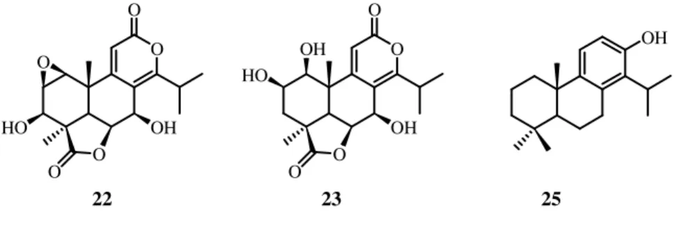
Nagilactones are a kind of nor and bisnorditerpene dilactones known as allelochemical and cytotoxic compounds, sometimes with antibacterial, antifungal, antifeedant activity (Hayashi et al., 1972; Hayashi et al., 1992; Kubo
et al., 1993; Kuo et al., 2008); the nagilactone C is the most studied compound (Figure 1.5). The dilactones are classified into three major subgroups in accordance with the structure of the B/C ring part: i) α-pyrone [8(14),9(11)-dienolide] type; ii) 7α,8α-epoxy-9(11)-enolide type; iii) 7(8),9(11)-dienolide type. It should be noted that the dilactone members in each of these structural types show fine differences in their biological activity (Hayashi and Matsumoto, 1982). O O O O OH HO O
9 1.4 Interaction with biomolecules: potential biological activities
Over the past years there has been a rapid escalation in the discovery of molecular targets that may be applied to the discovery of novel tools for the diagnosis, prevention, and treatment of human diseases. With the sequencing of the human genome, there has been an explosion in the knowledge of the protein products associated with the constituent genes and the discovery of molecular targets associated with various disease types, such as cancer, diabetes, and obesity. Natural products research plays a highly significant role in the drug discovery and development process, through the analysis of the molecular mechanism responsible of biological activity (Chin et al., 2006).
Even though natural products may not have coevolved with human proteins, they have emerged in nature to interact with biomolecules and it should be considered that varied genomes, based on similar chemistry, have spread across the Earth and human genus shares its gene families with other organisms. The production of metabolites with a certain affinity of interaction with a large variety of proteins and biological targets is improved with the evolution. Mainly the great homology between structural and functional domains of proteins from animal, vegetable or marine world allows the interaction among each other with a comparable affinity (Caporale, 2002; Koehn and Carter, 2005; Zhang, 2007).
Natural products should be able to penetrate biological barriers and make their way to certain cells or organs in which they will exert the effect. Thus, natural products are already biologically validated to reach and to bind specific proteins. Looking at all proteins and analyzing them for structural features, elements of conservatism and diversity may be found. The conservative elements are the domains: the parts that fold to compact secondary structures. Among the hundreds of thousands of human proteins, the number of distinct domains is only about 600 to 8000. Consequently, proteins that may seem altogether different are quite similar when viewed structurally, but diversity lies in the precise details
10
since similar domains may have very different amino acid sequences (Kirkpatrick, 2003).
1.5 Target discovery strategies
Especially for complex diseases, a lot of compounds are involved in the modulation of several targets and the exact targets of some drugs often remain unknown. The simplified “one molecule, one target” concept seems to be by now out-of date (Rix and Superti-Furga, 2009). Even if the multi-target interaction could also lead to harmful side effects, reducing the potency and efficiency of the drugs there is a possible application in multifactorial diseases (Bantscheff et al., 2009).
The identification of a drug interactome followed by biological assays for the measure of its activity against the identified target proteins, is actually one of the key processes in the drug development study. Several approaches carried out in the absence of the biological context have been developed to reach this goal even if gaining only a partial coverage of the full interactome. Recent developments in analytical methods, mainly in mass spectrometry and liquid chromatography, have had an important impact in the field of target discovery. In particular, mass spectrometry-based chemical proteomics has been emerging as a powerful tool to a comprehensive characterization of a drug interactome under physiological relevant conditions. Moreover, such method enables the analysis of the drug mechanism of action, in the context of a complex proteome accelerating the difficult process of target validation and drug discovery (Jeffery and Bogyo, 2003).
11
1.5.1 Proteomics techniques
Proteomics, as a scientific field, is defined as the study of the protein products of the genome, and their interactions and functions. Similarly, the proteins expressed at a given time in a given environment constitute a proteome. The techniques being developed in proteomics are applicable to all areas of drug discovery, from target identification, to assessment of drug efficacy, both in pre-clinical (target selectivity) and pre-clinical situations (patient response), to protein profiles for diagnostics.
For drug discovery, the ideal proteomics method would be one that is:
1. Sensitive enough to detect low-abundance proteins. 2. Able to detect activity over in addition to abundance. 3. Able to detect protein–protein and protein–small molecule interactions.
4. Easily implemented and performed quickly (Burbaum and Tobal, 2002).
Several proteomic techniques are based on two-dimensional gel-electrophoresis, isotope-coded affinity-tagging, “drug on-beads” immobilization, affinity chromatography.
1.5.2 Chemical proteomics
A new research field is chemical proteomics, or activity-based proteomics, that is considered a combination of affinity purification and mass spectrometric identification of a set of proteins targeted (or captured) by a small molecule. It is a multidisciplinary technique, involving different research areas such as biochemistry, cell biology, organic synthesis and mass spectrometry. This technique allows the analysis of all potential targets of a molecule in a single
12
experiment, leading to a complete and selective target mapping of a drug candidate. This means that the drug is able to fish out its own targets directly from the biological sample of interest.
Chemical proteomics approaches include several different experimental procedures, however three of them have emerged as most popular: (i) fishing for partners (or pull down), which employs a ligand immobilized onto a solid support, (ii) global proteomics, in which isobaric tags are used to asses changing in protein concentration upon drug treatment, and (iii) ABPP, activity-based protein profiling, which uses active site-directed probes to record variations in the activity of enzymes in a whole proteome (Sadaghiani et al., 2007; Rix et al., 2007).
The pull down assay is based on the non covalent and specific interactions occurring between the small molecule and its potential binders, and the following purification and mass spectrometric identification of these interacting proteins (Jeffery and Bogyo, 2003); (Figure 1.6).
One of the methods includes the compound of interest (with known or unknown biological activity) anchoring to a solid support through a spacer arm. This matrix is then treated with a lysate, from cells or tissue, to promote the interaction between the anchored compound and its specific binding proteins. After several washing steps, useful to reduce the amount of non-specific binding proteins, the captured proteins are eluted and subjected to either Sodium Dodecyl Sulphate Polyacrylamide gel electrophoresis (SDS PAGE), or gel free chromatography. The peptide mixtures obtained after trypsin digestion are then submitted to MS analysis and database search for the final protein identification.
The first step of the procedure is the drug on-beads immobilization: molecules containing one or more reactive sites or after synthetic modifications can be attached to a such solid support. To avoid or reduce steric interferences between the targets and the ligand-bearing matrix, the solid support is usually modified with an opportune spacer arm that provides a suitable distance between the beads
and the small molecule. The choice of the solid support is one of the most important steps of the experiment
support is then added to an opportune protein extract from a cell culture, tissue or serum, to promote ligand protein interaction. After the treatment, the matrix is washed to reduce the amount of the non
elution of the retained ones is carried on. The eluted proteins are sequentially separated by gel electrophoresis,
finally analyzed and identified by HPLC
There are proteins exclusively interacted with the linker or the beads, proteins highly abundant with low affinity for the drug
high affinity for matrix with the drug.
Figure
and the small molecule. The choice of the solid support is one of the most portant steps of the experiment. The small molecule anchored to the solid support is then added to an opportune protein extract from a cell culture, tissue or serum, to promote ligand protein interaction. After the treatment, the matrix is washed to reduce the amount of the non-specific binding proteins and finally the elution of the retained ones is carried on. The eluted proteins are sequentially separated by gel electrophoresis, in situ or in solution digested with trypsin, and finally analyzed and identified by HPLC-MS/MS and data base search.
here are proteins exclusively interacted with the linker or the beads, proteins highly abundant with low affinity for the drug-support system, and proteins with high affinity for matrix with the drug.
Figure 1.6: Fishing for partners strategy
13
and the small molecule. The choice of the solid support is one of the most olecule anchored to the solid support is then added to an opportune protein extract from a cell culture, tissue or serum, to promote ligand protein interaction. After the treatment, the matrix is eins and finally the elution of the retained ones is carried on. The eluted proteins are sequentially or in solution digested with trypsin, and
here are proteins exclusively interacted with the linker or the beads, proteins support system, and proteins with
14
Another key step is the protein identification: one of the procedure involves the previous enzymatic digestion of the proteins and the tandem mass spectrometric analysis of the complex peptide mixtures (Chait, 2006). The usual proteolytic enzyme used in proteomics is trypsin, a protease that cleaves peptides at C-terminus of arginine and lysine residues. The peptides originated by proteolytic cleavage have a size less than 3 kDa and a free amino group at C-terminus, and give rise to doubly and triply charged peptides, when exposed to a mass spectrometer.
The peptide mixture is subjected to liquid chromatography coupled tandem MS analysis, and the raw data are processed to give a peak list submitted to a protein search in a data base, for instance Mascot that is a web program comparing the experimental MS2 spectra with theoretical fragment spectra generated “in silico” and needing of parameters such as peptide tolerance, proteolytic enzyme, miss cleavage and variable modifications (Perkins et al., 1999).
1.5.3 Target validation
The results obtained by this technique are only indicative and useful to have an idea of the possible target of a compound but it is necessary to determine the biological relevance and the kind of the interaction.
In the first case biological assays aimed to identity enzymatic inhibition, activity ability, in vivo and in vitro effects, etc. according to the kind of the identified proteins, are necessary.
The physical interaction could be determined by measuring the quantitative binding parameters with different techniques like surface plasmon resonance (SPR), isothermic titration calorimetry (ITC), STD-NMR, and cocrystallization experiments.
15 1.6 Objectives of the thesis project
As part of an ongoing research program on plants biosynthesizing diterpenes, it was performed a project aimed to create a diterpenes library from different plant’s species. These compounds were subjected to a chemical proteomic study as a strategy for the molecular target identification of natural bioactive compounds, setting up a method for the research of their molecular targets.
The attention was paid on plants known to be rich on diterpenes, since they are a class of natural terpenoids with a great structural variability and a wide spectrum of biological activities.
Particularly two diterpenes included in our library were considered: a semi synthetic compound 15-ketoatractyligenin methyl ester, and the hardwickiic acid. From an experimental point of view, this work was divided into the following steps: (i) isolation and structural characterization of new and known diterpenes through a phytochemical study in order to enrich a pre-existing diterpenes library with new structures (ii) chemical proteomic approach as a strategy for the research of the molecular target of the ent-kaurane 15-ketoatractyligenin methyl ester (iii) validation techniques of the protein Hsp27 as a target for the clerodane diterpene hardwickiic acid.
According to the primary purpose, Podocarpus elongatus (Aiton) L’Hèr. ex Pers, Podocarpus gracilior Pilger (Podocarpaceae), Clerodendrum splendens G. Don. (Verbenaceae), and Sideritis pullulans Vent. (Lamiaceae) were selected.
In chapters 2-4 the extraction and isolation methods and the structural characterization procedures of new and known compounds from the previously listed plants are described.
In chapter 2 the diterpenes isolated from P. elongatus leaves are reported: the
new diterpenes nagilactone C 7-O-α-L-arabinopyranosyl-(1→4)-β-D
-xylopyranoside, nagilactone C 7-O-β-D-glucopyranosyl-(1→4)-β-D
-16
arabinopyranosyl-(1→4)-β-D-xylopyranoside,
2β,15S,16,17,19-pentahydroxy-isopimar-8(14)-ene 17-O-β-D-glucopyranoside,
2β,15R,16,17,19-pentahydroxy-isopimar-8(14)-ene 17-O-β-D-glucopyranoside, and the known diterpenes
4-carboxy-19-nor-totarol, nagilactone B, and nagilactone C. In this chapter the new diterpenes found in P. gracilior leaves, 2α,16-dihydroxy-4β-carboxy-O-β-D -glucopyranosyl-19-nor-totarol, nagilactone K together with the known nagilactone B, nagilactone C, nagilactone D, and podolactone B are also mentioned.
In chapter 3
2α-acetoxy-3β-(2’,3’-diacetoxy-2’-methyl)-butanoyloxy-14-hydro-15-hydroxyclerodin, 3β,15-dihydroxy-14-hydro-clerodin,
2α,15-
dihydroxy-3β-(2’-hydroxy-3’-acetoxy-2’-methyl)-butanoyloxy-6α,18-diacetoxy-4α,17-epoxy-clerodan-11,16-lactone,
3β,14S,15-trihydroxy-6α,18-diacetoxy-4α,17-epoxy-clerodan-11,16-lactone, and the only known 14,15-dihydroxy-3-epicarioptin isolated from C. splendens are described.
In chapter 4 the new diterpenes isolated from S. pullulans, 1α,3α,7β,18-tetrahydroxykaur-16-ene, 3α,11α,18-trihydroxy-17-norkauran-16-ene,
ent-3α,7β,18-trihydroxy-17-norkauran-16-one, ent
-3α,7β-dihydroxy-18-acetyloxy-17-norkauran-16-one, 3α,7β,16α,17-tetrahydroxy-18-acetyloxy-kaurane, ent-7β,16α,17,18-tetrahydroxykaurane, and the known foliol, linearol, ent-3α,7β,17-trihydroxy-18-acetyloxy-15α,16α-epoxykaurane are described.
Chapters 5 and 6 are related to studies aimed to the research of the proteic
target for small molecules through chemical proteomics techniques.
In chapter 5 a fishing for partners study as a chemical proteomics strategy to identify the proteic target of 15-ketoatractyligenin methyl ester (SR2017) is described. SR2017 is semi-synthetic ent-kaurane synthetized at Dipartimento di Chimica Organica, Università di Palermo (Rosselli et al., 2007).
This approach revealed the heat shock protein 70 (Hsp70) and peroxisomeproliferator-activated receptor gamma (PPARγ) as potential targets. To confirm results a SPR based binding assay was used.
17
In chapter 6 the study of hardwickiic acid (HAA) and its heat shock protein 27 (Hsp27) interaction is reported. HAA is a diterpene chosen as a representative of the clerodane diterpenes library isolated from Salvia spp. Hsp27 was identified as potential target of HAA through a fishing for partner studies based on a chemical proteomics approach and in a previous study this was confirmed by SPR experiments.
As first step to evaluate the effect of the interaction between HAA and Hsp27 on protein structure and activity, a limited proteolysis-mass spectrometry based strategy was used.
The ability of HAA to modulate the in vitro chaperone activity of Hsp27 was evaluated by monitoring substrates induced aggregation in the presence of Hsp27, with or without HAA. Finally to give a cellular significance to Hsp27/HAA interaction, it was explored the pro-apoptotic activity of the small molecule in human monocytes.
Chapter 2
Phytochemical study of Podocarpus elongatus (Aiton) L’Her. ex Pers and Podocarpus gracilior Pilger
Based on:“Faiella L., et al., Phytochemistry, 2012, 76, 172-177“; “Faiella L., et al.,
18
Chapter 2
2.1 Introduction: Podocarpus genus
Podocarpaceae is an ancient and large family of mainly Southern Hemisphere conifers, comprising about 156 species of evergreen trees and shrubs. It is especially spread in tropical and subtropical forests of the Austral Hemisphere: New Zealand, New Caledon, and Tasmania are particularly rich of genera. The Podocarpaceae family shows great diversity, both morphologically and ecologically (Eckenwalder, 2009).
Podocarpus species are important timber trees in their native areas. The timber is fine-grained, nonresinous, light, and moderately strong. Some species are used as ornamental trees, mostly for landscaping. The fruits of other species are eaten raw or cooked or used locally for jams and preserves. Other products obtained from this genus include dyes, tannins, and waxes. Some species of
Podocarpus are used for cultural purposes in South Africa (Abdillahi et al, 2010). Podocarpus spp. are also traditionally used in the native regions for the treatment of many diseases like fevers, asthma, coughs, cholera, arthritis, rheumatisms, and venereal diseases (Abdillahi et al., 2011). The presence or absence of different compounds can be used to relate the new and the old taxa. For example, flavonoids can be a useful chemotaxonomic tool in this group of plants, since biflavonoids of the amentoflavone and hinokiflavone groups have been shown to be good taxonomic markers in the great majority of Podocarpus (Abdillahi et al., 2010).
Phytochemical and pharmacological studies of Podocarpus genus led to the isolation of a number of bioactive diterpenes, including nor- and
bisnor-19
diterpenoid lactones (Park et al., 2004; Xuan et al., 1995), flavonoids (Markham
et al., 1985), and ecdysones (Nakanishi et al., 1966). Since nor- and bis-norditerpenes are taxonomic markers of these species, the possibilities that the related taxa will have similar biological activities or bioactive compounds cannot be overruled. The nagilactone C is the most studied. It had shown an antiproliferative activity against different type of cell lines such as fibrosarcoma and murine colon carcinoma. A study on Hela cells shows that it inhibits the eukaryotic proteic synthesis, but its molecular target is unknown (Chan et al., 2004; Shrestha et al., 2001).
Podocarpus elongatus (Aiton) L’Her. Ex Pers. and Podocarpus gracilior Pilger were selected for this study mainly because of the relevant presence of diterpenes in plants belonging to this genus.
2.2 Podocarpus elongatus (Aiton) L’Her. ex Pers
Podocarpus elongatus (Aiton) L’Her. ex Pers (Figure 2.1) is a tree native to South Africa and West Cape, also known as Cape yellowwood, yellow pine, fern pine, breade river yellowwood or westelike geelhout. It is mainly used as ornamental tree mostly for landscaping. The light-brown, soft durable, moderately strong, elastic, resinous, pale yellow brown wood is used for building houses, railway sleepers, beams, planks, rafters, and furnitures (Abdillahi et al, 2010).
There are only few and old previous phytochemical studies of P. elongatus that report the isolation of biflavones, namely amentoflavone, podocarpusflavone A, bilobetin, isoginkgetin (Prasad and Krishnamurty, 1976), hinokiflavone, and tetra-O-methyl-amentoflavone (Iqbal et al., 1982) and abietane diterpenes, such as totarol, 16-oxytotarol, and 16-hydroxytotarol (Taylor, 1965).
Figure 2.1
2.3 Podocarpus gracilior
Podocarpus gracilior
and East Africa, known as African fern p insect attack (Kubo et al.,
reported the isolation of diterpenes such as podolactones (Hayashi nagilactones C, D, and F (Kubo
Kupchan et al., 1975), phenolic diterpenoids (Cambie ponasterone A (Kubo et al.
and dimethylamentoflavone (Kubo
Figure 2.1: Podocarpus elongates (Aiton) L’Her ex Pers
Podocarpus gracilior Pilger
Pilger (Figure 2.2) is a conifer that grows in Central known as African fern pine and characterized by a resistance to
et al., 1984). Previous phytochemical studies on P. gracilior reported the isolation of diterpenes such as podolactones (Hayashi et al., 1980), nagilactones C, D, and F (Kubo et al., 1984), podolide (Bryan and Smith, 1975; ., 1975), phenolic diterpenoids (Cambie et al., 1983), the ecdysone
et al., 1984) and biflavonoids such as podocarpusflavone A and dimethylamentoflavone (Kubo et al., 1983; 1984).
20
) is a conifer that grows in Central ine and characterized by a resistance to
P. gracilior
., 1980), , 1975; , 1983), the ecdysone , 1984) and biflavonoids such as podocarpusflavone A
Figure
2.4 Plant materials
Leaves of P. elongatus Cairo, Egypt, on May 2008, same place on March 2010, b
Zoharia Research Garden, Cairo, Egypt). The voucher
elongatus Ait./1 and No
Herbarium Hortii Botanici Pisani, Flora Aegyptiaca, Pisa, Italy.
2.5 Extraction and iso
Dried powdered leaves of
were successively and separately extracted for 48 h with
Figure 2.2: Podocarpus gracilior Pilger
P. elongatus were collected in El Zoharia Research Garde Cairo, Egypt, on May 2008, while leaves of P. gracilior were collected in same place on March 2010, both were identified by Dr. Mamdouh Shokry (El
den, Cairo, Egypt). The voucher specimens (No. 15 Ait./1 and No. 15, P. gracilior Pilger/1) were deposited at the Herbarium Hortii Botanici Pisani, Flora Aegyptiaca, Pisa, Italy.
2.5 Extraction and isolation
Dried powdered leaves of P. elongatus (800 g) and of P. gracilior (950 g) were successively and separately extracted for 48 h with n-hexane, CHCl
21
were collected in El Zoharia Research Garden of were collected in the Mamdouh Shokry (El (No. 15, P. ) were deposited at the
(950 g) hexane, CHCl3,
22
CHCl3–MeOH (9:1), and MeOH, by exhaustive maceration (2 l), to give 14.9, 19.7, 27.2, and 35.2 g of the respective residues for P. elongatus and 11.5, 17.3, 32.6, and 42.4 g of the respective residues for P.gracilior.
The MeOH extract of P. elongatus was partitioned between n-BuOH and
H2O, to afford a n-BuOH residue (15 g). Part of the n-BuOH fraction (6.0 g) was
submitted to Sephadex LH-20 using MeOH as eluent to obtain seven major fractions (A-G) grouped by TLC. Fraction B (120 mg) was purified by RP-HPLC
with MeOH-H2O (25:75) as eluent to give pure nagilactone C 7-O-β-D
-glucopyranosyl-(1→4)-β-D-xylopyranoside (compound 2, 1.6 mg, tR = 13 min),
nagilactone C 7-O-α-L-arabinopyranosyl-(1→4)-β-D-xylopyranoside (compound
1, 10.3 mg, tR = 15 min), 3,4,5-trihydroxydehydro-α-ionol 9-O-β-D
-glucopyranoside (compound 12, 2.3 mg, tR = 18 min), roseoside (compound 13,
5.6 mg, tR = 20 min), and
1-(2,6,6-trimethyl-4-hydroxycyclohexenyl)-1-hydroxy-buta-1-en-3-one 4-O-β-D-glucopyranoside (compound 7, 1.5 mg, tR = 24 min). Fraction C (244 mg) was subjected to RP-HPLC with MeOH-H2O (3.5:6.5) as
eluent to give nagilactone A 7-O-α-L-arabinopyranosyl-(1→4)-β-D
-xylopyranoside (compound 4, 1.5 mg, tR = 13 min), linarionoside C (compound
14, 1.8 mg, tR = 23 min), 2β,15S,16,17,19-pentahydroxy-isopimar-8(14)-ene 17-O-β-D-glucopyranoside (compound 5, 2.0 mg, tR = 24 min),
2β,15R,16,17,19-pentahydroxy-isopimar-8(14)-ene 17-O-β-D-glucopyranoside (compound 6, 2.3
mg, tR = 25 min), and 20-hydroxyecdysone (compound 15, 8.6 mg, tR = 45 min).
Fraction D (300 mg) was purified over RP-HPLC with MeOH-H2O (3:7) as
eluent to yield pure erythro-4,7,9,9'-tetrahydroxy-3,3'-dimethoxy-8-O-4'-oxyneolignan 9'-O-β-D-glucopyranoside (compound 16, 5.2 mg, tR = 14 min), threo-4,7,9,9'-tetrahydroxy-3,3'-dimethoxy-8-O-4'-oxyneolignan
9'-O-β-D-glucopyranoside (compound 17, 6.4 mg, tR = 15 min), compound 13 (3.4 mg, tR =
18 min), and icariside E3 (compound 18, 3.6 mg, tR = 25 min). The CHCl3-MeOH extract (6.0 g) was chromatographed over Sephadex LH-20 to give eight major
23
fractions (A-H) grouped by TLC, together with pure isoginkgetin (compound 19, 120 mg) and bilobetin (compound 20, 25 mg). Fractions B (123 mg) and G (163 mg) were separately purified by RP-HPLC with MeOH-H2O (3.5:6.5) as eluent to yield blumenol C glucoside (compound 21, 2.7 mg, tR = 11 min) and compound 13 (3.1 mg, tR = 13 min), from fraction B and vitexin 2''-O-β-D -(6'''-acetyl)-glucopyranoside (compound 8, 14.4 mg, tR = 41 min) from fraction G, respectively. Fraction D (60 mg) was also purified by RP-HPLC with
MeOH-H2O (3:7) as eluent to yield nagilactone C 7-O-β-D-xylopyranoside (compound
3, 3.7 mg, tR = 13 min) and compound 13 (2.0 mg, tR = 18 min). Finally, fractions C (390 mg), E (185 mg), and F (220 mg) were separately subjected to RP-HPLC
with MeOH-H2O (2:3) as eluent to give compounds 18 (2.3 mg, tR = 20 min) and
15 (2.8 mg, tR = 45 min) from fraction C, nagilactone C (compound 22, 2.5 mg, tR = 8 min), compounds 16 (3.5 mg, tR = 9 min) and 17 (2.7 mg, tR = 10 min) from fraction E, and nagilactone B (compound 23, 3.0 mg, tR = 9 min) and
glochidiobioside (compound 24, 2.0 mg, tR = 15 min) from fraction F,
respectively.
Part of the CHCl3 extract (10.0 g) was chromatographed over silica gel column eluting with CHCl3 followed by increasing concentrations of MeOH in CHCl3 (between 1% and 50%). Fractions of 25 ml were collected, analyzed by TLC (silica gel plates, in CHCl3 or mixtures of CHCl3-MeOH 99:1, 98:2, 97:3, 9:1, 4:1), and grouped into 10 fractions (A-L). Fraction G (290 mg) was purified
by RP-HPLC with MeOH-H2O (8.5:2.5) as eluent to obtain
4β-carboxy-19-nor-totarol (compound 25, 3.1 mg, tR = 10 min).
The MeOH extract of P. gracilior was partitioned between n-BuOH and H2O,
to afford a n-BuOH residue (5 g) that was submitted to a Sephadex LH-20 using MeOH as eluent to obtain eight major fractions (A-H) grouped by TLC. Fraction B (372 mg) was purified by RP-HPLC with MeOH-H2O (2.5:7.5) as eluent to
give15-hydroxy phaseic acid (compound 11, 1.7 mg, tR = 10 min), compound 22
-glucopyranosyl-19-24
nor-totarol (compound 9, 1.6 mg, tR = 38 min), and 24 (1.2 mg, tR = 40 min). Part
of the CHCl3-MeOH extract (10.0 g) was chromatographed over Sephadex
LH-20 to give ten major fractions (A-K) grouped by TLC. Fractions C (275 mg) and
E (350 mg) were separately purified by RP-HPLC with MeOH-H2O (3.5:6.5) as
eluent to yield compounds 21 (1.2 mg, tR = 36 min) and 15 (5.4 mg, tR = 38 min) from fraction C and compounds 22 (8.4 mg, tR = 11 min), nagilactone D (compound 26, 1.8 mg, tR = 17 min), 17 (3.5 mg, tR = 21 min), and 18 (2.7 mg, tR = 24 min) from fraction E, respectively. Fraction F (208 mg) was also purified by
RP-HPLC with MeOH-H2O (25:65) as eluent to yield pure podolactone B
(compound 27,1.2 mg, tR = 15 min), compounds 22 (4.3 mg, tR = 19 min), nagilactone K (compound 10, 1.7 mg, tR = 25 min), and 26 (4.0 mg, tR = 42 min). Part of the CHCl3 extract (10.0 g) was chromatographed over silica gel column eluting with CHCl3 followed by increasing concentrations of MeOH in CHCl3 (between 1% and 50%). Fractions of 25 ml were collected, analyzed by TLC (silica gel plates, in CHCl3 or mixtures of CHCl3-MeOH 99:1, 98:2, 97:3, 9:1, 4:1), and grouped into 13 fractions (A-M). Fractions C (363 mg), D (100
mg) and E (182 mg) were subjected to RP-HPLC with MeOH-H2O (3.5:6.5) as
eluent to give pure compound 26 (8.9 mg, tR = 17 min) from fraction C,
compounds 22 (6.1 mg, tR = 10 min) and 26 (5.0 mg, tR = 17 min) from fraction
D, and compound 23 (6.2 mg, tR = 11 min) from fraction E, respectively. The structures of new compounds 1-11 are showed in Figure 2.3 and Figure 2.4 respectively. The structures of the diterpenes 22, 23, and 25 are showed in
Figure 2.5 while 26 and 27 are reported in Figure 2.6. The structures of the other
25 2.6 New isolated compounds
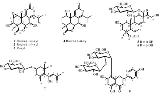
Nagilactone C 7-O-α-L-arabinopyranosyl-(1→4)-β-D-xylopyranoside (1)
Amorphous powder;
[ ]
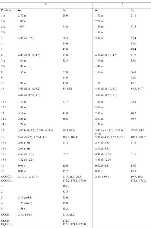
α 25D-10 (c 0.1, MeOH); UV (MeOH) λmax (log ε) 285 (3.80) nm; 1H and 13C NMR, see Table 2.1; HRESIMS m/z 649.2122 [M+Na]+ (calcd. for C29H38O15Na, 649.2108); ESI-MS m/z 649 [M+Na]+, 517 [M+Na-132]+, 385 [M+Na-132-132]+.Nagilactone C 7-O-β-D-glucopyranosyl-(1→4)-β-D-xylopyranoside (2)
Amorphous powder;
[ ]
α 25D+1.6 (c 0.05, MeOH); UV (MeOH) λmax (log ε) 290 (3.80) nm; 1H and 13C NMR, see Table 2.1; HRESIMS m/z 679.2261 [M+Na]+ (calcd. for C30H40O16Na, 679.2214); ESI-MS m/z 679 [M+Na]+, 517 [M+Na-162]+, 385 [M+Na-162-132]+.Nagilactone C 7-O-β-D-xylopyranoside (3)
Amorphous powder;
[ ]
α 25D+5 (c 0.1, MeOH); UV (MeOH) λmax (log ε) 280 (4.12) nm; 1H and 13C NMR, see Table 2.1; HRESIMS m/z 517.1712 [M+Na]+ (calcd. for C24H30O11Na, 517.1686); ESI-MS m/z 517 [M+Na]+, 385 [M+Na-132]+.Nagilactone A 7-O-α-L-arabinopyranosyl-(1→4)-β-D-xylopyranoside (4)
Amorphous powder;
[ ]
α 25D-16 (c 0.1, MeOH); UV (MeOH) λmax (log ε) 282 (4.08) nm; 1H and 13C NMR, see Table 2.1; HRESIMS m/z 635.2357 [M+Na]+ (calcd. for C29H40O14Na, 635.2316); ESI-MS m/z 635 [M+Na]+, 503 [M+Na-132]+.26
2β,15S,16,17,19-Pentahydroxy-isopimar-8(14)-ene 17-O-β-D -glucopyranoside (5)
Amorphous powder;
[ ]
α 25D+1.3 (c 0.08, MeOH); 1H and 13C NMR, see Table2.2; HRESIMS m/z 539.2888 [M+Na]+ (calcd. for C26H44O10Na, 539.2832); ESI-MS m/z 515 [M-H]-, 353 [M-H-162]-, 293 [M-H-162-60]-.
2β,15R,16,17,19-Pentahydroxy-isopimar-8(14)-ene 17-O-β-D -glucopyranoside (6)
Amorphous powder;
[ ]
α 25D+3.6 (c 0.2, MeOH); 1H and 13C NMR, see Table
2.2; HRESIMS m/z 539.2895 [M+Na]+ (calcd. for C26H44O10Na, 539.2832); ESI-MS m/z 515 [M-H]-, 353 [M-H-162]-.
1-(2,6,6-Trimethyl-4-hydroxycyclohexenyl)-1-hydroxy-buta-1-en-3-one
4-O-β-D-glucopyranoside (7)
Amorphous powder;
[ ]
α 25D-42.9 (c 0.08, MeOH); UV (MeOH) λmax (log ε)284 (3.78) nm; 1H and 13C NMR, see Table 2.2; HRESIMS m/z 409.1870
[M+Na]+ (calcd. for C19H30O8Na, 409.1839); ESI-MS m/z 409 [M+Na]+, 325 [M+Na-84]+.
Vitexin 2''-O-β-D-(6'''-acetyl)-glucopyranoside (8)
Amorphous powder;
[ ]
α 25D-5 (c 0.1, MeOH); UV (MeOH) λmax (log ε) 272(3.90), 335 (3.22) nm; 1H and 13C NMR, see Table 2.2; HRESIMS m/z 659.1600
[M+Na]+ (calcd. for C29H32O16Na, 659.1588); ESI-MS m/z 659 [M+Na]+, 569
27
2α,16-Dihydroxy-4β-carboxy-O-β-D-glucopyranosyl-19-nor-totarol (9)
Amorphous powder;
[ ]
α
25D +50.2 (c 0.05, MeOH); UV (MeOH) λmax (log ε) 230 (3.89), 278 (4.40) nm; 1H and 13C NMR, see Table 2.3; HRESIMS m/z 511.2537 [M+H]+ (calcd. for C26H38O10, 511.2543); ESI-MS m/z 533 [M+Na]+,371 [M+Na-162]+.
Nagilactone K (10)
Amorphous powder;
[ ]
α
25D +4.1 (c 0.2, MeOH); UV (MeOH) λmax (log ε) 246 (4.03) nm; 1H and 13C NMR, see Table 2.3; HRESIMS m/z 319.1160 [M-H] -(calcd. for C17H20O6, 319.1182); ESI-MS m/z 319 [M-H]
-, 275 [M-H-44]-.
15-Hydroxy phaseic acid (11)
Amorphous powder;
[ ]
α
25D +2.1 (c 0.06, MeOH); UV (MeOH) λmax (log ε)260 (3.95) nm; 1H and 13C NMR, see Table 2.3; HRESIMS m/z 297.1320 [M-H]
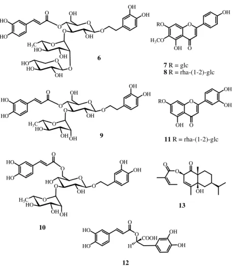
28 Figure 2.3: New isolated compounds from P. elongatus
HO 11 O O O 10 O HO 1 2 3 4 19 5 6 7 8 911 15 16 18 20 10 14 1 2 3 4 5 6 7 8 10 9 11 12 13 O COOR HO R = HO O HO OH CH2OH 1 2 3 4 18 19 5 10 20 7 6 14 8 9 11 1213 15 16 CH2OH OH 17 O CH2OH COOH 11 OH 15 9
Figure 2.4: New isolated compounds from P. gracilior
2.7 Structural elucidation
As described before, from P. elongatus leaves eight new (1–8) and 13 known compounds were isolated. From P. gracilior were isolated three new compounds
CH2OH HO O OH R O O OH 7 O O HO OH OH 8 O O OR O O 1 R=ara-(1-4)-xyl 2 R=glc-(1-4)-xyl 3 R=xyl O HO 1 2 3 4 19 5 6 7 8 9 11 15 16 17 12 18 20 10 14 5 R = α OH 6 R = β OH O HO HO OH CH2OH O HO HO OH CH2OH O HO HO O CH2OH O HO OH CH2OAc 4 R=ara-(1-4)-xyl O O OR OH O O 1 2 3 4 18 19 5 10 20 7 6 14 8 9 11 12 13 17 15 16 1 2 3 4 5 6 7 8 9 10 11 12 13 HO 2 3 4 10 5 6 7 8 9 1' 2' 3' 4' 6' 5'
29
(9-11) together with nine known compounds, some of which are in common with
P.elongatus. The following known compounds were identified by comparison with published spectroscopic data: nagilactone B (Hayashi et al., 1980), nagilactone C (Kubo and Ying, 1991), nagilactone D (Hayashi et al., 1980), podolactone B (Hayashi et al., 1980), 4β-carboxy-19-nor-totarol (Kubo and Ying, 1991), roseoside (Otsuka et al., 1995), blumenol C glucoside (Pabst et al., 1992), linarionoside C (Otsuka, 1994), 3,4,5-trihydroxydehydro-α-ionol
9-O-β-D-glucopyranoside (Rastrelli et al., 1998), icariside E3 (Kuono et al., 1991), erythro-4,7,9,9'-tetrahydroxy-3,3'-dimethoxy-8-O-4'-oxyneolignan 9'-O-β-D-glucopyranoside (Matsuda and Kituchi, 1996), threo-4,7,9,9'-tetrahydroxy-3,3'-dimethoxy-8-O-4'-oxyneolignan 9'-O-β-D-glucopyranoside (Matsuda et al., 1996), glochidiobioside (Takeda et al., 1998), isoginkgetin (Markham et al., 1987), bilobetin (Markham et al., 1987), and 20-hydroxyecdysone (Miller et al., 1985).
The molecular formula of compound 1 (C29H38O15) was established by 13
C
NMR and ESIMS (m/z 649 [M+Na]+) spectra, indicating eleven degrees of
unsaturation. In the ESIMS spectrum of compound 1 two ion fragments at m/z 517 [M+Na-132]+ and 385 [M+Na-132-132]+, due to the subsequent losses of two pentose residues, were observed. The α,β,γ-unsaturated lactone carbonyl functionality was evidenced by the presence of an ester carbonyl resonance at
164.2 ppm in the 13C NMR spectrum (Table 2.1). The 1H NMR spectrum showed
the presence of two methyls singlet at δ 1.43 (Me-18), 1.49 (Me-20), two methyl doublets at δ 1.25 (Me-16) and 1.38 (Me-17), five oxymethines at δ 3.43 (H-2), 3.59 (H-1), 4.39 (H-3), 5.12 (Η−6), 5.56 (H-7), and one singlet olefinic proton at δ 6.38 (H-11) (Table 2.1). COSY and 1D-TOCSY measurements showing couplings between H-1―H-3, H-5―H-7, H-15―H-17 led to the assignement of the spin systems. HMBC correlations from H-11 to C-8, C-12, C-14 confirmed
30
correlations of H-1 signal centered at δ 3.59 with C-3, C-5, and C-10 and HMBC correlations of H-2 (δ 3.43) with C-4, C-10, and Me-20 established the presence of a 1β,2β-epoxide. The elucidation of the whole skeleton from the above subunits was achieved on the basis of HSQC and HMBC correlations, which also allowed the assignment of all the resonance in the 13C NMR spectrum of the pertinent carbons. By this way, the aglycon moiety of 1 was characterized as nagilactone C (Kubo and Ying., 1991). 1D-TOCSY, DQF-COSY, and HSQC experiments provided evidence for the presence in the molecule of two pentose residues. On the basis of chemical shifts, multiplicity, values of the coupling constants, and magnitude in the 1H and 13C NMR spectra, the two sugar moieties
were characterized as one β-xylopyranose and one α-arabinopyranose (Table
2.1) (De Tommasi et al., 1997). The HMBC experiment provided the sugar
sequence through unambiguous correlations between H-7C-1xyl and H-1ara
C-4xyl. The configuration of the sugar units was assigned after hydrolysis of 1 with 1N HCl and GC analysis of trimethylsilyated sugars through a chiral column. The sugar units were so determined to be D-xylopyranose and L-arabinopyranose. On the basis of the above evidences, compound 1 was identified as nagilactone C
7-O-α-L-arabinopyranosyl-(1→4)-β-D-xylopyranoside.
The molecular formula of compound 2 (C30H40O16) was established by 13C
NMR and ESIMS spectrum (m/z 679 for [M+Na]+). In the ESIMS spectrum were
also observed two main fragments at m/z 517 [M+Na-162]+ and 385
[M+Na-162-132]+) due to the subsequent loss of one hexose and one pentose. Its NMR spectral data (Table 2.1) suggested that the structure of 2 resembled that of 1, but differed in the sugar chain. Comparison of chemical shifts of 2 with those of 1 suggested that the arabinopyranosyl residue was replaced by a β-glucopyranosyl unit. The absolute configuration of the sugar units was determined as reported for
1. Thus, the structure of 2 was identified as nagilactone C 7-O-β-D
31
The ESIMS of compound 3 (C24H30O11) showed a main signal at m/z 517 ([M+Na]+) and a fragmentation pattern similar to that of 1. Furthermore, in the MS2 spectrum a fragment ion peak at m/z 385 [M+Na-132]+ was detected, indicating the loss of one pentose unit. Analysis of the NMR data (Table 2.1) of compound 3 and comparison with those of 1 revealed 3 to differ from 1 only by the absence of the terminal arabinopyranose unit. Thus, compound 3 was
determined as nagilactone C 7-O-β-D-xylopyranoside.
Compound 4 was isolated as a colourless optically active amorphous powder, with [α]D = -16 (c = 0.1, MeOH). In the ESIMS spectrum, the [M+Na]+
signal was observed at m/z 635, consistent with the molecular formula C29H40O14. A
peak at m/z 503 [M+Na-132]+ was also observed, due to the loss of one pentose
unit. The observation of signals in the 13C NMR spectrum (Table 2.1) consistent
with δ-lactones and the presence of 19 carbon signals for the aglycon moiety led to the conclusion that this compound was again a norditerpene lactone glycoside
(Kubo and Ying, 1991). The 1H and 13C NMR data (Table 2.1) of 4 indicated the
presence of two double bond and two hydroxyl group (δH 3.93, δC 72.0; δH 5.51, δC 65.0). An isopropyl group was clearly evident in the
1
H NMR spectrum (two methyl doublets at δ 1.25 and 1.38 and a one proton signal at δ 3.40). The absence of further coupling of the isopropyl proton at δ 3.40 indicated C-14 was fully substituted. The 1H NMR spectral data combined with 1D TOCSY and DQF-COSY experiments suggested the sequence C-1C-3 and C-5C-7. Analysis of the proton coupling of the carbinolic protons allowed the remaining substitution pattern to be determined. Thus, the aglycon of compound 4 was recognized to be nagilactone A (Xuan et al., 1995). Structural elucidation of the sugar moiety was performed on the basis of DQF-COSY and 1D TOCSY; starting from the well isolated anomeric signal at δ 4.45 (d, J = 7.5 Hz), the first sugar spin system was clarified to be β-xylose, while from the anomeric signal at δ 4.37 (d, J = 7.0 Hz), the other saccharide was identified as α-arabinose. The
32
sequence of the sugar chain could be assigned by the correlations in the HMBC spectrum between signal at δ 5.51 (H-7) and C-1xyl (δ 100.8) and δ 4.37 (H-1ara) and C-4xyl (δ 77.0). The absolute configuration of the sugar units was determined as reported for 1. From the foregoing evidence, the structure of 4 was established
as nagilactone A 7-O-α-L-arabinopyranosyl-(1→4)-β-D-xylopyranoside.
The molecular formula of 5 (C26H44O10) was determined by ESIMS ([M-H]- at m/z 515). The 1H NMR spectrum (Table 2.2) showed signals for two tertiary methyl groups, two pairs of doublets centered at δ 3.05 (J = 11.0 Hz), 3.30 (J = 11.0 Hz), 3.76, and 3.85 (J = 12.0 Hz) for two hydroxymethylene groups, signals for protons at δ 3.79, 3.67, and 3.47 (br d, J = 9.0 Hz), and a hexose residue. The 1
H NMR spectral data combined with 1D TOCSY and DQF-COSY experiments suggested the sequence C-1C-3, C-5C-7, C-9C-12, and C-15C-16. The 13
C NMR spectrum (Table 2.2) showed the presence of two hydroxymethines, one trisubstituted double bond, three hydroxymethylenes, and was in good agreement with that of a pimarane derivative (Nishiya et al., 1991; Politi et al., 2002). Structural elucidation of the glucopyranose moiety was performed on the basis of 1D TOCSY; starting from the well isolated anomeric proton signal at δ 4.28, the sugar spin system was clarified. The configuration of the glucose unit of
5 was obtained as reported before. The 13C NMR signals were assigned on the basis of a HSQC experiment. Location of the hydroxyl groups, the double bond, and the sugar unit was obtained by analysis of the HMBC spectrum. In fact, the signals at δ 3.47 and 3.67 (H2-16) correlated with carbon resonances at δ 43.3 (C-13), 75.0 (C-15), and 125.3 (C-14), the signal at δ 3.79 (H-15) correlated with a signal at δ 65.5 (16), leading to the location of one diol group at 15 and C-16; the signals at δ 3.76 and 3.85 (H2-17) correlated with δ 25.7 12), 43.3 (C-13), and 75.0 (C-15), establishing the location of one hydroxymethylene at C-17, and finally the signals at δ 3.05 and 3.30 (H2-19) correlated with δ 19.2 (Me-18), 40.0 (C-4), 45.3 (C-3), 47.5 (C-5), positioning the third hydroxymethylene
33
function at 19. The HMBC correlations between H-14 and 7, 12, and C-16 confirmed the C-8/C-14 double bond, while correlation between δ 4.28 (H-1glc) and 72.6 (C-17) established the sugar location. 2D ROESY measurements supported the proposed structure and proved the relative stereochemistry at C-2, C-4, C-5, C-10, and C-13. Thus, the signal of the proton at δ 0.89 (Me-20) correlated with H-19a (δ 3.05) and H-17b signals (δ 3.85), the proton signal at δ 3.84 (H-2) affected the Me-18 (δ 0.85) signal, the proton signal at δ 1.44 (H-5) correlated with the H-9 (δ 1.90) signal, while the signal at δ 3.76 (H-17a) influenced H-12a (δ 1.36), H-19a (δ 3.05), and H-1glc (δ 4.28). The absolute configuration of the 15,16-diol moiety of 5 was determined by the circular dichroism (CD) induced after in-situ complexation with dimolybdenum tetracetate in DMSO solution (Di Bari et al., 2001). According to a rule proposed by Snatzke (Frelek et al., 1999), the sign of the diagnostic band at about 305 nm correlated with the absolute configuration of the chiral centers in the 1,2-diol moiety. In particular a S-monosubstituted glycol gives rise to a positive Cotton effect at 305 nm. Thus the positive sign observed in the CD spectrum of 5 allowed us to assign the S-configuration to C-15. Consequently, 5 was
characterized as 2β,15S,16,17,19-pentahydroxy-isopimar-8(14)-ene 17-O-β-D
-glucopyranoside.
Compound 6 was assigned molecular formula C26H44O10 (ESIMS
pseudomolecular negative ion peak at m/z 515 [M-H]-), being an isomer of 5. The
spectral data (Table 2.2) of compound 6 indicated its diterpene skeleton. Comparison of its NMR spectra with those of 5 showed that 6 differed from 5 in
the signals due to the 1,2-diol moiety and the 13C NMR resonances of 12,
C-13, C-14, and C-17. The absolute configuration of the 15,16-diol moiety of 6 was determined by the same method reported for 5. The negative sign observed in the CD spectra of 6 led to the assignment of R-configuration to C-15. Thus, 6 was
34
identified as 2β,15R,16,17,19-pentahydroxy-isopimar-8(14)-ene 17-O-β-D
-glucopyranoside.
The ESIMS spectrum of compound 7 showed a quasi molecular ion peak at
m/z 409 [M+Na]+ appropriate for a molecular formula C19H30O8. The 13C NMR data of 7 revealed 19 carbon signals, with 6 of them ascribable to one hexose unit and 13 to an α-ionol aglycon (Table 2.2) (De Tommasi et al., 1996). Analysis of 1
H NMR spectrum (Table 2.2) confirmed the presence of four methyl groups (δH
1.24, 1.30, 1.56, 2.23), one (δH 5.96) olefinic proton, and one carbinol proton (δH 4.16). The DQF-COSY spectrum of 7 indicated for the aglycon moiety, two spin
system corresponding to the –CH2CHOHCH2- and the -CH3COCH=COH-
moieties, respectively. The three-proton singlet at δ 2.23 and the sharp one-proton singlet at δ 5.96 indicated that the side chain consisted in a β-diketone functionality present in one of the two enolized tautomeric forms. One additional tetrasubstituted double bond was apparent from the 13C NMR resonances at δ 123.7 and 124.0. A HSQC experiment established the association of the protons with the corresponding carbons and HMBC spectrum led to the location of the five remaining quaternary carbons at C-1, C-5, C-6, C-7, and C-9. Definitive evidence in the compound 7 structure was derived by the HMBC spectrum, which clearly showed cross peaks between H-8 (δ 5.96) and C-1 (δ 36.0), C-6 (δ 124.0), C-9 (δ 202.0), and C-10 (δ 26.6); 10 (δ 2.23) and C-9 (δ 202.0); Me-13 (δ 1.56) and C-3 (δ 72.7), C-4 (δ 48.0), and C-5 (δ 123.7); H-1glc (δ 4.41) and C-3 (δ 72.7). The 1
H NMR and 13C NMR data of the sugar portion of 7 led to
conclude that the hexose moiety was β-glucopyranose. The absolute
configuration of the sugar units was determined as reported for 1. From all these
data, the structure of 7 was determined as
1-(2,6,6-trimethyl-4-hydroxycyclohexenyl)-1-hydroxy-buta-1-en-3-one 4-O-β-D-glucopyranoside.
The aglycon of 7 was previously isolated in its β-diketone form as a metabolite from the dinoflagellate Prorocentrum minimum (Andersen et al., 1980).