https://doi.org/10.1007/s00262-019-02352-6
ORIGINAL ARTICLE
Pembrolizumab as first‑line treatment for metastatic uveal melanoma
Ernesto Rossi1 · Monica Maria Pagliara2 · Daniela Orteschi4 · Tommaso Dosa1 · Maria Grazia Sammarco2 ·Carmela Grazia Caputo2 · Gianluigi Petrone3 · Guido Rindi3 · Marcella Zollino4 · Maria Antonietta Blasi2 ·
Alessandra Cassano1 · Emilio Bria1 · Giampaolo Tortora1 · Giovanni Schinzari1
Received: 25 July 2018 / Accepted: 30 May 2019 / Published online: 7 June 2019 © The Author(s) 2019
Abstract
Background No standard treatment has been defined for metastatic uveal melanoma (mUM). Although clinical trials testing Nivolumab/Pembrolizumab for cutaneous melanoma did not include mUM, anti PD-1 agents are commonly used for this disease.
Patients and methods In this prospective observational cohort single arm study, we investigated efficacy and safety of Pem-brolizumab as first-line therapy for mUM. The efficacy was evaluated in terms of progression-free survival (PFS), response rate and overall survival (OS). Toxicity was also assessed.
Results Seventeen patients were enrolled. A median of 8 cycles were administered (range 2–28). Two patients achieved partial response (11.7%), 6 a disease stabilization (35.3%), whereas 9 (53%) had a progression. No complete response was observed. PFS of the overall population was 3.8 months. PFS was 9.7 months for patients with an interval higher than 5 years from diagnosis of primary tumor to metastatic disease and 2.6 months for patients with an interval lower than 5 years [p = 0.039, HR 0.2865 (95% CI 0.0869–0.9443)]. Median OS was not reached. The two responding patients were still on treat-ment with Pembrolizumab at the time of data analysis. Survival was 12.8 months for patients with clinical benefit, while OS for progressive patients was 3.1 months. PD-L1 expression and genomic abnormalities predictive of relapse after diagnosis of primary tumor were not associated with PFS. Toxicity was mild, without grade 3–4 side effects.
Conclusions The efficacy of Pembrolizumab does not seem particularly different when compared to other agents for mUM, but responding patients had a remarkable disease control.
Keywords Uveal melanoma · Immunotherapy · Pembrolizumab · First line · Liver metastases
Abbreviations
LDH Lactate dehydrogenase mUM Metastatic uveal melanoma OS Overall survival
PD-1 Programmed death 1 PD-L1 Programmed death ligand 1 PFS Progression free survival UM Uveal melanoma
Introduction
Uveal melanoma is a rare tumor, but the most common malignancy of the eye [1]. Metastatic spread is frequent [2] despite radical treatment of the primary tumor. The liver is the first metastatic site as hepatic involvement represents about 90% of the metastatic disease [3, 4]. Liver metas-tases are usually multifocal, limiting the possibility of a
Some of the data presented in this paper appeared as a poster presentation at the “XX National AIOM (Italian Association of Medical Oncology) Congress” in Rome, Italy, November 16–18, 2018.
* Ernesto Rossi
1 Department of Medical Oncology, Fondazione Policlinico Universitario “A. Gemelli” IRCCS, Università Cattolica del Sacro Cuore, L.go F. Vito 1, 00168 Rome, Italy
2 Department of Ophthalmology, Fondazione Policlinico Universitario “A. Gemelli” IRCCS, Università Cattolica del Sacro Cuore, L.go F. Vito 1, 00168 Rome, Italy
3 Department of Pathology, Fondazione Policlinico
Universitario “A. Gemelli” IRCCS, Università Cattolica del Sacro Cuore, L.go F. Vito 1, 00168 Rome, Italy
4 Institute of Genomic Medicine, Fondazione Policlinico Universitario “A. Gemelli” IRCCS, Università Cattolica del Sacro Cuore, L.go F. Vito 1, 00168 Rome, Italy
loco-regional procedure [5]. The prognosis of metastatic disease is dismal for the majority of patients and no stand-ard treatments have been established. Several approaches for metastatic uveal melanoma have been proposed, although they often followed clinical trials on cutaneous melanoma. Among the systemic therapies, chemotherapy was used with poor results, considering the median overall survival (OS) lower than 15 months [6–8]. Target agents were tested with-out benefits [9]. After the introduction for the treatment of cutaneous melanoma, immune checkpoint inhibitors have also become a common therapy for uveal melanoma, despite controlled trials with immunotherapy did not include ocular melanoma. Ipilimumab is associated with a slight activity both in pre-treated and in naïve patients with metastatic uveal melanoma [10–12]. In pre-treated patients, Karydis et al. [13] reported a partial response rate of 8% and dis-ease stabilization of 24% with Pembrolizumab, while the median progression-free survival (PFS) was 3 months. Similar results were described by Algazi et al. [14], who employed Pembrolizumab, Nivolumab or atezolizumab in 48 pre-treated patients and 8 naïve patients. There are cur-rently no available prospective clinical data with immune checkpoint inhibitors in a population of only naïve patients.
We have hereinafter described the preliminary results of a prospective observational cohort single-arm study of Pem-brolizumab as first-line treatment in patients with metastatic uveal melanoma.
Patients and methods
Patients
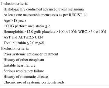
This prospective study included patients with histologically proven advanced uveal melanoma. An Eastern Cooperative Oncology Group (ECOG) performance status of 0–2, ade-quate bone marrow, liver and renal functions were required. No prior systemic anticancer treatments were allowed. Inclu-sion and excluInclu-sion criteria are outlined in Table 1.
Pembrolizumab was administered intravenously at the dose of 2 mg/kg every 3 weeks, until disease progression, unacceptable toxicity or consent withdrawal. Toxicity was reported according to Common Terminology Criteria for Adverse Events (CTCAE vs 4.0). Baseline tumor assess-ment with brain–chest–abdominal computed tomography (CT) scan or brain and abdominal magnetic resonance imaging (MRI) was required within 3 weeks before the first Pembrolizumab administration. Subsequent tumor assess-ment was scheduled every 9 weeks until disease progression or discontinuation for any reason, with the possibility of anticipating the radiological evaluation in case of signs and symptoms of tumor progression.
The primary endpoint of the study was PFS. An interim analysis was planned after the observation of at least 50% cases of progression. Secondary endpoints were response rate, clinical benefit, OS and tolerability. Response was assessed according to RECIST criteria 1.1. The response rate included complete and partial responses. Clinical benefit was defined as the percentage of patients with a complete or partial response or disease stabilization. PFS was calculated from the first day of treatment to progression or death for any reason. Survival was defined as the interval from the first day of treatment to death for any cause. PFS and OS were calculated with the Kaplan–Meier method. The log-rank test was employed to analyze the differences between patient subgroups.
Immunohistochemical evaluation
Formalin-fixed paraffin-embedded (FFPE) tissue blocks were retrieved from the archives of the Department of Pathology, Fondazione Policlinico Universitario Agostino Gemelli IRCCS, Rome. PD-1 and PD-L1 expression was evaluated using the primary Abs PD-1 (monoclonal mouse, NAT105, Ventana, prediluted) and PD-L1 (Kit DAKO, Monoclonal mouse, clone 22C3 PharmDx, prediluted). The slides were independently evaluated and subsequently analyzed by two experienced pathologists. The expression of PD-L1 and PD-1 was tested on primary tumor or liver metastasis or both, when available.
PD-L1 positivity was defined by a threshold of 5% of tumor cell expression. Positive cells had a strong cyto-plasmic expression with membrane-accentuating or single
Table 1 Inclusion and exclusion criteria
RECIST response evaluation criteria in solid tumors, ECOG Eastern
Cooperative Oncology Group, AST aspartate aminotransferase, ALT alanine aminotransferase
Inclusion criteria
Histologically confirmed advanced uveal melanoma At least one measurable metastases as per RECIST 1.1 Age ≥ 18 years
ECOG performance status ≤ 2
Hemoglobin ≥ 12.0 g/dl; platelets ≥ 100 × 109/l; WBC ≥ 3.0 × 10⁹/l AST and ALT ≤ 2.5 ULN
Total bilirubin ≤ 2.0 mg/dl Exclusion criteria
Prior systemic anticancer treatment History of other neoplasm Instable heart failure Serious respiratory failure History of rheumatic disease
membrane pattern [15]. Inner control was determined by cytoplasmic positivity for retinal pigmented epithelium cells as previously described [16].
Genetic analysis
DNA from uveal melanoma samples, fresh frozen or for-malin fixed/paraffin embedded tissues, was extracted using DNeasy Blood & Tissue Kit (QIAGEN, Hilden, Germany). Oligonucleotide aCGH was performed using the Agilent Human Genome CGH microarray 8X60K (Agilent Tech-nologies Santa Clara, CA, USA), with an average resolution of 75 kb, following the manufacturer’s instructions.
The arrays were analyzed with the SureScan Microarray Scanner, a graphical overview of the results was obtained using CytoGenomics V2.7.22.0 software (Agilent Technolo-gies Santa Clara, CA, USA).
Results
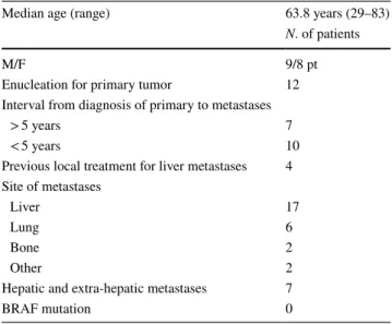
From January 2016 to April 2018 the study enrolled 17 patients, whose characteristics are summarized in Table 2. The median age was 64.7 years (ranging between 29 and 83). Nine subjects were males and 8 females. All the patients had hepatic metastases. None of them had resectable liver metastases. Seven patients had both hepatic and extra-hepatic disease. Ocular enucleation was previously per-formed for the treatment of primary tumor in 12 patients, while Ruthenium 106 brachytherapy was carried out in 5 patients. Four patients received a previous local treatment for liver metastases (metastasectomy or local ablation) at least 6 months before being recruited in the study. These patients progressed following local treatment and had a measurable disease when enrolled.
A median of 8 cycles per patient was administered (range 2–28).
Two patients achieved a partial response (11.7%), 6 patients obtained disease stabilization (35.3%), while 9 (53%) patients showed disease progression at first tumor assessment (Fig. 1). The two responding patients without progression after 19.4 and 28.9 months, respectively, were still on treatment at the time of data analysis. Eight patients (47%) achieved clinical benefit. No complete response was reported.
Table 2 Patients’ characteristics
Median age (range) 63.8 years (29–83)
N. of patients
M/F 9/8 pt
Enucleation for primary tumor 12
Interval from diagnosis of primary to metastases
> 5 years 7
< 5 years 10
Previous local treatment for liver metastases 4 Site of metastases
Liver 17
Lung 6
Bone 2
Other 2
Hepatic and extra-hepatic metastases 7
BRAF mutation 0
Fig. 1 Waterfall chart of response. Black column: progression. Dark gray column: stable disease. Light gray col-umn: partial response. Circles identify patients with an interval longer than 5 years from diagnosis of primary tumor to metastases
PFS of the whole population (Fig. 2) was 3.8 months (95% CI 2.9–9.7). Patients with an interval from diag-nosis of primary tumor to metastatic disease longer than 5 years had a PFS of 9.7 months, whereas for patients with an interval lower than 5 years PFS was 2.6 months [p = 0.039, HR 0.2865 (95% CI 0.0869–0.9443) Fig. 3a]. PFS was 3.1 months for patients with only liver metas-tases, while, for patients with liver and extrahepatic metastases, it was 8.4 months [p = 0.65, HR 0.5811 (95% CI 0.1782–1.8950) Fig. 3b]. One of the two responding patients had only hepatic disease, while the other patients had both liver and extra-hepatic metastases. For the first patient the response was observed in liver metastases and in all the metastatic sites (liver, pancreas, pleura and lung) for the other patients. Ocular enucleation, previous local treatment and LDH did not influence PFS.
At the time of data cut-off for interim analysis, five deaths were reported. Therefore, median OS of the whole population was not reached. Survival for the patients with clinical benefit was 12.8 months and 3.1 months for patients with disease progression [p = 0.047, HR 0.1543 (95% CI 0.0254–0.9377) Fig. 3c].
No grade 3–4 adverse events were observed. Grade 2 hypophysitis was observed in one patient and grade 1 hypothyroidism was reported in 2 patients. No diarrhea, rash or other cutaneous adverse events occurred.
PD‑1 and PD‑L1
We evaluated PD-1 and PD-L1 expression in 14 pri-mary tumors and 10 metastases. PD-1 was negative in 100% of primary tumors and 90% of metastases. PD-L1
Fig. 2 PFS of the entire population
Fig. 3 Stratification of time-dependent clinical endpoints. a PFS of patients with an interval longer than 5 years from diagnosis of pri-mary tumor to metastatic disease (dashed line) vs patients with an interval lower than 5 years (solid line)—p = 0.039, HR 0.2865 (95% CI 0.0869–0.9443). b PFS of patients with only liver metastases (solid line) vs PFS for patients with liver and extrahepatic disease (dashed line)—p = 0.65, HR 0.5811 (95% CI 0.1782–1.8950). c Sur-vival of patients with clinical benefit (dashed line) vs surSur-vival of pro-gressive patients (solid line)—p = 0.047, HR 0.1543 (95% CI 0.0254– 0.9377)
was negative in 92.9% of primary tumors and 90% of liver metastases. One of the responding patients did not express PD-L1 and PD-1 either in primary uveal mela-noma or liver metastases. Liver metastases of the other responding patient were PD-1 and PD-L1 negative, while his primary tumor sample was not available.
Genetic analysis
Genomic abnormalities are reported in Table 3. Samples for genetic analysis were available for 12 patients and no sig-nificant correlation was found between genomic alterations and PFS. Among the responding patients, chromosome 3 monosomy was found in association with 8q duplication in one of the patients.
Discussion
Metastatic uveal melanoma is a poor-prognosis disease. To date, remarkable advantages of a systemic therapy have not been reported and a standard treatment has not been estab-lished. Although uveal melanoma was ruled out in controlled clinical trials, this neoplasm is commonly treated as a cuta-neous melanoma, despite the different clinical and biological features. BRAF is often wild type, thus also excluding the possibility of a treatment with BRAF/MEK inhibitors.
Immunotherapy is one of the options for this disease, par-ticularly chosen for the favorable toxicity profile. The avail-able studies on anti-PD-1 agents in mUM are retrospective and included pre-treated patients [13, 14, 17], while there is a lack of prospective data on naïve patients only. We have
evaluated Pembrolizumab, an PD-1 monoclonal anti-body, as first-line treatment for advanced uveal melanoma.
In the present study, the response rate was 11.7%. More than half of the patients experienced a progression at first tumor assessment. The objective response rate was lower than in cutaneous melanoma treated with pembrolizumab (32.9%), despite the different schedule used [18]. In our previous study on the efficacy and safety of a triple-agent chemotherapy, we found a 20% response rate, a 68% clini-cal benefit and a survival advantage for patients achieving tumor control [19]. The results in the chemotherapy study could be justified by the selected population suitable for a cisplatin-based combination chemotherapy.
In our population treated with Pembrolizumab, two responding patients were still alive for at least 19 months, suggesting a possible survival advantage in case of tumor shrinkage.
The median PFS of the entire population in our study (3.8 months) was similar to the PFS reported by Karidis et al. [13] (91 days) and slightly longer than the PFS described by Algazy et al. [14] (2.6 months) in their retro-spective analyses.
The patients with an interval from the diagnosis of pri-mary tumor to metastases longer than 5 years had a sig-nificant prolonged PFS compared to patients with an inter-val lower than 5 years (9.8 months vs 3.7 months). In UM, relapse may occur more than 5 years after treatment of the primary tumor [20]. It was suggested that immune-surveil-lance through T cell activity is a mechanism which explains the late recurrence [20, 21]. Therefore, our aim was to verify the different efficacy of immunotherapy in patients relapsing within 5 years compared to patients with later recurrence.
Table 3 Genetic abnormalities
CNVs chromosome number variants, TOT VAR total variants, WCAD whole chromosome arm deletion/duplication, PCAD partial chromosome
arm deletion/duplication
a Entire arm, partial or complete chromosome 8 trisomy
Pts Monosomy 3 8q duplicationa Gross rearrangements: monosomy, trisomy,
WCAD and/or PCAD Other CNVs TOT VAR
#2 Y Y 5 (1q+,8p−,16p+,16q−,18q−) 2 9 #3 Y Y 3 (1p−,16q−,21q−) 20 25 #4 Y Y 2 (8p−,−19) 28 32 #5 Y Y 2 (1p−,4q−) 4 #6 Y Y 1 (17p−) 1 4 #7 Y 6 (1p−,+7,−11,−14,−15,−21) 7 #8 Y Y 4 (4p+,7p+,8p−,16q−) 8 14 #9 5 (1p−,6p+,6q−,13q+,16q−) 5 #10 Y Y 2 (6q−,8p−) 2 6 #11 Y 8 (1q+,4q+,6p+,6q−8p−,9p−,9q+,12p−) 4 13 #14 Y 5 (1p−,6p+,6q−13q+,16q−) 8 14 #17 Y Y 4 (4p+,8p−,16q−,+19) 15 21
Based on our data, relapse after 5 years from the diagnosis of primary tumor could be considered a parameter to select patients who may benefit more from immunotherapy.
In our study, patients with extra-hepatic disease showed a longer PFS than patients with only liver metastases. Although not statically significant, this datum seems to confirm the results in pre-treated patients [3], suggesting a role of immunotherapy mainly in patients with extra-hepatic disease. The possibility of immune escape in patients with only liver metastases could justify this observation [22].
We confirmed the favorable toxicity profile of Pembroli-zumab in metastatic uveal melanoma.
The small number of patients responding to Pembroli-zumab showed a remarkable survival advantage. Therefore, the identification of predictive factors for response is cru-cial. In our study, basal LDH did not correlate with PFS, in contrast with the results reported by Heppt et al. [17]. As possible predictive factors, we also evaluated both PD-L1 expression and genomic abnormalities commonly involved in the risk of relapse after the diagnosis of primary UM.
In our series, we found a limited expression of PD-L1 both in primary tumor and in metastases (7.1% and 10%, respectively). Few reports are available on PD-L1 expres-sion in UM. Zoroquiain et al. [15] reported a 40% PD-L1 expression on tumor cells of primary UM in patients with metastatic disease. On the other hand, similar to our data, Javed et al. [23] observed that only 5% of uveal melanoma expresses PD-L1 in the metastatic sites. The responders in our study did not express PD-L1 either in primary tumor or metastases, thus PD-L1 could not be considered a predictive factor for these patients.
Regarding the genetic results, different genomic altera-tions, such as chromosome 3 monosomy and gains of chro-mosome 8q, are predictive of relapse after primary treatment [24–28]. In this study, we analysed the association between these genomic alterations with PFS. The genomic alterations found in 12 metastatic patients did not allow to identify spe-cific abnormalities correlated with PFS. This result seems to suggest that the genetic prognostic factors, commonly pre-dictive of relapse after treatment of the primary tumor, are not useful in predicting the efficacy of Pembrolizumab for metastatic disease.
It has recently been reported that a defect of MBD4, a transcriptional factor of gene promoters, is associated with a hypermutated CpG > TpG pattern which generates multi-ple subclones of the primary UM with more heterogeneous metastases. A patient with this MBD4-related hypermuta-tor phenotype showed a remarkable response to immune checkpoint inhibitors [29]. Indeed, this evidence implies that specific and selected subgroups of UM could benefit from immunotherapy.
Due to the rarity of the disease, the small sample size of the study limits the interpretation of the data. Results
regarding survival and patients still on treatment could pro-vide further information.
Pembrolizumab is a well-tolerated agent which allows to obtain an objective response in metastatic uveal mela-noma only in a minority of patients. These patients could benefit from a long-term disease control. Further investiga-tions on the biological characteristics of uveal melanoma are essential to select patients who could benefit more from immunotherapy.
Acknowledgements We thank all the patients.
Author contributions ER, GS designed and supervised the study. ER, GS, MMP, MGS, CGC, MAB, AC recruited patients. ER, GS, TD and AC treated and followed patients. DO, MZ, GP, GR analyzed samples. ER, GS, DO, GP, GR, MZ gathered and analyzed the data. ER, GS drafted the manuscript. GT and EB supervised the project and revised the manuscript.
Funding No sponsor was involved.
Compliance with ethical standards
Conflict of interest The authors declare that they have no conflict of interest.
Ethical approval and ethical standards This clinical study was con-ducted in accordance with the Helsinki declaration of 1975. The “Ethi-cal Committee of the Fondazione Policlinico Universitario Agostino Gemelli IRCCS” approved the study with number 0049309/16. Informed consent All the patients included in the study provided informed consent for the treatment in addition to the use of their speci-mens and data for research and publication purposes.
Open Access This article is distributed under the terms of the Crea-tive Commons Attribution 4.0 International License (http://creat iveco mmons .org/licen ses/by/4.0/), which permits unrestricted use, distribu-tion, and reproduction in any medium, provided you give appropriate credit to the original author(s) and the source, provide a link to the Creative Commons license, and indicate if changes were made.
References
1. McLaughlin CC, Wu XC, Jemal A, Martin HJ, Roche LM, Chen VW (2005) Incidence of non cutaneous melanomas in the US. Cancer 103:1000–1007. https ://doi.org/10.1002/cncr.20866
2. Kujala E, Makitie T, Kivela T (2003) Very long-term prognosis of patients with malignant uveal melanoma. Investig Ophthalm Vis Sci 44:4651–4659. https ://doi.org/10.1167/iovs.03-0538
3. Rietschel P, Panageas KS, Hanlon C, Patel A, Abramson DH, Chapman PB (2005) Variates of survival in metastatic uveal melanoma. J Clin Oncol 23:8076–8080. https ://doi.org/10.1200/ JCO.2005.02.6534
4. Diener-West M, Reynolds SM, Agugliaro DJ, Caldwell R, Cum-ming K, Earle JD et al (2005) Development of metastatic disease after enrollment in the COMS trial for treatment of choroidal melanoma: Collaborative Ocular Melanoma Study Group Report
No 26. Arch Ophthalmol 123:1629–1643. https ://doi.org/10.1001/ archo pht.123.12.1639
5. Agarwala SS, Eggermont AM, O’Day S, Zager JS (2014) Meta-static melanoma to the liver: a contemporary and comprehensive review of surgical, systemic, and regional therapeutic options. Cancer 120:781–789. https ://doi.org/10.1002/cncr.28480
6. Flaherty LE, Unger JM, Liu PY, Mertens WC, Sondak VK (1998) Metastatic melanoma from intraocular primary tumors: the South-west Oncology Group experience in phase II advanced melanoma clinical trials. Am J Clin Oncol 21:568–572
7. Nathan F, Sato T, Hart E (1994) Response to combination chemo-therapy of liver metastasis from chorhoidal melanoma compared with cutaneous melanoma [abstract]. Proc Am Soc Clin Oncol 13:1994
8. Bedikian AY, Legha SS, Mavligit G, Carrasco CH, Khorana S, Plager C et al (1995) Treatment of uveal melanoma metastatic to the liver; a review of the MD Anderson Cancer Center experi-ence and prognostic factors. Cancer 76:1665–1670. https ://doi. org/10.1002/1097-0142(19951 101)76:9%3c166 5:AID-CNCR9 %3e3.0.CO;2-J
9. Carvajal RD, Piperno-Neumann S, Kapiteijn E, Chapman PB, Frank S, Joshua AM, Piulats JM, Wolter P, Cocquyt V, Chmielowski B et al (2018) Selumetinib in combination with dac-arbazine in patients with metastatic uveal melanoma: a phase III, multicenter, randomized trial (SUMIT). J Clin Oncol 36:1232– 1239. https ://doi.org/10.1200/JCO.2017.74.1090
10. Danielli R, Ridolfi R, Chiarion-Sileni V, Queirolo P, Testori A, Plummer E et al (2012) Ipilimumab in pretreated patients with metastatic uveal melanoma: safety and clinical efficacy. Cancer Immunol Immunother 61:41–48. https ://doi.org/10.1007/s0026 2-011-1089-0
11. Khattak MA, Fisher R, Hughes P, Gore M, Larkin J (2013) Ipili-mumab activity in advanced uveal melanoma. Melanoma Res 23:79–81. https ://doi.org/10.1097/CMR.0b013 e3283 5b554 f
12. Zimmer L, Vaubel J, Mohr P, Hauschild A, Utikal J, Simon J et al (2015) Phase II DeCOG-study of Ipilimumab in pretreated and treatment-naïve patients with metastatic uveal melanoma. PLoS One 10:e0118564. https ://doi.org/10.1371/journ al.pone.01185 64
13. Karydis I, Chan PI, Wheater M, Arriola E, Szlosarek PW, Ottensmeier CH (2016) Clinical activity and safety of Pembroli-zumab in Ipilimumab pre-treated patients with uveal melanoma. Oncoimmunology 5:e1143997. https ://doi.org/10.1080/21624 02X.2016.11439 97
14. Algazi PA, Tsai KK, Shoushtari AN, Munhoz RR, Eroglu Z, Piu-lats JP (2016) Clinical Outcomes in metastatic uveal melanoma treated with PD-1 and PD-L1 antibodies. Cancer 122:3344–3353.
https ://doi.org/10.1002/cncr.30258
15. Zoroquiain P, Esposito E, Logan P, Aldrees S, Dias AB, Mansure JJ, Santapau D, Garcia C, Saornil MA, Belfort Neto R, Burnier MN (2018) Programmed cell death ligand-1 expression in tumor and immune cells is associated with better patient outcome and decreased tumor-infiltrating lymphocytes in uveal melanoma. Mod Pathol 31:1201–1210. https ://doi.org/10.1038/s4137 9-018-0043-5
16. Yang W, Li H, Chen PW, Alizadeh H, He Y, Hogan RN, Nieder-korn JY (2009) PD-L1 expression on human ocular cells and its possible role in regulating immune-mediated ocular inflammation. Investig Ophthalmol Vis Sci 50:273–280. https ://doi.org/10.1167/ iovs.08-2397
17. Heppt MV, Heinzerling L, Kähler KC et al (2017) Prognostic factors and outcomes in metastatic uveal melanoma treated with
programmed cell death-1 or combined PD-1/cytotoxic T-lym-phocyte antigen-4 inhibition. Eur J Cancer 82:56–65. https ://doi. org/10.1016/j.ejca.2017.05.038
18. Robert C, Schachter J, Long GV, Arance A, Grob JJ, Mortier L, Daud VA, Carlino MS, McNeil C, Lotem M (2015) Pem-brolizumab versus Ipilimumab in advanced melanoma. NEJM 372:2521–2532. https ://doi.org/10.1056/NEJMo a1503 093
19. Schinzari G, Rossi E, Cassano A, Dadduzio V, Quirino M, Pagli-ara M, Blasi MA, Barone C (2017) Cisplatin, dacarbazine and vinblastine as first line chemotherapy for liver metastatic uveal melanoma in the era of immunotherapy: a single institution phase II study. Melanoma Res 27:591–595. https ://doi.org/10.1097/ CMR.00000 00000 00040 1
20. Komatsubara KM, Carvajal RD (2017) Immunotherapy for the treatment of uveal melanoma: current status and emerging therapies. Curr Oncol Rep 19:45. https ://doi.org/10.1007/s1191 2-017-0606-5
21. Eyles J, Puaux AL, Wang X, Toh B, Prakash C, Hong M et al (2010) Tumor cells disseminate early, but immunosurveillance limits metastatic outgrowth, in a mouse model of melanoma. J Clin Investig 120:2030–2039. https ://doi.org/10.1172/JCI42 002
22. Amaro A, Gangemi R, Piaggio F, Angelini G, Barisione G, Fer-rini S, Pfeffer U (2017) The biology of uveal melanoma. Can-cer Metastasis Rec 36:109–140. https ://doi.org/10.1007/s1055 5-017-9663-3
23. Javed A, Arguello D, Johnston C et al (2017) PD-L1 expression in tumor metastasis is different between uveal melanoma and cutaneous melanoma. Immunotherapy 9:1323–1330. https ://doi. org/10.2217/imt-2017-0066
24. Moore AR, Ceraudo E, Sher JJ et al (2016) Recurrent activating mutations of G-protein-coupled receptor CYSLTR2 in uveal mela-noma. Nat Genet 48:675–680. https ://doi.org/10.1038/ng.3549
25. Robertson AG, Shih J, Yau C et al (2017) Integrative analysis identifies four molecular and clinical subsets in uveal mela-noma. Cancer Cell 32:204–220. https ://doi.org/10.1016/j.ccell .2017.07.003
26. Tyagi S, Chabes AL, Wysocka J, Herr W (2007) E2F activation of S phase promoters via association with HCF-1 and the MLL family of histone H3K4 methyltransferases. Mol Cell 27:107–119.
https ://doi.org/10.1016/j.molce l.2007.05.03
27. Lake SL, Damato BE, Kalirai H, Dodson AR, Taktak AF, Lloyd BH, Coupland SE (2013) Single nucleotide polymorphism array analysis of uveal melanomas reveals that amplification of CNKSR3 is correlated with improved patient survival. Am J Pathol 182:678–687. https ://doi.org/10.1016/j.ajpat h.2012.11.036
28. Park JJ, Diefenbach RJ, Joshua A, Kefford RF, Carlino MS, Rizos H (2018) Oncogenic signaling in uveal melanoma. Pigment Cell Mel Res 31:661–672. https ://doi.org/10.1111/pcmr.12708
29. Rodrigues M, Mobuchon L, Houy A et al (2018) Outlier response to anti-PD-1 in uveal melanoma reveals germline MBD4 muta-tions in hypermutated tumors. Nat Commun 9:1866. https ://doi. org/10.1038/s4146 7-018-04322 -5
Publisher’s Note Springer Nature remains neutral with regard to jurisdictional claims in published maps and institutional affiliations.


![CI 0.1782–1.8950) Fig. 3b]. One of the two responding patients had only hepatic disease, while the other patients had both liver and extra-hepatic metastases](https://thumb-eu.123doks.com/thumbv2/123dokorg/8337875.132825/4.892.461.805.88.821/responding-patients-hepatic-disease-patients-liver-hepatic-metastases.webp)
