DOCTORAL SCHOOL IN BIOLOGY Section: Biodiversity and Ecosystem Analysis
XXVIII Cycle – Academic Year 2015/2016
Reproductive endocrinology and stress physiology
in Galápagos land iguanas
Endocrinologia riproduttiva e fisiologia dello stress nelle
iguane terrestri delle Galápagos
CANDIDATE Ph.D. STUDENT: Michela Onorati
TUTORS: Leonardo Vignoli, Monica Carosi CO-TUTOR: Gabriele Gentile
Onorati M (2016). Reproductive endocrinology and stress physiology in Galápagos land iguanas. Doctoral thesis. Department of Science, Roma Tre University, Rome, Italy.
Michela Onorati
Department of Science, Roma Tre University, Viale Marconi, 446,
00146 Rome, Italy.
e-mail: [email protected]; [email protected]
Thesis defense 15th February 2016 in front of the following jury:
Prof. Ferdinando Boero (Università del Salento) Prof. Giovanna Abbate (Università Sapienza di Roma) Prof. Romolo Fochetti
Your reason and your passion are the rudder and the sails of your seafaring soul.
INDEX
CHAPTER 1General introduction………... 1
1) Multidisciplinary is the key for conservation………..……. 1
2) Reproductive endocrinology and aims………... 1
3) Stress physiology and aims……….…….. 3
References………. 9
CHAPTER 2 Plasma levels of progesterone and estradiol and their relation to reproduction in Galápagos land iguanas, Conolophus marthae and Conolophus subcristatus……… 17
1) Introduction………..…...19
2) Materials and Methods………22
2.1) Ethic statement……….. 22 2.2) Samples………. 22 2.3) Ultrasound analysis……… …... 23 2.4) Hormone assays……… 26 2.5) Statistical analysis………. 27 3) Results………. 27 3.1) Reproductive status………... 27 3.2) Progesterone (P4)……….. 28 3.3) 17β-estradiol (E2)………. 31 4) Discussion………... 32 References………... 37 CHAPTER 3 Relationships between leukocyte profiles and Hepatozoon infection in Galápagos land iguanas, Conolophus marthae and Conolophus subcristatus………. 45
2) Materials and Methods………..…... 50
2.1) Study area and species………. 50
2.2) Sampling and blood collection………. 51
2.3) Leukocyte formula and detection of parasites……… . …... 51
2.4) Statistical analysis……… 52
3) Results………. 53
3.1) Interspecific comparisons (Volcán Wolf)……… 53
3.2) Site comparisons (C. subcristatus)………...57
4) Discussion……… …... 58
References………. .. …... 61
CHAPTER 4 Effects of parasitic infection and reproduction on corticosterone plasma levels in Galápagos land iguanas, Conolophus marthae and Conolophus subcristatus……… 71
1) Introduction………. 73
2) Materials and Methods………75
2.1) Ethic statement……….….... 75
2.2) Field sites and sampling sessions………. 75
2.3) Field phase………... 76
2.4) Laboratory phase: haematological analysis………. 77
2.5) Laboratory phase: hormonal analysis……….. …... 77
2.6) Statistical analysis……… 78
3) Results………. 79
3.1) Corticosterone and parasitemia……… 79
3.2) Corticosterone plasma levels in females……….. 81
3.3) Corticosterone and testosterone plasma levels in males….. 84
4) Discussion……… …... 85
References………... 89
CHAPTER 5 Conclusions……….. 101
CHAPTER 1
General introduction
1) Multidisciplinary is the key for conservation
During the last century, threatening processes such as invasive species and pathogenic diseases have been working synergistically to biodiversity loss (Brook et al. 2008). Free-living animals must struggle against a variety of challenges and man-mediated alterations that can cause high stress conditions. Disruption of behaviour and reproductive physiology and the alteration of population fitness are among the most hostile consequences of prolonged stress. Thus, understanding if and how threatened free-living populations reproduce and respond to external changes is necessary to determine vulnerability and set conservation priorities. In that respect, the use of an integrative and functional approach, direct also to the comprehension of physiological and endocrine individual dynamics, can help to characterize and alleviate problems that could threaten a species, population, community.
Conservation physiology is an evolving important field of conservation science that takes advantage from this integrative approach (Wikelski and Cooke 2006). Conservation biologists are progressively using different techniques that range from endocrinology to stress physiology, to develop and choose solutions. This combination of conservation tools, with supplementary knowledge of the basic biology of organisms, is fundamental for the safety of both captive and wild populations. Multidisciplinary is the key to more efficient problem solving in conservation. For this reason, in this occasion, I quote Wildt et al. (2003) who stated: “conservation can be likened to a complex jigsaw puzzle where the puzzle pieces are issues, stakeholders or scientific disciplines themselves”.
2) Reproductive endocrinology and aims
Reproduction is the foundation on which a species survives. Understanding the complexities of when and how individuals reproduce is basal for the perpetuation of natural populations and their future management. Therefore, knowledge about the reproductive
endocrinology of a species can fill an important gap in our understanding of timing and modalities of reproduction. Reproductive endocrinology offers the possibility to better realize factors impairing species vitality, and it may even offer early-warning signals of a risk before survivorship or reproductive rates plummet.
Much of our knowledge of vertebrate reproductive endocrinology has been collected from studies of mammals. Anyhow, there are sufficient structural and functional similarities between mammals and non-mammals to indicate that many of mechanisms regulating reproduction are probably common to all vertebrates (Crews and Silver 1985).
Reproductive activity is associated with an essential variation in circulating concentrations of the primary sex steroid hormones: progesterone (P4) and 17β-estradiol (E2) (Jones and Guillette 1982; Crews and Silver 1985; Norris and Lopez 2010). Measuring the plasma levels of these sexual steroid hormones can be useful to track the reproductive hormone profiles of free-living animals and this could be crucial for species conservation especially when direct observations on field are strongly limited by logistic constraints and ex-situ captive breeding programs may become necessary. This is the case of two Galápagos land iguanas species that occur in Volcán Wolf (Isabela Island): Conolophus marthae and C. subcristatus, among the most representative species of the Galápagos Islands.
Conolophus marthae, the Galápagos Pink Land Iguana (also simply known as pink iguana), was only recently described (Gentile and Snell 2009; Gentile et al. 2009) and listed as Critically Endangered in the IUCN Red List (Gentile 2012). Current data suggests that this species lives, with an extremely small population, exclusively on the top and along the northwest slopes of Wolf volcano, the highest peak (1,707m) in the archipelago. Just because only recently discovered, information about its ecology is limited to circumstantial observations and reproductive biology is completely unknown. Newly hatched individuals and juveniles of the species were never observed.
Contrary to the pink iguana, the Galápagos common iguana Conolophus subcristatus (for convenience here also referred to as yellow iguana) currently inhabits six islands in the archipelago, including Isabela Island and the Wolf volcano where occupies an area larger than C. marthae. The yellow iguana is listed as vulnerable in the IUCN Red List and experienced various disturbances by direct and
indirect human activity so that several populations became dramatically reduced in size or were extirpated (Snell et al. 1984). About this species, some studies were produced sustaining that clutch size and mating season vary across islands (Werner 1983; Snell et al. 1984). However, little is known about the reproductive biology of this species on Wolf volcano.
To pinpoint species-specific times of reproduction in the volcano and possible interspecific interactions, I examined and explained baseline steroid levels of progesterone (P4) and 17β-estradiol (E2) in both iguana species. The existence of previous studies on sex steroid hormones of the Galápagos marine iguana Amblyrhynchus cristatus, the sister taxon of Conolophus spp. (Rassmann 1997), offered a unique opportunity to use an appropriate reference model for the much less investigated land iguanas.
3) Stress physiology and aims
Free-living animals periodically experience a multiplicity of internal/external environmental challenges and man-mediated alterations that can produce stress condition.
The term “stress” has become popular thanks to the pioneering work of Hans Selye (1946), who described the stress condition as “a general adaptation syndrome (GAS)” in which a rapid initial reaction ("alarm") was followed by sustained glucocorticoid secretion ("phase of resistance") and eventually by a dangerous debility when corticoid output could not be sustained ("phase of exhaustion").
Stress is a term used across a broad spectrum of scientific researches; however, its definition is often ambiguous and sometimes not defined at all. Nowadays, biologists distinguish between “stressor” and “stress response”. Stressor is any noxious stimulus (Romero 2004) or exceptional event that disturbs an animal’s homeostasis generating the so-called emergency life-history stage (ELHS) (Wingfield et al. 1998; McEwen and Wingfield 2003). Free-living animals experience many stressors during their life including physical factors (i.e., change in temperature, oxygen, and salinity), climatic stressors (drought and storms) and biotic stressors (predation, competition and social dynamics, parasitism) that challenge their homeostasis (Romero 2004; Jessop et al. 2013). These disturbance phenomena may have effects on the ecology and evolution of organisms (Hoffmann and Hercus 2000;
Badyaev 2005; Jessop et al. 2013) and, depending on their pervasiveness, magnitude and frequency, can influence the individual fitness (Bonier et al. 2009; Busch and Haiward 2009). Thus, in response to a stressor, animals mount stress responses, which work for neutralizing the effects of the stressor to regenerate homeostasis. The stress response is constituted by all physiological, endocrinological, immunological and behavioural adaptations, which can be concurrently used to cope with the stress condition limiting the negative consequences on fitness (Wingfield et al. 1998; Wikelski and Cooke 2006).
One main feature of stress response in vertebrates is the release of glucocorticoids (i.e. cortisol and corticosterone), steroid hormones whose synthesis is regulated by hypothalamic–pituitary–adrenal axis (HPA). Fish and most mammals generally release cortisol, whereas most birds, reptiles, amphibians, and many rodents release corticosterone (Johnson et al. 1992; Sapolsky 1992; Romero 2004; Romero and Butler 2007; Crespi et al. 2013). Glucocorticoids (GCs) are the final product of the HPA axis; these stress hormones participate in the control of homeostasis activating immediate life-saving processes (Romero et al. 2009). Upon perception of stress, the hypothalamus is activated to secrete arginine vasotocin (AVT, homologous of the mammalian arginine vasopressin) and corticotropin-releasing factor (CRF), which stimulate the pituitary gland to release adrenocorticotropin (ACTH). This in turn, causes the release of glucocorticoids from the adrenal glands (Rich and Romero 2005). The cessation of the pathway leading to GCs production occurs through a negative feedback under the control of the GCs themselves. Stress-induced concentrations of GCs interact with glucocorticoid receptors in the hippocampus, hypothalamus and pituitary gland to suppress the initial steps of the HPA axis (De Kloet et al. 1998). The level at which GCs are elevated depend on the severity of the stressor; therefore, under acute stress conditions, the feedback mechanism operates efficiently and the system rapidly returns to normal; under chronic stress conditions, feedback signals are weak and the system remains activated for longer periods (Sapolsky 1992).
Generally, short-term glucocorticoid releases are helpful for organisms because stimulate emergency mechanisms such as mobilizing glucose (gluconeogenesis) and protein catabolism to immediately increase energetics availability to overcome the
perturbation (Wingfield and Ramenofsky 1999; Wingfield and Romero 2001; Wingfield 2013). However, chronic activation of the HPA axis and prolonged elevated GC concentrations may have large deleterious effects on fitness (Romero 2004; Blas et al. 2007) resulting in stress-related disease (Sapolsky 1992; Romero et al. 2009). Indeed, long-term activation of the stress response can expose the individual to a long-term overstimulation of survive mechanisms with consecutive inhibition of many fundamental functions including immunocompetence and reproduction (Sapolsky 1987; Wingfield et al. 1997; Dhabhar 2000; Sapolsky et al. 2000; Dallman and Bhatnagar 2001; Wingfield and Romero 2001).
Overall, GCs concentrations are being used increasingly in ecological and conservation studies as indices of animal well-being (Wikelski and Cooke 2006; Busch and Hayward 2009; Sheriff et al. 2011). Measuring these hormones can help to understand how specific stressors affect the survival and reproductive success of free-living animals. However, although these hormones can be measured directly from many biological matrices as blood, saliva, faeces and urine (Wasser et al. 1997; Sapolsky et al. 2000; Narayan et al. 2010; Sheriff et al. 2011), measuring them under field conditions is very difficult and may require caution.
Historically, blood is the traditional biological matrix used to assess GCs concentration. Blood collection allows the measurement of instantaneous and direct product of the adrenal cortex. Moreover, this method permits a simultaneous collection of blood components with a comprehensive assessment of the state of the animal, including indices of condition (haematocrit), immune function (leukocyte profiles), and reproductive status (reproductive hormones). The most appropriate method for blood collection varies across species. However, despite the method used, the collection of blood sample is itself invasive. Thus, when planning researches on basal-levels of stress, the effects of sample collection must be considered as they may bias the hormonal response of the examined animals. To avoid this problem blood samples are generally taken before the adrenal cortex has been activated, that is within few minutes from capture (Wingfield and Romero 2001; Romero and Reed 2005). Other, less sensitive, indicators of stress may also be used, such as leukocytes profiles or more in detail the heterophils (or neutrophils in mammals) and
lymphocytes ratio. In fact, the immune response is another part of the adaptive responses to stressful situations.
The immune system is the primary defence mechanism through which the organism protects itself from stressors represented by pathogens. Leukocytes or white blood cells (WBC) are fundamental mediators of the immune response (Lobato et al. 2005; Davis et al. 2011); they circulate continuously in the blood stream and various organs, actively destroying invading microorganisms. This circulation is essential for maintaining an effective immune defence network.
Most vertebrates have five types of WBCs: lymphocytes, neutrophils, eosinophils, basophils and monocytes, each one with specific morphology and function. The morphology of each cell type appears to be conserved across taxa, except in the case of neutrophils; indeed, in birds and reptiles neutrophils are replaced with heterophils, which perform the same immunological function (Hawkey and Dennett 1989; Jain 1993).
Neutrophils/heterophils and lymphocytes make up the highest percentage (i.e. nearly 80% combined) in WBCs of many vertebrates including reptiles (Eliman 1997; Fisse et al. 2004; Davis et al. 2008). Specifically, neutrophils/heterophils are the primary immune phagocytosing cells; they enter the tissues during the inflammatory response (Jain 1993; Campbell 1995; Davis et al. 2008) participating actively to the phagocytosis of organisms and other foreign material (Thrall et al. 2012). Lymphocytes are involved in a variety of immunological functions such as immunoglobulin production and modulation of immune defence (Campbell 1996). Generally, the numbers and proportions of leukocytes in blood provide an important representation of leukocyte distribution in the body and of the activation state of the immune system. Leukocyte profiles have a recognized predictive power for interpreting individuals’ health status; indeed, the observations of variations in leukocytes numbers are particularly useful in the field of conservation biology to describe an altered health status (Wakelin 1996; Davis et al. 2004; Davis et al. 2008). Usually, during a stress condition, an increase in neutrophyls (N)/heterophils (H) and a decrease in lymphocytes (L) are observed (Maxwell and Robertson 1998; Ots et al. 1998; Davis et al. 2008). Since numbers of these types cells are affected by stress in opposite directions, the relative proportion of neutrophils/heterophils to lymphocyte (N-H/L) is commonly used as a composite measure of the
stress response (Gross and Siegel 1983; Maxwell 1993; Maxwell and Robinson 1998; Lobato et al. 2005; Davis et al. 2008; Xuereb et al. 2012; Lentfer et al. 2015). Differently from the glucocorticoids measurements, the WBC approach offers the advantage that it does not require prohibitively rapid sampling and is relatively inexpensive. Moreover, leukocyte profiles are particularly useful in the field of stress physiology because they can be directly related to stress hormone levels (Davis et al. 2008). Many studies have described the stress-induced change in leukocyte distribution mediated by hormones released by the adrenal gland (Dhabhar et al. 1996; Dhabhar and McEwen 1999). Chronically elevated glucocorticoid levels may cause long-term elevations in N-H/L ratio as they simultaneously induce a reduction in the number of circulating lymphocyte, with a redistribution from circulatory to bone marrow. At the same, they cause increase in the number of neutrophils/heterophils, by stimulating their influx into the blood and attenuating their egress from the blood to other compartments (Sapolsky et al. 2000). Therefore, trafficking and function of blood cells are altered transiently by GCs.
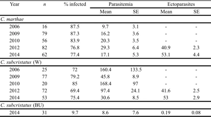
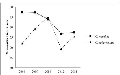
Because stress in animal populations is an important factor to consider when evaluating their welfare in both captive and wild condition, using different stress markers is fundamental to obtain a reliable assessment of the stress condition. For this, in this part of the PhD project, using both haematologic and hormonal profiles, I specifically explored how parasites affect iguanas’ life traits as leukocyte profiles and glucocorticoids levels. In fact, the two populations of C. marthae and C. subcristatus have to overcome the strong impact of ticks, which seem to be more abundant in Volcán Wolf than elsewhere in the archipelago. The site is characterized by a massive occurrence of ticks Amblyomma spp., ectoparasites already described infecting marine iguanas (Wikelski 1999). Probably, ticks are the major vectors of Hepatozoon (Apicomplexa: Adeleorina) in Galápagos reptiles (Bataille et al. 2012). Hepatozoon, with over 300 species described, is the most common among intracellular blood parasites in reptiles (Telford 1984; Smith 1996). They infect host erythrocytes and their effects on host fitness are still debated.
In general, ecto and endoparasites are considered potential sources of biotic stress for all organisms (Lozano 1998). They can coexist with their hosts without causing any measurable deleterious effects, but
they can also increase in numbers and overwhelm a host already weakened by other forms of stress such as malnutrition or reproduction (Walzer and Genta 1989). Finally, they can directly provoke inflammatory responses and disease affecting individual health and fitness-related traits (Schwanz 2008). Moreover, parasites and glucocorticoid hormones interact and affect a multiplicity of processes, such as immune response and reproduction (Wingfield et al. 1997; Sapolsky et al. 2000). However, the nature of the relationship between parasitic infection and levels of glucocorticoids and their possible covariation with haematological profiles has received relatively little attention in wild animals, and with equivocal results. For this, aware of the poor knowledge about patterns of natural variation of haematological parameters and glucocorticoid levels and their relationship with parasites in reptile wild populations, in the second part of the project I analysed the relation between ecto- and endoparasites and the stress physiology of these two Galápagos land iguanas. For this purposes I used: (i) leukocyte profiles and specifically the heterophils/lymphocytes ratio (H/L), commonly used as diagnostic tool for assessing long-term stress in vertebrates (Davis et al. 2008), (ii) endocrinological markers as baseline corticosterone plasma levels, the primary adrenal glucocorticoid hormone produced in response to stressful events in reptiles (Greenberg and Wingfield, 1987).
I used a separate population of C. subcristatus, occurring in a coastal area where notoriously ecto-parasites and haemoparasites are marginally present (Bahia Urbina), as “blank” condition for haematologic and hormonal comparisons.
References
Badyaev AV (2005). Stress-induced variation in evolution: from behavioural plasticity to genetic assimilation. Proceedings of the Royal Society of London B: Biological Sciences 272(1566): 877-886. Bataille A, Fournié G, Cruz M, Cedeño V, Parker PG, Cunningham AA, Goodman SJ (2012). Host selection and parasite infection in Aedes taeniorhynchus, endemic disease vector in the Galápagos Islands. Infection, Genetics and Evolution 12(8): 1831-1841.
Blas J, Bortolotti GR, Tella JL, Baos R, Marchant TA (2007). Stress response during development predicts fitness in a wild, long lived vertebrate. Proceedings of the National Academy of Sciences 104(21): 8880-8884.
Bonier F, Martin PR, Moore IT, Wingfield JC (2009). Do baseline glucocorticoids predict fitness?. Trends in Ecology and Evolution, 24(11): 634-642.
Brook BW, Sodhi NS, Bradshaw CJ (2008). Synergies among extinction drivers under global change. Trends in Ecology and Evolution 23(8): 453-460.
Busch DS, Hayward LS (2009). Stress in a conservation context: a discussion of glucocorticoid actions and how levels change with conservation-relevant variables. Biological Conservation 142(12): 2844-2853.
Campbell TW (1995) Avian Hematology and Cytology (No. Ed. 2). Iowa State University Press, Ames.
Campbell TW (1996). Clinical pathology. In: Reptile Medicine and Surgery. WB Sauders Co, Philadelphia.
Cash WB, Holberton RL, Knight SS (1997). Corticosterone secretion in response to capture and handling in free-living red-eared slider turtles. General and Comparative Endocrinology 108(3): 427–433.
Christian KA, Bedford GS (1995). Physiological consequences of filarial parasites in the frillneck lizard, Chlamydosaurus kingii, in northern Australia. Canadian Journal of Zoology 73(12): 2302-2306. Campbell TW (1995) Avian Hematology and Cytology (No. Ed. 2). Iowa State University Press, Ames.
Campbell TW (1996). Clinical pathology. In: Reptile Medicine and Surgery. WB Sauders Co, Philadelphia.
Crespi EJ, Williams TD, Jessop TS, Delehanty B (2013). Life history and the ecology of stress: how do glucocorticoid hormones influence life-history variation in animals?. Functional Ecology 27(1): 93-106. Crews D, Silver R (1985). Reproductive physiology and behavior interactions in nonmammalian vertebrates. In: Handbook of Behavioral Neurobiology, pp. 101-182. Plenum Press, New York. Dallman MF, Bhatnagar S (2001). Chronic Stress and Energy Balance: Role of the Hypothalamo-Pituitary-Adrenal Axis. In: Handbook of Physiology, pp. 179-210. Oxford University Press, New York.
Davis AK, Cook KC, Altizer S (2004). Leukocyte profiles in wild House Finches with and without mycoplasmal conjunctivitis, a recently emerged bacterial disease. EcoHealth 1(4): 362-373.
Davis AK, Maney DL, Maerz JC (2008). The use of leukocyte profiles to measure stress in vertebrates: a review for ecologists. Functional Ecology 22(5): 760-772.
Davis AK, Ruyle LE, Maerz JC (2011). Effect of trapping method on leukocyte profiles of black-chested spiny-tailed iguanas (Ctenosaura melanosterna): implications for zoologists in the field. ISRN Zoology, 2011.
Dhabhar FS (2000). Acute stress enhances while chronic stress suppresses skin immunity: the role of stress hormones and leukocyte trafficking. Annals of the New York Academy of Sciences 917(1): 876-893.
Dhabhar FS, McEwen BS (1999). Enhancing versus suppressive effects of stress hormones on skin immune function. Proceedings of the National Academy of Sciences 96(3): 1059-1064.
Dhabhar FS, Miller AH, McEwen BS, Spencer RL (1996). Stress-induced changes in blood leukocyte distribution. Role of adrenal steroid hormones. The Journal of Immunology 157(4): 1638-1644. De Kloet ER, Vreugdenhil E, Oitzl MS, Joels M (1998). Brain corticosteroid receptor balance in health and disease. Endocrine reviews 19(3): 269-301.
Eliman MM (1997) Hematology and plasma chemistry of the inland bearded dragon, Pogona vitticeps. Bulletin of the Association of Reptile and Amphibian Veterinarians 7(4): 10-12.
Fisse A, Draud M, Raphael B, Melkonian K (2004). Differential leukocyte counts of critically endangered grand cayman blue iguanas, Cyclura nubila lewisi. Journal of Herpetological Medicine and Surgery 14(4): 19-21.
Gentile G (2012). Conolophus marthae. The IUCN Red List of Threatened Species 2012: http://dx.doi.org/10.2305/IUCN.UK.2012-1.RLTS.T176672A1414375.en
Gentile G, Fabiani A, Marquez C, Snell HL, Snell HM, Tapia W, Sbordoni V (2009). An overlooked pink species of land iguana in the Galápagos. Proceedings of the National Academy of Sciences 106(2): 507-511.
Gentile G, Snell H (2009). Conolophus marthae sp. nov. (Squamata, Iguanidae), a new species of land iguana from the Galápagos archipelago. Zootaxa 2201: 1-10.
Greenberg N, Wingfield JC (1987). Stress and reproduction: reciprocal relationships. In: Hormones and reproduction in fishes, amphibians, and reptiles, pp. 461-503. Springer, New York.
Gross WB, Siegel HS (1983). Evaluation of the heterophil/lymphocyte ratio as a measure of stress in chickens. Avian Diseases 27(4): 972-979.
Hawkey CM, Dennett TB (1989). Color atlas of comparative veterinary hematology. Veterinary Clinical Pathology 18(4): 108-108. Hoffmann AA, Hercus MJ (2000). Environmental stress as an evolutionary force. Bioscience 50(3): 217-226.
Jain NC (1993). Essentials of veterinary hematology. Blackwell Publishing, Philadelphia.
Jessop TS, Woodford R, Symonds MR (2013). Macrostress: do large-scale ecological patterns exist in the glucocorticoid stress response of vertebrates?. Functional Ecology 27(1): 120-130.
Johnson EO, Kamilaris TC, Chrousos GP, Gold PW (1992). Mechanisms of stress: a dynamic overview of hormonal and behavioral homeostasis. Neuroscience and Biobehavioral Reviews 16(2): 115-130.
Jones RE, Guillette LJ (1982). Hormonal control of oviposition and parturition in lizards. Herpetologica 38(1): 80-93.
Lentfer TL, Pendl H, Gebhardt-Henrich SG, Fröhlich EKF, Von Borell E (2015). H/L ratio as a measurement of stress in laying hens– methodology and reliability. British Poultry Science 56(2): 157-163. Lobato E, Moreno J, Merino S, Sanz JJ, Arriero E (2005). Haematological variables are good predictors of recruitment in nestling pied flycatchers (Ficedula hypoleuca). Ecoscience 12(1): 27-34.
Lozano GA (1998). Parasitic stress and self-medication in wild animals. Advances in the Study of Behaviour 27: 291-318.
Maxwell MH (1993). Avian blood leucocyte responses to stress. World's Poultry Science Journal 49(01): 34-43.
Maxwell MH, Robertson GW. (1998). The avian heterophil leucocyte: a review. World's Poultry Science Journal 54(02): 155-178.
McEwen BS, Wingfield JC (2003). The concept of allostasis in biology and biomedicine. Hormones and Behavior 43(1): 2-15. Narayan E, Molinia F, Christi K, Morley C, Cockrem J (2010). Urinary corticosterone metabolite responses to capture, and annual patterns of urinary corticosterone in wild and captive endangered Fijian ground frogs (Platymantis vitiana). Australian Journal of Zoology 58(3): 189-197.
Norris DO, Lopez KH (2010). Hormones and reproduction of vertebrates (Vol. 3). Academic Press. Elsevier, San Diego.
Ots I, Murumägi A, Horak P (1998). Haematological health state indices of reproducing great tits: methodology and sources of natural variation. Functional Ecology 12(4): 700-707.
Rich EL, Romero LM (2005). Exposure to chronic stress downregulates corticosterone responses to acute stressors. American Journal of Physiology-Regulatory, Integrative and Comparative Physiology 288(6): R1628-R1636.
Romero LM (2004). Physiological stress in ecology: lessons from biomedical research. Trends in Ecology and Evolution 19(5): 249-255. Romero LM, Butler LK (2007). Endocrinology of stress. International Journal of Comparative Psychology 20(2): 89-95.
Romero LM, Dickens MJ, Cyr NE (2009). The reactive scope model—a new model integrating homeostasis, allostasis, and stress. Hormones and Behavior 55(3): 375-389.
Romero LM, Reed JM (2005). Collecting baseline corticosterone samples in the field: is under 3 min good enough?. Comparative Biochemistry and Physiology Part A: Molecular and Integrative Physiology 140(1): 73-79.
Sapolsky RM (1987). Stress, social status, and reproductive physiology in free-living baboons. In D. Crews (ed.), Psychobiology of reproductive behavior: An evolutionary perspective. Prentice-Hall, Englewood Cliffs, New Jersey.
Sapolsky RM (1992). Neuroendocrinology of the stress response. In: Behavioral Endocrinology, pp. 287-324. MIT Press, Cambridge. Sapolsky RM, Romero LM, Munck AU (2000). How do glucocorticoids influence stress responses? Integrating permissive, suppressive, stimulatory, and preparative actions 1. Endocrine Reviews 21(1): 55-89.
Schwanz LE (2008). Chronic parasitic infection alters reproductive output in deer mice. Behavioral Ecology and Sociobiology 62(8): 1351-1358.
Selye H (1946). The general adaptation syndrome and the diseases of adaptation. The Journal of Clinical Endocrinology and Metabolism 6(2): 117-230.
Sheriff MJ, Dantzer B, Delehanty B, Palme R, Boonstra R (2011). Measuring stress in wildlife: techniques for quantifying glucocorticoids. Oecologia 166(4): 869-887.
Smith TG (1996). The genus Hepatozoon (Apicomplexa: Adeleina). The Journal of Parasitology 82(4): 565-585.
Snell HL, Snell HM, Tracy CR (1984). Variation among populations of Galápagos land iguanas (Conolophus): contrasts of phylogeny and ecology. Biological Journal of the Linnean Society 21(1-2): 185-207. Telford JrSR. (1984). Haemoparasites of reptiles. In: Diseases of amphibians and reptiles, pp. 385-517. Plenum Press, New York.
Thrall MA, Weiser G, Allison R, Campbell TW (2012). Veterinary hematology and clinical chemistry. John Wiley & Sons, Hoboken. Wakelin D (1996). Immunity to parasites: how parasitic infections are controlled. Cambridge University Press, Cambridge.
Walzer PD, Genta RM (1989). Parasitic infections in the compromised host. Dekker, New York.
Wasser SK, Bevis K, King G, Hanson E (1997). Noninvasive physiological measures of disturbance in the northern spotted owl. Conservation Biology 11(4): 1019-1022.
Werner DI (1983). Reproduction in the iguana Conolophus subcristatus on Fernandina Island, Galápagos: clutch size and migration costs. American Naturalist 121(6): 757-775.
Wikelski M (1999). Influences of parasites and thermoregulation on grouping tendencies in marine iguanas. Behavioral Ecology 10(1): 22-29.
Wikelski M, Cooke SJ (2006). Conservation physiology. Trends in Ecology and Evolution 21(1): 38-46.
Wildt DE, Ellis S, Janssen D, Buff J (2003). Toward more effective reproductive science for conservation. In: Reproductive Science and Integrated Conservation, pp. 2-20. Cambridge University Press, Cambridge.
Wingfield JC (2013). Ecological processes and the ecology of stress: the impacts of abiotic environmental factors. Functional Ecology 27(1): 37-44.
Wingfield JC, Hunt K, Breuner C, Dunlap K, Fowler GS, Freed L, Lepson J (1997). Environmental stress, field endocrinology, and conservation biology. Behavioral approaches to conservation in the wild, pp 95-131. Cambridge University Press, Cambridge.
Wingfield JC, Maney DL, Breuner CW, Jacobs JD, Lynn S, Ramenofsky M, Richardson RD (1998). Ecological bases of hormone—behavior interactions: the “emergency life history stage”. American Zoologist 38(1): 191-206.
Wingfield JC, Ramenofsky M (1999). Hormones and the behavioral ecology of stress. In: Stress Physiology in Animals, pp. 1-51. Sheffield Academic Press, Sheffield.
Wingfield JC, Romero LM (2001). Adrenocortical responses to stress and their modulation in free-living vertebrates. In: Handbook of Physiology; Section 7: The Endocrine System; Volume IV: Coping with the Environment: Neural and Endocrine Mechanisms, pp. 211-234. Oxford University Press, New York.
Xuereb A, Row JR, Brooks RJ, MacKinnon C, Lougheed SC (2012). Relation between parasitism, stress, and fitness correlates of the eastern foxsnake (Pantherophis gloydi) in Ontario. Journal of Herpetology 46(4): 555-561.
CHAPTER 2
Plasma levels of progesterone and estradiol and their relation to reproduction in Galápagos land iguanas, Conolophus marthae and Conolophus subcristatus
Onorati M1, Sancesario G2, Carrion J3, Bernardini S2, Lauro D2, Carosi M1, Vignoli L1, and Gentile G4 (2016).
1Università degli Studi Roma Tre, Rome, Italy
2Dipartimento di Medicina Interna, Università Tor Vergata, Rome, Italy
3Dirección del Parque Nacional Galápagos, Puerto Ayora, Isla Santa Cruz, Galápagos Islands, Ecuador
4Dipartimento di Biologia, Università Tor Vergata, Rome, Italy
1) Introduction
Reproductive endocrinology is a fundamental resource for scientists interested in comprehending different aspects of reproductive biology in vertebrates. Endocrinological mechanisms controlling the reproductive biology have been studied more widely in mammalian organisms than in reptiles (Bronson 1989) and birds (Tsutsui et al. 2000; Wikelski et al. 2000). Despite this, although differences that reflect the evolutionary processes among classes exist, several studies on vertebrate reproduction and endocrinological processes indicate a notable homogeneity throughout the subphylum (Nandi 1967; Bentley 1998).
Reproductive activity in reptiles is associated with an essential variation in circulating concentrations of the primary sex steroid hormones: progesterone (P4) and 17β-estradiol (E2) (Jones and Guillette 1982; Crews and Silver 1985; Wibbels et al. 1992; Edwards and Jones 2001; Taylor et al. 2004; Norris and Lopez 2010). Reproductive rhythms, sexual behaviours, physiological processes correlated to reproduction such as mating, gestation and oviposition are under a complex endocrine control which involves the activity and regulation of hypothalamic-pituitary-gonadal axis (HPG) on sex steroids hormones production (Licht 1979; Crews and Silver 1985). In reptiles, surveys on sex steroid hormones openly declare the importance of progesterone in regulating the oocyte maturation and in maintaining gestation (Callard et al. 1992; Custodia-Lora and Callard 2002). Progesterone has a role in determining the timing of oviposition (Norris 2007), inhibits oviductal contractility (Guillette and Jones 1985; Edwards and Jones 2001), and delays parturition (Guillette et al. 1991). The pattern of progesterone production during the reproductive cycle differs between viviparous and oviparous reptiles (Callard et al. 1992). While in live-bearing reptiles the highest concentration is reached during mid-pregnancy, in those laying eggs progesterone shows a pre-ovulatory and early-pregnancy rise with a strong decrease before oviposition (Crews and Silver 1985; Taylor et al. 2004). The role of P4 in pregnancy maintenance has been well-studied especially in viviparous reptiles where its function in inhibiting follicular development and maintaining oviductal vascularity is deeply described (Guillette et al. 1981; Mead et al. 1981).
The 17β-estradiol (E2) is the primary estrogenic steroid hormone in reptiles (Norris 2007). Ovarian development and vitellogenesis process (yolk production) are usually associated with elevated plasma concentrations of estradiol in squamates (Bonnet et al. 1994), turtles (Ho et al. 1981), and alligators (Guillette et al. 1997). Vitellogenesis is clearly an estrogen-dependent process; estradiol regulates the synthesis of vitellogenin by the liver and the yolk protein accumulation in blood and oocytes (Licht 1979; Ho 1987). Moreover, estradiol plays an important role in inducing sexual behaviours during mating period (Whittier and Tokarz, 1992; Rhen and Crews 2000); indeed, exogenous administrations of estrogen are known to have a stimulatory effect on female sexual receptivity in some species of lizards (Crews 1975a; Valenstein and Crews 1977).
Thus, investigating circulatory levels of sexual hormones can be very informative of the reproductive status of wild reptiles and may prove very useful when conservation is also an issue, especially when the duration of field investigations, that allow direct observations, is strongly limited by logistic constraints. This is the case of the pink iguana from the Galápagos (Conolophus marthae), a species recently discovered (Gentile and Snell 2009; Gentile et al. 2009) and listed as Critically Endangered in the IUCN Red List (Gentile 2012). The species occurs only on the top and along the northwest slopes of Volcán Wolf (Isabela Island), the highest peak (1,707m) and one of the most remote and difficult field sites in the Galápagos archipelago (Fig. 1). Threatens include small population size, extremely limited distribution, possible competition with a syntopic population of C. subcristatus, and introduced predators (Gentile et al. 2016).
Contrary to the pink iguana, C. subcristatus is widely distributed across the archipelago, including Isabela Island and Wolf volcano. A third Galápagos land iguana species, C. pallidus, occurs only in the island Santa Fe. Little is known about the reproductive biology of these species. The available information is incomplete and regards only C. subcristatus and C. pallidus for which previous studies indicated that clutch size and mating season vary across islands (Werner 1983; Snell et al. 1984). Information on the reproductive biology and ecology of C. marthae is limited to circumstantial observations.
As the two syntopic species on Wolf volcano may compete for nesting sites, comprehending times and modes of reproduction is crucial to
understand whether the two populations have complete overlapping reproductive seasons. Additional needed sensible data are also the densities of reproducing females, particularly important in the light of the fact that clutch size are very different in the two species (Gentile et al. 2016). Clearly, information gained from hormonal surveys may potentially allow addressing these issues.
Unfortunately, no previous studies of sexual hormones of Conolophus species exist. Sexual steroids were instead investigated in the marine iguana Amblyrhynchus cristatus (Rubenstein and Wikelski 2005; Vitousek et al. 2010; Vitousek and Romero 2013), the sister taxon of Conolophus (Rassmann 1997). Such studies focused precisely on how baseline patterns of sex steroids vary during the breeding season in relation to female aggression (Rubenstein and Wikelski 2005), receptivity (Vitousek et al. 2010), and mate selection (Vitousek and Romero 2013). In A. cristatus physiological changes in circulating hormones affect reproductive biology. Progesterone and estradiol were reported to be associated to different reproductive processes and work independently showing distinctive patterns during mating and nesting periods. Progesterone was elevated at the beginning of mating period but decreased towards the end, increased again at the beginning of nesting period, related to pregnancy maintenance, and then incessantly decreased throughout nesting phases (Rubenstein and Wikelski 2005). Moreover, in A. cristatus progesterone seemed to be a potential inhibitor of vitellogenesis as its plasma levels increased during follicular atresia (Vitousek et al. 2010). On the contrary, estradiol apparently stimulated attractivity and receptivity of female marine iguanas; plasma concentration of estradiol was extremely low during all nesting phases but peaked during the mating period when it stimulated the vitellogenesis process and modulated the aggressive behaviour (Rubenstein and Wikelski 2005).
Considering the evidences obtained in marine iguanas and the sister taxon relationship between Amblyrhynchus and Conolophus, in this study we used A. cristatus as a reference biological system and used a combined approach of biometric, endocrinological and ultrasound analyses to examine and explain baseline steroid plasma levels of P4 and E2 in the two syntopic populations of terrestrial Galápagos iguanas C. marthae and C. subcristatus.
Figure 1. Galápagos Islands. The triangle indicates the volcano where
C. marthae and C. subcristatus were studied.
2) Materials and methods
2.1) Ethic statement
Animal manipulation and blood sampling were performed according to a protocol that minimized animal stress, in accordance with the European Community guidelines and with the approval of the Galápagos National Park. Samples were exported and imported under the CITES permits 101/BG and IT/IM/2015/MCE/01711, respectively. 2.2) Samples
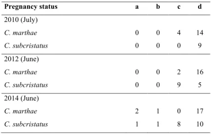
Samples of C. marthae and C. subcristatus females were collected in three different years: July 2010, June 2012 and 2014. Sample sizes and reproductive status considered in the present investigation are summarized in Table 1.
During all field sessions, approximately 2ml of blood were drawn from each iguana within 5 minutes from capture, using a 5 ml heparinized syringe. Blood was collected from the caudal vein and
kept on ice. A few hours later blood was centrifuged and plasma was separated. Plasma was stored at -10°C while in the field and at -40°C once back in the laboratory.
All captured iguanas were weighed and measured. The body condition index (BCI) was then estimated as the ratio of body mass/snout-vent length (SVL)3 x 10⁶ (the ratio was multiplied by 106 to reduce the number of decimals). Although simple, this index has been already used to describe the physical condition of marine (Laurie 1989; Wikelski and Trillmich 1997, Romero and Wikelski 2001) and land iguanas (Costantini et al. 2009).
2.3) Ultrasound analysis
For each female we determined the number of eggs, egg size, and the stage of development of follicles using a Sonosite portable ultrasound machine (FUJIFILM SonoSite, Inc.). Technical characteristic of the device and probe used, as well as the protocol applied, can be found in Gentile et al. (2016). Although abdomen palpation can be a possible method for diagnosing pregnancy, the use of ultrasound machine offers clear advantages. In fact, several studies identified it as the most reliable method to realize an accurate evaluation of reproductive conditions in reptiles (lizard: Gartrell et al. 2002; Gilman and Wolf 2007; tortoise: Robeck et al. 1990). The analysis allowed us to determine the reproductive status of each female, differentiating between development stages of eggs (Fig. 2): stage “a”, females showing follicles with eggs of homogenous, spherical and small dimensions; stage “b”, females with larger, yet not fully formed, unshelled eggs; stage “c”, females with large, fully formed, shelled eggs; stage “d”, non-reproductive females carrying no visible eggs inside follicles. An examination of the corpus luteum would prove useful to assess whether a female has laid a clutch of eggs in the recent past. However, while the corpora lutea persist in ovoviparous reptiles after egg-laying (Glasser and Bullock 2012), in oviparous reptiles as Conolophus they regress shortly after deposition (Yadav 2008). Additionally, along with its advantages, the ultrasound approach has the disadvantage that it does not allow the visualization of the corpus luteum (Norris and Lopez 2010).
Pregnancy status a b c d 2010 (July) C. marthae 0 0 4 14 C. subcristatus 0 0 0 9 2012 (June) C. marthae 0 0 2 16 C. subcristatus 0 0 9 5 2014 (June) C. marthae 2 1 0 17 C. subcristatus 1 1 8 10
Table 1. Pregnancy status of each species during three sampling
seasons. (stage a) Females with only follicular eggs; (stage b) females with not fully-formed eggs with only shell membrane; (stage c) females with fully-formed eggs; (stage d) non-reproductive females when no follicles or eggs were visualised.
Figure 2. Original ultrasound images.
Stages: “a” follicles with eggs of homogenous, spherical and small dimensions; “b” large, yet not fully-formed, unshelled eggs; “c” large, fully-formed, shelled eggs; “d” no visible eggs.
2.4) Hormone assays
Plasma levels of sexual steroids hormones progesterone (P4) and 17β-estradiol (E2) were determined by using competitive enzyme-linked immunosorbent assays (ELISA).
Indeed, several studies have analysed steroid hormones in reptiles using radioimmunoassay (RIA) (turtles: Mahmoud et al. 1989; snakes: Highfill and Mead 1975; Taylor et al. 2004; lizards: Judd et al. 1976; Arslan et al. 1978; Amey and Whittier 2000; Husak et al. 2007). The only works on iguanine lizards regard A. cristatus and use RIA (Rubenstein and Wikelski 2005; Vitousek et al. 2010; Vitousek and Romero 2013).
Radioimmunoassay is a common method for quantifying the steroids hormones in vertebrates, however some problems associated to this method exist. The need of special facilities for handling radioactivity, the short stability time of the radiolabeled ligands and potential health risks are commonly associated to this methodology (Andoh 2006; Sink et al. 2008). On the contrary ELISA is generally faster and safer than RIA, it is less expensive and shows a greater stability of reagents. Overall, there is still lack of data on how these two methods are comparable. The little information available suggests that differences may be observed when comparing results from RIA and ELISA. Problems may especially reside in differences in protocols of analysis (Sink et al. 2008).
All ELISA immunoassays were performed at the Laboratory of Clinical Biochemistry (Tor Vergata University Hospital). Plasma samples were preserved at -40°C until assayed.
We used 50 µl of plasma for the determination of each hormone. Only for E2, plasma was diluted 1:2 with assay buffer (containing proteins and sodium azide) to remove matrix interference. All samples were assayed in duplicate and randomly distributed between plates.
We used the enzyme-linked immunosorbent assay kit (CEA459Ge) pre-coated with a monoclonal antibody for P4. The detection range of progesterone ELISA kits (CEA459Ge) was 1.23-100 ng/mL. The intra-assay variation was < 10%, the inter-assay variation < 12%. We could not use ELISA kits of the same lot throughout the whole study, despite it was recommended (Sink et al. 2008). In fact, we used ELISA kits belonging to two different lots. To evaluate inter-lot variation, 5 individuals were analysed by using both lots. The power
curve y = axb, where a = 0.059006 and b = 1.4786, fitted data with a
proportion of variance explained (R2) equal to 0.995. We used such a curve to adjust readings from the second lot. In order to account for experimental error, a randomly generated number comprised within the minimum and maximum residual values of the regression was added to each predicted value.
For the 17β-estradiol (E2) we used the immunoassay KA2535 pre-coated with a polyclonal antibody. The detection limit of all estradiol ELISA kits was 14 pg/mL. The intra-assay variation was around 3%, the inter-assay variation 9%. Also for this hormone we used kits belonging to two different lots. As full correspondence in concentrations of retested animals was found, no adjustment procedure was applied in this case.
Both progesterone and 17β-estradiol assays were performed according to the instructions of manufacturers.
2.5) Statistical analysis
We used parametric and nonparametric test in order to analyse differences in hormonal plasma levels among years, between the two species, and between egg-carrying females (stages a, b, and c) and non-egg-carrying females (stage d), and to test the difference in clutch size between species. When data presented normal distribution we used Student’s unpaired t-test and ANOVA, and when normality assumption was not achieved Mann-Whitney U-test and Kruskal-Wallis ANOVA were applied. Pearson correlation analyses were performed to test the relationship between clutch size and body metrics (BCI, SVL, and weight).
Statistical analyses were performed by using software Past (version 3.07 for MAC) with two tails and alpha set to 0.05.
3) Results
3.1) Reproductive status
Of the 18 C. marthae females sampled in 2010, 4 (22%) showed fully formed eggs (stage c). No eggs were present in any of the other 14 females (78%) (stage d). Two females (11%) sampled in 2012 carried
not fully formed eggs (stage c); the other 16 (89%) carried no eggs (stage d). Two females (10%) sampled in 2014 showed follicles with small, spherical eggs (stage a), one female (5%) carried not fully formed eggs (stage b) while the remaining 17 (85%) carried no eggs (stage d).
In 2010 all C. subcristatus females sampled carried no eggs (stage d). In 2012 we observed nine females (64%) with fully formed eggs (stage c), and five (36%) without eggs (stage d). In 2014, one female (5%) showed follicles with small, spherical eggs (stage a), one female (5%) carried not fully formed eggs (stage b), eight females carried fully formed eggs (stage c) (40%), and 10 females (50%) were found without eggs (stage d).
Considering both fully and not fully formed eggs, we estimated clutch sizes equal to 8.4 ± 3.4 and 5.4 ± 1.5 for C. subcristatus and C. marthae, respectively. The difference between sizes was statistically significant (U = 20.5, P = 0.01). For both species, we did not observe a significant linear relationship between clutch size and body measures (BCI, SVL, and Weigh) (for all tests P > 0.05).
3.2) Progesterone (P4)
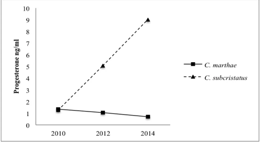
Progesterone plasma levels in C. marthae and C. subcristatus are shown in Fig. 3. Overall, considering all sampled females, the C. subcristatus presented higher P4 concentration than C. marthae (U = 764, P = 0.01).
When sampling years were treated separately, C. subcristatus showed significantly higher levels than C. marthae in June 2014 (U = 110, P = 0.042).
In C. marthae, P4 plasma levels were higher in July 2010 than in June 2012 and 2014 (although not statistically supported), when instead C. subcristatus showed low values.
When analysing the difference between egg-carrying females (cumulating females at stages a, b, and c) and non-egg-carrying females (stage d), C. marthae presented higher concentration of P4 plasma levels in egg-carrying females (U = 109, P = 0.042). Moreover, we observed a significant difference in P4 concentration among females with eggs at three different stages (a, b, and c) (H = 6.533; P = 0.036, Tab. 2). Mann-Whitney post-hoc tests, after Bonferroni correction, showed a significant difference between females at stages
a and c (H = 2. 252; P = 0.035). The remaining pairwise tests resulted not significant, (Pa vs b = 0.136 and Pb vs c = 0.081). However, when we pooled females at stages a and b to increase the statistical power due to the small sample used, P4 levels resulted higher in females at stage c than in the pooled sample (U = 0, P = 0.019).
In C. subcristatus we did not observe a significant difference between egg-carrying and non-egg-carrying females (U = 157, P = 0.18), but we found different variances (F = 3.11, P = 0.02). Considering the June (2012+2014) and July (2010) sampling sessions separately, F test indicated a larger variance in June than in July (F = 13.38, P = 0.001). We also observed a statistically significant difference between variances when comparing females without eggs sampled in June (2012+2014) with females without eggs sampled in July (2010) (F = 25.99, P = 0.0002; Fig. 4). We observed a statistically significant difference between total egg-carrying females and non-egg-carrying females of 2010 (U = 27, P = 0.018), with egg-carrying females showing higher P4 levels. However, this significance disappeared when total egg-carrying females were compared with non-egg-carrying females of June (2012+2014) (U = 88; P = 0.36).
Figure 3. Progesterone patterns in Conolophus marthae and C.
subcristatus during three sampling seasons. Concentrations are reported as median.
Figure 4. Progesterone plasma levels in non-pregnant females of July
(2010) and June (2012+2014) in C. subcristatus (median ± SD).
Progesterone concentration Follicular eggs (a) Not fully-formed eggs (b) Fully-formed eggs (c) N 2 2 5 Min 0.479 1.025 3.061 Max 0.503 2.358 19.045 Variance 0.0003 0.888 45.134 Stand. dev. 0.017 0.942 6.718 Median 0.491 1.691 5.059
Table 2. Variability in progesterone concentrations between three
3.3) 17β-estradiol (E2)
In C. subcristatus we found significant differences in 17β-estradiol plasma levels among years (H = 12.44, P = 0.002) (Fig. 5). Bonferroni post-hoc test indicated that estradiol plasma levels in 2010 were significantly lower than either 2012 (P = 0.004) or 2014 (P = 0.02). Instead, C. marthae did not show significant difference in estradiol levels among years (H= 2.21, P = 0.3).
Overall, we observed a higher concentration of estradiol in pink than in yellow iguanas (U = 273.5, P = 0.03).
In both species we did not observe a significant difference in estradiol plasma concentrations between females at reproductive stages a-c and stage d (C. subcristatus P = 0.4; C. marthae P = 0.5). However, in C. marthae, a statistically significant difference emerged when comparing females at stages b+c with females at stage a, these latter exhibiting higher estradiol concentration (t = - 2.9817, P = 0.02). In C. subcristatus, females carrying no eggs showed estradiol levels and variances higher in June (2012+2014) than in July (2010) (U = 6, P = 0.001; F = 15.193, P = 0.001; Fig. 6).
Figure 5. 17β-estradiol patterns in Conolophus marthae and C.
subcristatus during three sampling seasons. Concentrations are reported as median.
Figure 6. 17β-estradiol plasma levels in non-pregnant females of July
(2010) and June (2012+2014) in C. subcristatus (median ± SD).
4) Discussion
One of the main findings of our study is represented by the observation of a contrasted pattern of progesterone plasma levels in the congeneric land iguanas C. subcristatus and C. marthae. As for C. subcristatus, despite difference in the analytical tools, the observed patterns and order of magnitude of hormone concentration levels are consistent with those found by Rubenstein and Wikelski (2005) in A. cristatus, predictably changing during the mating and nesting periods. In fact, in A. cristatus progesterone increases during the mating period as related to oviduct vascularity and pregnancy maintenance. Indeed, a role of progesterone in pregnancy maintenance has been documented in many reptiles (Highfill and Mead 1975; Arslan et al. 1978; Naulleau and Fleury 1990; Bonnet et al. 2001; Taylor et al. 2004). Colonophus subcristatus showed significant changes of progesterone and estradiol levels throughout the three reproductive seasons considered in our study. High plasma progesterone levels in June 2012 and 2014 contrasted the low progesterone levels observed in July 2010.
The ultrasound analyses indicated that in July 2010 no sampled females carried eggs, whereas those sampled in June 2012 and 2014 were at different reproductive stages, with many females carrying eggs at various stages of development (64% in 2012, 50% in 2014). The high number of females carrying eggs in 2012 and 2014 associated with high progesterone plasma levels, and the correspondent absence of egg-carrying females in July 2010, when we observed low progesterone levels, could be sufficient to assert that reproduction of C. subcristatus from V. Wolf is still ongoing in June and has ended by July. The difference in the variance of progesterone levels of females at stage d (2012+2014 versus 2010) is consistent with such a scenario. In fact, while the ultrasound analysis did not allow to discriminate if the lack of eggs would indicate mating phase or occurred deposition, hormonal profiles were informative especially when we pooled June 2012 and 2014 together and compared the pooled sample with July 2010. The higher variance in progesterone plasma levels exhibited by non-carrying-egg females in June could testify for the presence of females in different reproductive conditions: (i) females in which progesterone could have dropped after deposition (Taylor et al. 2004); (ii) females that did not reproduce, suffering the low hormonal levels typical of non-receptive females (Vitousek et al. 2010); (iii) females still in a mating phase, when hormone concentration is lower than in early nesting period but higher than in a post deposition condition (Rubenstein and Wikelski 2005). The presence in June of females still in a mating phase is also suggested by the lack of a difference in plasma P4 levels between total egg-carrying females and non-egg-carrying females of June. Such a difference emerged instead when total egg-carrying females and non-egg-carrying females of July were compared.
Further support to the described scenario is also provided by the analysis of estradiol. In fact, we know that in A. cristatus estradiol peaks during the mating period and strongly declines during all nesting phases (Rubenstein and Wikelski 2005). In C. subcristatus sampled in July, we did not observe egg-carrying females and both progesterone and estradiol levels were very low. Furthermore, estradiol concentration in non-egg-carrying females was lower in July than in June, when also higher variance was observed. This strongly suggests that in June many females had high levels of estradiol because still in a mating phase (probably in last copulation stage),
when E2 influences the vitellogenesis process (Edwards and Jones 2001; Guillette et al. 1997; Ott et al. 2000; Rubenstein and Wikelski 2005), receptivity, attractivity (Mason and Adkins 1976; Rhen and Crews 2000; Winkler and Wade 1998), and aggressive behaviours against males attempting to copulate again (Rhen et al. 1999; Woodley et al. 2000a; Woodley and Moore 1999b; Rubenstein and Wikelski 2005). In July, all females had concluded the breeding season and abandoned the nest sites, showing very low levels of estradiol. Other studies on reptiles showed that extremely low estradiol levels emerge especially in post-parturition phase (Jones and Guillette 1982; Taylor et al. 2004).
Of course, reproduction in Conolophus may vary between years and locations as influenced by environmental conditions and resource availability (Snell et al. 1984), as in many reptiles (Laurie 1990; Vitousek et al. 2010). Our data indicate that the laying season in C. subcristatus from V. Wolf may occur in June-July, as in Fernandina Island. Interestingly, these are the only two known sites where C. subcristatus reproduces in those months. Fernandina and V. Wolf are also among the westernmost volcanos in Galápagos. If this correlates with particular climatological and environmental conditions that may affect reproduction remains to be uncovered.
The pattern of progesterone plasma levels in C. marthae, opposed to that of C. subcristatus, and the presence of egg-carrying females in 2010 could suggest a slightly delayed reproduction in the pink species compared to the congeneric syntopic population.
Overall, in C. marthae we observed a significant increase of progesterone levels in egg-carrying females. Also in this case the role of P4 in pregnancy maintenance was clear. Furthermore, plasma progesterone concentration significantly varied with stage of gravidity; in fact, it increased when eggs reaching complete maturation (shell and yolk formation). This observation suggests a role of the corpus luteum and its primary product (progesterone) in the shell secretion as occur in many reptiles during pregnancy (Ferguson 1985; Guillette and Jones 1985; Guilette et al. 1989). Anyway, although in C. marthae we found a positive relationship between egg-development stages and hormone levels, we observed constantly low comparable levels of progesterone through the three sampled years (2010-2012-2014), limited number of egg-carrying females and reduced number of eggs, compared to C. subcristatus. The
concentration of estradiol was higher in C. marthae than in C. subcristatus, but no difference across years was observed in C. marthae. The association of progesterone with the pregnancy status was a signal of the ovarian system functioning. In oviparous reptiles, the corpora lutea persist during pregnancy producing progesterone (Norris and Lopez 2001) and, in C. marthae a higher production of progesterone during pregnancy appeared. Furthermore, in the pink species, estradiol level seemed higher at early stage of egg maturation; this is in agreement with the evidence that in reptiles estrogens are secreted by vitellogenic ovarian follicles to then decline as eggs remain in the uterus (McNicol and Crews 1979; Etches and Petitte 1990; Norris and Lopez 2010). However, although in C. marthae, hormonal profiles of progesterone and estradiol provide physiological evidence of a hormonal change at different reproductive stages, they did not allow the identification of a specific reproductive period. Based on our data, for C. marthae, we could hypothesize the absence of a specific breeding season. The pink iguana could employ opportunistic reproductive strategies dependent on environmental conditions or interspecific interactions with C. subcristatus. Generally, reptiles tend to time egg incubation when a favourable season with minimum physiological stresses and maximum food resources is present. In pink iguana, we could hypothesize an individual opportunism as commonly occurs in other vertebrates (Milton et al. 2004) with females respond to stressful conditions varying reproductive period and clutch size. We could hypothesize that the lack of observed recruitment for this species (Gentile 2012) may be due to a limiting factor as for example predation on hatchlings and juveniles by hawks (Buteo galapagoensis) or feral cats (Felis catus). Indeed hawks and feral cats are constantly present on volcano and they are already described as cause of mortality in the marine iguana (Laurie and Brown 1990).
However, no C. marthae female resulted in reproductive conditions after a recent ultrasound surveys performed in November 2015 on a small number of healthy pink iguanas, or indirect evidence of reproduction activity (homospecific pairs, sperm at the cloaca of males and females) was found. This is in contrast with observation of such evidence in June/July (Gentile et al. 2016). Thus, for this reason, it is most likely that at present C. marthae may suffer from lack of
effective reproduction and the population results in attrition (Gentile 2012).
To conclude, our results suggest that hormonal profiles are fundamental to improve the knowledge on the reproductive biology of wild populations, especially when long-term observations are impossible. Our data indicate that C. subcristatus presents a specific and recognizable breeding season on Volcán Wolf. This season reaches its peak in June and concludes in July. In the same period, C. marthae shows reproductive activity, but the combination of hormonal profiles and ultrasound analysis demonstrated that such activity does not result in high numbers of reproductive females. Although opportunistic reproductive strategy cannot be completely ruled out for C. marthae, effective reproduction in this species seems hampered, determining attrition. In this regard, it is clear that further investigations are needed, especially aimed at uncovering the relationship between area of distribution, habitat characteristics and its usage, by tracking movements of individuals over time. This will help to locate nesting sites, currently unknown, and guide future conservation action for this critically endangered species.