Full Terms & Conditions of access and use can be found at
http://www.tandfonline.com/action/journalInformation?journalCode=uarc20
Download by: [Universita Di Ferrara] Date: 17 March 2016, At: 04:22
International Journal of Architectural Heritage
Conservation, Analysis, and Restoration
ISSN: 1558-3058 (Print) 1558-3066 (Online) Journal homepage: http://www.tandfonline.com/loi/uarc20
Oxalate Patinas on Stone Monuments in the
Venetian Lagoon: Characterization and Origin
Alessandra Bonazza, Claudio Natali, Nadia Ghedini, Carmela Vaccaro &
Cristina Sabbioni
To cite this article: Alessandra Bonazza, Claudio Natali, Nadia Ghedini, Carmela Vaccaro & Cristina Sabbioni (2015) Oxalate Patinas on Stone Monuments in the Venetian Lagoon: Characterization and Origin, International Journal of Architectural Heritage, 9:5, 542-552, DOI: 10.1080/15583058.2013.837546
To link to this article: http://dx.doi.org/10.1080/15583058.2013.837546
Accepted author version posted online: 10 Nov 2014.
Submit your article to this journal
Article views: 120
View related articles
Calcium oxalate patinas have been sampled on architectural elements of two ancient churches located on Torcello Island (Venetian Lagoon) and subsequently analyzed. The site had been selected presenting patinas of exceptional amount and thickness, elsewhere generally found as thin alteration layers covering stone surfaces or within black crusts on monuments in urban envi-ronments. Optical and mineralogical analyses suggest that these patinas are not the result of a simple deposition process, but origi-nate as surface “transformations” of the substrate, and are mainly composed of dihydrate calcium oxalate (weddellite) and gypsum. Among the experimental techniques, isotope analyses (C and S) have been specifically carried out aiming at achieving a better understanding of their origin and possible causes of formation. The observed carbon isotopic fingerprint reveals in fact a strong biological fractionation recorded in the oxalate patina (δ13C
rang-ing from−22.3‰ to −28.0‰), almost exclusively attributable to C3 pathway photosynthesis processes. Slightly negative sulfur iso-tope ratio values seem to be inversely correlated with patina expo-sure to atmospheric agents and pollutants. The results obtained prove that calcium oxalate originates from a biomineralization process induced by lichen colonization in specific environmental conditions.
Keywords oxalate patina, cultural heritage, isotope analyses, biomineralization, lichen
1. INTRODUCTION
Stones of monuments and historic buildings experience dif-ferent chemical, physical, and biological processes during their exposure to the atmosphere, which leads to the formation of a surface layer, depending on both the stone characteristics and the microclimate interacting with materials. A number
Received January 14, 2013; accepted August 20, 2013.
Address correspondence to Alessandra Bonazza, Institute of Atmospheric Sciences and Climate, ISAC-CNR, via Gobetti 101, 40129 Bologna, Italy. E-mail:[email protected]
Color versions of one or more of the figures in the article can be found online atwww.tandfonline.com/uarc.
of surface typologies have been identified in the literature on carbonate and silicate stones of the built cultural heritage in dif-ferent geographical areas, and their origin has been linked to the effects of air pollution, rain leaching, biodeterioration, tourism pressure, or conservation treatments (Viles et al.1997).
Although investigation has been made concerning the com-position and formation mechanisms of most damage layers found on historic stone monuments and buildings in polluted and unpolluted areas, an open problem still exists with regard to the origin of oxalate patinas.
Oxalate patinas have been found on cultural heritage from a variety of historical periods (from Greek-Roman to modern architecture) and on different materials (Del Monte et al.1987). A comprehensive review and discussion on oxalate patina com-position in diverse settings is presented by Matteini and Moles (1986) and Brimblecombe (1992), but most of the debate still focuses on stone monuments.
Calcium oxalate patinas are alteration layers characterized by variable color, thickness and texture. Two main typologies are reported in the literature: thin layers, orange-brownish in color, with high consistency and coherency, and white patinas of considerable thickness and scarce consistency (Ariño et al.
1995). Some patinas show a homogeneous structure, others a stratified one, but the mineralogical composition is in both cases very similar: monohydrate and dihydrate calcium oxalates (i.e., whewellite and weddellite) are the main crystalline phases associated with gypsum and quartz in different amounts and ratios.
The low solubility of whewellite and weddellite protects the underlying substratum against atmospheric weathering (Alessandrini1989). Therefore, it may be argued that the for-mation of calcium oxalate patinas mostly produces an aesthetic impact with chromatic alterations of the monument surface on which it develops.
As widely reported in the literature, three are the possible origins of calcium oxalate patinas on architectural surfaces in marble and limestone: biodeterioration, air pollution impact,
542
and degradation of organic treatments (Sabbioni et al. 2003; Vazquez-Calvo et al.2007). A considerable number of papers ascribe oxalate patinas to a biological origin. The chemical-physical interaction between colonizing microorganisms secret-ing oxalic acid and the limestone substrate leads to the dissolu-tion of carbonates. The calcium cadissolu-tions freed by the process are involved into the formation of calcium oxalate (Del Monte and Sabbioni1983). This occurrence is confirmed by laboratory experiments, which demonstrated that the interaction between oxalic acid (Kouzeli et al. 1996) or colonial bacteria (Monte
2003) and samples of Carrara marble, produces calcium oxalate layers. The neoformation of thin oxalate layers occurs along the lichen-substrate interface, where the pH, humidity and reduc-tion potential values are suitable (Adamo et al.1997). In high humidity and basic pH conditions weddellite crystallizes prefer-entially, whereas in acid and hydrated environments whewellite is more stable (Frey-Wyssling1981; Ascaso et al.1982; Horner et al.1985).
Because of its low solubility, the calcium oxalate tends to amass in the thallus of the lichen to form a thin crystalline layer around it (Jackson1981), but also at the lichen-substrate interface (Ascaso and Galvan1976). Del Monte et al. (1987) reported that the interface that subdivides the lichens from the unaltered substrate is extremely irregular, implying a pervasive interaction between oxalate and carbonate (Ariño et al.1995). Therefore, oxalate patinas cannot be considered as a simple
deposition on the stone, but a transformation of the stone itself.
The literature also mentions weddellite and whewellite on basalt stone in natural outcrops as a consequence of lichen colonization (i.e., Pertusaria corallina). The action of myco-bionts on primary minerals, such as Ca-rich plagioclases, allows the availability of calcium ions and the subsequent formation of calcium oxalate (Jones et al. 1980; Arocena et al. 2007). Lichens colonizing serpentinites lead to the precipitation of magnesium oxalate, as glushinskite (Wilson et al.1980,1981; Adamo et al.1993; Favero-Longo et al.2005). The neoforma-tion of Mg-oxalate has also been encountered during in vitro experiments on fungi-mineral interaction on dolomite (Kolo and Claeys2005). Lichen colonization on carbonate structures pro-duces not only calcium oxalates, but also metallic oxides and hydroxides, as well as aluminum-silica compounds, may form (Adamo and Violante2000).
De Santis and Allegrini described, in1989, oxalic acid asso-ciated to air pollution deposition as a second hypothesis for oxalate patina origin. Sabbioni et al. (2003) focused a review on small (C1-C2) organic anions, including oxalate, in black crusts on monuments and buildings. They showed that these short chain ions are almost constantly associated with sulfur and nitrogen compounds linked to airborne gas and particle deposition emitted by combustion sources and producing black crust formation on stone substrates. In aerosol composition the positive correlation between non-sea salt sulfate and oxalate could indeed be ascribed to similar pathway during in-cloud formation (Yu et al.2005).
As a third hypothesis, the decomposition of organic treat-ments applied in the past to protect the surface of monutreat-ments is considered to be an origin of calcium oxalate formation. Since the classical antiquity, animal- and vegetal-derived components have been used as protective compounds (Rampazzi et al.2004; Gabrielli and Parenti1992; Cennini1991; Frazzoni1988; Piva
1988; Fassina1989). Berlucchi and Ginanni Corradini (1996) interpreted the presence of joints in some oxalate patinas, rec-ognized by thin section optical microscope (OM) analyses, as post-application pull back structures of the protective treatment layer. Moreover, infrared spectrometer analyses of the same samples revealed the presence of barium silicates used in the past to whiten the compound.
Oxalate patinas have also been found indoors, on surfaces not directly exposed to atmospheric weathering. The few stud-ied cases ascribe the presence of calcium oxalate mainly to the alteration of protective treatments (Alessandrini et al.1989; Chiari et al.1996).
The identification of the origin of oxalate patinas on stone monuments continues to be a challenging area of research, in spite of more than twenty years of debate. Today, the growth mechanism of oxalate patinas on monuments may be gaining in importance, in view of the different future trends assumed in the near and far future by climate parameters linked to biodeterioration (i.e., temperature and precipitation), pollution and anthropic pressure (Grossi et al.2008; Bonazza et al.2009a;
2009b, Gomez-Bolea et al. 2012). Within the perspective of global change, the need for an effective analytical technique with which to solve the remaining problem of oxalate patina formation mechanisms is therefore particularly pressing in the field of cultural heritage conservation.
Stable carbon isotopic ratios due to the precise fingerprint drawn by biological processes in both polluted and unpolluted areas may be a suitable indicator for interpreting the formation process of these specific surface layers. With this perspective, the aim of the present work is, besides an exhaustive chemical and mineralogical characterization of the alteration patinas onto monuments in Torcello Island, to explore if isotope analyses can constitute a powerful tool for discriminating the origin of surface oxalate patinas formed on heritage building stones. The work focuses on the results achieved by a first-time application of isotope analyses to this particular typology of neoformation surface layer, with the aim of providing a contribution to the identification of oxalate patina genesis.
2. MATERIALS AND METHODS 2.1. Sampling
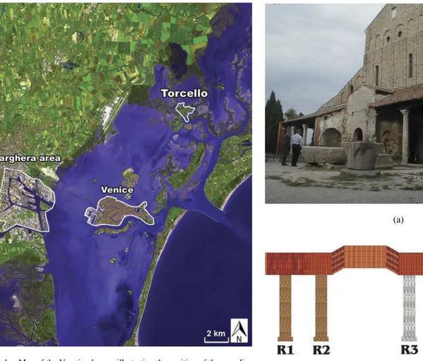
The sampling was performed on Torcello Island, located in the Venetian Lagoon, 9 km northeast of Venice (Figure 1). The site was selected because of the outstanding abundance and thickness of calcium oxalate layers on the surfaces of the island’s monuments, allowing both a complete characterization of this specific typology of surface layer and the application
FIG. 1. Map of the Venetian lagoon illustrating the position of the sampling site of Torcello Island, Venice, and the power plant and oil refinery of Porto Marghera.
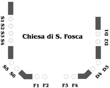
of isotope analysis, which requires homogeneous surface lay-ers. The samples were collected from the columns and capitals of different lithologies belonging to the external perimeter log-gias of the two churches of Santa Maria Assunta (Figure 2a) and Santa Fosca (Figures 3 and4). Most of the columns are constituted by impure greenish marble (Greek Marble s.s.) and nodular reddish limestone (Rosso di Verona, Figure 2b), only two being composed of micritic limestone. The description and localization of the samples on which the experimental work was based are summarized inTable 1. Most of the patinas observed on the columns and capitals of the two churches were white-gray in color, with a thickness of up to 0.5 cm, scarcely coherent and revealing a plastic behavior during sampling (Figure 5).
2.2. Analytic Procedures
Mineralogical and petrographic investigation of the samples was carried out by an Olympus BX51 polarized light opti-cal microscope (OM) and X-ray diffraction (XRD) using a Philips PW1710 Based diffractometer, equipped with Cu pipe (Kα1 = 1.54060 Å, Kα2 = 1.54439 Å). Morphological obser-vations were performed by a Philips XL 20 scanning electron
(a)
(b)
FIG. 2. (a) Perspective view of the church of Santa Maria Assunta (7th cen-tury). (b) Santa Maria Assunta loggia before the church gate. Columns R1, R2, R6 are made of nodular limestone (Rosso di Verona), column R3 is made of impure marble (Greek marble s.s.), and column R4 and R5 are in limestone.
FIG. 3. Frontal view of Santa Fosca (11th century).
FIG. 4. Octagonal floor plan of Santa Fosca and of its loggia. The sampled columns are in marble.
microscope (SEM), equipped by x-ray energy dispersion ana-lyzer (EDX) to perform elemental analyses. Differential and gravimetric thermal analyses were carried out by a METLER TOLEDO TGA/SDTA 851, equipped with TSO 800GC1 pro-grammable gas switch. A combination of these techniques was employed to attain an accurate characterization of the surface layer and to test its homogeneous composition.
With the aim of quantifying the anion concentrations in the collected samples, ion chromatographic (IC) analyses were performed using a Dionex Chromatograph (Model 4500i), equipped with conductivity detector (Dionex CDII), following the methodology reported by Gobbi et al. (1995).
Isotopic analyses were carried out by a VARIO EL element analyzer coupled with a carbon, nitrogen and sulphur (CNS) mass spectrometer (accuracy of 0.2 ‰). The samples were pow-dered in an agate mortar and then treated with 37% HCl solution in order to eliminate carbonate phases, thus avoiding the car-bon signature of the substrate (marble and limestone). The CO2
freed by carbonate dissolution were trapped by NaOH grains within the stagnate glass dryer where the samples were stored.
3. RESULTS AND DISCUSSION 3.1. OM Analysis
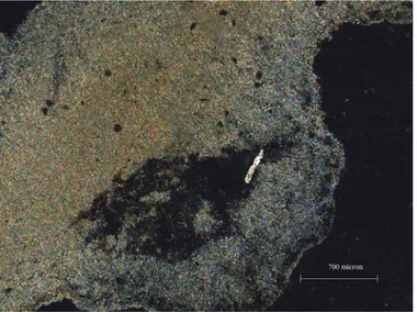
Thin sections of oxalate patinas, collected from different lithologies, and one of substrate (a marble column fragment) were analyzed by optical microscope (OM). In this way, two kinds of oxalate patina were recognized, in relation to the different substrate on which they form. The first, collected from architectural elements in limestone (Rosso di Verona), is mainly composed of a yellow-brownish microcrystalline matrix of calcium oxalate that included rare uncolored phyllosilicate (muscovite) and small rounded black oxides (Figures 6 and
7). The measured average thickness is around 1100 μm. The TABLE 1
Description and localization of samples from the two churches of Torcello Island (Venice)
Sample Localization Description
TOR2bis Santa Maria Assunta Cathedral. Column R2. Collected from various points all over the surface
Pink (bottom side) - grey patina 2 mm thickness scarcely coherent
TOR3-TORNEW3 Santa Maria Assunta Cathedral. Column R3. Collected from various points all over the surface
White (bottom side) – light grey patina about 1 mm thickness scarcely coherent
TOR4bis-TORNEW4 Santa Maria Assunta Cathedral. Column R3. Collected only from inner side
White patina of about 2-3 mm thickness scarcely coherent
TOR5-TORNEW5 Santa Maria Assunta Cathedral. Column R3. Collected only from outer side
White (bottom side) – grey patina about 1 mm thickness, scarcely coherent
TORNEW 6 Santa Maria Assunta Cathedral. Column R3. Collected only from inner side
White (bottom side) – grey patina of about 2-3 mm thickness. Portion of conspicuously altered substrate was also collected during sampling
TOR7 Santa Maria Assunta Cathedral. Column R3 Marble fragment belonging to the column TORNEW7 Santa Maria Assunta Cathedral. Column SMA1 in
Rosso di Verona belonging to the inner loggia
Greyish patina of about 2 mm thickness scarcely coherent
TOR8 S. Fosca Church. Capital of column D2 in marble White patina scarcely coherent TOR11 S. Fosca Church. Upper part of Abside behind the
church (marble)
White patina
FIG. 5. Sample of oxalate patina collected from a column in marble at Santa Fosca church.
FIG. 6. Sample TOR2bis: microphotograph showing the high porosity of the oxalate patina (cross polarized light).
second kind, collected from impure marble columns and cap-itals, shows a similar microcrystalline matrix to the former, and includes a remarkable amount of phyllosilicate, calcite and quartz crystals (Figures 8 and9). In the figure, it is possible to identify a patina’s growth pattern perpendicular to the sur-face layer-substrate intersur-face, reflected by the isorientation of the phyllosilicate crystals. The average thickness measured in this case is around 900–950μm.
The substrate sample shows an inequigranular texture with isorientation of the mineral grains typical of metamorphic processes. It was found to be mainly composed of ehuedral white calcite, under plane polarized light with the character-istic rombohedral cleavage, and of greenish elongated chlorite crystals. These fibrous minerals are of discrete dimensions,
FIG. 7. Sample TOR2bis: small phyllosilicate crystal incorporated in homo-geneous oxalate patina (cross polarized light).
FIG. 8. Sample TOR4bis: microphotograph showing an orthogonal isorienta-tion of phyllosilicates highlighting the growth direcisorienta-tion of oxalate patina (cross polarized light).
and are homogeneously present in the section, showing low interference colors by cross-polarized light, typical of Mg-rich chlorites (pennine). Quartz is also present in a smaller amount in anhedral crystals with undulated extinction. Elongated mus-covite crystals constitute the second phyllosilicate phase rec-ognized. On the basis of the results obtained, the substrate sample was classified as low-grade metamorphic rock (impure marble). Following the petrographic characterization, it may be argued that the significant presence of phyllosilicate, cal-cite, and quartz found on the oxalate patina collected from the column in impure marble of Santa Maria Assunta Cathedral is linked to the underlying rock, particularly in the process of alteration.
FIG. 9. Sample TOR4bis: microphotograph highlighting the presence of quartz crystals (white–yellow color) and calcite (pink color, on left) (cross polarized light).
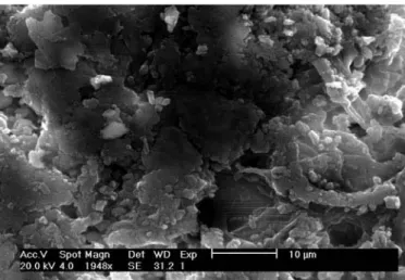
3.2. Scanning Electron Microscope (SEM)-X-Ray Energy Dispersion (EDX) Analysis
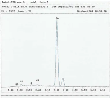
Scanning electron microscope (SEM) analysis were per-formed on three samples of oxalate patinas (TORNEW5, TORNEW6, TORNEW7), in order to investigate their morphol-ogy from micro to nanoscale and, through the x-ray energy dispersion (EDX) probe, to evaluate their elemental composi-tion. In all the investigated samples, the patina’s surface was mostly composed of a homogeneous microcrystalline matrix, with globular morphology and diffuse porosity, sometimes bro-ken by fractures. The EDX analyses of the surfaces showed them to be mainly composed of Ca, with smaller amounts of Si, Cl, S, and Al, while no particles from atmospheric deposi-tion were found to be present (Figures 10and11). Increasing
FIG. 10. Scanning electron microscope (SEM) microphotograph showing the patina morphology of sample TORNEW5.
FIG. 11. X-ray energy dispersion analyzer (EDX) spectrum of TORNEW5 patina surface.
magnification led to the identification of phyllosilicates, includ-ing muscovite (Figures 12and13) and feldspar crystals of about 100 μm in length in samples TORNEW5 and TORNEW6, where smaller amphiboles, chlorite and cubic halite were also found. As already evidenced by OM observation the identified phyllosilicate are likely to be related to the accessory miner-als of the substrate (impure marble) more than to atmospheric soil dust deposition. This finding allows us to advance the hypothesis that in the oxalate formation process, silicate min-erals belonging to the substrate, such as micas and quartz, are embedded in the newly formed patina without undergoing any substantial chemically alteration. In addition in the analyzed
FIG. 12. Scanning electron microscope (SEM) microphotograph of sample TORNEW5 showing a pair of muscovite crystals.
FIG. 13. X-ray energy dispersion analyzer (EDX) microanalysis of crystals in Figure 12.
FIG. 14. Scanning electron microscope (SEM) microphotograph of sample TORNEW5 showing numerous organic filaments resembling fungal hyphae with crystals on surface.
samples it is worth noting the presence of organic filaments emerging by about 100μm from the surface. A large number of smaller fragments very similar to fungal hyphae appears net-worked within the matrix (Figures 14). In sample TORNEW7, these organic fragments are covered by a thin layer of calcium oxalate (Figure 15).
3.3. X-Ray Diffraction Analysis
The X-ray diffraction analysis of five samples of calcium oxalate patina was carried out, in order to determine their mineralogical composition by means of a qualitative and semi-quantitative approach. The obtained results are reported in
Table 2. It should be noted in the table, that most of the patinas
FIG. 15. Scanning electron microscope (SEM) microphotograph of sample TORNEW7 showing numerous organic filaments resembling fungal hyphae covered by the layer of calcium oxalate.
are mainly composed of tetragonal calcium oxalate dihydrate and subordinate gypsum. Minor amounts of quartz and cal-cite are usually recognized, whereas halite, iron and manganese oxide/hydroxide are always present in traces. As well as cal-cium oxalate above discussed, the formation of gypsum in these patinas can be ascribed to several origins. Dry atmospheric deposition of SO2on carbonate substrates followed by
rainwa-ter acidification processes lead to the formation of CaSO4·2H2O
due to the dissolution of calcite that releases calcium cation. Moreover, it should be pointed that aerosol sulfate concentra-tions have been found in positive correlation with oxalate in different environments, suggesting the possibility of a common in-cloud pathway for these compounds (Crahan et al.2004; Yu et al.2005), and pointing to a common atmospheric source of the gypsum and calcium oxalate in patinas. Sea spray depo-sition of sulfate, usually occurring in brackish environments, is another possible source in the formation of this compound. Phyllosilicate (chlorite, mica, illite, clinochlore) were found to be present in some of the samples (TOR4bis, TOR5) collected from marble columns.
The constant presence of halite is due to sea spray recrystallization, whereas iron, magnesium and manganese oxide/hydroxide could derive from both the substrate and deposited atmospheric aerosol. However, a number of authors interpret these phases as neoformation minerals linked to the interaction between lichen biological activity and rock sub-strate through the mobilization of metallic ion (Barker and Banfield1996; Prieto et al.1997; Adamo and Violante 2000; Arocena et al.2003). In particular, the presence of wustite in one sample (TOR11) should be ascribed to “de-dolomitization” processes, leading to the oxidation of bivalent iron contained in the substrate.
Calcium oxalate monohydrate appeared only in sample TOR2bis, probably due to crystallization of calcium oxalate in
TABLE 2
Semi-quantitative mineral phases estimated by x-ray diffraction (XRD) of surface patina samples.
Sample Wd Gy Q C Ch Mi/Il H Cc Do Pe Wu Pc Py/Mn Wh TOR2bis +++++ ++ ± ± ± ± ± ± ± TOR4bis +++++ +++ ++ +++++ + ± + + ± ± ± TOR5 +++++ ++ +++ ± ++ ++ ± ± ± TOR8 +++++ +++ ± ± ± ± ± ± TOR11 +++++ +++ ± ± + ± ± ± ±
Wd, weddellite, Gy, gypsum, Q, quarz, C, calcite, Ch, chlorite, Mi/Il, mica/illite, H, halite, Cc, clinoclore, Do, dolomite, Pe, periclase, Wu, wustite, Pc, pirocroite, Py/Mn, pirolusite/manganosite, Wh, whewellite. Legend: +++++ = principal mineral present; ++++ = very much abundant;+++ = abundant; ++ = scarcely abundant; + =present; ± =traces.
both low humidity environments and low pH conditions (Horner et al.1985). In this case, subsequent hydration, which leads to the transformation into the dihydrate form, could therefore not be completed.
3.4. DTA-TGA Analysis
Differential thermal coupled with gravimetric analysis was conducted on two samples (TOR4bis and TOR8) represen-tative of the two oxalate patina types. The analyses were performed under O2 constant flux conditions. In matrixes
containing calcium oxalates, the quantitative interpretation of DTA-TGA results is particularly complex, because hydrated oxalate forms (mono, bi- and trihydrates) loose water crystal-lization at approximately 200 ◦C, while on reaching 500 ◦C they decompose into CaCO3, developing CO2, as highlighted
by Riontino et al. (1998). The data obtained showed the pres-ence of organic carbon with a exothermic peak at 310 ◦C, producing a weight loss of between 11 and 14 wt%. The decom-position of anhydrous oxalate (CaC2O4) into calcite (CaCO3)
occurs through a exothermic reaction at 460◦C, with the loss of 9 wt% in TOR4bis and 14 wt% in TOR8. Decomposition of carbonate phases at temperatures between 760-780◦C is cou-pled with loss of weight of 14% (TOR4bis) and 16% (TOR8). It should be emphasized that the weight loss incurred by car-bonate phase decomposition give an overestimation of calcite, because of the above mentioned calcium oxalate decomposition at 460◦C.
3.5. IC Analysis
The IC analyses provided the concentration of the ion soluble fraction of the patinas. The results obtained indicate that oxalate anion concentration ranges from 1 to 7%. The experimental data proved that the sampled patinas are formed by consider-able higher amounts of oxalates than those measured in damage layers from monuments in different locations, built in different materials, which were constantly lower than 1.6 % (Riontino et al.1998; Sabbioni et al.2003), with exception of black crusts sampled in Venice, which showed a maximum concentration of 3.4 % (Bonazza et al.2005). These results demonstrate that the
selected samples were particularly suitable for testing isotope analyses.
In addition to oxalate, other organic anions were found: acetate and formate with concentrations of one and two order of magnitude lower than oxalate anion, being respectively 0.4% and 0.001%. Among the non-organic anions, sulfate predomi-nates followed by nitrate and chloride. Phosphates are present in variable concentrations, and nitrate and fluoride varying from trace to negligible concentrations.
3.6. Isotopic Analysis
The isotope analyses were performed on three surface patina samples from the church of Santa Maria Assunta, collected from impure marble (TORNEW5 and TORNEW6) and limestone (TORNEW7), both internal and external. The data obtained for
δ13C and forδ34S are reported inTable 3.
The values ofδ13C ranging from –22‰ to –28‰ indicate
that carbon fractionation is not affected by different substrate lithology or the position of the patina. Considering that the
δ13C in carbonate rocks varies from –1 to 6‰, and that the
only carbon source able to affect this value is atmospheric CO2 with average δ13C equal to –7‰, it can be concluded
that the only process in nature responsible for the large nega-tiveδ13C recorded on the Torcello oxalate patinas, is biological
carbon fractionation. Colonizing organisms, i.e., lichens and fungi residing on rocky substrates, produce similar paragen-esis of mineral neoformation having different carbon isotopic fractionation. Kolo et al. (2007) observed slightly negativeδ13C (–4.2‰) in Mg and Ca oxalates neomineral originating from fungi attack on limestone and dolomite substrates (δ13C ≈ +2.1‰ ca.). More negative δ13C values have been reported
for lichen biological activity. Using atmospheric carbon dioxide as the only nutrient for the photosynthesis process (C3 path-way), the lichen phycobiont (green or blue algae) preferentially select12CO
2instead of13CO2through a diffusion process. Such
fractionation results in δ13C around –27‰ for higher plants
adopting this kind of photosynthesis cycle (Fogel and Cifuentes
1993), whereas this value can range between –17‰ and –34‰ for lichens depending on the intensity of the diffusion process
(2002) found that calcium oxalate pruina is enriched in 13C,
compared to lichen tissues producing higher mean values of
δ13C of between 6 and 7‰.
Based on such assumptions, it may be argued that TORNEW5, undergoing greater carbon fractionation, was sam-pled in the area mostly exposed to moist conditions: values in neomineral calcium oxalate were extremely low (δ13C = −28‰), implying that the lichen tissues potentially have mean values ofδ13C ranging from –34 to –37‰, which are ascribed in the literature to the presence of photosymbiodemes, a more generalist lichen association (Maguas et al.1993;1995; Smith and Griffiths1996). Assuming that atmospheric CO2is the only
source of carbon utilized by lichen biological activity, exclud-ing the uptake of substrate rock carbon (Beazley et al.2002), the other possible cause of such low values of δ13C is the
starting value of atmospheric CO2. Widory and Javoy (2003
and reference therein) found an inverse correlation between the concentration of CO2 in the atmosphere and its δ13C value,
asserting that all anthropogenic carbon dioxide contributions are
13C depleted.
Sulfur isotope ratioδ34S is slightly negative, ranging from −0,2‰ (TORNEW7) to −2,5‰ (TORNEW5). Sulfate of oceanic sea spray (δ34S = +21‰ [Rees et al.1978]) or bio-genic sulfur (DMS,δ34S=+15±3,1‰ [Calhoun et al.1991]) are characterized by much heavier compositions. It has to be underlined that even if negligible, a sulfur contribution from sea salt deposition in the patinas cannot be completely excluded (aerosol marine effect is proved by halite determination by XRD and SEM analyses). Gypsum has already been recognized by some authors as biomineral, in association with calcium oxalate, related to the biological activity of lichens in unpol-luted (Prieto et al.1999) and polluted areas (Garty et al.2002), but no isotopic data have been reported. Biological fractionation should not affect the sulfur isotope ratio, particularly in epi-phytic lichens and mosses, where the values ofδ34S reflect the
composition of atmospheric SO2, suggesting that no
discrimi-nation between32S relative to34S is involved during metabolic cycles (Krouse 1977; Winner et al. 1978; Case and Krouse
1980). The values recorded in the patinas, given the widespread
2 2
concentration. Indeed, lichens living in excess of atmospheric SO2 concentration proportionally enhance the production of
volatile H2S weighting the sulfur residue (Krouse1977; Krouse
et al.1991; Newman et al.1991). The lowestδ34S values found
in lichens living in similar conditions on Newfoundland by Wadleigh (2003) are partially comparable (slightly higher e.g.
δ34S≈ 4–7‰) to those encountered in the Torcello patinas, the
main difference being the healthy condition of the lichens at the Italian site.
4. CONCLUSION
The analyses performed on the patinas from the loggias of two investigated churches on Torcello Island reveal that they are mainly composed of dihydrate calcium oxalate (weddellite). The particular abundance of the metastable form of this mineral reflects the specific environmental characteristics of the stud-ied site: a lagoon environment characterized by high relative humidity and sea spray deposition.
The integrated methodological approach carried out allowed us to assume a major biological origin of the calcium oxalate patina sampled. The SEM analyses performed in particular show a significant presence of fungal hyphae, suggesting a direct involvement of present or past biological activity in the formation of the patinas. The results led also to the identifi-cation of a thin microcrystalline weddellite/gypsum veil that sometimes covers the hyphae, pointing to a possible extracel-lular mineralization already observed in lichen thallus (Garty et al. 2002). The study of sample thermal behavior confirms a significant presence of organic fraction and, in addition, ion chromatography shows a remarkably high concentration of oxalate anions.
Isotope analyses were performed to investigate the origin of surface oxalate layers on monuments. The isotope measure-ments forδ13C of the oxalates in the patinas, indicate
photo-synthesis and a fixation processes based on the C3 pathway (Calvin-cycle), involving the Rubisco enzyme. Small variations can occur due to biogeochemical interactions on the microscale
and different diffusion with related fractionation prior to fixa-tion. In addition,δ34S measurements suggest that a correlation
between atmospheric exposure of patinas and sulfur isotope ratio might exist. Even if it is impossible to exclude a marine contribution to patina formation, gypsum can also be associ-ated with Ca-oxalates, as a biomineral product deriving from lichen activity in polluted environments. The possible marine contribution is limited by the isotopic fingerprint that should be characterized by heavier sulfur isotopic ratios. Isotopic results confirm that calcium oxalate, the main components of the Torcello Island alteration patina, are related to a biomineral-ization process induced by colonizing lichens in a peculiar environment.
Therefore, the results obtained contribute to the debate on the origin of oxalate patina, demonstrating, for the first time using quantitative data, the biological origin of oxalate patina growth on stone monuments. In addition, the present work proves isotopic analyses to be a suitable methodology for discriminat-ing the origin of oxalate patinas, which application should be further developed. Within a perspective of changing climate, the identification of the growth mechanisms of surface layers on monuments is essential for the purpose of developing suitable strategies for the protection of cultural heritage.
FUNDING
This work was performed within the EC Project “Global cli-mate change impact on built heritage and cultural landscapes - NOAH’S ARK”, supported by the European Commission, within the 6th Framework Program on research (Contract nr. SSPI-CT-2003-501837-NOAH’S ARK) and is funded within EC Project TeACH “Technologies and tools to prioritize Assessment and diagnosis of air pollution impact on immovable and movable Cultural Heritage” (Contract nr. 212458 - TeACH), European Commission 7FP.
REFERENCES
Adamo, P., A. Marchetiello, and P. Violante. 1993. The weathering of mafic rocks by lichens. Lichenologist 25: 285–97.
Adamo, P., C. Colombo, and P. Violante. 1997. Occurrence of poorly ordered Fe–rich phases at the interface between the lichen Stereocaulon vesuvianum and volcanic rock from Mt Vesuvius. Clay Minerals 32:453–461. Adamo, P., and P. Violante. 2000. Weathering of rocks and neogenesis of
minerals associated with lichen activity. Applied Clay Science 16:229–256. Alessandrini, G., R. Bonecchi, R. Peruzzi, L. Toniolo, and E. Fedeli. 1989. Caratteristiche composizionali e morfologiche di pellicole ad ossalato: Studio comparato su substrati lapidei di diversa natura. In Proceedings of the
1st International Symposium on the Oxalate Films: Origin and Significance in the Conservation of Works of Art, eds., Centro CNR “Gino Bozza” and
Politecnico di Milano, 137–50. October 25–26, 1989, Milan, Italy. Rome, Italy: CNR.
Ariño, X., J. J. Ortega-Calvo, A. Gomez-Bolea, and C. Saiz-Jimenez. 1995. Lichen colonization of the roman pavement at Baelo Claudia (Cadiz, Spain): Biodeterioration vs. bioprotection. Science of the Total Environment 165:353–363.
Arocena, J. M., L. P. Zhu, and K. Hall. 2003. Mineral accumulations induced by biological activity on granitic rocks in Qindhai Plateau, China. Earth
Surface Processes Landform 28:1429–1437.
Arocena, J. M., T. Siddique, R. W. Thring, and S. Kapur. 2007. Investigation of lichens using molecular techniques and associated mineral accumu-lations on a basaltic flow in a Mediterranean environment. Catena 70: 356–365.
Ascaso, C., and J. Galvan. 1976. Studies on the pedogenic action of lichen acid.
Pedobiologia 16:321–331.
Ascaso, C., J. Galvan, and C. Rodriguez-Pascal. 1982. The weathering of calcareous by lichens. Pedobiologia 24:219–229.
Barker, W. W., and J. F. Banfield. 1996. Biologically versus inorganically mediated weathering reactions: relationships between minerals and extra-cellular microbial polymers in lithobiontic communities. Chemical Geology 132:55–69.
Beazley, M. J., R. D. Richman, D. K. Ingram, T. W. Boutton, and J. Russ. 2002. Natural abundances of carbon isotopes (14C,13C) in lichens and cal-cium oxalate pruina: implications for archeological and paleoenvironmental studies. Radiocarbon 44:675–83.
Berlucchi, N., and R. Ginanni Corradini. 1996. Patine ad ossalati su alcuni mon-umenti di Roma. In Proceedings of the 1st International Symposium on the
Oxalate Films: Origin and Significance in the Conservation of Works of Art, eds., Centro CNR “Gino Bozza” and Politecnico di Milano, 137–50.
October 25–26, 1989, Milan, Italy. Rome, Italy: CNR.
Bonazza, A., C. Sabbioni, and N. Ghedini. 2005. Quantitative data on carbon fractions in interpretation of black crusts and soiling on European built heritage. Atmospheric Environment 39:2607–2618.
Bonazza, A., P. Messina, C. Sabbioni, C. M. Grossi, and P. Brimblecombe. 2009a. Mapping the impact of climate change on surface recession of carbonate buildings in Europe. Science of the Total Environment 407:2039–2050.
Bonazza, A., C. Sabbioni, P. Messina, C. Guaraldi, and P. De Nuntiis. 2009b. Climate change impact on stones: Mapping thermoclastism for European monuments. Science of the Total Environment 407:4506–4512.
Brimblecombe, P. 1992. The balance of environmental factors attacking arti-facts. In Durability und Change, eds., W. E. Krumbein, P. Brimblecombe, D. E. Cosgrove and S. Staniforth, 67–135. Sussex, UK: Wiley.
Brugnoli, E., K. T. Hubick, S. Von Caemmerer, S. C. Wong, and G. D Farquhar. 1988. Correlation between the carbon isotope discrimination in leaf starch and sugars of C3 plants and the ratio of intercellular and atmospheric partial pressures of carbon dioxide. Plant Physiology 88:1418–1424.
Calhoun, J., T. Bates, and R. Charlson. 1991. Sulphur isotope measure-ment of submirometre sulphate aerosol particles over the Pacific Ocean.
Geophysical Research Letters 18:1877–1880.
Case, J. W., and H. R. Krouse. 1980. Variations in sulphur content and stable isotopic composition of vegetation near a SO2source at Fox Creek, Alberta, Canada. Oecologia 44:248–257.
Cennini, C. 1991. Il libro dell’Arte. Florence, Italy: Le Monnier.
Chiari, G., N. Gabrielli, and G. Torraca. 1996. Calcium oxalates on mural paint-ings in internal exposure. Sistine chapel and others. In Proceedpaint-ings of the
1st International Symposium on the Oxalate Films: Origin and Significance in the Conservation of Works of Art, eds., Centro CNR “Gino Bozza” and
Politecnico di Milano, 137–50. October 25–26, 1989, Milan, Italy. Rome, Italy: CNR.
Crahan, K. K., D. Hegg, D. S. Covert, and H. Jonsson. 2004. An explo-ration of acqueous oxalic acid production in the coastal marine atmosphere.
Atmospheric Environment 38:3757–3764.
Del Monte, M., and C. Sabbioni, 1983. Weddellite on limestone in the Venice environment. Environmental Science & Tecnology 17:518–522.
Del Monte, M., C. Sabbioni, and G. Zappia. 1987. The origin of calcium oxalate on historical buildings, monuments and natural outcrops. Science of Total
Environment 67:17–39.
De Santis F., and I. Allegrini. 1989. Determinazione di acido ossalico e di ossalati in atmosfera mediante tubi di diffusione. In Proceedings of the 1st
International Symposium on the Oxalate Films: Origin and Significance in the Conservation of Works of Art, eds., Centro CNR “Gino Bozza” and
Politecnico di Milano, 137–50. October 25–26, 1989, Milan, Italy. Rome, Italy: CNR.
Fassina, V. 1989. Le pellicole ad ossalato. Lo stato delle conoscenze attuali. In Proceedings of the 1st International Symposium on the Oxalate Films:
Origin and Significance in the Conservation of Works of Art, eds., Centro
sulphate-containing structures on the thallial surface of the lichen Ramalina
lacera: response to polluted air and simulated acid rain. Plant, Cell & Environment 25(12):1591–604.
Gobbi, G., G. Zappia, and C. Sabbioni. 1995. Anion determination in damage layers of stone monuments. Atmospheric Environment 29(A):703–707. Gomez-Bolea, A., E. Llop, X. Ariño, C. Saiz-Jimenez, A. Bonazza, P. Messina,
and C. Sabbioni. 2012. Mapping the impact of climate change on biomass accumulation on stone. Journal of Cultural Heritage 13:254–258. Grossi, C. M., A. Bonazza, P. Brimblecombe, I. Harris, and C. Sabbioni.
2008. Predicting twenty-first century recession of architectural limestone in European cities. Environmental Geology 56(3–4):455–4561.
Hoefs, J. 1997. Stable isotope geochemistry, 4th ed. Berlin, Germany: Springer-Verlag.
Horner, H. T., L. H. Tiffany, and A. M. Cody 1985. Calcium oxalate bipyra-midal crystals on the Basidiocarps of Geastrum minus (Lycoperdales).
Proceedings of the Iowa Academy of Sciences 92:70–77.
Jackson, D. W. 1981. An SEM study of lichen pruina crystal morphology.
Scanning Electron Microscopy 3:279–284.
Jones, D., M. J. Wilson, and M. J. Tait 1980. Weathering of a basalt by
Pertusaria corallina. Lichenologist 12:277–289.
Kolo, K., and P. Claeys, 2005. In vitro formation of Ca-oxalates and the mineral glushinskite by fungal interaction with carbonate substrates and seawater.
Biogeosciences 2:277–293.
Kolo, K., E. Keppens, A. Preat, and P. Claeys. 2007. Experimental observa-tions on fungal diagenesis of carbonate substrates. Journal of Geophysical
Research–Biogeosciences 112:G01007.
Kouzeli, K., C. Lazari, A. Economopoulos, and C. Pavelis. 1996. Phosphatic patinas on Greek monuments (Acropolis of Athens and other ancient and byzantine monuments): General discussion and further documentation on the presence of oxalate. In Proceedings of the 1st International Symposium
on the Oxalate Films: Origin and Significance in the Conservation of Works of Art, eds., Centro CNR “Gino Bozza” and Politecnico di Milano, 137–50.
October 25–26, 1989, Milan, Italy. Rome, Italy: CNR.
Krouse, H. R., J. W. B. Stewart, and V. A. Grinenko. 1991. Pedosphere and biosphere. In Stable isotopes: Natural and anthropogenic sulphur in the
environment, eds., H. R. Krouse and V. A. Grinenko, 267–306. New York,
NY: John Wiley.
Krouse, H. R. 1977. Sulphur isotope abundance elucidate uptake of atmospheric emissions by vegetation. Nature 265:45–46.
Lange, O. L., and H. Ziegler 1986. Different limiting processes of photosynthe-sis in lichens. In Biological control of photosynthephotosynthe-sis, eds., R. Marcelle, H. Clijsters and M. Van Poucke, 147–161. Dordrecht, The Netherlands: Martinus Nijhoff Publishers.
Maguas, C., H. Griffiths, J. L. Ehleringer, and J. Serodio. 1993. Characterization of photobiont association in lichens using carbon isotope discrimination techniques. In Stable isotopes and plant carbon–water relations, eds., J. L. Ehleringer, A. E. Hall and G. D. Farquhar, 201–212. San Diego, CA: Academic Press.
Maguas, C., H. Griffiths, and M. S. J. Broadmeadow. 1995. Gas exchange and carbon isotope discrimination in lichens: Evidence for interactions
tion by lichens growing on granitic rocks. Spectrochimica Acta: Part A 55:211–217.
Rampazzi, L., A. Andreotti, I. Bonaduce, M.P., Colombini, C. Colombo, and L. Toniolo. 2004. Analytical investigation of calcium oxalate films on marble monuments. Talanta 63:967–977.
Rees, C. E., Jenkins, W. J., and Monster, J. 1978. The sulphur isotopic composition of ocean water sulphate. Geochimica et Cosmochimica Acta 42:377–381.
Riontino, C., C. Sabbioni, N. Ghedini, G. Zappia, G. Gobbi, and O. Favoni. 1998. Evaluation of atmospheric deposition on historical buildings.
Thermochimica Acta 224:235–244.
Russ, J., R. L. Palma, L. Loyd, T. W. Boutton, and M. A. Coy. 1996. Origin of the Whewellite–Rich rock crust in the Lower Pecos Region of Southwest Texas and its significance to paleoclimate reconstructions. Quaternary
Research 46:27–36.
Sabbioni, C., N. Ghedini, and A. Bonazza. 2003. Organic anions in dam-age layers on monuments and buildings. Atmospheric Environment 37: 1261–1269.
Smith, E. C., and H. Griffiths. 1996. The occurrence of chloroplast pyrenoid is correlated with the activity of a CO2-concentrating mechanism and carbon isotope discrimination in lichens and bryophytes. Planta 198:6–16. Vazquez-Calvo, C., M. Alvarez De Buergo, and R. Fort. 2007. Overview of
recent knowledge of patinas on stone monuments: the Spanish experience.
Geological Society, London, Special Publications 271:295–307.
Viles, H. A., D. Camuffo, S. Fitz, B. Fitzner, O. Lindqvist, R. A. Livingston, et al. 1997. What is the state of our knowledge of the mechanisms of deterio-ration and how good are our estimates of rates of deteriodeterio-ration? Saving our architectural heritage. In Conservation of Historic Stone Structures, eds., N. S. Baer and R. Snethlage. Dahlem Workshop Report. Toronto, Ontario, Canada: Wiley.
Wadleigh, M. A. 2003. Lichens and atmospheric sulphur: What stable isotopes reveal. Environmental Pollution 126:345–351.
Widory, D., and M. Javoy. 2003. The carbon isotope composition of atmo-spheric CO2in Paris. Earth and Planetary Science Letters 215:289–298. Wilson, M. J., D. Jones, J. D. Russell. 1980. Glushinskite, a naturally occurring
magnesium oxalate. Mineralogical Magazine 43:837–840.
Wilson, M. J., D. Jones, and W. J. McHardy. 1981. The weathering of serpentinite by Lecanora. Lichenologist, 13:167–181.
Winner, W. E., J. D. Bewley, H. R. Krouse, and H. M. Brown, 1978. Stable sulphur isotope analysis of SO2pollution impact on vegetation. Oecologia 36: 351–61.
Yu, Z. J., X.-F. Huang, J. Xu, and M. Hu. 2005. When aerosol sulfate goes up, so does oxalate: Implication for the formation mechanisms of oxalate.
Environmental Science & Technology 39:128–133.
Zitelli, A., and O. Salvadori. 1982. Studio dell’origine biodeteriogena e
descrizione del lichene colonizzante le colonne della Basilica di S. Maria Assunta dell’isola di Torcello (VE): Considerazioni e proposte. Rapporti e Studi, vol. VIII. Venice, Italy: Istituto Veneto di Scienze Lettere ed Arti,
153–169.