Acetylation and subnuclear
localization regulate P-TEFb activity
Arianna Sabò
Ph.D Thesis in Molecular Biology
Supervisor: Prof. Mauro Giacca
Scuola Normale Superiore
A Samuele Alla mia famiglia
Contents ... 5
Introduction... 11
Transcription Elongation ... 11
Promoter escape (or promoter clearance)...11
Promoter-proximal pausing...13
The CTD code ...15
P-TEFb... 18
The P-TEFb core complex...18
P-TEFb regulatory partners ...20
P-TEFb and HIV-1 transcription...22
P-TEFb and transcription...23
P-TEFb and cellular differentiation ...25
Cdk9 post-translational modifications ...27
Subcellular localization of P-TEFb...28
PML bodies ... 31
PML protein ...32
PML bodies and transcription ...34
PML bodies and chromatin ...35
Function of PML bodies ...40
Acetylation ... 41
HATs...42
Regulation of HAT activity ...43
HDACs...44
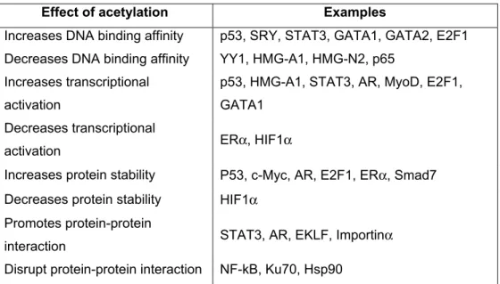
Effects of acetylation on protein function ...45
Acetylation and transcription...47
GCN5 ... 48
Yeast GCN5-containing complexes...50
Human GCN5-containing complexes ...53
Materials and methods... 57
Plasmids and siRNAs ...57
Cell culture and transfection ...58
Recombinant proteins...58
In vitro acetylation assay ...58
In vitro binding assays ...59
Antibodies ...59
Immunofuorescence ...61
Biochemical fractionation...61
Chromatin immunoprecipitation ...62
Results... 67
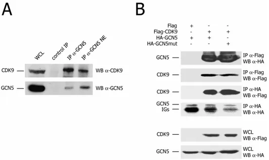
Cdk9 interacts with and is acetylated by GCN5 ... 67
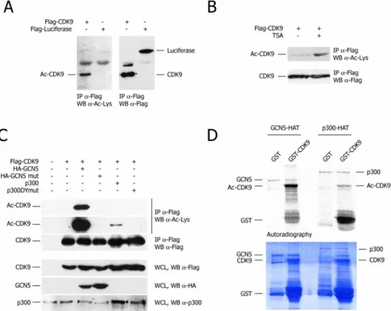
Cdk9 is acetylated by GCN5 acetyltransferase ...67
Cdk9 interacts with GCN5 ...69
Acetylation modifies crucial residues of Cdk9 ... 72
Cdk9 is acetylated at lysine 44 and 48 by GCN5 ...72
Cdk9 K44,48R has impaired catalytic activity...75
Acetylation inhibits Cdk9 kinase and transcriptional activity... 78
GCN5 mediated acetylation inhibits Cdk9 kinase activity...78
Inhibition of Cdk9 kinase activity by GCN5 impairs Cdk9 function in transcription...80
Acetylated Cdk9 accumulates at PML bodies ... 82
Anti Ac-Cdk9 specific antibody detects endogenous acetylated Cdk9 ...82
Acetylated Cdk9 localizes in speckled structures inside the nucleus. ...84
Cdk9 interacts with PML ...87
Acetylation increases Cdk9 affinity for PML bodies...93
Cdk9 localization at PML bodies is independent from acetylation...97
GCN5 interacts with PML ...99
Selective binding of acetylated Cdk9 to the HIV-1 promoter and HIV-1 genome during transcriptional latency ... 101
Discussion... 109
Acetylation modifies crucial residues of Cdk9 ... 109
Acetylation inhibits Cdk9 kinase activity... 111
Acetylation does not impair the binding of Cdk9 to its regulatory partners... 112
Acetylation re-localizes Cdk9 at PML bodies ... 115
What is the functional consequence of Cdk9 acetylation? ... 117
HIV-1 Latency ... 119
Dissection of protein-protein interactions that govern HIV-1 transcription by biophysical methods... 123
Introduction ... 123
Tat-associated factors and HIV-1 transcription...123
Fluorescence Resonance Energy Transfer (FRET) ...125
Materials and Methods... 130
Plasmids ...130
FRET ...130
Results and Discussion... 132
Cdk9 directly interacts with PML, but not with Tat ...132
A structural model of the N-terminus of CyclinT1 ...135
Insights into HIV-1 Tat:P/CAF bromodomain interaction ...139
Chapter 1
I
NTRODUCTIONTranscription Elongation
The regulation of gene expression is one of the most intensely investigated areas in all of life sciences. Differential gene expression in multicellular organisms forms the foundation of cell-type specificity. Deregulation of the appropriate pattern of gene expression has profound effects on cellular function and underlies many diseases. Although there are many cellular processes that control gene expression, the most direct regulation occurs during transcription. The transcription of protein-coding genes in eukaryotes is carried out by RNA polymerase II (RNAPII). Until recently, the vast majority of studies aimed at elucidating the molecular mechanisms of transcription regulation have focused on early stages, such as the formation of a transcription initiation complex (pre-initiation) or initiation. For several years, transcript elongation has been thought of as the trivial addition of ribonucleoside triphosphates to the growing mRNA chain, but this process is actually a dynamic and highly regulated stage of the transcription cycle, capable of coordinating downstream events. In particular, recent studies have challenged the once commonly held view that transcription is predominantly regulated at the level of RNA polymerase II (RNAPII) recruitment to the promoter and it is now clear that many genes in organisms from flies to men are regulated by reversing early blocks to RNAPII elongation (Chao and Price, 2001; Price, 2000).
Promoter escape (or promoter clearance)
Transcription starts with pre-initiation complex (PIC) assembly at the promoter. The PIC includes RNAPII and several general transcription factors (GTFs) (Orphanides et al., 1996). After melting of the double stranded DNA, transcription initiation occurs upon addition of two initiating nucleotides triphosphates and formation of the first phosphodiester bond (Goodrich and Tjian, 1994).
The first block to elongation occurs after this step: before RNAPII becomes engaged into productive transcript elongation, it needs to escape the ties that bind it to the promoter (promoter clearance). An important but poorly understood aspect of promoter clearance is that RNAPII now needs to start moving through chromatin,
rather than merely be embedded in it at the promoter (Thoma, 1991). Therefore the PIC must eventually be disassembled: a subset of GTFs remains at the promoter, serving as a scaffold for the formation of the next transcription initiation complex (Yudkovsky et al., 2000; Zawel et al., 1995). During promoter clearance, RNAPII itself must undergo structural and functional maturation in order to break its contacts with promoter-sequence elements and to tighten its grip on the nascent RNA.
The main structural change that occurs to RNAPII during early stages of transcription is the hyper-phosphorylation of the C-terminal domain (CTD) of its largest subunit, Rpb1. RNAPII CTD contains multiple repeats of the heptapeptide sequence YSPTSPS, each of which can be phosphorylated. The number of repeats increases with genomic complexity: 26 in yeast (Allison et al., 1985), 45 in Drosophila (Allison et al., 1988; Zehring et al., 1988), and 52 in mammals (Corden et al., 1985). The existence of the hypo- and hyper-phosphorylated forms of RNAPII (IIA and IIO, respectively) was first described in the early 1980s (Dahmus, 1981). Studies using functional assays together with antibodies specific to one or the other form of RNAPII demonstrated that RNAPII in the pre-initiation complex (PIC) was unphosphorylated (Laybourn and Dahmus, 1989; Lu et al., 1991), whereas transcription-competent RNAPII was heavily phosphorylated on its CTD (Christmann and Dahmus, 1981); subsequently, Ser2 and Ser5 residues were identified as the major modification sites.
The three kinases that target the RNAPII CTD are the transcriptional cyclin-dependent kinases Cdk7, Cdk8, and Cdk9 (Prelich, 2002). These enzymes are evolutionarily conserved from yeast to mammals, and all are components of protein complexes.
Cdk8 associates with the Srb/Mediator complex and functions in transcriptional events prior to elongation (Cho et al., 1998; Maldonado et al., 1996). Cdk7 is a subunit of the general transcription factor TFIIH (Orphanides et al., 1996) and phosphorylates RNAPII CTD on Ser5 immediately after the formation of the first phosphodiester bond of the nascent transcript, thus regulating RNAPII promoter clearance (Akoulitchev et al., 1995; Orphanides and Reinberg, 2002; Rodriguez et al., 2000). Cdk9, is part of the P-TEFb (positive transcription elongation factor b)
complex (Zhu et al., 1997) and phosphorylates RNAPII CTD on Ser2 after the positioning of the 5’ cap to the mRNA (Cho et al., 2001).
There are also CTD phosphatase that reverse the status of the CTD. Among them, the FCP1 phosphatase targets the CTD of RNAPII (Archambault et al., 1997; Archambault et al., 1998; Chambers and Dahmus, 1994) and participates in RNAPII recycling (Cho et al., 1999; Mandal et al., 2002).
Promoter-proximal pausing
Before becoming a fully productive elongation complex, the early elongation complex undergoes continued adjustments and this process is often accompanied by transcriptional pausing (or stalling) near the promoter. Promoter-proximal pausing is a phenomenon whereby RNAPII pauses in the 5’ region of the transcription unit and only progresses efficiently into productive elongation upon stimulation by appropriate signals. It constitutes the second block to transcription elongation and functions as a checkpoint before committing to productive elongation.
The most notable examples of genes that harbor a paused polymerase include the heat-shock-inducible genes and the mammalian proto-oncogenes MYC and FOS (Lis, 1998); this mechanism, however, is more widely exploited, since it that takes place at many genes in eukaryotes (Raschke et al., 1999) and during viral transcription (Barboric and Peterlin, 2005).
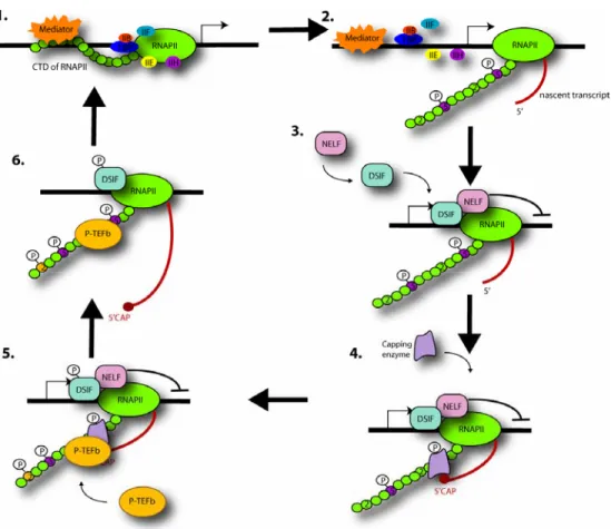
Candidate pausing factors were discovered during attempts to reconstitute in vitro transcription that displays the same sensitivity to the inhibitor 5,6-dichloro-1-β-D-ribofuranosylbenzimidazole (DRB) as seen in vivo. DRB sensitivity inducing factor (DSIF) and negative elongation factor (NELF) (Wada et al., 1998; Yamaguchi et al., 1999) were both required for the inhibition of transcription by DRB in vitro and their negative effect could be overcome by the action of the protein that was actually inhibited by DRB, namely P-TEFb.
DSIF and NELF, bind to RNAPIIA, the hypo-phosphorylated form of the polymerase, that contains Ser5P but not Ser2P (Wada et al., 1998; Yamaguchi et al., 1999) and they cooperate to inhibit the elongation rate of RNAPII by increasing the duration of time spent at paused sites (Renner et al., 2001).
The paused RNAPII is then joined by the capping enzyme that interacts with the Ser5P CTD and DSIF (Kim et al., 2002; Wada et al., 1998; Wen and Shatkin, 1999) and the nascent RNA becomes capped. Following the addition of a cap to the 5’ end of the nascent RNA, the negative effect of DSIF and NELF are relieved by the action of P-TEFb, which phosphorylates DSIF, NELF and RNAPII CTD on Ser2 (Cho et al., 2001). Once the transition to productive elongation has taken place, NELF leaves the elongation complex while DSIF remains associated and becomes a positive factor.
It is not clear how P-TEFb is recruited to the transcription complexes. P-TEFb may be recruited to RNAPII at the promoter, and its kinase activity may be inactive until the cap has been added. Indeed, the observation that transcriptional activators can interact with P-TEFb suggests that this enzyme may be recruited to the initiation complex at the promoter (Barboric et al., 2001; Kanazawa et al., 2000). Alternatively, P-TEFb may join the paused RNAPII complex after capping: the capping enzyme may assist the recruitment of Cdk9 as in S. pombe, in which the Cdk9 ortholog directly binds a subunit of the capping apparatus (Pei et al., 2003). In all likelihood, efficient elongation requires that the CTD is kept hyper-phosphorylated during the entire length of the run, but the precise mechanism of action of the kinases responsible, and indeed, the processes leading to CTD de-phosphorylation are not yet well defined. CTD de-de-phosphorylation is likely to be a continuing occurrence during elongation, as kinase inhibitors such as DRB and H8, inhibit elongation both in vivo and in vitro (Chodosh et al., 1989; Marshall and Price, 1995; Yankulov et al., 1995; Yankulov et al., 1996), and CTD kinases, such as Ctk-1 and P-TEFb, stimulate transcript elongation (Chodosh et al., 1989; Lee and Greenleaf, 1997; Marshall and Price, 1995; Yankulov et al., 1995; Yankulov et al., 1996). This indicates that the hyper-phosphorylated state of the CTD, acquired at the initiation–elongation transition, needs to be maintained, perhaps in order to keep the integrity of elongating RNAPII holoenzyme.
Figure 1. A “Checkpoint” model for the coupling of 5’ pre-mRNA capping and early
transcription initiation. Adapted from (Orphanides and Reinberg, 2002).
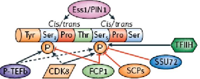
The CTD code
As transcription elongation proceeds, the RNA transcript is matured by capping and splicing, and these events, as well as the termination-coupled processes leading to mRNA polyadenylation, all happen co-transcriptionally, that is, coupled to the progress of RNAPII along the gene (Kornberg, 2001; Proudfoot et al., 2002). It is now established that the RNAPII CTD plays a central role in the ordered recruitment of protein factors involved in elongation, as well as in mRNA maturation, surveillance, and export (Hirose et al., 1999; Orphanides and Reinberg, 2002; Proudfoot et al., 2002).
The Mediator complex binds RNAPII at least partly via the CTD and enhances TFIIH-mediated CTD phosphorylation up to several hundred-fold (Kim et al., 1994).
Mediator is absent from elongation complexes containing hyper-phosphorylated RNAPII (Pokholok et al., 2002; Svejstrup et al., 1997), thus CTD phosphorylation probably induces the disruption of CTD-Mediator interactions, enabling recycling of Mediator to a new initiating polymerase (Svejstrup et al., 1997); in all likelihood, other RNAPII-GTF interactions are also broken by CTD phosphorylation (Usheva et al., 1992).
While leading to disruption of one set of interactions, CTD phosphorylation might concomitantly establish another set. The phosphorylated CTD recruits capping enzymes (Cho et al., 1997; Rodriguez et al., 2000; Yue et al., 1997) and the SR proteins involved in splicing (Greenleaf, 1993; Kim et al., 1997; Patturajan et al., 1998).
Similar to capping and splicing, transcription termination and 3’-end processing require factors that associate with the RNAPII CTD. Several components of the 3’-end processing machinery interact with the Ser2P CTD in vitro (Barilla et al., 2001; Licatalosi et al., 2002), and RNAPII has been shown to stimulate 3’-end processing (Hirose and Manley, 1998; Proudfoot et al., 2002). Moreover 3’-end processing defects were the major alterations resulting from a loss of Ser2 phosphorylation (Ahn et al., 2004; Ni et al., 2004). In addition, the Ser2 kinases Ctk1 and Cdk9 have been shown to be essential for the 3’-end processing in both S.cerevisiae (Skaar and Greenleaf, 2002) and mammals (Medlin et al., 2005; Medlin et al., 2003).
Figure 2. The CTD can be modified by phosphorylation, glycosylation, and
The CTD is a simple repetition of an heptapeptide sequence, so how does such a simple sequence interact with so many targets? A series of different phosphorylation and conformation changes generates configurations specific for binding of particular factors. In essence, the existence of a “CTD code” has been inferred, that specifies the position of RNAPII in the transcription cycle (Buratowski, 2003).
The two phosphorylations on Ser5 and Ser2 help distinguish early and late phases of transcription. Chromatin Immunoprecipitation (ChIP) data on the fate of Ser5 and Ser2 phosphorylation during transcription have indicated that Ser5 phosphorylation is high at the promoter, and then decreases towards the 3’-end of the gene, while Ser2 phosphorylation increases towards the 3’-end of the gene (Cho et al., 2001; Komarnitsky et al., 2000; O'Brien et al., 1994; Schroeder et al., 2000). Thus, it appears that CTD phosphorylation at Ser5 correlates with transcription initiation and early elongation (promoter clearance), whereas Ser2 phosphorylation is associated with RNAP II farther away from the promoter.
It is important to note that a recent report characterizing the antibodies frequently used to distinguish between Ser5 and Ser2 CTD phosphorylation revealed that the Ser2-specific antibody can recognize in some circumstances both the Ser5 and Ser2 phosphorylated forms (Jones et al., 2004). Hence it may be necessary to re-examine some previously published information, although the general trends are likely to remain valid.
In addition to phosphorylation, the CTD code probably also includes cis-trans isomerization at two prolines that follow the phosphorylated serines. The proline isomerase Pin1/Ess1 acts at prolines preceded by a phosphorylated residue and has been involved in mRNA 3’-end formation. It remains to be determined whether phosphorylation changes the equilibrium between the cis and trans form of the CTD prolines.
Just considering the possible patterns of phosphorylation and proline configurations, sixteen distinct states can be specified within a single CTD repeat. Each state is potentially a specific recognition site for an interacting factor (Buratowski, 2003).
P-TEFb
P-TEFb (positive transcription elongation factor b) was originally identified based on its ability to stimulate DRB-sensitive transcription of long transcripts in vitro (Marshall et al., 1996; Marshall and Price, 1992). The heterodimeric P-TEFb complex consists of the Cdk9 kinase that associates either with Cyclin T1, Cyclin T2a, Cyclin T2b, or Cyclin K (Peng et al., 1998; Peng et al., 1998). The elongation activity of P-TEFb is dependent on its kinase activity on RNAPII CTD and DSIF/NELF during promoter clearance (Price, 2000). P-TEFb targets specifically Ser2 of the CTD, as demonstrated by RNA interference studies in C. elegans (Shim et al., 2002) and by exploiting the highly specific P-TEFb inhibitor flavopiridol (Ni et al., 2004).
Upon heat shock, P-TEFb is rapidly recruited to transcriptionally active loci on Drosophila polytene chromosomes and frequently co-localizes with the promoter-paused hypo-phosphorylated form of RNAPII (Lis et al., 2000). These results suggest that P-TEFb is recruited to facilitate productive elongation upon RNAPII pausing. Additional experiments revealed that P-TEFb appeared to track along with RNAPII during elongation with similar kinetics (Andrulis et al., 2000; Boehm et al., 2003). Therefore P-TEFb seems to act not only during promoter escape but also during transcriptional elongation.
The P-TEFb core complex
Cdk9 is a 43 kDa protein that has been originally identified during a cDNA screening aimed at isolating novel regulators of the mammalian cell cycle (Grana et al., 1994). As no cyclin partner or cell cycle function was demonstrated at that time, Cdk9 was temporarily designated PITALRE for its PSTAIRE-like sequence, a conserved motif found in CDC2 and related kinases (Pines, 1994). The cellular function of Cdk9 remained unknown until the Drosophila homologue of human PITALRE, Cdk9, was identified as the small subunit of the P-TEFb factor by Marshall and Price (Marshall and Price, 1995).
Recently, a novel isoform of Cdk9 has been identified, which is transcribed from an alternative upstream promoter (Shore et al., 2005; Shore et al., 2003). The
resulting 55 kDa Cdk9 isoform interacts with Cyclin T1 and 7SK and is able to phosphorylate RNAPII CTD like the 43 kDa isoform.
The two isoforms of Cdk9 have a differential expression. Cdk955 is the minor form of Cdk9 in HeLa and NIH/3T3 cells, comprising less than 20% of total Cdk9. However, the relative amount of the two forms is altered in cultured human macrophages, with Cdk955 predominating. Interestingly, this ratio is altered upon
LPS (lipopolysaccharide) treatment or HIV-1 infection of the macrophages, leading to a change in the relative amounts of the two forms with Cdk942 becoming the
major form (Shore et al., 2003).
Western analysis of murine tissues has shown that the relative abundance of the two forms of Cdk9 varies across different tissues, with liver having more Cdk955 than Cdk942. During adaptation of primary rat hepatocytes to culture, the ratio of the two forms of Cdk9 changed. Initially, Cdk955 was the predominant form, but as the cells began to enter the cell cycle Cdk942 became the major form. This
suggests that Cdk942 and Cdk955 may have specific functions in dividing and
nondividing cells respectively, or that dividing cells have a higher need for P-TEFb and that induction of the Cdk942 promoter provides the extra Cdk9 required (Shore et al., 2005).Moreover the two isoforms have different localization: epitope tagged transiently expressed Cdk942 localized diffusely in the nucleoplasm, while Cdk955 accumulated in nucleolus (Liu and Herrmann, 2005).
Cdk9 has two homologs in yeast, Ctk1 and Bur1 (Yao and Prelich, 2002). The two yeast kinases likely contain distinct functional activities and may have different targets in vivo. Whereas Bur1 is essential for cell viability, Ctk1 is not (Lee and Greenleaf, 1991; Patturajan et al., 1999; Prelich and Winston, 1993; Yao et al., 2000). Even though Bur1 phosphorylates the RNAPII CTD, genetically interacts with CTD truncations, and co-localizes with elongating RNAPII, mutations in the Bur1 gene do not appear to affect either Ser2 or Ser5 phosphorylation in the cells (Keogh et al., 2003; Murray et al., 2001).
Ctk1 is responsible for elongation-associated Ser2 phosphorylation of the CTD and is localized to the coding regions of genes (Cho et al., 2001; Patturajan et al., 1999). Deletion of Ctk1 results in loss of histone H3-K36 methylation and Set2 recruitment (Krogan et al., 2003; Xiao et al., 2003), further highlighting the role of
Ctk1 in the regulation of transcript elongation. However, the fact that Ctk1 is not essential suggests that other kinases, likely Bur1, can compensate for its deficiency.
The Cyclin T1 regulatory subunit of Cdk9 was independently identified by the groups of Katherine Jones and David Price using different strategies (Peng et al., 1998; Wei et al., 1998). Subsequently, three T-type cyclins were identified in human cells and were named Cyclins T1, T2a and T2b (T-type cyclins; (Peng et al., 1998). Cyclins T2a and T2b are splice variants of a primary transcript. The two cyclins share the first 642 amino acids, but Cyclin T2b contains a larger C-terminal domain. The three T-type cyclins share a highly conserved N-terminus containing an 81% identical cyclin box. However, the C-terminus is much less conserved (46% identity) (Peng et al., 1998). In addition to the N-terminal cyclin domain, Cyclin T1 contains a putative coiled-coil motif, a His-rich motif and a carboxy-terminal PEST sequence (Wei et al., 1998). PEST sequences have been previously identified in proteins with high turnover rates, including G1 cyclins, and appear to regulate protein turnover by ubiquitin-dependent proteolysis (Rechsteiner and Rogers, 1996). However, the role of the PEST sequence present in Cyclin T1 is not well understood.
A more distantly related cyclin, Cyclin K, has also been shown to both interact and form an active complex with Cdk9 (Fu et al., 1999). Interestingly, in contrast to T-type cyclin/Cdk9 complexes, Cyclin K/Cdk9 can only activate transcription when tethered to RNA but not DNA. Cyclin K lacks an essential His-rich region in its carboxy terminus that, in Cyclin T1, helps recognizing the RNAPII CTD (Lin et al., 2002).
P-TEFb regulatory partners
In human HeLa cells, more than half of the P-TEFb heterodimer is associated with large ribonucleoprotein (RNP) complexes which also contain the 7SK small nuclear RNA (Nguyen et al., 2001; Yang et al., 2001) and the HEXIM1 or HEXIM2 protein (Byers et al., 2005; Michels et al., 2003; Yik et al., 2003; Yik et al., 2005). Actually the large complex seems to be formed by one 7SK molecule, multimers of HEXIM1 and 2 and multiple P-TEFb complexes (Dulac et al., 2005; Li et al., 2005).
Association of P-TEFb with 7SK/HEXIM1 is specific and reversible. Inhibition of cellular transcription by chemical agents or ultraviolet irradiation triggers the disruption of the P-TEFb/7SK complex (Nguyen et al., 2001; Yang et al., 2001). In contrast to its free form, the 7SK/HEXIM-associated fraction of P-TEFb shows little kinase activity, indicating that the 7SK snRNA, in collaboration with HEXIM1, functions as an inhibitory factor of P-TEFb. In HeLa cells, the overexpression of HEXIM1 inhibits the activity of a variety of reporter promoters whereas the siRNA-mediated knockdown of HEXIM1 expression results in transcriptional activation (Fraldi et al., 2005; Yik et al., 2003).
Likewise, the depletion of 7SK snRNA increases the CTD kinase activity of P-TEFb and stimulates transcription from RNAPII specific promoters, including the HIV-1 long terminal repeat (Nguyen et al., 2001; Yang et al., 2001). Thus, it is conceivable that certain cellular processes marked by an increase in global transcription are modulated by changes in the availability of free P-TEFb. An example of such a global process leading to an increase in the synthesis of total RNA is illustrated by cardiac muscle hypertrophy, which is due to increased cardiac workload or defective mechanical performance (Sano et al., 2002). Hypertrophic signals dissociate the 7SK snRNA from T-type cyclins/Cdk9 complexes, leading to an increase in Cdk9 activity that results in hyper-phosphorylation of the CTD of RNAPII (Sano et al., 2002).
Recently two groups have identified a new component of the P-TEFb active complex, the Brd4 protein. Brd4 is a mammalian bromodomain protein that binds to acetylated chromatin and interacts with P-TEFb (specifically with Cyclin T1) through its bromodomain (Jang et al., 2005). It interacts with active P-TEFb free of 7SK/HEXIM and stimulates its kinase and transcriptional activity (Jang et al., 2005; Yang et al., 2005). Moreover chromatin immunoprecipitation (ChIP) assays revealed that the recruitment of P-TEFb to promoters was dependent on Brd4, which contacts DNA through acetylated histones as well as through the Mediator complex (Jang et al., 2005; Yang et al., 2005).
P-TEFb and HIV-1 transcription
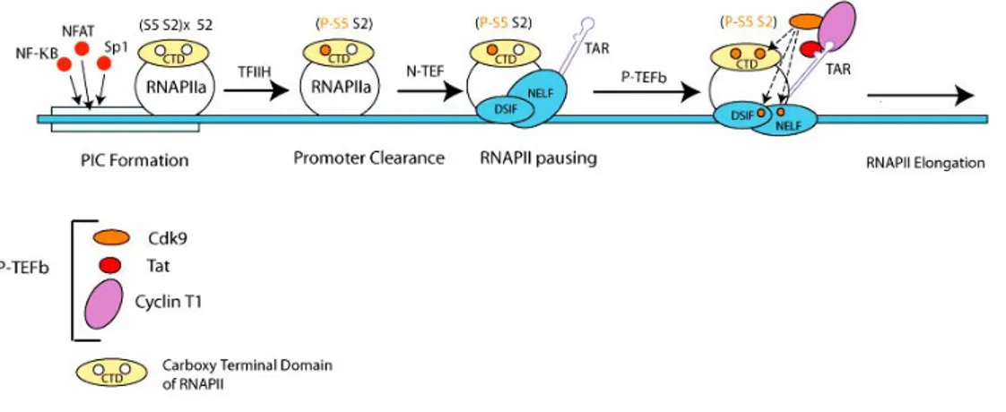
In mammalian cells, the role of P-TEFb has been initially highlighted by the association of Cyclin T1 with the human immunodeficiency virus type 1 (HIV-1) Tat transactivator (Mancebo et al., 1997; Wei et al., 1998; Zhu et al., 1997).
The HIV-1 long terminal repeat (LTR) promoter depends on P-TEFb activity more than normal cellular promoters, since activation of that promoter by the viral transactivator Tat is blocked by concentrations of P-TEFb inhibitors that do not affect regular cellular transcription (Chao et al., 2000; Chao and Price, 2001; Flores et al., 1999).
Tat is unique among transcriptional activators in eukaryotic cells in that it functions via RNA rather than DNA promoter elements. It binds the transactivation response element (TAR) that forms a stable RNA stem loop at the 5′ end of all viral transcripts. Thus, Tat requires minimally the transcription of TAR before it can stimulate HIV transcription from the long terminal repeat (LTR). Indeed, in the absence of Tat, RNAPII clears the HIV LTR successfully but soon arrests, yielding predominantly short viral transcripts (Kao et al., 1987). Tat binds the 5′ bulge in TAR via its arginine-rich motif from positions 49 to 57. However, this binding is not sufficient for Tat's function in vivo. The N-terminal core and cysteine-rich regions, which form the activation domain of the protein, lie adjacent to the arginine-rich motif. This activation domain binds Cyclin T1 (Wei et al., 1998). As a consequence, a tripartite complex is formed between Tat, Cyclin T1 and TAR at the 5’ end of each viral mRNA. The formation of the P-TEFb-TAR-Tat complex is an essential step towards the assembly of the processive RNAPII machinery at the LTR promoter (Bieniasz et al., 1998; Fujinaga et al., 1998; Garber et al., 1998; Zhou et al., 1998).
The assembly and disassembly of the complex between P-TEFb, Tat, and TAR is a regulated process in vivo. Whereas the phosphorylation of Cdk9 (Garber et al., 2000) and the P/CAF-mediated acetylation of the lysine 28 of Tat strengthens this complex, the p300-mediated acetylation of the lysine 50 of Tat weakens it (Kiernan et al., 1999).
Figure 3. Early phases of HIV-1 transcription. Adapted from (Barboric and Peterlin,
2005).
Interestingly, the HEXIM1 protein binds the same region at the N-terminus of Cyclin T1 that is responsible for Tat binding (Michels et al., 2003). As a matter of fact, GST pull-down experiments and size exclusion chromatography reveal a mutually exclusive binding of the two effectors to Cyclin T1, suggesting a model where HIV-1 Tat competes with HEXIMHIV-1 for CyclinTHIV-1 binding (Schulte et al., 2005). Furthermore, the 7SK-binding motif in HEXIM1 contains clusters of positively charged residues reminiscent of the arginine-rich RNA-binding motif found in a wide variety of proteins. Part of it is highly homologous to the TAR RNA-binding motif of Tat. A similar RNA-protein recognition mechanism may regulate the formation of both the Tat-TAR-P-TEFb and the HEXIM1-7SK-P-TEFb ternary complexes, which may help convert the inactive HEXIM1/7SK-bound P-TEFb into an active complex for Tat-activated and TAR-dependent HIV-1 transcription (Michels et al., 2004; Yik et al., 2004). Therefore it has been speculated that the TAR RNA/Tat lentiviral system has evolved to subvert the cellular 7SK RNA /HEXIM1 system.
P-TEFb and transcription
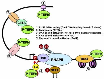
Although Tat was the first activator known to recruit P-TEFb to initiating RNAPII, additional members of this group were soon identified. They include another viral
transactivator, HTLV Tax (Zhou et al., 2006), and mammalian activators such as the androgen receptor (Lee et al., 2001), c-Myc (Eberhardy and Farnham, 2001), the class II transactivator (CIITA) (Kanazawa et al., 2000), myoblast determination protein (MyoD) (Giacinti et al., 2006), and nuclear factor κB (NF-κB) (Barboric et al., 2001). These forms of recruitment of P-TEFb are specific and tailored to individual transcription units. Nevertheless P-TEFb involvement in transcription appears to be a more general feature. In fact, in C. elegans, genetic inactivation of Cdk9 or Cyclin T1 and Cyclin T2 resulted in the inhibition of all RNAPII transcription (Shim et al., 2002). Although no murine knockouts of subunits of P-TEFb have been reported, DRB and flavopiridol, two ATP analogs that inhibit the kinase activity of Cdk9, can inhibit nearly all transcription by RNAPII in human cells (Chao and Price, 2001). Therefore there should also be a general mechanism for bringing P-TEFb to RNAPII. The association of P-TEFb with the Brd4 protein, which associates with the Mediator and binds acetylated histones, (Jang et al., 2005; Yang et al., 2005), might provide one possible general tethering system.
In any case, these observations do not rule out the possibility that specific P-TEFb complexes could be rate-limiting for the expression of a subset of genes under certain cellular instances.
Figure 4. Mechanisms of recruitment of P-TEFb to promoters (Peterlin and Price,
P-TEFb and cellular differentiation
In contrast to Cdks with known cell cycle regulatory functions, the Cdk9-associated kinase activity is fairly constant throughout both the cell cycle and during cell cycle entry from a quiescent state (Garriga et al., 2003; Grana et al., 1994), but it changes during cell differentiation (De Falco et al., 2005; Foskett et al., 2001; Sano et al., 2002; Simone et al., 2002) and viral infection (Bark-Jones et al., 2005; Liou et al., 2004; Tamrakar et al., 2005; Zhou et al., 2000).
The discovery of Cdk9 as the catalytic subunit of the Tat-Associated Kinase (Herrmann and Rice, 1993; Herrmann and Rice, 1995), the activity of which appeared to be dramatically upregulated during T-cell activation (Yang et al., 1997), prompted the examination of the regulation of P-TEFb expression in cells that are target for HIV-1 infection.
Cyclin T1 protein levels have been shown to increase following activation of peripheral blood lymphocytes (PBLs) and the upregulation of Cyclin T1 to correlate with hyper-phosphorylation of RNAPII and increased HIV replication in these cells (Garriga et al., 1998; Ghose et al., 2001; Herrmann et al., 1998; Marshall et al., 2005).
Cdk955 is expressed at relatively high levels in resting lymphocytes and is not regulated by activation, while Cdk942 is expressed at low levels in resting lymphocytes and seems to be upregulated by activation (Liu and Herrmann, 2005) In human monocytes and macrophages, Rice and colleagues have observed complex patterns of P-TEFb regulation. CyclinT1 mRNA levels are high but little protein expression can be observed in monocytes freshly isolated from healthy blood donors (Liou et al., 2002). When monocytes are instead cultured under conditions that induce macrophage differentiation, CyclinT1 protein expression is induced to high levels within one or two days. In contrast, Cdk9 protein levels are generally high in freshly isolated monocytes and are not strongly upregulated during differentiation. However, after approximately seven to ten days of macrophage differentiation in culture, CyclinT1 protein expression is shut-off by proteasome-mediated proteolysis (Liou et al., 2004). Macrophages activators such as lipopolysaccharide or other pathogen-associated molecular patterns (PAMPs) can reinduce expression of CyclinT1 after the shut-off, suggesting that induction of
CyclinT1 is a component of the innate immune response (Liou et al., 2004). Interestingly, HIV infection can also induce CyclinT1 expression in the late differentiated-macrophages (Liou et al., 2004). In this respect, however, it should be mentioned that these findings have been challenged by a study that reported no significative differences in CyclinT1 expression levels between unstimulated and stimulated primary lymphocytes (Martin-Serrano et al., 2002).
Besides cells that are target for HIV infection, there are other examples of differentiating cells in which P-TEFb activity appears to be regulated.
In the mouse, the kinase activity and protein expression of Cdk9 are highest in terminally differentiated tissues such as the muscle and brain (Bagella et al., 1998). In keeping with this notion, C2C12 cells induced to differentiate along muscle lineages peaked in Cdk9 kinase activity during differentiation. Overproduction of Cdk9 and of its associated cyclin (Cyclin T2a) stimulates myogenic differentiation in both MyoD-converted fibroblasts and C2C12 muscle cells. Conversely, inhibition of Cdk9 activity by a dominant negative form (Cdk9-dn) represses the myogenic program (Simone et al., 2002). Furthermore, Cdk9, Cyclin T2a and MyoD can be detected in a multimeric complex on promoters where Cdk9 strengthens MyoD-dependent transcription (Giacinti et al., 2006; Simone et al., 2002)
Cardiac Cdk9 kinase activity declines during normal cardiac maturation but is reactivated by hypertrophic signals. In these cases little or no change occurs in the levels of the kinase or of its activator, CyclinT. Instead, Cdk9 activation involves the dissociation of the inhibitor 7SK small nuclear RNA (Sano et al., 2002).
In contrast to this system, the Rice’s group has observed that 7SK RNA and HEXIM1 protein expression, and the association of 7SK with P-TEFb, are induced upon activation of quiescent lymphocytes, parallel, however, to the increase of Cdk9 kinase activity (Haaland et al., 2003; Haaland et al., 2005). On the other hand, increased HEXIM1 expression has been shown to correlate with inhibition of P-TEFb activity during erythroleukemia cell differentiation (Turano et al., 2006). The Cdk9/CyclinT1 complex seems to be also required for neuron differentiation induced by retinoic acid, since the expression level of the complex increases during differentiation. In addition, in samples of neuroblastoma and PNET (Primary Neuroectodermal Tumor), the levels of Cdk9 expression parallel the differentiation
state of the tumor (De Falco et al., 2005). HEXIM1 expression appears to be regulated during neuroblastoma cell differentiation as well (Turano et al., 2006). Finally P-TEFb has been found to participate in the process of adipogenesis. Cdk9, as well as CyclinT1 and CyclinT2, show differences in nuclear localization at distinct stages of adipogenesis. Overexpression of Cdk9 increases the adipogenic potential of 3T3-L1 cells, whereas the inhibition of Cdk9 by specific Cdk inhibitors and by a dominant-negative Cdk9 mutant impairs adipogenesis. The positive effects of Cdk9 on the differentiation of 3T3-L1 cells are mediated by a direct interaction with, and by the phosphorylation of, peroxisome proliferator-activated receptor gamma (PPARgamma), which is the master regulator of this process, on the promoter of PPARgamma target genes. PPARgamma-Cdk9 interaction results in increased transcriptional activity of PPARgamma and therefore increased adipogenesis (Iankova et al., 2006).
Not only cellular differentiation but also viral infection can modify P-TEFb activity. HIV-1 infection induces CyclinT1 expression (Liou et al., 2004), while HCMV infection increases Cdk7 and Cdk9 protein levels and kinase activity (Tamrakar et al., 2005). Besides regulating the overall level of kinase activity, cell infection by different viruses may also change the substrate specificity of Cdk9: in presence of HIV Tat or EBV EBNA2, Cdk9 phosphorylates RNAPII CTD on both Ser2 and Ser5 (Bark-Jones et al., 2005; Zhou et al., 2000).
Cdk9 post-translational modifications
The stability and activity of P-TEFb are regulated by multiple mechanisms.
The maturation of active CyclinT1/Cdk9 complexes is mediated by a chaperone pathway that sequentially transfers newly synthesized Cdk9 from HSP70 to HSP90/Cdc37 and finally to Cyclin T1 (O'Keeffe et al., 2000). A factor in this pathway appears to be rate-limiting for the stabilization of Cdk9, since ectopic expression of an HA-tagged version of Cdk9 leads to downregulation of endogenous Cdk9 (Garriga et al., 2003; Garriga et al., 1996). This downregulation of endogenous Cdk9 when Cdk9 is ectopically overexpressed is due to an increased rate of Cdk9 protein turnover, both endogenous and exogenous (Garriga
et al., 2003; O'Keeffe et al., 2000), suggesting that saturation of the pathway mediating stabilization of Cdk9 induces the degradation of free Cdk9.
Kiernan et al. hypothesized that Cdk9 stability could be regulated by an ubiquitin-proteasome system. In fact, they found that Cdk9 is modified by ubiquitin in an unusual way: CyclinT1 recruits the ubiquitin ligase through its PEST domain, but then it is Cdk9 that is ubiquitinated and degraded by the proteasome (Kiernan et al., 2001). While subsequent work has questioned this conclusion (Garriga et al., 2003), it is commonly accepted that Cdk9 activity can be modulated by ubiquitination. As a matter of fact, Barboric et al. have demonstrated that ubiquitination of Cdk9 by Skp2 facilitates optimal Tat transactivation because it strengthens the formation of the ternary complex between P-TEFb, Tat and TAR (Barboric et al., 2005).
Another important post-translational modification of P-TEFb is phosphorylation. Cdk9 can be phosphorylated on both serine and threonine residues. In particular, like all the other Cdks, it must be phosphorylated on Thr 186 of the T-loop for the catalytic pocket to become correctly folded (Ramanathan et al., 1999). Other sites of Cdk9 phosphorylation lie within the C-terminus of the protein and can be modified by Cdk9 itself or by other protein kinases such as PKA (Garber et al., 2000).
Evidence has been provided that Cdk9 autophosphorylation represents an important step in binding its cyclin partner and forming a stable interaction with the HIV-1 Tat/TAR complex (Fong and Zhou, 2000; Garber et al., 2000). Cdk9 phosphorylation is regulated during HIV transcription: in fact TFIIH in the pre-initiation complex inhibits Cdk9 phosphorylation, whereas Cdk9 is phosphorylated and active during transcription elongation when TFIIH is absent from the transcription complex (Zhou et al., 2001).
Subcellular localization of P-TEFb
Transcriptional regulation in mammalian cells is a highly dynamic process, requiring temporal and spatial coordination of functional protein complex assembly (Carmo-Fonseca, 2002; Stein et al., 2000). In addition, transcription overlaps extensively with downstream processes of mRNA maturation (Hirose and Manley,
2000). For these reasons, several of the factors participating in these events are found localized in specific subnuclear compartments; among them, CyclinT1 and Cdk9 co-localize in the non-nucleolar nucleoplasm with an evident speckled pattern indicative of distribution within compartments (Herrmann and Mancini, 2001). In particular, CyclinT1 is exclusively present in nuclear speckled structures while Cdk9, albeit mainly nuclear, can also be visualized in the cytoplasm where it is actively exported from the nucleus through a leptomycin B-sensitive pathway. Interestingly, enforced expression of CyclinT1 enhances nuclear localization of Cdk9 and this effect requires the catalytic activity of the kinase (Napolitano et al., 2002; Napolitano et al., 2003). Cdk9 localization also depends on the differentiation status of the cell: in fact, Cdk9 undergoes a change in subcellular localization from nucleus to cytoplasm during in vitro-induced myogenesis (MacLachlan et al., 1998).
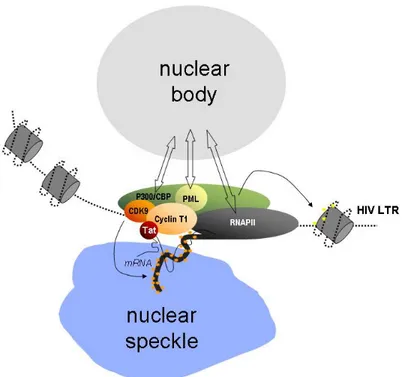
The subnuclear foci in which CyclinT1 resides appear juxtaposed, although not exactly coincident, with nuclear speckles, while they co-localize with promyelocytic leukaemia (PML) bodies, either upon overexpression of PML or, at least for a subset of them, at the levels of expression of the endogenous proteins. Localization of CyclinT1 into PML nuclear bodies depends on the physical interaction between the two proteins. In fact, CyclinT1 specifically binds PML not only in in vitro binding and co-immunoprecipitations experiments, but also in fluorescence resonance energy transfer (FRET) experiments that (as explained in chapter 5) indicate a direct interaction between the two proteins. Accordingly, CyclinT1 deletion mutants unable to associate with PML are also less prone than the wild-type protein to form nuclear foci in vivo. Most notably, human CyclinT1 has a diffuse localization in the nucleoplasm of fibroblasts derived from PML-knockout mice (Marcello et al., 2003).
The overexpression of Tat, which specifically binds CyclinT1 in FRET experiments, determines the recruitment of CyclinT1 outside the Cyclin T1 bodies and promotes HIV-1 transcriptional activation (Marcello et al., 2001), whereas CyclinT1 or PML overexpression forces CyclinT1 in nuclear bodies and correlates with an inhibition of HIV transcription (Marcello et al., 2003).
Together, these observations favor a model by which nuclear bodies modulate the activity of the HIV promoter by coordinating the availability of several factors that act in concert and are transiently part of the same complex assembled onto the LTR, including P-TEFb, p300/CBP, RNAPII and PML itself. Forced expression of PML or of other factors regulated by nuclear bodies such as CyclinT1 or PML might shift this dynamic equilibrium toward the formation of larger bodies that do not participate in transcription. In contrast, the expression of Tat might recruit these factors outside nuclear bodies in a region of nucleoplasm favorable for transcription. This model, which is schematically depicted in Figure 5, implies that nuclear body proteins shuttle in and out of these domains (Boisvert et al., 2001; Phair and Misteli, 2000), and is in agreement with the notion that transcription itself might occur at the periphery of the nuclear bodies (Boisvert et al., 2000).
PML bodies
The mammalian nucleus is a complex organelle organized into chromatin territories and discrete nuclear compartments or bodies. One of these is the promyelocytic leukemia (PML) body, also known as the PML oncogenic domain (POD), nuclear domain 10 (ND 10) or Kremer (Kr) body. There are approximately 5-30 bodies observed per nucleus, ranging in size from ~0.2 to 1 μm (Melnick and Licht, 1999). The major structural component of the PML bodies is the PML protein (Dyck et al., 1994; Koken et al., 1994; Weis et al., 1994).
The importance of PML bodies in cell differentiation and cell growth was first indicated in studies of promyelocytes from patients suffering from acute promyelocytic leukemia (APL). APL is a common form of acute myeloid leukemia (AML) that can be morphologically characterized by a distinct blockage of myeloid differentiation and accumulation of immature promyelocytes in patient bone marrow and peripheral blood (Warrell et al., 1993). In 99% of APL cases, a fusion of the PML protein and the retinoic acid receptor a (RARa) occurs as a result of the chromosomal translocation t(15:17) leading to the production of a PML-RARa chimeric protein (de The et al., 1991; Goddard et al., 1991; Kakizuka et al., 1991). Through its ability to heterodimerize with PML and RXR, PML-RARa interferes with both the PML and RAR/RXR-RA pathways, thus acting as a double dominant negative oncogenic product (Dyck et al., 1994; Kastner et al., 1992).
The role of PML in cell growth and tumorigenesis has been further characterized by overexpression and knock out of the PML protein. Stable overexpression of PML significantly reduced the growth rate of HeLa cells lengthening the G1 phase of cell cycle (Mu et al., 1997); in contrast, PML-/- MEFs grew faster than PML+/+ MEFs
(Wang et al., 1998). These findings demonstrate that PML can function in vivo as a negative growth regulator. Furthermore, PML-/- mice are highly susceptible to
develop tumors in several in vivo models of physical or chemical induced carcinogenesis (Wang et al., 1998), thus indicating that PML can act as a tumor suppressor in vivo and that PML functional inactivation may be critical in APL leukemogenesis.
Notwithstanding these observations, the following considerations should also be taken into account in an assessment of PML nuclear bodies function. First, the expression of the PML gene is not required for viability, since PML-/- mice develop, in essence, normally (Wang et al., 1998). Second, the PML gene is not evolutionarily conserved among eukaryotes, being absent in Drosophila melanogaster, Saccharomyces cerevisiae, and Arabidopsis thaliana (Borden, 2002). Third, unlike other nuclear organelles, there appears to be no PML bodies in Xenopus laevis.
PML protein
The PML protein contains at its N-terminus several important functional domains which collectively form the RBCC or TRIM motif (Jensen et al., 2001). This motif is characterized by the presence of a zinc-binding domain that includes the RING finger motif followed by two additional zinc fingers (B-boxes) and an a-helical coiled-coil motif. The RING finger is a specialized type of zinc finger that is found in factors involved in transcription, tumor suppression and genomic stability and that may confer E3 ubiquitin-protein ligase activity (Jackson et al., 2000). RING and B-box motifs are thought to be involved in protein interactions and do not appear to bind nucleic acid directly (Borden, 2000). The helical coiled-coil region consists of eight heptad repeats (Kastner et al., 1992) and is responsible for multimerization of PML (Kastner et al., 1992; Le et al., 1996) as well as its heterodimerization with PML-RARa (Grignani et al., 1996; Perez et al., 1993).
Alternative splicing generates seven major isoforms of PML, which all share the N-terminal region motif but differ in their C-N-terminal portions and subcellular distribution (Jensen et al., 2001). The majority of PML protein forms nuclear bodies (Melnick and Licht, 1999); however, there is some PML located in cytoplasmic bodies and a soluble component in the nucleus (Flenghi et al., 1995; Melnick and Licht, 1999). A partial cytoplasmic localization is a feature shared even by the PML isoforms that are predominantly nuclear (Flenghi et al., 1995; Melnick and Licht, 1999).
Post translational modifications of PML regulate PML bodies formation. In particular, SUMO-1 modification of PML appears to regulate the dynamics of
protein localization within PML nuclear bodies (Eskiw et al., 2003; Maul et al., 2000; Zhong et al., 2000). For example, although SUMO-1 modification of the PML protein is not required for the formation of PML aggregates, the accumulation of many other PML nuclear body associated proteins within these structures appears to require the sumoylation of PML (Zhong et al., 2000).
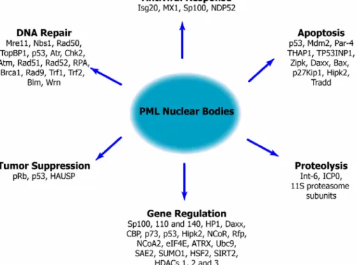
Although the PML protein is the only essential protein for PML body formation (Dyck et al., 1994), many other proteins localize to these structures. Up to date over 77 proteins are known to localize to this compartment (see the Nuclear Protein Database http://npd.hgu.mrc.ac.uk/) (Dellaire et al., 2003), implicating these structures in virtually every nuclear activity, including transcription (reviewed by (Zhong et al., 2000)), DNA repair (reviewed by (Dellaire and Bazett-Jones, 2004)), apoptosis (reviewed by (Takahashi et al., 2004)), tumor suppression (reviewed by (Salomoni and Pandolfi, 2002)), proteolysis (Lallemand-Breitenbach et al., 2001), and the antiviral response (reviewed by (Everett, 2006; Regad and Chelbi-Alix, 2001)).
Figure 6. Functions of proteins that localize at PML NBs or associate with PML
PML bodies and transcription
PML has been touted as both a transcriptional activator and repressor. Although PML itself does not exhibit DNA binding activity, it can regulate transcription through interaction with various transcription factors/cofactors (reviewed in (Zhong et al., 2000)).
PML shows intrinsic repression activity when tethered to a promoter through fusion with the Gal4 DNA binding domain (Ahn et al., 1998; Vallian et al., 1997). Consistent with this repression activity, overexpression of PML led to inhibition of various promoters, including the MDR and EGF-receptor (EGF-R) promoters (Mu et al., 1994). In particular for the EGF-R promoter, the mechanism of repression was attributed to the PML-dependent disruption of the Sp1-DNA interaction (Vallian et al., 1998). PML was also reported to directly interact with the non-phosphorylated form of Rb and to abrogate the enhancing effect of Rb on glucocorticoid receptor-mediated transactivation (Alcalay et al., 1998). Moreover, PML has been shown to interact with histone deacetylases (Wu et al., 2001) and other co-repressors (c-Ski, N-CoR, and mSin3A) (Khan et al., 2001).
On the other hand, several lines of evidence suggest that PML can also act as a transcription co-activator. PML has been shown to upregulate the transcription of genes related to the major histocompatibility complex (Zheng et al., 1998), GATA-2 (Tsuzuki et al., 2000), AP-1 (Vallian et al., 1998). The positive effect of PML on transcription can be partially explained by the fact that it can recruit transcriptional co-activators such as CBP and p300 histone acetyltransferases (Doucas et al., 1999; LaMorte et al., 1998; von Mikecz et al., 2000). In particular for p53 mediated transcription, PML is required for the appropriate formation of certain p53 post-translational modifications (phosphorylation, acetylation, sumoylation) that potentiate its activity (Fogal et al., 2000; Guo et al., 2000; Pearson et al., 2000). At the same time, it can bind and inactivate the negative p53 regulator, Mdm2, thus protecting p53 from Mdm2-mediated degradation (Kurki et al., 2003; Louria-Hayon et al., 2003; Wei et al., 2003).
Recently, the contribution to transcription of the PML nuclear body itself (and not only of the PML protein) has been addressed by artificially tethering reporter plasmids to PML bodies (Block et al., 2006). In these conditions, the PML nuclear
body environment has been shown to modulate the expression of the reporter gene in a promoter-dependent manner (the SV40 promoter was repressed, the CMV promoter was activated and a minimal eukaryotic promoter was not affected).
PML bodies and chromatin
The structure of PML nuclear bodies has been extensively studied using both the light and electron microscope to examine their protein composition and ultrastructure (Boisvert et al., 2000; Eskiw et al., 2003; Grande et al., 1996; Ishov et al., 1999). The bodies are spherical or ring-shaped (Boisvert et al., 2000; Eskiw et al., 2003) and often appear as spheroid shells of proteins, with little protein mass at their core (Boisvert et al., 2000). Like many nuclear bodies, PML nuclear bodies reside in the interchromatin domain (ICD) space (Boisvert et al., 2000).
While LaMorte et al. reported the existence of nascent RNAPII transcripts within this compartment (LaMorte et al., 1998), subsequent analysis demonstrated that the core of PML nuclear body is a dense protein-based structure which does not contain detectable nucleic acid (Boisvert et al., 2000). Although neither chromatin nor RNA are found within the central core of these bodies, newly synthesized RNA is associated with their periphery (Boisvert et al., 2000) and both proteins and chromatin-like fibers appear to connect the bodies via multiple contacts to the surrounding chromatin (Eskiw et al., 2004; Eskiw et al., 2003). These contacts with chromatin at the periphery of the PML nuclear bodies seem to be partly responsible for the positional stability over long time intervals during interphase (Eskiw et al., 2004; Eskiw et al., 2003).
The integrity and stability of PML bodies are lost when chromatin is disrupted by stress, transcriptional repression or early apoptotic events. Under such conditions, PML bodies become mobile (Eskiw et al., 2004; Eskiw et al., 2003; Maul et al., 1995; Nefkens et al., 2003). Contacts between chromatin and PML bodies could have a more profound role than just the maintenance of their positional stability. One hypothesis is that the disruption of PML bodies might directly affect the availability of PML body-associated proteins within the nucleus leading to the release of these proteins. It is equally possible that the association of PML bodies
with chromatin also brings together specific gene loci regulated by PML body-associated proteins.
Figure 7. Ultrastructure of PML nuclear bodies (Dellaire and Bazett-Jones, 2004).
(A) An electron micrograph of a PML NB is shown by electron spectroscopic imaging (ESI). Nucleic acid is visualized in the net phosphorous image (P) and both nucleic acid and protein in the net nitrogen image (N). Overlay of the two images produces an image where protein-rich structures, such as the PML NB, appear green and structures with intermediate protein and nitrogen content, such as chromatin and ribonucleoprotein (RNP) complexes, appear yellow. In the segmented panel, the ratio of nitrogen and phosphorous is used to segment the image into chromatin (red), RNP (blue) and the PML body (green). The PML body has a ring-like appearance with a hole at the centre of the PML NB. In addition, a finger-like projection from the top of the body can be seen. The PML NB is in contact with the surrounding chromatin at several locations (small arrows). (B) A three-dimensional volume representation of the PML NB in A. (C) A cross-section
through the three-dimensional volume of the PML NB, along the plane shown in the back panel of B (white line), is shown.
Many examples of the non-random association of PML nuclear bodies in mammalian cells with specific chromosomal loci or chromatin have been reported, which include TP53 (Sun et al., 2003) and the MHC gene cluster (Shiels et al., 2001). Wang et al. have extended the analysis to multiple gene-rich and gene-poor regions on other chromosomes in order to determine whether there is a spatial organization of PML bodies relative to particular regions of the genome. These authors found that PML bodies associate with genomic regions of high transcriptional activity, which is a function of both the gene density and the proportion of genes that are active. Single genes that are highly expressed do not show significant associations with PML bodies alone. The organization of PML bodies in relation to transcriptionally active regions was further supported by the association of PML bodies with many genes on the active X chromosome compared with their homologues on the inactive X chromosome, and with replication-dependent histone genes on chromosome 6 in S-phase cells compared with those in G0/G1 phase cells (Wang et al., 2004).
PML bodies thus seem to associate with chromosomal regions of high transcriptional activity and/or gene density, and, in some cases, the association may be cell cycle-dependent. Yet, not all transcriptionally active regions of genome associate with PML bodies and PML bodies do not serve as obligate transcription sites for the associated genes that were tested, nor are basal transcription levels of these genes altered by knockdown of PML protein. Collectively, these observations suggest that the specificity of PML body association is determined by locus-specific factors rather then by the transcriptional level (Ching et al., 2005; Wang et al., 2004).
The genes brought into the vicinity of the PML body might then be jointly regulated through clustered co-activators such as CBP/p300, and co-repressors, such the histone deacetylases (HDACs). Such a role for PML bodies is reminiscent of that proposed for matrix attachment regions (MARs) in DNA (Cockerill and Garrard, 1986). Furthermore, Kiesslich et al. (Kiesslich et al., 2002) found that, in unsynchronized mammalian cells, approximately 30% of PML bodies were spatially
associated with transcription foci, marked by the presence of hyper-phosphorylated RNA polymerase II and fluorouridine-labeled newly synthesized RNAs; this percentage rose to 70% when cells were synchronized in G1.
These findings may suggest, for the PML bodies, a role analogous or coincident with the so-called “transcription factories”. These factories are discrete sites within the nucleus where RNA transcription occurs; growing evidence indicates that when a gene becomes active, its DNA moves to one of those factories. Genes on different chromosome whose proteins work together travel long distances within the nucleus to meet up in the same factories (Pennisi, 2006).
On the other hand, several observations are also in sharp contrast with this conclusion. Knockdown experiments of PML bodies by RNA interference showed no effect on the expression of genes non-randomly associated with PML bodies (Wang et al., 2004). Experiments in the Ana Pombo laboratory have demonstrated by high-resolution imaging of ultrathin cryosections that PML bodies contain no detectable RNAPII or nascent RNA in HeLa cells, but are often surrounded by these markers at a distance >25 nm (Xie and Pombo, 2006). This observation supports the view that, although PML bodies are present in transcriptionally active areas of the nucleus, they are not themselves sites of polymerase assembly, transport or activity. One possible explanation for the difference between these results and those of Kiesslich et al. (Kiesslich et al., 2002) is that in vivo labeling of newly made RNA with fluorouridine allows for some transcripts to be completed and move away from the sites of synthesis towards PML bodies. Alternatively, the insufficient resolution provided by confocal microscopy of whole cells may give the false impression that PML bodies are transcriptionally active. Thus, the association of active genes with PML bodies, although nonrandom, can be explained by the coincidental positioning of PML bodies, active genes and polymerases in regions of the nucleus that are transcriptionally active (Wang et al., 2004). Proposed functions of PML bodies would agree with such preferential localization as they include storage of proteins, such as many transcription factors and chromatin modifiers, post-translational modification, like acetylation, phosphorylation and SUMOylation, and proteosomal-dependent degradation.
These results do not exclude the possibility that PML proteins that are freely available throughout the nucleoplasm (i.e. not in PML bodies) interact with RNAPII or regulate gene expression.
Not only human genes, but also viral genomes associate with PML bodies. The genomes and/or replication complexes of HSV-1 and adenovirus were preferentially located in close association with PML bodies (Ishov and Maul, 1996; Maul et al., 1996). This observation has been extended to include the papovaviruses SV40 and polyomavirus, and members of all subfamilies of the herpesviridae (for reviews see (Everett, 2001; Maul, 1998)). More recently, similar observations were made in cells infected with the parvovirus AAV (Fraefel et al., 2004).
PML bodies not only are often associated with viral genomes but also facilitate their transcription and replication (Ching et al., 2005), again supporting the hypothesis that these bodies are sites of gene activity. Regarding HIV-1, the group of G. Maul approached the localization of proviral 1 DNA and transcribed HIV-1 mRNAs with respect to speckles and PML bodies in the nucleus of infected cells (Bell et al., 2001). To distinguish between unintegrated and integrated viral DNA, these investigators employed a technique called stress-induced chromosome condensation (SICC), which allows the visualization of the interchromatin space. Surprisingly, unintegrated HIV-1 DNA was found to accumulate within nuclear speckles, while no specific association of either unintegrated viral DNA, or transcription foci, or integrated proviral DNA could be detected with PML nuclear bodies. However, given the fact that these experiments were conducted with a FISH probe covering the whole HIV genome, it should be considered that multiply spliced subgenomic mRNAs might have escaped detection by this method. These considerations still leave the question of what are the sites in the nucleus at which HIV transcription occurs with respect to the known nuclear territories, largely unanswered.
Finally, it is of interest to note that, since PML bodies could also play a role in the mechanism of antiviral action of interferon, viruses have also evolved different ways to alter PML expression and/or localization (for review see (Everett, 2006; Regad and Chelbi-Alix, 2001)).
Function of PML bodies
All the above reported observations on the properties of PML bodies may collectively lead to three possible models to explain their actual function.
In the first model, the bodies are proposed to be aggregations of excess nucleoplasmic protein (Negorev and Maul, 2001): for example, they can sequester and inactivate co-activators, co-repressors or transcription factors (Li and Chen, 2000; Zhong et al., 2000). By titrating these factors from the active pool in the nucleoplasm, the PODs could interfere with transcription, resulting in either activation or repression.
In the second model, PML bodies are proposed to be sites of post-translational modification and degradation of proteins. Observations supporting this model include the acetylation (Pearson et al., 2000), phosphorylation (D'Orazi et al., 2002) and sumoylation (Fogal et al., 2000; Kwek et al., 2001) of p53 at PML bodies and the localization of ubiquitin/proteasome associated proteins at some PML bodies (Anton et al., 1999; Everett et al., 1997; Lafarga et al., 2002; Lallemand-Breitenbach et al., 2001).
In the third model, PML bodies are proposed to be sites of specific nuclear activities, such as transcriptional regulation and DNA replication. Evidence for this model includes the detection of nascent RNA around PML bodies (Boisvert et al., 2000), the association of PML bodies with regions of high transcriptional activity (Wang et al., 2004), and the non-random nature of PML body assembly (based on the conservation of their size and position) following dissociation and re-formation as a result of cellular stress (Eskiw et al., 2003).
Acetylation
Post-translational modification represents an important mechanism for regulating protein function. Lysine acetylation, or the transfer of an acetyl group from acetyl coenzyme A to the ε-amino group of a lysine residue, was initially discovered on histone proteins about four decades ago (Vidali et al., 1968). Intensive research in the past decade has shown that this modification is quite common and plays important roles in regulating the functioning of eukaryotic, viral and bacterial proteins (Kouzarides, 2000; Sterner and Berger, 2000). Importantly, a protein module termed bromodomain has been shown to possess specific acetyllysine-recognizing ability (Zeng and Zhou, 2002).
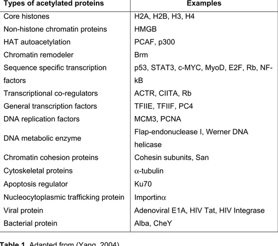
Types of acetylated proteins Examples
Core histones H2A, H2B, H3, H4
Non-histone chromatin proteins HMGB
HAT autoacetylation PCAF, p300
Chromatin remodeler Brm
Sequence specific transcription factors
p53, STAT3, c-MYC, MyoD, E2F, Rb, NF-kB
Transcriptional co-regulators ACTR, CIITA, Rb General transcription factors TFIIE, TFIIF, PC4 DNA replication factors MCM3, PCNA
DNA metabolic enzyme Flap-endonuclease I, Werner DNA helicase
Chromatin cohesion proteins Cohesin subunits, San Cytoskeletal proteins a-tubulin
Apoptosis regulator Ku70
Nucleocytoplasmic trafficking protein Importina
Viral protein Adenoviral E1A, HIV Tat, HIV Integrase
Bacterial protein Alba, CheY
HATs
Lysine acetylation is a reversible post-translational modification that is governed by the opposing action of acetyltransferases and deacetylases (Grozinger and Schreiber, 2002; Yang, 2004).
Since 1995 when the first acetyltransferase was discovered in yeast (Kleff et al., 1995), dozen of proteins have been discovered to possess intrinsic lysine acetyltransferase activity. Although most of these enzymes were first identified as histone acetyltransferases (HAT) and then tested for activities towards other proteins, acetyltransferases only modifying non-histone proteins have also been identified. According to sequence similarity, lysine acetyltransferases can be organized into different groups.
Figure 8. Schematic illustration of the Gcn5/PCAF (A) and p300/CBP (B) families
of HATs (Yang, 2004).
The Gcn5/PCAF family is composed of GCN5, PCAF and related proteins. Yeast Gcn5 possesses a HAT domain and a bromodomain and is highly homologous to the C-terminal halves of human PCAF and GCN5L (mammalian GCN5 long form)
(Georgakopoulos and Thireos, 1992; Smith et al., 1998; Wang et al., 1997; Xu et al., 1998). Numerous studies indicate that these HATs function as histone acetylating transcriptional co-activators but also that they can acetylate non-histone proteins as well (Yang, 2004).
The p300/CBP family is another major group of nuclear HATs that has been extensively characterized (Chan and La Thangue, 2001; Goodman and Smolik, 2000). Like PCAF, both p300 and CBP are transcriptional co-activators able to acetylate histones and non-histone proteins. Reminiscent of PCAF and GCN5L, p300 and CBP form a pair of homologous HATs in mammals.
The MYST family of proteins constitutes a third major group of nuclear HATs. The acronym MYST is from its four founding members: human MOZ (monocytic leukemia zinc finger protein), yeast Ybf2, yeast Sas2 and the mammalian Tip60. Compared with the GCN5/PCAF and p300/CBP groups, the MYST family is larger, more diverse and not so well characterized. Despite their similar HAT domains, MYST proteins play different roles in various cellular processes (Yang, 2004). In addition to these three major groups of HATs, more than a dozen other proteins have been shown to possess acetyltransferase activity.
Most HATs exist as stoichiometric multisubunit complexes in vivo. The complexes are typically more active than their respective catalytic subunits and display distinct substrate specificities, suggesting that associated subunits regulate the activities of the respective catalytic subunits. In addition, non-catalytic subunits are also involved in recruiting substrates for targeted action to ensure the specificity. Amazingly, one HAT can be the catalytic subunit of multiple complexes. GCN5L forms at least two distinct multisubunit complexes, and yeast GCN5 is the catalytic subunit of four complexes (reviewed in (Carrozza et al., 2003)).
Regulation of HAT activity
Multiple mechanisms are involved in the control of HAT activity (see (Yang, 2004) and references therein).
First, as an essential cofactor for different acetyltransferases, acetyl-CoA also stabilizes GCN5 and PCAF.
Second, as described above, formation of stoichiometric multisubunit complexes modulates the specific activities and substrate specificities of different HATs. Third, the enzymatic activities of PCAF, p300 and CBP are regulated by interaction with transcription factors and viral proteins.
Fourth, HATs are subject to covalent modifications such as phosphorylation, acetylation, ubiquitination and sumoylation.
Fifth, HATs are degradated by caspases, calpains and ubiquitin-dependent proteasomes.
Sixth, subcellular compartmentalization is an important regulatory mechanism for HATs. For example, HAT1 binds to 14-3-3 proteins and TIP60 is sequestered to the cytoplasm in a signal-dependent manner.
Finally, while p300, CBP, MOZ and MORF possess PHD fingers, yeast Esa1 and Sas3 associate with PHD finger-containing subunits. PHD fingers are implicated in phosphoinositide binding and may thus provide structural modules for integrating nuclear lipid signals, so that activities of these acetyltransferases may be regulated by nuclear signaling events.
HDACs
Two families of deacetylating enzymes have been identified in eukaryotes: the histone deacetylases, or HDACs, and the Sir2 (silent information regulator-)-like family of NAD-dependent deacetylases, or sirtuins. Both families have been evolutionary conserved from prokaryotes to humans, and both consist of several different proteins with non-redundant cellular functions, many of which involve transcriptional regulation.
The HDAC family members can be divided into two classes based on their similarity to yeast histone deacetylase Rpd3 (class I) or Hda1 (class II) (Grozinger et al., 1999). Four class I (HDAC 1, 2, 3 and 8) and five class II (HDAC 4, 5, 6, 7, 9 and 10) HDACs have been identified and partially characterized in humans (Gray and Ekstrom, 2001; Guardiola and Yao, 2002; Zhou et al., 2001). A third group of HDACs has been formed after the discovery of HDAC 11, which contains conserved residues in the catalytic core shared by both class I and class II HDACs