NADPH oxidase activation and 4-hydroxy-2-nonenal/aquaporin-4
adducts as possible new players in oxidative neuronal damage
presents in drug-resistant epilepsy
A. Pecorelli
a,b,1, F. Natrella
c,1, G. Belmonte
c, C. Miracco
c, F. Cervellati
d, L. Ciccoli
a, A. Mariottini
c, R. Rocchi
c,
G. Vatti
c, A. Bua
c, R. Canitano
b, J. Hayek
b, H.J. Forman
e,f, G. Valacchi
d,⁎
a
Department of Molecular and Developmental Medicine, University of Siena, Via A. Moro 2, 53100 Siena, Italy
b
Child Neuropsychiatry Unit, University Hospital, Azienda Ospedaliera Universitaria Senese (AOUS), Viale M. Bracci 16, 53100 Siena, Italy
c
Department of Medicine, Surgery and Neuroscience, University of Siena, Viale M. Bracci 16, 53100 Siena, Italy
dDepartment of Life Science and Biotechnologies, University of Ferrara, Via L. Borsari 46, 44121 Ferrara, Italy e
Life and Environmental Sciences Unit, University of California, 5200 North Lake Road, Merced, CA 95344, USA
f
Andrus Gerontology Center of the Davis School of Gerontology, University of Southern, 3715 McClintock Ave., Los Angeles, CA 90089-0191, USA
a b s t r a c t
a r t i c l e i n f o
Article history: Received 8 August 2014
Received in revised form 27 October 2014 Accepted 17 November 2014
Available online 22 November 2014
Keywords: 4-Hydroxy-2-nonenal Matrix metalloproteinase NADPH oxidase Aquaporin-4 Drug-resistant epilepsy
A correlation between epilepsy and cellular redox imbalance has been suggested, although the mechanism by which oxidative stress (OS) can be implicated in this disorder is not clear. In the present study several oxidative stress markers and enzymes involved in OS have been determined. In particular, we examined the levels of 4-hydroxy-2-nonenal protein adducts (HNE-PA), a by-product of lipid peroxidation, and the activation of NADPH oxidase 2 (NOX2), as cellular source of superoxide (O2−), in surgically resected epileptic tissue from
drug-resistant patients (N = 50). In addition, we investigated whether oxidative-mediated protein damage can affect aquaporin-4 (AQP4), a water channel implicated in brain excitability and epilepsy. Results showed high levels of HNE-PA in epileptic hippocampus, in both neurons and glial cells and cytoplasmic positivity for p47phoxand p67phoxsuggesting NOX2 activation. Interestingly, in epileptic tissue immunohistochemical
localiza-tion of AQP4 was identified not only in perivascular astrocytic endfeet, but also in neurons. Nevertheless, nega-tivity for AQP4 was observed in neurons in degeneration. Of note, HNE-mediated post-translational modifications of AQP4 were increased in epileptic tissues and double immunofluorescence clearly demonstrated co-localization of AQP4 and HNE-PA in epileptic hippocampal structures. The idea is that sudden, disorderly, and excessive neuronal discharges activates NOX2 with O2−production, leading to lipid peroxidation. The resulting
generation of HNE targets AQP4, affecting water and ion balance. Therefore, we suggest that seizure induces ox-idative damage as well as neuronal loss, thereby promoting neuronal hyperexcitability, also affecting water and ion balance by AQP4 modulation, and thus generating a vicious cycle.
© 2014 Elsevier B.V. All rights reserved.
1. Introduction
Epilepsy, a brain disorder characterized by recurrent, unprovoked and unpredictable occurrence of seizures, is one of the most common serious neurological disorders, affecting more than 50 million people worldwide. As is well known, impaired redox status constitutes a com-mon mechanism of injury for many neurodegenerative disorders[1]. Oxidative stress (OS) occurs when antioxidant defense against oxidant production is in imbalance towards oxidation of cellular targets[2].
Due to the low regenerative capacity of neurons, high metabolic rate, limited antioxidant capacity, high oxygen consumption, and elevated concentrations of polyunsaturated fatty acids (PUFA) and iron, the brain is particularly vulnerable to OS[3]. Emerging works indicate an in-volvement of redox imbalance in epileptogenesis[4]. Increased oxidant generation has been demonstrated to be induced in epilepsy either by recurrent seizures, as well as, by mitochondrial dysfunction[5]with high levels of OS biomarkers and low antioxidant defenses present in epileptic subjects[6]. Furthermore, treatment with the antioxidants vi-tamin E[7]and vitamin C[8], and the component of glutathione perox-idase, selenium[9], demonstrated protective effects on the oxidative-induced injury in epilepsy. However, whether OS is a causative factor or rather a consequence (or both) in mechanisms involved in seizures is not completely clear[10].
⁎ Corresponding author at: Department of Life Sciences and Biotechnology, University of Ferrara, Via Luigi Borsari, 46, 44121 Ferrara, Italy. Tel.: +39 0532 455 482.
E-mail address:[email protected](G. Valacchi).
1
These authors contributed equally to this work.
http://dx.doi.org/10.1016/j.bbadis.2014.11.016
0925-4439/© 2014 Elsevier B.V. All rights reserved.
Contents lists available atScienceDirect
Biochimica et Biophysica Acta
j o u r n a l h o m e p a g e : w w w . e l s e v i e r . c o m / l o c a t e / b b a d i sAlthough mitochondria are generally thought to be the main source of OS, a significant involvement for nicotinamide adenine dinucleotide phosphate (NADPH) oxidase (NOX) in O2•−and H2O2production
appear evident in brain[11]. NOX2, which catalyzes the reduction of molecular oxygen to form O2•−, is a multi-subunit enzyme composed
of the cytosolic proteins, p40phox, p47phox, and p67phox, and at least
two membrane proteins, including gp91phoxand p22phox. The assembly
of the NOX2 complex is regulated by p47phoxphosphorylation[12,13].
In the brain, normal NOX function appears to be required for processes such as neuronal signaling and memory, but overproduction of oxidants contributes to neurotoxicity and neurodegeneration[14]. Literature data indicate clearly a NOX-induced O2•−and H2O2 production in
several epilepsy models (animal and in vitro)[15–21], but as of today, because of difficulty in recruiting the samples, little has been done in humans.
The production of O2•−and H2O2, the latter mostly through
superox-ide dismutase catalyzed dismutation of O2•−to H2O2and O2, can
con-tribute to functional molecular impairment and cellular damage, either directly or through iron-initiated (Fenton chemistry) lipid perox-idation, which occurs in brain due to its high content in PUFA and relatively low antioxidant capacity[22]. Lipid peroxidation alters mem-brane structure, affecting itsfluidity and permeability and the activity of membrane-bound proteins, and produces many cytotoxic and reactive by-products. Among these, the well known 4-hydroxy-2-nonenal (HNE) is able to form adducts with biomolecules, including proteins, lipids and nucleic acids, thereby propagating oxidative damage[23, 24]. HNE-mediated damage to proteins is a known oxidative post-translational modification (PTM) that leads to functional changes or deactivation of enzymes, transporters, ion channels and receptors[25]. Furthermore, the accumulation of HNE protein adducts (HNE-PA) occurs in various pathological conditions, including neurological diseases, where it contributes to cell death and neurodegeneration
[25,26]. A recent study investigated the generation and potential role of protein nitration in epileptogenesis[27], another oxidative PTM of proteins (noted as 3-nitrotyrosine) a common biomarker of disease. In addition, there is only one work conducted on an epilepsy animal model, i.e. the kainate-treated rat, which has examined the time course and distribution of HNE-PA in the damaged hippocampus after kainate injections[28]. Nevertheless, to our knowledge, there is no published information regarding HNE-PA levels and localization in epileptic human tissues.
Although oxidative generation in cells wasfirst described as contrib-uting to damage, it is now well-established that low level production of H2O2participates as a second messenger in gene expression and several
signaling pathways[29]. Oxidant generation associated with damage, can also participate in signaling, although it may be deregulated. It is also well known that oxidative stimuli can modulate the expression/ activity of extracellular matrix enzymes, including matrix metallopro-teinases (MMPs)[30]. MMPs have been shown to play an important role also in neuropathological processes and neurodegenerative disor-ders[31]. A link between MMPs activity and epilepsy has been re-peatedly reported both in humans and in animal models[32].
Aquaporin-4 (AQP4) is a member of a family of bidirectional, high-capacity water channels. In the brain, AQP4 is primarily expressed in the astrocyte endfeet membranes adjacent to blood vessels and plays a key role in brain water homeostasis. Emerging evidences demonstrat-ed dysregulation of water and ions homeostasis in patients with mesial temporal lobe epilepsy (MTLE)[33]. Moreover, expression and subcel-lular localization of AQP4 have been shown recently to be altered in sclerotic hippocampus obtained from patients with mesial temporal sclerosis[34,35].
Based on these data, the main goal of this study was to elucidate the presence and the possible source of the redox imbalance in surgically resected epileptic tissues from drug-resistant patients and to investigate whether oxidative imbalance could affect AQP4 expression and/or function.
2. Materials and methods 2.1. Patients and controls
Fifty patients operated on for drug-resistant epilepsy between 2003 and 2013 at the University Hospital of“Santa Maria alle Scotte” of Siena by a same neurosurgeon (A.M.) were included in our study. Main pa-tient data and clinicalfindings are reported inTable 1.
Presurgical evaluation included careful analyses of seizure semeiology, continuous scalp video-EEG monitoring using noninvasive or invasive (subdural grids) methods, anatomical neuroimaging (MRI) and functional neuroimaging (SPECT, functional MRI), and neuropsy-chological assessment.
All patients of this series were submitted to a two-step anterior tem-poral lobectomy with the temtem-poral neocortex removedfirst, followed by resection of the deep structures (uncus, amygdala, hippocampus and paraippocampus gyrus). In tumoral cases (epilepsy-associated tumors), the gross-total neoplastic removal was part of the wider antero-mesial temporal resection.
The main portion of the resected tissues was submitted for patholog-ical evaluation. Samples of temporal neocortex and, in some cases, of sclerotic hippocampus (approximately 1 cm3) were separated from each resected tissue and immediately snap-frozen by immersion in liq-uid nitrogen and stored at−80 °C until analysis as above.
The temporal neocortex offive non-epileptic patients, suffering from other pathologies (a case of cavernous hemangioma, two melanoma me-tastases and a case of glioblastoma) was used as control sample. In all cases an“in block” resection has been performed, and control samples were taken from areas distant from the lesion sites, which were normal at histopathological evaluation. In addition, non-sclerotic hippocampal tissues of epileptic patients were compared to hippocampal sclerotic tis-sues of the other epileptic patients in immunohistochemistry assay.
This study was approved by the Ethics Committee of the Hospital of Siena, and informed consent was acquired from enrolled subjects.
It should be noted that due to the difficulty in obtaining fresh and relatively healthy brain tissue, only 5 cortical control samples were obtained to be compared with 50 cortical biopsies from patients with drug-resistant epilepsy. Moreover, as there are very limited non-epileptic indications allowing removal of temporal mesial structures (hippocampus, amygdala), relative differences between hippocampus and neocortex could only be made within the epilepsy group. Nonethe-less, as the hippocampus was involved in the pathology of 70% of the ep-ilepsy patients, it was important to evaluate the hippocampus despite this limitation. Then, comparisons were made between the group of ep-ileptic patients with healthy hippocampus (which served as control) and patients with hippocampal sclerosis (HS).
2.2. Samples
Epileptic surgical samples for histopathological examination always included temporal neocortex, amygdale, uncus, and hippocampus. Di-agnoses (seeTable 1) were performed according the ILAE classifications
[36,37]. Hippocampal sclerosis (HS) was diagnosed in 34 patients, and probable HS (only CA1 and CA4 regions were evaluable) in one patient. HS was subclassified according to Blümcke et al.[37]on the bases of the site of neuronal cell loss in the Cornus Ammonis (CA): type I, in CA1 and CA4; type II, in CA1; type III, in CA4. There were 18 focal cortical dyspla-sia (FCD), subclassified according to the new ILAE consensus classifica-tion system[36].
2.3. Western blotting
Brain samples (temporal neocortex from non-epileptic and epileptic patients, and epileptic sclerotic hippocampus) were lysed in RIPA buffer containing protease inhibitors and equal amounts of proteins (40μg) were separated by SDS–PAGE on a 10% gel and transferred to
nitrocellulose membranes. The Western blotting procedure was per-formed as previously described[38]using the following antibodies: anti-4-hydroxynonenal (Millipore Corporation, Billerica, MA, USA), anti-aquaporin 4 (Millipore Corporation and Proteintech Group, Inc., Manchester, United Kingdom) andβ-actin (Millipore Corporation) as the loading control. The bands were visualized by autoradiography. Quantification of the bands was performed by digitally scanning the Amersham Hyperfilm™ ECL and by measuring immunoblotting image densities with ImageJ software.
2.4. Immunoprecipitation
For immunoprecipitation, 1 mg of brain tissue protein (temporal neocortex from non-epileptic and epileptic patients, and epileptic scle-rotic hippocampus) was incubated with 3 μg of anti aquaporin 4
antibody (Proteintech Group, Inc.) overnight at 4 °C on a rotator. Then, the immune complex was incubated with 25μl of Protein A/G agarose beads (Thermo Fisher Scientific Inc., Waltham, MA, USA) and rotated at RT for 2 h. Samples were briefly centrifuged and washed six times with ice-cold RIPA buffer. The pellet was mixed with reducing sample buffer, boiled and loaded on a 15% SDS-PAGE gel for immunoblotting with anti-4-hydroxynonenal antibody (Millipore Corporation), as de-scribed in previous section. Samples processed with normal rabbit IgG (Sigma-Aldrich S.r.l., Milan, Italy), instead of appropriate antibody, were used as negative control.
2.5. Gelatin zymography
As previously described[39], tissues lysates (15μg of protein from temporal neocortex from non-epileptic and epileptic patients, and
Table 1
Main demographic and clinicalfindings of epileptic drug-resistant patients included in the study. Patient Age at surgery
(years)/sex Age at seizure onset Type of epilepsy Lobe Engel's class⁎
MRIfindings Type of surgery Histopathological classification
#1 57/M 6 PC R TL Ia MTS R T_Lobect. HS 2
#2 46/M 19 PC R TL Ia MTS R T_Lobect. HS 1
#3 55/F 51 PC L TL Ia Cripto L T_Lobect. FCD Ia
#4 35/F 34 PC R TL Ia Cripto R T_Lobect. HS 1
#5 29/M 7 PC/Sotos R TL Ia MTS R T_Lobect. FCDIIIa(FCDIb + HS 1)
#6 28/F 18 PC L TL II MTS+FCD L T_Lobect. FCDIIIa(FCDIb + HS 1)
#7 19/F 19 P+GTC L TL Ia NDMTA +Tum L T_Lobect. + Lesionect. GG
#8 18/F 18 PC L TL Ia NDMTA +Tum L T_Lobect. + Lesionect. GG
#9 43/M 12 PC R TL Ia MTS R T_Lobect. HS 1
#10 36/M 8 PC R TL Ia MTS R T_Lobect. HS 1
#11 34/F 33 PC L TL III NDMTA +Tum L T_Lobect. + Lesionect. GG
#12 13/F 3 PC L TL Ia MTS L T_Lobect. FCDIIIa(FCDIIa + HS 1) #13 18/M 18 PC L TL Ia MTS L T_Lobect. HS1 #14 28/M 9 PC R TL Ia FCD R T_Lobect. FCD Iia #15 35/F 8 PC R TL Ia MTS R T_Lobect. HS 1 #16 39/F 7 PC L TL II MTS L T_Lobect. HS 1 #17 5/M 1 PC L TL Ia FCD L T_Lobect. FCDIIIb(FCDIb + GG) #18 25/F 19 PC L TL Ia MTS L T_Lobect. FCDIIIa(FCDIb+HS1) #19 28/F 13 PC L TL Ia FCD L T_Lobect. FCDIIIc(FCDIb+VM)
#20 39/M 17 PC L TL Ia DNET L T_Lobect. DNET
#21 41/F 13 PC R TL Ia MTS L T_Lobect. HS 1
#22 37/M 15 PC R TL Ia MTS R T_Lobect. HS 1
#23 56/M 20 PC R TL II MTS R T_Lobect. HS 1
#24 51/F 48 PC L TL Ia NDMTA + Tum L T_Lobect. GG
#25 54/M 20 PC R TL Ia MTS R T_Lobect. HS 1 #26 39/M 33 PC L TL Ia MTS L T_Lobect. HS 3 + PA #27 41/F 13 PC R TL Ia MTS R T_Lobect. HS 1 #28 39/F 34 PC R TL Ia FCD R T_Lobect. FCDIIIa(FCDIb+HS2) #29 41/M 34 PC R TL Ia MTS R T_Lobect. HS 1 #30 29/M 3 PC R TL Ia FCD R T_Lobect. FCDIIIa(FCDIb+HS1) #31 30/M 10 PC L TL Ia MTS L T_Lobect. HS 2 #32 44/F 35 PC L TL Ia Cripto L T_Lobect. CHG #33 35/F 6 PC R TL Ia FCD R T_Lobect. FCD Iib #34 42/F 35 PC L TL Ia MTS L T_Lobect. HS 1 #35 41/M 12 PC L TL Ia MTS L T_Lobect. HS 1 #36 34/M 15 PC L TL Ia MTS L T_Lobect. HS 1 #37 42/F 34 PC L TL Ib Cripto L T_Lobect. CHG #38 28/F 29 PC L TL Ia FCD L T_Lobect. FCDIIIa(FCDIb+HS 1) #39 21/F 10 PC R TL Ia FCD L T_Lobect. FCDIIIc(FCDIb+VM) #40 45/F 11 PC R TL Ia MTS+FCD R T_Lobect. FCDIIIa(FCDIb+HS 1) #41 27/F 17 PC L TL Ia MTS L T_Lobect. HS 1 #42 48/F 46 PC R TL Ia Cripto R T_Lobect. CHG
#43 30/F 17 PC R TL Ib FCD Right Cortic. FCD IIa
#44 45/M 11 PC R TL Ib MTS R T_Lobect. HS 1
#45 29/F 15 PC L TL II MTS L T_Lobect. HS 1
#46 31/F 9 PC L TL IV Post-encephalitic scar L T_Lobect. HS 1
#47 37/F 4 PC L TL Ia MTS L T_Lobect. HS 1
#48 33/M 10 PC R TL Ia MTS+FCD R T_Lobect. FCDIIIa(FCDIa+HS 1)
#49 20/F 12 PC L TL Ia MTS+FCD L T_Lobect. FCDIIIa(FCDIb+HS 1)
#50 57/F 20 PC L TL Ia MTS+FCD L T_Lobect. FCDIIIa(FCDIb+HS 1)
Legend: M, male; F, female; PC, partial complex epilepsy; GTC, generalized tonic clonic seizure; Sotos, Sotos Syndrome; R TL, right temporal lobe; L TL, left temporal lobe; MTS, mesial tempora sclerosis; FCD, focal cortical dysplasia; Cripto, criptogenetic; Tum, tumors; NDMTA, nondefinite mesial temporal abnormalities; R T_Lobect., right temporal lobectomy; L T_Lobect., left temporal lobectomy; Lesionect., lesionectomy; Cortic., corticectomy; FCD, focal cortical dysplasia; GG, ganglioglioma; DNET, dysembrioplastic neuroepithelial tumor; PA, pilocytic as-trocytoma; HS1, 2, 3, hippocampal sclerosis types 1, 2, 3; VM, vascular malformation; CHG, cortical and hippocampal gliosis.
epileptic sclerotic hippocampus) were analyzed by gelatin zymography in SDS-PAGE gels (8%) containing 0.1% gelatin. Prestained molecular weight standard (Bio-Rad) were also run with samples. MMPs activity was visualized as clearance zones at the appropriate molecular weights in the stained gels. Coomassie-stained gels were scanned and the inten-sity of molecular forms of MMPs was analyzed by image analysis soft-ware ImageJ.
2.6. Immunohistochemistry and double immunofluorescence
Paraffin embedded tissue sections (3–4 μm) (temporal neocortex from non-epileptic and epileptic patients, and epileptic non-sclerotic and sclerotic hippocampus) were deparaffinized and rehydrated. After antigen retrieval and blocking, as previously described[40], the slides were incubated with the following antibodies: anti-4-hydroxynonenal, anti-p47phox, anti-p67phox, anti-Glial Fibrillary Acidic
Protein, Tubulin, beta III isoform (Millipore Corporation), aquaporin 4 (Millipore Corporation and Proteintech Group, Inc.), anti-α-smooth muscle actin and anti-CD34 (Leica Microsystems Srl, Milano, Italy).
For immunohistochemistry, the slides were incubated with EnVision + System-HRP (DAKO, Glostrup, Denmark). The reaction products were stained with diaminobenzidine (DAB), counterstained with Mayer's hematoxylin, and mounted with Eukitt mounting medium.
For double immunofluorescence, the slides were incubated with fluorochrome-conjugated secondary antibodies (Alexa Fluor 488 and 568; Thermo Fisher Scientific Inc.). The nuclei were counterstained by incubating the sections with 4′,6-diamidino-2-phenylindole (DAPI). Slides were mounted with Antifade. Negative controls were generated by omitting the primary antibody. Images were acquired and analyzed with a microscope Leica AF CTR6500HS (Microsystems).
For automatic visualization of co-localizedfluorescent signals were used ImageJ Software, once determined and set the threshold values for the two channels, so as to eliminate background noise. White spots represent positive correlation (co-localization).
2.7. Statistical analysis
Data are presented as means ± SD for 3 experiments in triplicate. One-way analysis of variance and Student's t-test were used where ap-propriate, and pb 0.05 between two experimental groups was regarded as significant.
3. Results
3.1. Impaired redox status and MMPs activity upregulation in cerebral tissues from drug-resistant epilepsy patients
To determine the occurrence of redox imbalance in human drug-resistant epilepsy, the presence of HNE-PA in brain tissues was evaluat-ed. As it is shown inFig. 1A, epileptic temporal neocortex had greater HNE-PA levels compared to control neocortex samples (circa + 44%). Densitometric analysis of bands also indicates a small but significant in-crease of HNE-PA content (pb 0.05) in epileptic sclerotic hippocampus respect to the epileptic neocortex (Fig. 1A).
It is well known that oxidants can activate MMPs, which subse-quently induce the degradation of extracellular matrix[41]. Moreover, both OS and MMPs have been implicated in a variety of neurological dis-eases[31]. On this basis, as the relatively greater HNE-PA suggests redox imbalance, we therefore assessed the gelatinase activity. As shown in
Fig. 1B, we observed an overall increase of MMP-9 activity in epileptic neocortex with respect to control neocortex (circa + 215%). Epileptic sclerotic hippocampus showed relatively greater MMP-9 activity com-pared with epileptic neocortex.
3.2. 4-Hydroxy-2-nonenal protein adducts localize on neurons and glial cells in epileptic sclerotic hippocampus
Given the relatively larger amount of HNE-PA detected in sclerotic hippocampal tissue of drug-resistant epilepsy compared with epileptic neocortex and because 35 of the 50 epilepsy patients had hippocampal sclerosis, sections from epileptic sclerotic hippocampus were processed by immunohistochemistry to determine the tissue distribution and cellular localization of HNE-PA between relatively healthy and damaged areas. Differences in localization of the proteins between apparently normal and damaged cells within the same sclerotic hippocampal sam-ples were clearly observed. Immunostaining revealed the diffuse pres-ence of HNE-PA in sclerotic epileptic hippocampus (Fig. 1C-a). Cytoplasmic HNE-PA protein expression was seen in both astroglia (star-shaped cells), especially in gliotic areas (Fig. 1C-b), and in neurons (Fig. 1C-c). In contrast, in degenerating neurons showing morphological features of apoptosis with shrunken cytoplasm and extensively dark pyknotic nuclei, it was possible to observe positivity for HNE-PA in the plasma membrane (Fig. 1C-d).
To confirm the cellular localization of HNE-PA in the epileptic scle-rotic hippocampus compared to non-sclescle-rotic hippocampal tissue, a double immunofluorescence was performed for HNE-PA with Glial Fi-brillary Acidic Protein (GFAP), an astrocyte-specific marker, and Tubu-lin, beta III isoform (β-III TUB), a neuron-specific marker. As shown in
Fig. 2, there was a more diffuse immunoreactivity for HNE-PA (green fluorescence) in the sclerotic hippocampus (Fig. 2B) respect to the non-sclerotic epileptic sample (Fig. 2A). A clear localization of HNE-PA in both glial cells (GFAP, redfluorescence) and neurons (β-III TUB, red fluorescence) was evident in merged images (yellow color) of the scle-rotic hippocampus (Fig. 2B).
3.3. NOX2 associated p47phoxand p67phoxtranslocate in plasma membrane
of degenerating hippocampal neurons
To determine the possible source of O2•−production, the presence
and the cellular localization of NOX2 associated proteins in epileptic lesional tissues were investigated. Intense immunopositivity for p47phoxand p67phoxwas found especially in neurons of the Ammon's
horn CA1 area in HS (Fig. 3). Immunohistochemical staining reveals a preferential cytosolic distribution of NOX2 associated proteins in appar-ently normal neuronal cells, indicating NOX2 inactivity (Fig. 3a, b and c). On the other hand, at higher magnification, it is possible to appreciate that degenerating neurons have immunoreactivity for p47phox and
p67phoxin plasma membrane (arrows), implying the activation of the
enzyme (Fig. 3b and d).
Fig. 1. Western blotting and immunohistochemistry for HNE-PA and gelatin zymography of MMP-9 in temporal neocortex and sclerotic hippocampus from drug-resistant epileptic patients.Panel A shows up-regulation of HNE-PA levels in representative samples of temporal neocortex and sclerotic hippocampus from patients with drug-resistant epilepsy (n = 50) respect to temporal neocortex from control subjects (n = 5). Quantification of HNE-PA, normalized to β-actin and determined by densitometric analysis of the scanned images, is shown in the bottom panel. Panel B depicts a representative gelatin zymogram gel of brain samples from control subjects (n = 5) and epileptic patients (n = 50). All samples were normalized per protein concentration (15μg). Densitometry of MMP-9 gelatinolytic activity in control and epileptic temporal neocortex and in epileptic sclerotic hippocampus is shown in the bottom panel. The values represent mean ± SD of three independent experiments. Averages of the values from all the samples are expressed in arbitrary units. *pb 0.05. Panel C shows immunohistochemical localization of HNE-PA in epileptic sclerotic hippocampus. Diffuse positivity for HNE-PA in area of gliosis (a) with hypertrophic, reactive, star-shaped astro-cytes (b). Apparently normal neurons (c) show intense reaction for aldehyde-protein adducts in the cytoplasm; degenerating neurons present residual HNE-PA immunopositivity in plas-ma membrane (d). Scale bar: a = 100μm; b, c, d = 50 μm.
3.4. Expression of water channel aquaporin 4 does not change in brain of drug-resistant epileptic patients
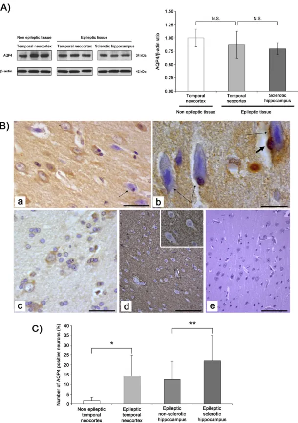
Evidences indicate the potential involvement of water channel AQP4 in modulation of brain excitability and in epilepsy[42]. However, its pattern of expression and distribution in epileptic tissues remains con-troversial (downregulation vs mislocalization)[43]. The immunoblot-ting results indicated that there were not significant changes in AQP4 expression between control neocortex and epileptic brain tissues (temporal neocortex and hippocampus) (Fig. 4A), although a trend showing a decreased expression in epileptic sclerotic hippocampus was noticed.
After determining by immunoblotting that there are no significant quantitative variations of AQP4 expression in the analyzed tissues, we have proceeded in identifying AQP4 tissue distribution. In all cases, in-cluding epileptic lesional tissues and controls, AQP4 was positive in subpial areas and, as shown inFig. 4B, in the neuropil. Of note, although AQP4 is a water channel predominantly found in astrocytes in the CNS, our immunohistochemicalfindings revealed its presence also in some apparently healthy neurons (Fig. 4B-a, -b and -c), while AQP4 positivity in degenerating neurons was mainly observed in the cell periphery (Fig. 4B-a, and -b, thin arrows). On the other hand, negativity for AQP4 was usually observed in neurons of nonepileptic temporal neo-cortex (Fig. 4B-d). When the number of AQP4 positive neurons was quantified (Fig. 4C), there was a clear and strong difference between the control and epileptic neocortex (means ± SD: 1.45 ± 1.9% vs 14.2 ± 10.5% respectively; pb 0.001). Furthermore, the number of pos-itive neurons for AQP4 was also significant different between non-sclerotic and non-sclerotic hippocampus (means ± SD 12.5 ± 9.2% vs 22.0 ± 12.7%; pb 0.05). Furthermore, AQP4 was also detected in the perivascular astrocytic endfeet and in few endothelial cells of several small vessels (Fig. 1s).
Double AQP4-Beta III tubulin immunoflorescence showed colocalization of both the proteins in several cortical (Fig. 5a–o) as well as hippocampal (Fig. 5p–t) neurons. In degenerating neurons (Fig. 5p–t) the positivity was limited to the cytoplasm periphery. 3.5. Oxidative post-translational modification of aquaporin-4 and its co-localization with 4-hydroxy-2-nonenal protein adducts in sclerotic hippocampus from drug-resistant epileptic patients
To explore the possible formation of HNE-AQP4 adducts, we per-formed the immunoprecipitation of AQP4 from samples obtained from non-epileptic and epileptic tissues, followed by Western blotting for HNE. As it is evidenced inFig. 6A, our results revealed a significant in-crease of HNE-AQP4 adducts levels in epileptic temporal neocortex compared to the control temporal neocortex. In addition, we evaluated in situ co-localization of HNE-PA and AQP4 in sclerotic hippocampus from drug-resistant epileptic patients. A clear co-localization of AQP4 (redfluorescence) with HNE-PA (green fluorescence) was evident in the same structures in epileptic hippocampal tissue (Fig. 6B-c, yellow in merged image). These data suggest a possible HNE-mediated oxida-tive post-translational modification of AQP4.
4. Discussion
Several reports have already indicated the presence of OS in epilep-sy, mainly using animal models and to a lesser extent by means of studies in humans[44]. In the present work, we report the evidence of lipid peroxidation and oxidative-mediated protein damage, i.e.
significantly higher levels of HNE-PA, in human neocortical tissue from drug-resistant epileptic patients versus neocortical control tissue and alteration in location and expression of HNE-PA in non-sclerotic vs sclerotic hippocampus. Moreover, the present study uncovers anoth-er importantfinding related to the source of OS, as it has been shown by the activation of NOX2 in neurons within the epileptic area. Finally, al-though we did not document significant changes in the AQP4 protein expression between control and epileptic brain specimens, a critical ob-servation is that the water channel is a target for HNE, which therefore can negatively modulate its structure, stability and functions. The impli-cations of all these observations can bring new insight to better under-stand the mechanisms of epileptogenesis.
Oxidant production is clearly implicated in the pathogenesis of sev-eral neurodegenerative disorders[1]. Research into pathophysiology of epileptic seizures is primarily done in experimental models and only rarely on human samples because of the obviously limited availability and the greater difficulty in collection of human brain samples. Although seizure models have made important contributions to the un-derstanding of the role of redox imbalance in epileptogenesis, evidence also suggests that OS does not always appear to follow the same pattern in all models[45]. In addition, animal studies do not always reliably pre-dict human outcomes. For this reason the use of samples from epilepsy-surgical brain specimens of drug-resistant patients is one of the most re-liable models to study the mechanism underlying this pathology. Alter-natively, it is possible to collect samples from post-mortem tissues, although also in this case there are several limitations due to timing is-sues; the tissue needs to be collected within a few hours after the de-cease and often the cause of death can affect brain tissue protein expression independently of what epilepsy has caused[46].
Several mechanisms related to OS have been proposed to be in-volved in epileptic seizures, such as impairment of antioxidant systems and mitochondrial dysfunction. Moreover, different strategies using so-called“antioxidants” have proven effective for seizure treatments[45], although the actual mechanism cannot involve radical scavenging ex-cept by vitamin E[47].
In particular, numerous data support a key role of mitochondrial dysfunction in the increased susceptibility to seizures[48,49]. Although the brain is particularly rich in mitochondria and suggesting that mod-ifications of mitochondrial activity can be the main source of OS in this tissue, a growing body of evidence supports also the role for abnormal NOX activation as an important potential source of O2•−and H2O2
gen-eration in neuronal cells[14,50]. In line with our work, there is the study of Shimohama et al. that has demonstrated in post-mortem brain tissue from Alzheimer patients an increased accumulation of neu-ronal cytosolic NOX2 subunits p47phoxand p67phoxat the cell surface of diseased brain regions, suggesting chronic activation of this enzyme
[51]. It is possible that the repeated seizures can induce the upregulation and activation of NOX2 in hippocampal neurons of epileptic brain. Effec-tively, in our study neurons with a normal aspect showed an intense re-activity for p47phoxand p67phox, suggesting high NOX2 expression that
may be involved in the degeneration and loss of hippocampal neurons. This proposed mechanism is corroborated by some studies in various experimental forms of epilepsy models. The translocation of NOX sub-units from hippocampal cytosol to membrane fractions has been dem-onstrated in kainate-injected rats[16]. Furthermore, the involvement of NOX in oxidant-mediated damage and neuronal death has been sug-gested in pilocarpine[17,18,20,52]and kainic acid models[16,19]. In ad-dition, the inhibition of NOX by apocynin reduce O2•− and H2O2
production and lipid peroxidation after seizure and decrease the num-ber of degenerating hippocampal neurons in the pilocarpine rat model
Fig. 2 Double immunofluorescence for HNE-PA/GFAP and HNE-PA/β-III TUB in sclerotic hippocampus compared with the non-sclerotic hippocampal tissue.A) Representative images depicting very low HNE-PA expression in non-sclerotic hippocampal tissue (greenfluorescence; boxes: a, b, c); in the merged images (boxes: a, b, c), colocalization of HNE-PA with both GFAP (glial cells, redfluorescence; boxes: a, b) and β-III TUB (neurons, red fluorescence; box: c) is virtually absent. B) Increased, diffuse positivity for HNE-PA (green fluorescence; boxes: d, e, f) in sclerotic hippocampus is seen in both astrocytes (GFAP, redfluorescence; boxes: d, e) and neurons (β-III TUB, red fluorescence; box: f), as evident in merged images (yellow color; boxes: d, e, f). Scale bar: a, d = 100μm; b, c, e, f = 25 μm.
[21]. However, there were no reports regarding the NOX2 contribution to O2•−and H2O2generation in epilepsy using human samples prior to
the present studies.
Beside direct actions, the harmful effects of oxidants generated by NOX may be linked to the reaction with the PUFA which, leads to the production of a great variety of reactive species. Among them, HNE is considered as a“second toxic messenger,” that can propagate and am-plify initial oxidative injury. HNE can form covalent bonds with three different amino acyl side chains, i.e., lysyl, histidyl, and cysteinyl resi-dues, via Michael addition with cysteine favored kinetically, but lysine favored by abundance. In addition, HNE can modify protein structure through Schiff base formation with lysyl residues, leading to formation of pyrrole, and/or form intra- and/or intermolecular cross-links. Due to its amphiphilic nature, the hydroxy aldehyde can diffuse across mem-branes and covalently modify proteins in the cytoplasm and nucleus, far from their site of origin[25].
High levels of HNE-PA have been found in brain tissues and body fluids in several neurological and neurodegenerative diseases[25,26, 53–55]. Indeed, our study shows high HNE-PA levels in the brain of drug-resistant epileptic patients. This confirms the evidence of lipid per-oxidation and indicates the presence of an oxidative damage of proteins in human epileptic brain as reported by other authors in human plasma
[6,56–58]or in brain specimens. To date only a limited number of stud-ies have evaluated HNE levels in epileptic diseases. Frantseva et al. re-ported high levels of brain HNE during seizures in the kindling model
[59]. Similarly, Jacobsson et al. observed significant increase of brain HNE in a rodent model of soman-induced seizures[60]; although to our knowledge this is thefirst report showing the HNE-PA distribution in brain tissue of epileptic patients. This observation has important im-plications in understanding the epileptogenesis. Since the oxidative PTMs are known to alter protein functions and impair cellular mecha-nisms [61,62], HNE-PA in human epileptic brain may promote
deleterious subcellular events that trigger and progress the epileptogenesis. In addition, since it is well known that HNE can be an apoptotic inducer[63,64], in our study high levels of HNE-PA in appar-ently healthy neurons may eventually promote cell death, as suggested by the presence of several nearby neurons in degeneration, showing morphological features of possible apoptosis. Evidence that the HNE-PA plays a pivotal role in neuronal death has already been demonstrated in several neurological diseases[26,53,65–67].
Different molecular studies have provided potential insights into the pathogenesis of epilepsy, among them is the possible involvement of water channelopathies that can increase neuronal excitability[68]. In fact, alterations of water and ions homeostasis can dramatically affect seizure susceptibility[33]. Recent reports have highlighted the key role of the water channels AQPs in epilepsy[69]. Among the several AQPs, of particular interest in neuroscience is AQP4, since it is highly expressed in brain and spinal cord by glial cells[68]. Medici et al., eval-uating the AQP4 expression in epileptic and control human cerebral cor-tex, found a significant increase in AQP4 in the focal cortical dysplasia (FCD) type IIB samples, with a different protein distribution pattern
[70]. In comparison with the controls, AQP4 immunoreactivity was more diffuse in the neuropil, particularly around dysmorphic neurons, and less intense perivascularly. Similar remarks were observed also in experimental animal models. For example, in the kainic acid model of epileptogenesis, a loss of positivity for AQP4 occurred on both the endfeet andfine processes of astrocytes in different hippocampal layers
[71]. In addition, in AQP4 knockout mice, the susceptibility to seizure ini-tiation is increased compared with controls, as well as the seizure dura-tion and intensity[72]. All these data confirm that both the altered expression and mislocalization of AQP4 can lead to water and ion dys-regulation in epileptic brain, probably contributing to its hyperexcitabil-ity. It should be mentioned that the altered levels of AQP4 in epileptic brain tissues are still controversial, for instance in a recent work by
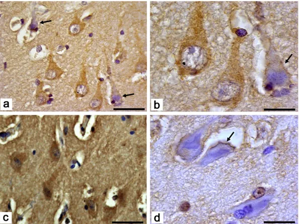
Fig. 3. Immunohistochemical localization of the NADPH oxidase subunits in epileptic sclerotic hippocampus. Panels a and b, normal neurons show cytosolic staining for p67phox
, whereas degenerating neurons maintain residual positivity on cell membrane (arrows). Likewise, p47phox
immunohistochemistry shows a similar pattern of stain (panels c and d). Arrow indicates plasma membrane positivity. Scale bar: a, c = 50μm; b, d = 25 μm.
Fig. 4. Western blotting and immunohistochemical localization of AQP4 in epileptic tissues. A) No change of AQP4 levels is observed in representative samples of control subjects (n = 5) and patients with drug-resistant epilepsy (n = 50) (left panel). Quantification of AQP4 normalized to β-actin and determined by densitometric analysis of the scanned images is shown in the right panel. Average values were expressed as arbitrary units. Data are means ± SD of three independent experiments. *pb 0.05. B) AQP4 positive neurons in the CA1 area of sclerotic hippocampus (panels a and b) and temporal neocortex (panel c) of an epileptic patient. In degenerating neurons (a, b, thin arrows), the positivity is usually limited to the cytoplasmic periphery. Thick, short arrow in b indicates an AQP4 positive oligodendrocyte. AQP4 negative neurons in temporal neocortex of a non-epileptic control patient (d, detail in the insert). Negativity for AQP4 in a negative control section with primary antibody omitted (e, temporal neocortex of a non-epileptic control). Scale bar: a, b = 25μm; c, d, e = 50 μm. C). For quan-tification, the neurons positive for AQP4 were counted in 10 randomly chosen fields, both in the epileptic temporal neocortex and in the CA area of epileptic hippocampus, with or without HS, and in the temporal neocortex of nonepileptic patients. Counts were expressed as percentages. Results are presented as mean percentage ± SD. *pb 0.001 and **p b 0.05 (panel C).
Bebek et al. it has been shown that AQP4 levels (mRNA and protein) did not differ between controls and patients. One reason for these results could be the limited number of samples, as the authors suggested[73]. In our study, we have found that the expression of AQP4 was not affected in epileptic specimens when compared with the control neo-cortex, but immunohistochemistry and double immunofluorescence showed that in epileptic tissues AQP4 was present not only in perivascular astrocytic endfeet, but also in apparently healthy neurons, whereas there was a clear loss of AQP4 positivity in degenerating neu-rons. Of note was also the fact that only few endothelial cells of several small vessels showed positivity to AQP4, whereas the endothelium of larger vessels and of most small vessels was negative, partly confirming the ongoing controversial debate present in the literature regarding the possible role of AQP4 in the endothelium [74,75]. Therefore, our results coincide with some, but not all previous studies. Most previous immunohistological brain studies showed AQP4 expression only in astrocytes and ependymal cells without strong evidence in
neurons[76]. Nevertheless, others have detected AQP4 in neurons of the supraoptic and paraventricular nuclei of the hypothalamus[77]
and in enteric neurons[78]. Moreover, in a study of in situ hybridization performed in the rat brain, Venero et al. showed mRNA staining for AQP4 in hippocampal pyramidal cells, granular cells of dentate gyrus, and neuronal cells of the cortex.[74]Although it is possible that the mRNA was not translated, it is more likely that the negative results stemmed from a failure of immunodetection due to low expression and/or a poor antibody. The AQP4 presence in neurons of epileptic brain could make these cells more vulnerable to water and ions trans-port, modulating neuronal transmission and excitability. Anyhow, fur-ther studies will need to be undertaken for the interpretation of our data in relation to epileptogenesis.
In addition our data clearly showed the formation of HNE-AQP4 ad-ducts by two different methods (immunoprecipitation and immuno flu-orescence). In fact, we observed that the water channel co-localized with HNE-PA in the same structures in epileptic sclerotic hippocampus
Fig. 5. AQP4 andβ-III TUB double immunofluorescence staining in epileptic tissues. The immunopositivity for AQP4 (green fluorescence; boxes: b, g, l and q) and β-III TUB (neurons, red fluorescence; boxes: c, h, m, and r) is dot-like and diffuse, respectively, in the cytoplasm of neurons; the neuropil is also positive to AQP4. The boxes inside the merged images of the tem-poral neocortex (boxes: a, f and k) and CA of sclerotic hippocampus (box p) of an epileptic patient have been enlarged to better visualize neurons showing AQP4 andβ-III TUB colocalization (yellow stain in boxes: d, i, n, and s; white stain after ImageJ software elaboration in boxes: e, j, o and t). Damaged neurons show AQP4 positivity at the cytoplasm periphery (boxes: f, j, k and o, arrow). A normal neuron, negative for AQP4, adjacent to a damaged neuron is also shown (boxes: n and o). Scale bar = 50μm.
and by Western blot we have demonstrated the formation of covalent binding between HNE and AQP4. Thesefindings support the idea that this protein could be a critical target for aldehyde adduction. The impairment of structural integrity resulting by the covalent bonds with the aldehyde can modify the biological activity of AQP4. The for-mation of HNE-AQP4 adducts might also be responsible for the changes in number and localization of water channel, promoting for example its greater proteolytic degradation. Indeed, Grune and coworkers have shown that HNE modification often leads to increased degradation
[79]. In this perspective, the HNE-mediated modulation of AQP4 would provide a new base for the altered neuronal excitability of epilep-tic brain.
Finally, we also observed an oxidant-dependent activation of MMP-9 in epileptic tissues. A key role for the aberrant activity of MMPs in chronic neurodegenerative diseases has been extensively reported
[31]. In addition, evidence from both in humans and animal models indicate the involvement of MMPs in epilepsy[32,80]. In the brain, the MMPs-mediated degradation of the extracellular matrix or membrane-bound proteins may increase the permeability of the blood brain barrier, resulting in edema and alterations of neuronal excitability[81]. In addi-tion, it is known that the dystrophin-dystroglycan complex has a critical role in the distribution and the maintenance of the AQP4 tetrameric complex on cell membranes[82]and MMP-9 is able to degrade dystro-glycan[83]. Therefore, the upregulation of MMPs could contribute to dis-organization of the tissue/cell distribution of water channel in epileptic brain.
5. Conclusions
In conclusion, our data reinforce the theory that OS may play a key role in epilepsy. The recurrent seizures can trigger a vicious circle, where the redox imbalance is both the cause and result of the
brain damage. The activation of NOX2 and MMPs could act in concert to cause neuronal loss and negatively modulate the AQP4, either inducing the structural and functional alterations due to the HNE-adduct formation than promoting its abnormal cellular distribution (Scheme1). Finally, any change in biologic activity of AQP4 that com-promises the ions and water balance, promoting neuronal hyperexcit-ability, could result in enhanced propensity for seizures and thereby contribute to the generation and perpetuation of the epileptogenesis process.
In addition, we should also remind that seizure can cause brain dam-age not only in epileptic patients but also in patients who do not suffer from epilepsy but are affected by seizure episodes. Of course to better understand the role of OS in epileptogenesis further mechanistic inves-tigations are needed, but this aspect can anyway bring new insights for therapeutic targets.
Supplementary data to this article can be found online athttp://dx. doi.org/10.1016/j.bbadis.2014.11.016.
References
[1] A. White, C. Culmsee, P. Beart, Oxidative stress and neurodegeneration, Neurochem. Int. 62 (2013) 521.
[2] H. Sies, Oxidative stress: oxidants and antioxidants, Exp. Physiol. 82 (1997) 291–295.
[3] B. Halliwell, Oxidative stress and neurodegeneration: where are we now? J. Neurochem. 97 (2006) 1634–1658.
[4] C.C. Aguiar, A.B. Almeida, P.V. Araújo, R.N. de Abreu, E.M. Chaves, O.C. do Vale, D.S. Macêdo, D.J. Woods, M.M. Fonteles, S.M. Vasconcelos, Oxidative stress and epilepsy: literature review, Oxidative Med. Cell. Longev. 2012 (2012) 795259.
[5] S. Rowley, M. Patel, Mitochondrial involvement and oxidative stress in temporal lobe epilepsy, Free Radic. Biol. Med. 62 (2013) 121–131.
[6] K. Sudha, A.V. Rao, A. Rao, Oxidative stress and antioxidants in epilepsy, Clin. Chim. Acta 303 (2001) 19–24.
[7] P. Ambrogini, A. Minelli, C. Galati, M. Betti, D. Lattanzi, S. Ciffolilli, M. Piroddi, F. Galli, R. Cuppini, Post-seizureα-tocopherol treatment decreases neuroinflammation and Fig. 6. Immunoprecipitation/Western blotting and double immunofluorescence for AQP4 and HNE in epileptic tissues. A) Representative images of the HNE-AQP4 adduct formation in control and epileptic temporal neocortex and in epileptic sclerotic hippocampus are shown in the left panel. Quantification of HNE-AQP4 adducts determined by densitometric analysis of the scanned images is shown in the right panel. Average values were expressed as arbitrary units. Data are means ± SD of three determinations. *pb 0.05. B) Representative AQP4 (a, red) and HNE-PA (b, green) immunofluorescence images in the epileptic hippocampus show co-localization of two antigens (c, yellow) in the same structures. Scale bar = 50 μm.
neuronal degeneration induced by status epilepticus in rat hippocampus, Mol. Neurobiol. (2014),http://dx.doi.org/10.1007/s12035-014-8648-2.
[8] R. Tomé Ada, C.M. Feitosa, R.M. Freitas, Neuronal damage and memory deficits after seizures are reversed by ascorbic acid? Arq. Neuropsiquiatr. 68 (2010) 579–585.
[9] V.A. Yürekli, M. Nazıroğlu, Selenium and topiramate attenuates blood oxidative toxicity in patients with epilepsy: a clinical pilot study, Biol. Trace Elem. Res. 152 (2013) 180–186.
[10] S. Waldbaum, M. Patel, Mitochondrial dysfunction and oxidative stress: a contribut-ing link to acquired epilepsy? J. Bioenerg. Biomembr. 42 (2010) 449–455.
[11] S. Sorce, K.H. Krause, NOX enzymes in the central nervous system: from signaling to disease, Antioxid. Redox Signal. 11 (2009) 2481–2504.
[12] J. El Benna, L.P. Faust, B.M. Babior, The phosphorylation of the respiratory burst ox-idase component p47phox
during neutrophil activation, J. Biol. Chem. 269 (1994) 23431–23436.
[13]A. van der Vliet, NADPH oxidases in lung biology and pathology: host defense en-zymes, and more, Free Radic. Biol. Med. 44 (2008) 938–955.
[14] D.W. Infanger, R.V. Sharma, R.L. Davisson, NADPH oxidases of the brain: distribution, regulation, and function, Antioxid. Redox Signal. 8 (2006) 1583–1596.
[15] A.P. Kudin, T.A. Kudina, J. Seyfried, S. Vielhaber, H. Beck, C.E. Elger, W.S. Kunz, Seizure-dependent modulation of mitochondrial oxidative phosphorylation in rat hippocampus, Eur. J. Neurosci. 15 (2002) 1105–1114.
[16]M. Patel, Q.Y. Li, L.Y. Chang, J. Crapo, L.P. Liang, Activation of NADPH oxidase and extracellular superoxide production in seizure-induced hippocampal damage, J. Neurochem. 92 (2005) 123–131.
[17] R.R. Pestana, E.R. Kinjo, M.S. Hernandes, L.R. Britto, Reactive oxygen species generat-ed by NADPH oxidase are involvgenerat-ed in neurodegeneration in the pilocarpine model of temporal lobe epilepsy, Neurosci. Lett. 484 (2010) 187–191.
[18] R. Di Maio, P.G. Mastroberardino, X. Hu, L. Montero, J.T. Greenamyre, Pilocapine al-ters NMDA receptor expression and function in hippocampal neurons: NADPH oxi-dase and ERK1/2 mechanisms, Neurobiol. Dis. 42 (2011) 482–495.
[19] C.Y. Tsai, J.Y. Chan, K.S. Hsu, A.Y. Chang, S.H. Chan, Brain-derived neurotrophic factor ameliorates brain stem cardiovascular dysregulation during experimental temporal lobe status epilepticus, PLoS One 7 (2012) e33527.
[20]J.E. Kim, H.J. Ryu, T.C. Kang, Status epilepticus induces vasogenic edema via tumor necrosis factor-α/endothelin-1-mediated two different pathways, PLoS One 8 (2013) e74458.
[21] J.H. Kim, B.G. Jang, B.Y. Choi, H.S. Kim, M. Sohn, T.N. Chung, H.C. Choi, H.K. Song, S.W. Suh, Post-treatment of an NADPH oxidase inhibitor prevents seizure-induced neu-ronal death, Brain Res. 1499 (2013) 163–172.
[22] J. Friedman, Why is the nervous system vulnerable to oxidative stress? in: N. Gadoth, H.H. Göbel (Eds.), Oxidative Stress and Free Radical Damage in Neurology, Springer Science Business Media, 2011, pp. 19–27.
[23] H. Esterbauer, R.J. Schaur, H. Zollner, Chemistry and biochemistry of 4-hydroxynonenal, malonaldehyde and related aldehydes, Free Radic. Biol. Med. 11 (1991) 81–128.
[24] G. Leonarduzzi, M.C. Arkan, H. Başağa, E. Chiarpotto, A. Sevanian, G. Poli, Lipid oxida-tion products in cell signalling, Free Radic. Biol. Med. 28 (2000) 1370–1378.
[25]G. Poli, F. Biasi, G. Leonarduzzi, 4-Hydroxynonenal-protein adducts: a reliable bio-marker of lipid oxidation in liver diseases, Mol. Asp. Med. 29 (2008) 67–71.
[26] K. Zarkovic, 4-Hydroxynonenal and neurodegenerative diseases, Mol. Asp. Med. 24 (2003) 293–303.
[27] K. Ryan, L.P. Liang, C. Rivard, M. Patel, Temporal and spatial increase of reactive ni-trogen species in the kainate model of temporal lobe epilepsy, Neurobiol. Dis. 64 (2014) 8–15.
[28]W.Y. Ong, X.R. Lu, C.Y. Hu, B. Halliwell, Distribution of hydroxynonenal-modified proteins in the kainate-lesioned rat hippocampus: evidence that hydroxynonenal formation precedes neuronal cell death, Free Radic. Biol. Med. 28 (2000) 1214–1221.
[29] H.J. Forman, F. Ursini, M. Maiorino, An overview of mechanisms of redox signaling, J. Mol. Cell. Cardiol. 73 (2014) 2–9.
[30] A.L. Jacob-Ferreira, R. Schulz, Activation of intracellular matrix metalloproteinase-2 by reactive oxygen-nitrogen species: consequences and therapeutic strategies in the heart, Arch. Biochem. Biophys. 540 (2013) 82–93.
[31] V.W. Yong, C. Power, P. Forsyth, D.R. Edwards, Metalloproteinases in biology and pa-thology of the nervous system, Nat. Rev. Neurosci. 2 (2001) 502–511.
[32]H. Mizoguchi, K. Yamada, Roles of matrix metalloproteinases and their targets in epileptogenesis and seizures, Clin. Psychopharmacol. Neurosci. 11 (2013) 45–52.
[33] D.K. Binder, C. Steinhäuser, Functional changes in astroglial cells in epilepsy, Glia 54 (2006) 358–368.
[34] T.S. Lee, T. Eid, S. Mane, J.H. Kim, D.D. Spencer, O.P. Ottersen, N.C. de Lanerolle, Aquaporin-4 is increased in the sclerotic hippocampus in human temporal lobe ep-ilepsy, Acta Neuropathol. 108 (2004) 493–502.
[35]T. Eid, T.S. Lee, M.J. Thomas, M. Amiry-Moghaddam, L.P. Bjørnsen, D.D. Spencer, P. Agre, O.P. Ottersen, N.C. de Lanerolle, Loss of perivascular aquaporin 4 may underlie
Scheme 1. Proposed sequence of events responsible for the redox imbalance and oxidative brain damage in drug-resistant epilepsy. The recurrent seizures activates NOX2 with O2−
pro-duction (1). This event will lead to the formation of lipid peroxidation-derived HNE that will form covalent adducts with cellular proteins, such as AQP4 (2). At the same time, the release and activation of MMP-9 are upregulated by OS (3). Both MMPs activation and HNE-AQP4 adducts will affect the water and the ion balance. Therefore, we suggest that seizure induces as well as neuronal loss (4), thereby promoting neuronal hyperexcitability, also affecting water and ion balance by AQP4 modulation, and thus generating a vicious cycle.
deficient water and K+
homeostasis in the human epileptogenic hippocampus, Proc. Natl. Acad. Sci. U. S. A. 102 (2005) 1193–1198.
[36] I. Blümcke, M. Thom, E. Aronica, D.D. Armstrong, H.V. Vinters, A. Palmini, T.S. Jacques, G. Avanzini, A.J. Barkovich, G. Battaglia, A. Becker, C. Cepeda, F. Cendes, N. Colombo, P. Crino, J.H. Cross, O. Delalande, F. Dubeau, J. Duncan, R. Guerrini, P. Kahane, G. Mathern, I. Najm, C. Ozkara, C. Raybaud, A. Represa, S.N. Roper, N. Salamon, A. Schulze-Bonhage, L. Tassi, A. Vezzani, R. Spreafico, The clinicopathologic spectrum of focal cortical dysplasias: a consensus classification proposed by an ad hoc Task Force of the ILAE Diagnostic Methods Commission, Epilepsia 52 (2011) 158–174.
[37]I. Blümcke, M. Thom, E. Aronica, D.D. Armstrong, F. Bartolomei, A. Bernasconi, N. Bernasconi, C.G. Bien, F. Cendes, R. Coras, J.H. Cross, T.S. Jacques, P. Kahane, G.W. Mathern, H. Miyata, S.L. Moshé, B. Oz, Ç. Özkara, E. Perucca, S. Sisodiya, S. Wiebe, R. Spreafico, International consensus classification of hippocampal sclerosis in tem-poral lobe epilepsy: a Task Force report from the ILAE Commission on Diagnostic Methods, Epilepsia 54 (2013) 1315–1329.
[38] A. Pecorelli, V. Bocci, A. Acquaviva, G. Belmonte, C. Gardi, F. Virgili, L. Ciccoli, G. Valacchi, NRF2 activation is involved in ozonated human serum upregulation of HO-1 in endothelial cells, Toxicol. Appl. Pharmacol. 267 (2013) 30–40.
[39]V. Fortino, E. Maioli, C. Torricelli, P. Davis, G. Valacchi, Cutaneous MMPs are differ-ently modulated by environmental stressors in old and young mice, Toxicol. Lett. 173 (2007) 73–79.
[40] G. Valacchi, Y. Lim, G. Belmonte, C. Miracco, I. Zanardi, V. Bocci, V. Travagli, Ozonated sesame oil enhances cutaneous wound healing in SKH1 mice, Wound Repair Regen. 19 (2011) 107–115.
[41]K. Jian Liu, G.A. Rosenberg, Matrix metalloproteinases and free radicals in cerebral ischemia, Free Radic. Biol. Med. 39 (2005) 71–80.
[42] D.K. Binder, E.A. Nagelhus, O.P. Ottersen, Aquaporin-4 and epilepsy, Glia 60 (2012) 1203–1214.
[43] H.E. Scharfman, D.K. Binder, Aquaporin-4 water channels and synaptic plasticity in the hippocampus, Neurochem. Int. 63 (2013) 702–711.
[44] N. Cardenas-Rodriguez, B. Huerta-Gertrudis, L. Rivera-Espinosa, H. Montesinos-Correa, C. Bandala, L. Carmona-Aparicio, E. Coballase-Urrutia, Role of oxidative stress in refractory epilepsy: evidence in patients and experimental models, Int. J. Mol. Sci. 14 (2013) 1455–1476.
[45] E.J. Shin, J.H. Jeong, Y.H. Chung, W.K. Kim, K.H. Ko, J.H. Bach, J.S. Hong, Y. Yoneda, H.C. Kim, Role of oxidative stress in epileptic seizures, Neurochem. Int. 59 (2011) 122–137.
[46] R. Ravid, D.F. Swaab, The Netherlands brain bank—a clinico-pathological link in aging and dementia research, J. Neural Transm. Suppl. 39 (1993) 143–153.
[47] H.J. Forman, K.J. Davies, F. Ursini, How do nutritional antioxidants really work: nu-cleophilic tone and para-hormesis versus free radical scavenging in vivo, Free Radic. Biol. Med. 66 (2014) 24–35.
[48]J. Gao, H. Yao, X.D. Pan, A.M. Xie, L. Zhang, J.H. Song, A.J. Ma, Z.C. Liu, Alteration of mitochondrial function and ultrastructure in the hippocampus of pilocarpine-treated rat, Epilepsy Res. 108 (2014) 162–170.
[49]D.S. Khurana, I. Valencia, M.J. Goldenthal, A. Legido, Mitochondrial dysfunction in epilepsy, Semin. Pediatr. Neurol. 20 (2013) 176–187.
[50] H.M. Gao, H. Zhou, J.S. Hong, NADPH oxidases: novel therapeutic targets for neuro-degenerative diseases, Trends Pharmacol. Sci. 33 (2012) 295–303.
[51]S. Shimohama, H. Tanino, N. Kawakami, N. Okamura, H. Kodama, T. Yamaguchi, T. Hayakawa, A. Nunomura, S. Chiba, G. Perry, M.A. Smith, S. Fujimoto, Activation of NADPH oxidase in Alzheimer's disease brains, Biochem. Biophys. Res. Commun. 273 (2000) 5–9.
[52] C. Hirotsu, G. Matos, S. Tufik, M.L. Andersen, Changes in gene expression in the fron-tal cortex of rats with pilocarpine-induced status epilepticus after sleep deprivation, Epilepsy Behav. 27 (2013) 378–384.
[53] D.A. Butterfield, T. Reed, R. Sultana, Roles of 3-nitrotyrosine- and 4-hydroxynonenal-modified brain proteins in the progression and pathogenesis of Alzheimer's disease, Free Radic. Res. 45 (2011) 59–72.
[54] A. Pecorelli, L. Ciccoli, C. Signorini, S. Leoncini, A. Giardini, M. D'Esposito, S. Filosa, J. Hayek, C. De Felice, G. Valacchi, Increased levels of 4HNE-protein plasma adducts in Rett syndrome, Clin. Biochem. 44 (2011) 368–371.
[55] A. Pecorelli, S. Leoncini, C. De Felice, C. Signorini, C. Cerrone, G. Valacchi, L. Ciccoli, J. Hayek, Non-protein-bound iron and 4-hydroxynonenal protein adducts in classic autism, Brain Dev. 35 (2013) 146–154.
[56]D. Turkdogan, S. Toplan, Y. Karakoc, Lipid peroxidation and antioxidative enzyme activities in childhood epilepsy, J. Child Neurol. 17 (2002) 673–676.
[57]D. Deepa, B. Jayakumari, S.V. Thomas, Lipid peroxidation in women with epilepsy, Ann. Indian. Acad. Neurol. 11 (2008) 44–46.
[58] B. Menon, K. Ramalingam, R.V. Kumar, Oxidative stress in patients with epilepsy is independent of antiepileptic drugs, Seizure 21 (2012) 780–784.
[59] M.V. Frantseva, J.L. Perez Velazquez, G. Tsoraklidis, A.J. Mendonca, Y. Adamchik, L.R. Mills, P.L. Carlen, M.W. Burnham, Oxidative stress is involved in seizure-induced neurodegeneration in the kindling model of epilepsy, Neuroscience 97 (2000) 431–435.
[60] S.O. Jacobsson, G.E. Cassel, S.A. Persson, Increased levels of nitrogen oxides and lipid peroxidation in the rat brain after soman-induced seizures, Arch. Toxicol. 73 (1999) 269–273.
[61] S. Grimm, A. Hoehn, K.J. Davies, T. Grune, Protein oxidative modifications in the age-ing brain: consequence for the onset of neurodegenerative disease, Free Radic. Res. 45 (2011) 73–88.
[62]A. Höhn, T. Jung, T. Grune, Pathophysiological importance of aggregated damaged proteins, Free Radic. Biol. Med. 71C (2014) 70–89.
[63] I. Kruman, A.J. Bruce-Keller, D. Bredesen, G. Waeg, M.P. Mattson, Evidence that 4-hydroxynonenal mediates oxidative stress-induced neuronal apoptosis, J. Neurosci. 17 (1997) 5089–5100.
[64] S. Dalleau, M. Baradat, F. Guéraud, L. Huc, Cell death and diseases related to oxida-tive stress: 4-hydroxynonenal (HNE) in the balance, Cell Death Differ. 20 (2013) 1615–1630.
[65] A. Yoritaka, N. Hattori, K. Uchida, M. Tanaka, E.R. Stadtman, Y. Mizuno, Immunohis-tochemical detection of 4-hydroxynonenal protein adducts in Parkinson disease, Proc. Natl. Acad. Sci. U. S. A. 93 (1996) 2696–2701.
[66] R.J. Mark, M.A. Lovell, W.R. Markesbery, K. Uchida, M.P. Mattson, A role for 4-hydroxynonenal, an aldehydic product of lipid peroxidation, in disruption of ion ho-meostasis and neuronal death induced by amyloid beta-peptide, J. Neurochem. 68 (1997) 255–264.
[67] K.S. Montine, E. Reich, M.D. Neely, K.R. Sidell, S.J. Olson, W.R. Markesbery, T.J. Montine, Distribution of reducible 4-hydroxynonenal adduct immunoreactivity in Alzheimer disease is associated with APOE genotype, J. Neuropathol. Exp. Neurol. 57 (1998) 415–425.
[68]I. Benga, O. Benga, Implications of water channel proteins in selected neurological disorders: epilepsies, muscular dystrophies, amyotrophic lateral sclerosis, neuromy-elitis optica, Parkinson's disease, and spongiform encephalopathies, Mol. Asp. Med. 33 (2012) 590–604.
[69] M.S. Hsu, D.J. Lee, D.K. Binder, Potential role of the glial water channel aquaporin-4 in epilepsy, Neuron Glia Biol. 3 (2007) 287–297.
[70]V. Medici, C. Frassoni, L. Tassi, R. Spreafico, R. Garbelli, Aquaporin 4 expression in control and epileptic human cerebral cortex, Brain Res. 1367 (2011) 330–339.
[71] D.J. Lee, M.S. Hsu, M.M. Seldin, J.L. Arellano, D.K. Binder, Decreased expression of the glial water channel aquaporin-4 in the intrahippocampal kainic acid model of epileptogenesis, Exp. Neurol. 235 (2012) 246–255.
[72] D.K. Binder, X. Yao, Z. Zador, T.J. Sick, A.S. Verkman, G.T. Manley, Increased seizure duration and slowed potassium kinetics in mice lacking aquaporin-4 water chan-nels, Glia 53 (2006) 631–636.
[73]N. Bebek, Ö. Özdemir, M. Sayitoglu, O. Hatırnaz, B. Baykan, C. Gürses, A. Sencer, A. Karasu, E. Tüzün, I. Üzün, S. Akat, N. Cine, G. Sargin Kurt, M. Imer, U. Ozbek, A. Canbolat, A. Gökyigit, Expression analysis and clinical correlation of aquaporin 1 and 4 genes in human hippocampal sclerosis, J. Clin. Neurosci. 20 (2013) 1564–1570.
[74] J.L. Venero, M.L. Vizuete, A.A. Ilundáin, A. Machado, M. Echevarria, J. Cano, Detailed localization of aquaporin-4 messenger RNA in the CNS: preferential expression in periventricular organs, Neuroscience 94 (1999) 239–250.
[75] N.N. Haj-Yasein, G.F. Vindedal, M. Eilert-Olsen, G.A. Gundersen, Ø. Skare, P. Laake, A. Klungland, A.E. Thorén, J.M. Burkhardt, O.P. Ottersen, E.A. Nagelhus, Glial-conditional deletion of aquaporin-4 (Aqp4) reduces blood–brain water uptake and confers barrier function on perivascular astrocyte endfeet, Proc. Natl. Acad. Sci. U. S. A. 108 (2011) 17815–17820.
[76] E.A. Nagelhus, T.M. Mathiisen, O.P. Ottersen, Aquaporin-4 in the central nervous sys-tem: cellular and subcellular distribution and coexpression with KIR4.1, Neurosci-ence 129 (2004) 905–913.
[77] C. Iacovetta, E. Rudloff, R. Kirby, The role of aquaporin 4 in the brain, Vet. Clin. Pathol. 41 (2012) 32–44.
[78] M.M. Thi, D.C. Spray, M. Hanani, Aquaporin-4 water channels in enteric neurons, J. Neurosci. Res. 86 (2008) 448–456.
[79] T. Grune, K.J. Davies, The proteasomal system and HNE-modified proteins, Mol. Asp. Med. 24 (2003) 195–204.
[80] A. Konopka, W. Grajkowska, K. Ziemiańska, M. Roszkowski, P. Daszkiewicz, A. Rysz, A. Marchel, L. Koperski, G.M. Wilczyński, J. Dzwonek, Matrix metalloproteinase-9 (MMP-9) in human intractable epilepsy caused by focal cortical dysplasia, Epilepsy Res. 104 (2013) 45–58.
[81] C. Lehner, R. Gehwolf, H. Tempfer, I. Krizbai, B. Hennig, H.C. Bauer, H. Bauer, Oxida-tive stress and blood–brain barrier dysfunction under particular consideration of matrix metalloproteinases, Antioxid. Redox Signal. 15 (2011) 1305–1323.
[82] M. Amiry-Moghaddam, D.S. Frydenlund, O.P. Ottersen, Anchoring of aquaporin-4 in brain: molecular mechanisms and implications for the physiology and pathophysi-ology of water transport, Neuroscience 129 (2004) 999–1010.
[83] K. Matsumura, D. Zhong, F. Saito, K. Arai, K. Adachi, H. Kawai, I. Higuchi, I. Nishino, T. Shimizu, Proteolysis of beta-dystroglycan in muscular diseases, Neuromuscul. Disord. 15 (2005) 336–341.