European Review for Medical and Pharmacological Sciences
Abstract. – OBJECTIVE: Laser therapy is known to stimulate cell proliferation and differ-entiation, an effect called “biostimulation”. Al-though many clinical applications of laser thera-py take advantage from such positive effect, the underlying molecular mechanisms are not fully understood. The aim of this work was to investi-gate the effect of near-infrared laser stimulation on rat pre-odontoblast cells (MDPC-23 cells) and the molecular mechanism/s involved.
MATERIALS AND METHODS: MDPC-23 cells were stimulated with a near-infrared (980 nm) laser source with different energy settings (1-50 J, corresponding to 0.65-32.47 J/cm2) and cell
proliferation was evaluated by manual count. ERK 1/2 pathway activation was evaluated by Western blot analysis.
RESULTS: 1-10 J stimulation (corresponding to 0.65-6.5 J/cm2) significantly increase
MDPC-23 cell proliferation and such effect seems to be mediated by ERK 1/2 signalling pathway ac-tivation, showing a key role of ERK 1/2 pathway in mediating the proliferative response induced by laser stimulation.
CONCLUSIONS: Near infrared laser stimula-tion with low energies (1-10 J) is able to increase cell proliferation through ERK 1/2 signalling pathway activation. At the same time, higher en-ergy stimulation (25-50 J) induces an initial toxic effect, probably activating pro-apoptotic sig-nalling molecules, downstream ERK 1/2 kinase. Such results foster the application of this thera-peutic approach in different clinical settings in which a regenerative tissue response is need-ed.
Key Words:
Biostimulation, MAPKs, Odontoblasts, 980 nm laser light.
Introduction
Diode lasers are characterized by a solid active medium (the semiconductor) transforming electric
Pre-odontoblast proliferation induced by
near-infrared laser stimulation
M. RIZZI
1, M. MIGLIARIO
2, V. ROCCHETTI
2, S. TONELLO
1, F. RENÒ
11Innovative Research Laboratory for Wound Healing, Health Sciences Department,
Università del Piemonte Orientale “A. Avogadro”, Novara, Italy
2Dental Clinic, Health Sciences Department, Università del Piemonte Orientale “A. Avogadro”,
Novara, Italy
energy in light energy. Diode lasers used for odon-toiatric purposes have a wavelength spanning be-tween visible and infrared portion of the light spectrum and can work both in continuous or gat-ed mode. Basgat-ed on their wavelength, energy is transported to the target tissue by an optic fiber which diameter can vary between 200 µm and 600 µm. Diode laser emission and the parameters char-acterizing the stimulation (power, wavelength, en-ergy) can be wisely tuned in order to obtain differ-ent clinical effects. The most important effect for odontoiatric procedures is the photothermic effect, mainly exploited for surgical purposes, along with the photochemical effect, mainly exploited to in-duce biomodulatory responses1. As energy absorp-tion varies depending on target tissue and wave-length used, energy settings have to be carefully controlled in order to avoid adverse effects2.
Laser therapy has been shown to modulate cel-lular metabolism in different celcel-lular models, thus resulting in an enhancement of cell prolifer-ation and differentiprolifer-ation, an effect currently re-ferred as “biostimulation”.
In dentistry, biostimulating effects of laser treatment are widely used to enhance healing processes along with the treatment of dentine hy-persensitivity4,8.
Being located at the dentin-pulp interface, odon-toblasts represent one of the first cellular popula-tions attained by odontoiatric laser stimulation9. Odontoblasts are the cell lineage responsible for dentine deposition: in mammalian teeth these cells are organized in a monolayer underlying dentine tissue, where they contribute to such mineralized tissue homeostasis by new dentine deposition in physiological and pathological conditions8-11.
The use of laser treatment to induce cell prolif-eration and differentiation in different cell lines is quite common, even if the underlying molecular mechanisms are poorly understood3-7.
Cellular responses such as proliferation, differ-entiation and death undergo a careful tuning: a key role in their regulation is played by MAPKs (mitogen activated protein kinases)12. In particu-lar, the timing of ERK (extra-cellular signaling regulated kinase) signaling pathway activation is critical in regulating cell mitogenesis and differ-entiation as its transient activation leads to cell proliferation, while persistent activation results in growth arrest and cell differentiation13.
The biological effect of laser stimulation on a number of cell types of different embryological origin has been well studied: nevertheless, only few studies focused on odontoblastic in vitro models8,14,15.
The aim of this study was to evaluate the in vitro biostimolatory ability of a near infrared (980 nm) diode laser treatment on a rat pre-odon-toblast cell line (MDPC-23). In particular, the ef-fect of increasing laser stimulation intensities (0-50 J) on cell proliferation has been studied, along with its ability to induce ERK signaling pathway activation.
Materials and Methods
Cell Culture
MDPC-23 odontoblast-like cells were a kind gift of Prof. Jacques Nör (University of Michigan Dental School, Ann Arbor, MI, USA). Cells were cultured in Dulbecco’s Modified Eagle’s Medium (DMEM, Euroclone, Milan, Italy) supplemented with 10% heat inactivated foetal bovine serum (FBS, Euroclone), 100 U/ml penicillin (Euro-clone), 100 mg/l streptomycin (Euroclone) and 2 mM glutamine (Euroclone) in a humidified incuba-tor with 5% CO2and 95% air at 37°C.
Laser Irradiation
Cells were irradiated using a DMT Giotto near-infrared (980 nm) laser equipment (DMT Srl, Lis-sone, Italy). Laser stimulation was performed in the continuous mode, with the light source posi-tioned vertically above each well (distance be-tween the light source and the bottom of the well = 9.7 cm). All experiments were performed in polystyrene tissue culture 24 wells plates (round wells) working at room temperature (~25°C) with no additional light sources other than environ-mental light. Polystyrene plates are characterized by a light transmittance of near to 90% when irra-diated at 980 nm16. Whole well area (1.54 cm2) coverage by the laser light has been evaluated
thanks to the instrument pilot light (635 nm, max-imum power output 5 mW). Laser irradiation was performed using the biostimulation frame equipped with a 600 µm optical fibre, setting the instrument’s power output to 1 W. Before irradia-tion the medium was removed from each culture well and the culture multiwells, with the lids off, were irradiated for 0, 1, 5, 10, 25, 50 sec, corre-sponding to an energy stimulation of 0, 1, 5, 10, 25, 50 Joules and to an energy density (spatial av-erage energy fluence) of 0, 0.65, 3.25, 6.50, 16.23, 32.47 J/cm2respectively. After laser stimu-lation the medium was immediately added again into each treated well and the plate was incubated again at 37°C. Laser stimulation was performed twice at 24 h intervals and 24 h after the last irra-diation cells were fixed and analysed.
Cell Proliferation
MDPC-23 were seeded at an initial density of 1.5x10⁴ cells/well in 24 wells cell culture plates and allowed to adhere overnight. Non-adherent cells were removed by gentle wash in phosphate buffer (PBS, pH = 7.4). Laser stimulation was performed in the absence of cell culture medium, which was added again immediately after laser stimulation. 24 hours after the last stimulation cells were fixed overnight at 4°C in 3.7% formaldehyde/3% sucrose solution and stained with 0.1% crystal violet alcoholic solution (20% methanol). Stained cells were photographed at 10X magnification using an optical microscope (Leica ICC50HD, Weitzlar, Germany). Laser stimulation effects on cell proliferation were evaluated by manual cell counting. Each experi-ment was performed three times in triplicate. The counting procedure has been performed by two different researchers blinded to experimental groups to assess reproducibility of the analysis. Interindividual variation was less than or equal to 20% so counting data from both researchers were analyzed. To minimize the effect of the gaussian profile of the laser source, cell counts were per-formed on at least 5 randomly chosen microscop-ic fields for each experimental condition. Cell density was expressed as % on control ± standard error of the mean (SEM).
Western Blot
For time course experiments, 2x10⁵cells were irradiated using the experimental settings giving the maximum proliferative response (10 J, 6.5 J/cm2) and then incubated for different times (0 min, 5 min, 15 min, 30 min, 60 min). In order to
confirm the specificity of ERK activation follow-ing laser stimulation, in some experiments cells were laser-stimulated in presence and absence of a specific ERK inhibitor (U0126). U0126 was added to cell culture medium at a 10 µM concen-tration12,17 10 min before laser stimulation. Cells were lysed in PBS containing 0.5% Triton X-100 and protease inhibitors cocktail (Roche Diagnos-tics GmbH, Mannheim, Germany). Total protein content of each sample was determined by means of BCA assay (Pierce, Rockford, IL, USA). 15 µg of proteins were separated onto a 12% SDS-PAGE gel under reducing conditions and blotted onto a nitrocellulose membrane (Amersham Bio-sciences, Little Chalfont, UK) using standard methods. Membranes were blocked in 7% milk in PBS with 0.1% Tween-20 at room temperature for 2 hour. Membranes were then incubated with primary specific anti-ERK1/2 (1:1000, Cell Sig-naling Technology cat #9102, Cell Signalling Inc, Beverly, MA, USA) or anti-phospho-ERK1/2 (1:5000, Cell Signaling Technology cat #9106) antibodies overnight at 4°C. Signals were revealed using the appropriate secondary peroxi-dase-conjugated antibodies, and the bands were visualized by chemoluminescence (Amersham Biosciences, Little Chalfont, UK). Western blot
experiments were quantified by densitometric analysis using ImageJ software.
Statistical Analysis
One-way ANOVA followed by Bonferroni’s post hoc tests were done for statistical analysis. Statistical procedures were performed with the Prism 4.0 statistical software (GraphPad Soft-ware Inc., CA, USA). Probability values of
p<0.05 were considered statistically significant.
Results
Laser Effect on MDPC-23 Cells Proliferation
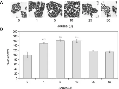
Laser stimulation was able to increase MDPC-23 cell proliferation only when performed at low energy, while higher energies did not affect cell proliferation. As shown in Figure 1, cells stimu-lated at 1, 5, 10 Joules, corresponding to 0.65, 3.25, 6.5 J/cm2respectively, display a statistically significant increase in cell proliferation com-pared to control levels. On the other hand, 25 and 50 Joules stimulation, corresponding to 16.23 and 32.47 J/cm2respectively, did not alter signifi-cantly cellular proliferation.
Figure 1. Laser effect on MDPC-23 cells proliferation A) representative images of MDPC-23 cells stimulated with near-in-frared (980 nm) laser every 24 hours for two days. Magnification 10X.B) Cellular proliferation quantification. Results are ex-pressed as mean values ± standard error of the mean.***p<0.01 compared to control.
Laser Effect on ERK 1/2 Signaling Pathway Activation
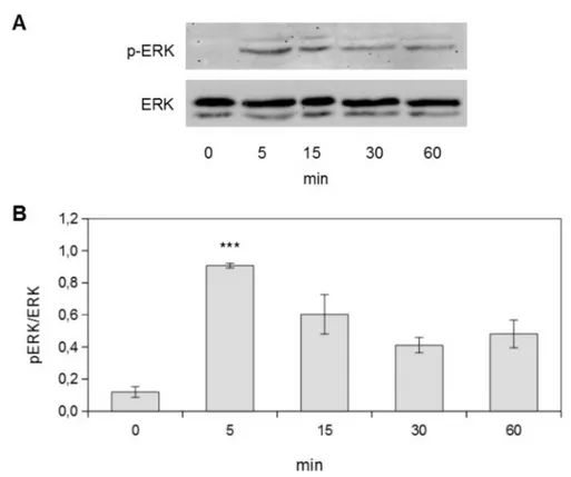
In order to evaluate the role of ERK (extracel-lular signaling regulated kinase) 1/2 signaling pathway in the observed increase in cell prolifer-ation, time course experiments on MDPC-23 cells stimulated with the energy (10 J, 6.5 J/cm2) giving the maximum proliferation response were carried out. As shown in Figure 2, 10 J (6.5 J/cm2) laser stimulation resulted in a transient in-crease in ERK 1/2 activation. In particular, this signaling pathway activation showed a maximum response 5 minutes after laser stimulation, as highlighted by phospho-ERK/ERK ratio.
In order to confirm the direct correlation be-tween laser stimulation and ERK signaling path-way activation, some experiments were per-formed in presence of a U0126, a highly selec-tive ERK inhibitor. As shown in Figure 3, U0126 pre-treatment completely abrogated laser induced ERK signaling activation.
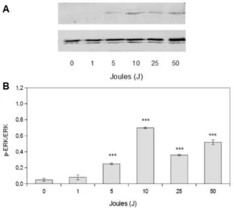
Once identified the time point corresponding to the maximum ERK activation, the effect on ERK phosphorylation of the whole stimulation
settings used in the study has been studied. As shown in Figure 4, cell proliferation increase at 5 and 10 J (3.25 and 6.5 J/cm2) corresponds to an increase in ERK phosphorylation. The in-crease in cell proliferation after 1 J (0.65 J/cm2) stimulation observed at the end of the experi-ment does not correspond to an increase in ERK activation, probably because the time point eval-uated is too short for such experimental condi-tion. When considering 25 and 50 J stimulation cell proliferation decreases: also ERK phospho-rylation decreases compared to the peak level, without disappearing completely. It is conceiv-able that such residual ERK activity results in the activation of apoptotic, rather than pro-liferative downstream signals.
Discussion
Laser therapy has been used as an alternative, noninvasive method to stimulate wound healing for the last 30 years and is also widely applied in different branches of regenerative medicine and
Figure 2. Laser stimulation induces ERK 1/2 signaling pathway activation A) representative Western blot images of MDPC-23 cells stimulated with 10 J energy. Cells were lysed at different time points.B) Densitometric quantification of ERK phos-phorylation. Results represent the mean values obtained from three independent experiments and are expressed as mean values ± standard error of the mean.***p<0.01 compared to control.
Figure 3. ERK 1/2 pathway inhibition completely abrogates laser-induced ERK 1/2 signaling activation A) Representative western blot images of MDPC-23 cells stimulated with 10 J energy in presence and absence of U0126 (10 µM). Cells were lysed 5 minutes after laser stimulation.B) Densitometric quantification of ERK phosphorylation. Results represent the mean values obtained from three independent experiments and are expressed as mean values ± standard error of the mean.***p<0.01
compared to control. In both figures: cnt = control (not stimulated); L = 10 J laser stimulation; U = 10 µM U126; L+U = 10 J laser stimulation after 10 µM U0126 pre-treatment (10 minutes).
Figure 4. Laser effect on ERK 1/2 signaling pathway activation A) Representative Western blot images of MDPC-23 cells stimulated with 1-50 J energy. Cells were lysed 5 minutes after laser stimulation.B) Densitometric quantification of ERK phosphorylation. Results represent the mean values obtained from three independent experiments and are expressed as mean values ± standard error of the mean.***p<0.01 compared to control.
dentistry, thanks to its beneficial effects on a va-riety of pathological conditions including pain relief and inflammation4,6.
Even if it is widely used for photodynamic therapy, to reduce inflammation and to stimulate cell differentiation and tissue repair, its use is still controversial6,14, as the mechanisms underlying its beneficial effects are poorly understood. Fur-thermore, the wide array of possible effects of laser stimulation observed in different cell lines by different research groups could be explained by differences in the irradiation parameters used, as the biological effects of laser irradiation de-pend on the properties of the light source (i.e. wavelength, output power and energy density).
As cell proliferation is a very important physi-ological response obtained in clinical practice by laser stimulation, such biological effect has been studied in vitro using different cellular models5. Laser irradiation has been shown to be able to in-crease cell proliferation, among the number of cell lines tested, in fibroblasts, keratinocytes, os-teoblasts, mesenchymal and cardiac stem cells, endothelial cells3-5. Moreover, it is known that such effects of photodynamic therapy result in reduction of inflammation and tissue repair15.
In the present study we focused our attention on a pre-odontoblast cell line (MDPC-23 cells): such cells have been chosen as they are the main responsible of the maintenance of tooth integrity by producing new dentin9-11.
All the experiments were performed using a near infra-red (980 nm) laser equipment with en-ergy output set to 1W in order to obtain a direct correlation between energy (Joules) and stimula-tion time (seconds). Laser equipment is provided with a pilot red light (635 nm) with a power out-put of 5 mW. Considering the experimental set-ting adopted for cell treatment, parameters re-ferred to pilot light did not affect the observed cellular responses (data not shown). Experimen-tal data presented herein show that laser stimula-tion with energy settings ranging from 1 to 10 J (0.65-6.5 J/cm2) are able to stimulate MDPC-23 cell proliferation, while higher energies do not have a statistically significant effect on such phe-nomenon, probably because of an initial toxic re-sponse, as previously observed by Migliario et al in a pre-osteoblast cell line3.
Once proved the ability of laser stimulation to increase cell proliferation, attention has been focused on the underlying molecular mecha-nism involved, as this item is currently poorly understood. In particular the role of MAPKs
(mitogen activated protein kinases) has been in-vestigated. Attention has been focused on MAPKs as this intracellular signaling pathway is known to control many aspects of mammalian cell physiology (i.e. cell proliferation, differen-tiation and death), acting by phosphorylating downstream transcription factors12. Among the components of MAPK family, ERK (extra-cel-lular signaling regulated kinase) signaling path-way is critical in regulating cell mitogenesis and differentiation: more in detail, transient activa-tion of such pathway leads to cell proliferaactiva-tion, while persistent activation results in growth ar-rest and cell differentiation13.
In the present study we demonstrated that the observed increase in cell proliferation after 5 and 10 J (3.25 and 6.5 J/cm2) stimulation corre-lated with ERK activation, a result in line with data available from the literature18. In our exper-imental model 1 J (0.65 J/cm2) laser stimulation was able to stimulate cell proliferation at the end of the experiment but this increase in cell proliferation did not reflect a ERK pathway ac-tivation: this is probably due to the timing adopted for ERK activation evaluation that could be too short for such stimulation parame-ter. Furthermore, our data show that laser stimu-lation with higher energies (25 and 50 J, corre-sponding to 16.23 and 32.47 J/cm2respectively) does not mediate a statistically significant in-crease in cell proliferation at the end of the ex-periment while determining ERK pathway acti-vation. It is conceivable that in such experimen-tal conditions ERK activation finally results in pro-apoptotic rather than prolifertive signaling pathway activation, with laser stimulation start-ing to cause toxic effects, maybe due to thermal damage or increased production of reactive oxy-gen species, as previously described for other cell lines3.
Conclusions
Even if more detailed studies will be necessary to investigate laser stimulation ability to positive-ly influence new dentin deposition, results dis-cussed herein show that near infrared (980 nm) irradiation with energy settings ranging from 1 to 10 J (0.65-6.5 J/cm2) positively influences odon-toblasts proliferation, suggesting useful applica-tions of this therapeutic approach in different clinical settings in which a regenerative tissue re-sponse is needed.
––––––––––––––––––––
Acknowledgements
The authors gratefully thanks Prof. Ercole Romagnoli for the interesting scientific discussions about laser biostimula-tion and for his helpful advice in paper preparabiostimula-tion. The study was supported by University local funds and Fon-dazione Comunità Novarese Onlus grant
–––––––––––––––––-––––
Conflict of Interest
The Authors declare that they have no conflict of interests.
References
1) LARREA-OYARBIDE N, ESPAÑA-TOST AJ, BERINI-AYTÉS L, GAY-ESCODAC. Aplicaciones del láser de diodo en
odontología. RCOE Revista del Consejo General de Colegios de Odontólogos y Estomatólogos de España 2004; 9: 529-534.
2) BENAAZZA D, CHERKAOUI A, ELMOUADDEN M,
Elmo-htarim B. Le laser en parodontie. AOS 2009; 247: 217-229.
3) MIGLIARIOM, PITTARELLAP, FANULI M, RIZZIM, RENÒ F. Laser-induced osteoblast proliferation is mediated by ROS production. Lasers Med Sci 2014; 29: 1463-1467.
4) ALGHAMDI KM, KUMAR A, MOUSSA NA. Low-level
laser therapy: a useful technique for enhancing the proliferation of various cultured cells. Lasers Med. Sci 2012; 27: 237-249.
5) GAOX, XINGD. Molecular mechanisms of cell
pro-liferation induced by low power laser irradiation. Journal of Biomedical Science 2009; 16: 1-16. 6) MOOREP, RIDGWAYTD, HIGBEE RG, HOWARD EW, LU
-CROY MD. Effect of wavelength on low-intensity laser irradiation-stimulated cell proliferation in vit-ro. Lasers Surg Med 2005; 36: 8-12.
7) GROSSMAN N, SCHNEID N, REUVENI H, HALEVY S,
LUBARTR. 780 nm low power diode laser irradiation
stimulates proliferation of keratinocyte cultures: involvement of reactive oxygen species. Lasers Surg Med 1998; 22: 212-218.
8) OLIVEIRACF, BASSOFG, LINSEC, KURACHI C, HEBLING
J, BAGNATOVS,DE SOUZACOSTACA. In vitro effect
of low-level laser on odontoblast-like cells. Laser Phys Lett. 2011; 8: 155-163.
9) LIUY, GAO J, GAO Y, XUS, ZHAN X, WUB. In vitro study of dentin hypersensitivity treated by 980-nm diode laser. J Laser Med ScI 2013; 4: 111-119. 10) DA SILVEIRA VARGAS F, SOARES DG, DIAS RIBEIRO AP,
HEBLINGJ, DESOUZACOSTACA. Protective effect of
alpha-tocopherol isomer from vitamin E against the H2O2induced toxicity on dental pulp cells.
Bio-Med Res Int 2014:895049.
11) LINDE A, GOLDBERG M. Dentinogenesis. Crit Rev
Oral Biol Med 1993; 4: 679-728.
12) RIZZIM, CRAVELLOB, RENÒF. Textile industry
manu-facturing by-products induce human melanoma cell proliferation via ERK 1/2 activation. Cell Prolif 2014; 47: 578-586.
13) CHUANG SM, LIOU GY, YANG JL. Activation of JNK, p38 and ERK mitogen-activated protein kinases by chromium (VI) is mediated through oxidative stress but does not affect cytotoxicity. Carcino-genesis 2000; 21: 1491-1500.
14) FONSECALLIMAA, DIASRIBEIROAP, GONÇALVESBASSOF, BAGNATOVS, HEBLINGJ, MARCHIGM,DESOUZACOSTA
CA. Effect of low-level laser therapy on
odonto-blast-like cells exposed to bleaching agent. Lasers Med Sci 2014; 29: 1533-1538.
15) PEREIRA LB, CHIMELLODT, FERREIRAMR, BACHMANNL, ROSA AL, BOMBONATO-PARDO KF. Low-level laser
therapy influences mouse odontoblast-like cell re-sponse in vitro. Photomed Laser Surg 2012; 30: 206-2013.
16) TRACHTENBERG A, VINOD TP, JELINEK R. Transparent,
conductive polystyrene in three dimensional con-figurations. Polymer 2014; 55: 5095-5101. 17) RIZZIM, PITTARELLAP, SABBATINIM, RENÒF. Epiregulin
induces human SK-N-BE cell differentiation through ERK1/2 signaling pathway. Growth Fac-tors 2013; 31: 90-97.
18) ALEKSIC V, AOKI A, IWASAKI K, TAKASAKI A, WANG CY, ABIKO Y, ISHIKAVA I IZUMI Y. Low-level Er:YAG laser
irradiation enhances osteoblast proliferation through activation of MAPK/ERK. Lasers Med Sci 2010; 25: 559-569.