Models of non-Alcoholic
Fatty Liver Disease and Potential Translational Value:
the Effects of 3,5-L-diiodothyronine
Elena Grasselli,* Laura Canesi,* Piero Portincasa,** Adriana Voci,* Laura Vergani,* Ilaria Demori* * Department of Earth, Environmental and Life Sciences-DISTAV, University of Genoa, Genoa, Italy. ** Clinica Medica “A. Murri”, Department of Biomedical Sciences and Human Oncology, University of Bari Medical School, Bari, Italy.September-October, Vol. 16 No. 5, 2017: 707-719
INTRODUCTION
Liver steatosis is the consequence of ectopic accumula-tion of excessive triacylglycerol (TAG) content (> 5% of liver volume or weight)1 in the liver, histologically seen when intracellular TAGs in more than 5% of the hepato-cytes.2 It is the result of an unbalance in lipid hepatic meta-bolic pathways following four possible mechanisms: • Increased delivery and/or uptake of fatty acids (FAs) or
carbohydrates due to excess dietary intake or release from adipose tissue.
• Increased de novo lipogenesis (DNL).
• Decreased very low-density lipoprotein (VLDL) syn-thesis and TAG export.
• Decreased FA catabolism due to impaired oxidation (Figure 1).
Non-alcoholic fatty liver disease (NAFLD) is consid-ered the hepatic phenotype of the metabolic syndrome (MS), associated with insulin resistance or established type 2 diabetes, increased visceral adiposity, overweight/ obesity, dyslipidemia and blood hypertension.3 NAFLD is associated with increased risk of cardiovascular, hepatic and metabolic diseases and is characterized by excessive TAG accumulation in the absence of other causes of chronic liver disease, such as hepatotoxic xenobiotics (al-cohol/drugs) and viral infections. NAFLD is recognized as the leading cause of chronic liver disease in adults and children4 and has an estimated prevalence of 20-40% in Western countries.5 The current Western diet, high in sat-urated fats and fructose, plays a significant role in the pathogenesis of NAFLD.6 NAFLD encompasses a wide spectrum of diseases ranging from simple steatosis to non-The Official Journal of the Mexican Association of Hepatology,
the Latin-American Association for Study of the Liver and the Canadian Association for the Study of the Liver
Manuscript received: Manuscript received:Manuscript received:
Manuscript received:Manuscript received: April 04, 2017. Manuscript accepted:Manuscript accepted:Manuscript accepted: June 06, 2017.Manuscript accepted:Manuscript accepted:
DOI:10.5604/01.3001.0010.2713. A B S T R A C T A B S T R A C T A B S T R A C T A B S T R A C T A B S T R A C T
Non-alcoholic fatty liver disease (NAFLD) is the most common liver disorder in industrialized countries and is associated with in-creased risk of cardiovascular, hepatic and metabolic diseases. Molecular mechanisms on the root of the disrupted lipid homeostasis in NAFLD and potential therapeutic strategies can benefit of in vivo and in vitro experimental models of fatty liver. Here, we describe the high fat diet (HFD)-fed rat in vivo model, and two in vitro models, the primary cultured rat fatty hepatocytes or the FaO rat hepatoma fatty cells, mimicking human NAFLD. Liver steatosis was invariably associated with increased number/size of lipid drop-lets (LDs) and modulation of expression of genes coding for key genes of lipid metabolism such as peroxisome proliferator-activated receptors (Ppars) and perilipins (Plins). In these models, we tested the anti-steatotic effects of 3,5-L-diiodothyronine (T2), a
metabo-lite of thyroid hormones. T2 markedly reduced triglyceride content and LD size acting on mRNA expression of both Ppars and Plins.
T2 also stimulated mitochondrial oxidative metabolism of fatty acids. We conclude that in vivo and especially in vitro models of
NAFLD are valuable tools to screen a large number of compounds counteracting the deleterious effect of liver steatosis. Because of the high and negative impact of liver steatosis on human health, ongoing experimental studies from our group are unravelling the ulti-mate translational value of such cellular models of NAFLD.
Key words.
Key words.Key words.
Key words.
alcoholic steatohepatitis (NASH) which can progress to more severe pathologies such as cirrhosis.7
At present, specific targeted pharmacological therapies are still lacking and the mainstay of therapy remains weight loss through lifestyle changes, including dietary modification and regular physical activity.5,8 The under-standing of the molecular pathways related to NAFLD is useful to avoid its harmful progression and represents the basis for finding new therapeutic strategies.9 Experimental models of NAFLD represent effectual tools for research in this field, and our group is actively involved in this re-spect.
Here, we provide a brief overview on the role of per-oxisome proliferator-activated receptors (PPARs) and li-pid droplet (LD)-associated perilipins (PLINs) in lili-pid homeostasis. We summarise and compare the features of the in vivo and in vitro models of NAFLD widely em-ployed by our group. Both models were emem-ployed to test the therapeutic potential of a thyroid hormone deriv-ative, 3,5-L-diiodothyronine, acting as a modulator of he-patic lipid metabolism. In perspective, similar models represent valuable tools to screen a large number of compounds counteracting the deleterious effect of liver steatosis.
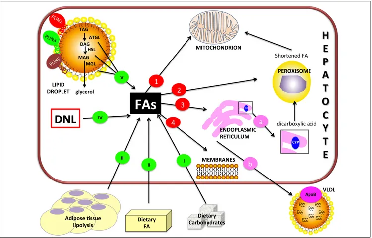
Figure 1. Figure 1. Figure 1. Figure 1.
Figure 1. Hepatic lipid metabolism/homeostasis. Free fatty acids (FAs) can derive from external sources such as dietary carbohydrates (I) and/or lipids (II)
and also from hydrolysis of fat in adipose tissue (III). Internal sources are de novo lipogenesis (DNL; IV) and release from storage organelles (V). In the cell,
FAs are stored in lipid droplet (LD) core mainly in the form of triglycerides (TAGs). LD surface is decorated with an array of proteins including members of the perilipin (PLIN) family, of which PLIN2, PLIN3 and PLIN5 are expressed in normal liver. Lipases are recruited on LD surface and this triggers TAG hydrolysis that occurs sequentially in 3 steps: TAGs are hydrolyzed to form diacylglycerol (DAG) and then monoacylglycerol (MAG), with the release of a FA at each step. These two reactions are catalyzed by adipose triglyceride lipase (ATGL) and hormone sensitive lipase (HSL) respectively. Finally, MAG is hydrolyzed by monoglyceride lipase (MGL) to release the last FA and glycerol. Glycerol can enter glycolytic or gluconeogenic pathways. FAs released from TAG hydrolysis can
be channelled to different organelles: 1. 1. 1. 1. 1. Mitochondria: short-chain (< C8), medium-chain (C8-C12), and long-chain (C12-C20) FAs, once transported by the
carnitine system, undergo β-oxidation. 2. 2. 2. 2. 2. Peroxisomes: very-long-chain (> C20) FAs undergo β-oxidation; shortened FAs released from peroxisomes are
sub-sequently oxidized in mitochondria. 3. 3. 3. 3. 3. Endoplasmic reticulum: a. a. a. a. a. in fatty liver, ω-oxidation, a minor pathway of FA metabolism, can be activated when the
mitochondrial oxidation system is inadequate to metabolize excess FAs. ω-oxidation produces long-chain dicarboxylic acids by CYP4A subfamily catalysis.
Long-chain dicarboxylic acids released from endoplasmic reticulum are subjected to further oxidation steps in peroxisomes. b. b. b. b. b. FAs can be again esterified and the
resulting TAGs are packaged into ApoB containing lipoproteins (VLDL) and consequently secreted from the cell. 4. 4. 4. 4. 4. Membranes: FAs can form phospholipids
HEPATIC LIPID HOMEOSTASIS IN HEALTH AND DISEASE
The key role of the liver in lipid homeostasis is main-tained throughout evolution. The major lipid metabolic pathways in hepatocytes are summarised in Figure 1.8
In mammals, the adipose tissue stores excess lipids in the form of TAGs in cytosolic LDs. LDs are present also in
hepatocytes, heart and skeletal myocytes, adrenocortical cells, enterocytes, and macrophages.10 LDs share their structural features with lipoproteins: both contain a neutral lipid core encased in a polar lipid monolayer, decorated by specific proteins. In the past, LDs were considered as mere fat depots, but they are now recognized as dynamic or-ganelles at the hub of lipid and energy metabolism.11 The dynamic features of LDs are confirmed by changes in the
Figure 2. Figure 2. Figure 2. Figure 2.
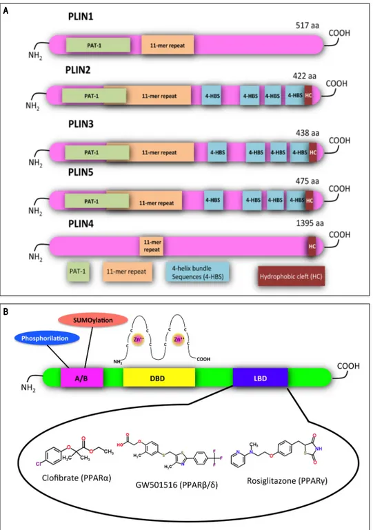
Figure 2. Structural features of
PLINs and PPARs. A.A.A.A.A. A schematic
di-agram of the structural features and the amino acid sequence similarities of PLINs. A ~100 amino acid region of high sequence similarity near their N-termini is defined as the PAT domain (PAT-1, green). Despite the absence of a clear PAT domain, PLIN4 retains sequence similarity to the other family members in other parts of the protein, including the 11-mer repeats (orange). X-ray crystallography has revealed a four-helix bundle sequence in PLIN3 (4-HBS, blue), which has been predicted also in PLIN2 and PLIN5. This domain is proposed to target the proteins to LDs. A hydrophobic cleft (HC, brown) is located at the C-termini of PLIN2, 3, 4 and 5. Adapted from references14,
84. B.B.B.B.B. Functional domains found in
PPARs and other nuclear receptors: N-terminal A/B domain (pink); DNA-bind-ing domain (DBD, yellow); ligand binding domain (LBD, blue). The DBD contains two zinc finger motifs, which bind to specific sequences of DNA known as peroxisome proliferator sponse element (PPRE) when the re-ceptor is activated. Selective agonists for the 3 PPAR isoforms are indicated. AAAAA
expression of their proteome that reflects the metabolic sta-tus of the cell.12 The most documented group of LD-asso-ciated proteins is the PLIN family,13 which comprises: perilipin (PLIN1), adipophilin/ADRP (adipose differentia-tion related protein; PLIN2), TIP47 (tail-interacting pro-tein of 47 kDa; PLIN3), S3-12 (PLIN4) and OXPAT (oxidative tissue-enriched PAT protein; PLIN5).14 The main function of PLINs is to regulate LD metabolism by selectively recruiting lipases and other proteins to LD sur-face. Their sequence features are depicted in figure 2A: a common feature of all PLINs is the propensity to coat LDs, whereas sequence differences confer specificity of function, transcriptional regulation and tissue expression.15 In the liv-er, PLINs are expressed with differences among humans and rodents. For example, PLIN1 is absent in normal and steatotic livers of mice,16 but it was found in hepatocytes isolated from humans with NAFLD. PLIN2 is constitu-tively expressed in human and murine liver. Expression of PLIN3 and PLIN5 has been widely documented in human and mouse liver, and, more recently, in rat hepatocytes.17 Upregulation of PLIN2 has been reported in the liver of rodents and humans with NAFLD.18,19 Actually, PLIN2 promotes TAG accumulation,20 inhibits FA oxidation,21 and impairs glucose tolerance.19,22 Plin2 knock-out mice are protected from hepatic steatosis when fed a HFD.23 PLIN3 is upregulated in fatty livers of HFD-fed mice and Plin3 anti-sense oligonucleotide reduces hepatic TAGs and im-proves insulin sensitivity.24 Expression of PLIN5 is in-creased in fatty livers of dystrophic mice,25 and Plin5 knock-out mice are protected from hepatic steatosis due to increased lipolysis and fat oxidation.26
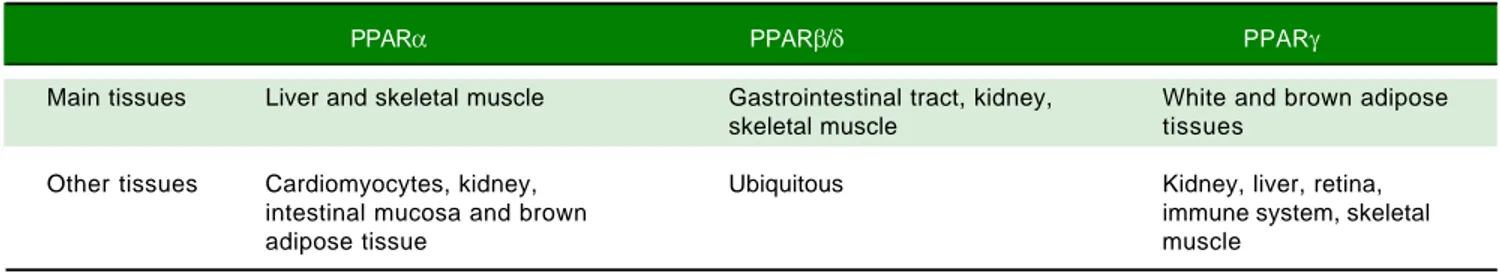
In the hepatocyte, genes coding for LD-associated Plins are under control of several transcription factors, among which a pivotal role is played by PPARs, that are ligand-activated transcription factors belonging to the nuclear hormone receptor superfamily.27 To date, three PPAR iso-forms have been identified: PPARα, PPARβ/δ and PPARγ. They share common structural features, which in-clude an amino-terminal modulatory domain, a DNA binding domain, a carboxyl-terminal ligand-binding do-main, and the ability to form heterodimers with the retin-oid X receptor. Activated heterodimers bind to peroxisome proliferator response element (PPRE) in the
Table 1. Tissue expression of peroxisome proliferator-activated receptor (PPAR) isoforms. Modified from reference 85.
PPARα PPARβ/δ PPARγ
Main tissues Liver and skeletal muscle Gastrointestinal tract, kidney, White and brown adipose
skeletal muscle tissues
Other tissues Cardiomyocytes, kidney, Ubiquitous Kidney, liver, retina,
intestinal mucosa and brown immune system, skeletal
adipose tissue muscle
promoter region of target genes 28 (Figure 2B). Many nat-ural and synthetic compounds have been identified as PPAR ligands, and this underlines the important role of PPARs as therapeutic targets.29 PPAR isoforms exhibit specific but partially overlapping tissue expression pat-terns (Table 1). In the liver, PPARα is the master regula-tor of both β- and ω-oxidation and PPARβ/δ is also required for hepatic FA oxidation.30 Conversely, PPARγ expression is induced when energy storage is required.8
PPARα expression is upregulated in the liver of high-fat diet (HFD)-fed rodents and in obese Wistar rats,31-33 as well as in human NAFLD biopsies,34 even if in some models it is reported to be decreased, possibly depending on different diet compositions and/or durations.35-36 PPARγ mediates the development of liver steatosis by mechanisms involving activation of lipogenic genes, as demonstrated both in animal models of obesity,37 and in obese patients with NAFLD.38 Treatment of ob/ob mice with the PPARγ agonist rosiglitazone increased oxidative stress and liver steatosis.39 Regarding PPARβ/δ, its contri-bution to NAFLD is still unclear. It has been reported that treatment with the PPARβ/δ agonist GW501516 amel-iorates hepatic steatosis and inflammation in the methio-nine-choline deficient (MCD) diet induced-mouse model of NASH40.
As aforementioned, genes coding for LD associated proteins are PPAR targets. Particularly, PPARγ appears to regulate the expression of Plin1, Plin2, Plin4 and Plin5 in adipose tissue, and Plin2 and Plin5 in liver.41-43 Moreover, the pharmacological PPARα agonist Wy-14643 is able to stimulate the expression of Plin5 in liver, heart, and skele-tal muscle.42,44 PPRE responsive to PPARα and PPARβ/δ were found upstream of Plin2 coding gene in both rat and human hepatocyte-derived cell lines.45
IN VIVO MODELS
OF NAFLD: THE HFD-FED RAT
Investigating the pathophysiology and possible treat-ments of NAFLD in humans is limited by the long time required for the occurrence of steatosis and the progres-sion to NASH, and also by ethical concerns regarding tis-sue collection and testing of drugs. Reliable and simple
tailed description of the HFD please refer to figure 3A legend).50,51 At the end of treatment, hepatic lipid accumu-lation was demonstrated by staining of liver sections with fat-soluble Oil red-O (ORO), that revealed the presence of numerous LDs indicating the microvesicular steatosis typical of NAFLD (Figure 4A, left panel).
IN VITRO MODELS OF NAFLD
Several in vitro models of NAFLD have been devel-oped, mainly consisting of primary hepatocyte cultures52,53 or hepatocyte cell lines54 treated with monounsaturated and/or saturated FAs (MUFAs and SFAs, respectively). These models usually employ palmitate (C16:0) or oleate (C18:1), or a mixture of the two,55 which are common long chain FAs in the Western diet56 and the most abun-dant FAs in liver of both normal subjects and patients with NAFLD.57 Adding oleate to palmitate-treated cells can ful-ly prevent palmitate lipotoxicity together with enhanced TAG synthesis.58 In this light, we used oleate:palmitate mixture in a 2:1 molar ratio to developed in vitro models of NAFLD in rat hepatic cells. For better resembling in vivo animal models resembling the most important features of
NAFLD are needed. Even though some studies have pro-posed non-rodent mammalian models, the most used are still those employing mouse and rat.46,47
In vivo NAFLD models are represented by two main
categories: genetic and dietary. The most frequent genetic models are the spontaneously diabetic rodents deficient in leptin or leptin receptors, which include ob/ob mouse, db/db mouse and Zucker fatty rat.48 Rodent dietary models encompass several treatments that differ in composition and duration, such as methionine and choline deficient (MCD) diet, high-fructose and/or sucrose diet, cafeteria diet, high cholesterol diet and HFD.49 Diversities in die-tary models also regard strain, sex and age of animals, the amount of energy derived from fats, the origin of fats, the ratio of saturated (SFA) to monounsaturated (MUFA) and polyunsaturated (PUFA) fatty acids, and the proportion between ω-3 and ω-6 PUFA. Such differences are reflect-ed in outcome variations ranging from mild hepatic steato-sis to severe NASH.47
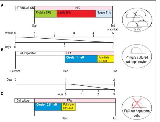
In our studies, we utilized male Wistar rats, which were administered for one month with a HFD (for a
de-Figure 3. Figure 3. Figure 3. Figure 3.
Figure 3. Experimental protocols for in vivo and in vitro models of NAFLD. In this figure we compare the number of animals needed and the duration of
the treatments used in vivo and in vitro
to obtain our 3 different models of NAFLD: a dramatic decrease in animal use and a high speeding up of the ex-perimental procedures can be
appreci-ated. AAAAA... In vivo model of NAFLD
(HFD). Male Wistar rats (aged 8 weeks) were housed in individual cages in a temperature-controlled room at 28
oC with a 12:12-h light:dark cycle. After
one week of acclimation, the animals were administered the HFD where most of the total metabolizable percent-age of energy derived from fats with re-spect to sugars (21% carbohydrates, 29% proteins, 50% lipids (19.85 kJ
gross energy/g).50,51 In the standard
control diet, total metabolizable per-centages of energy were: 60.4% carbo-hydrates, 29% proteins, 10.6% lipids (15.88 kJ gross energy/g). At least 5 animals must be included in each experimental group (Control and HFD-fed). At the
end of the treatments, all rats were sacrificed. The total time required to obtain hepatocellular steatosis was 5 weeks. B.B.B.B.B. In vitro model of NAFLD using rat
hepatocyte primary cultures. Hepatocytes were isolated from only one animal, plated and allow to attach and restore. The day after, cells were treated with
oleate:palmitate (FA) mixture 2:1 (1.5 mM) for 24 h. The total time required to obtain hepatocellular steatosis was 48 h. C.C.C.C. C.In vitro model of NAFLD using
FaO rat hepatoma cell line. Cells were routinely passaged and when at 70-80% confluence they were incubated with oleate:palmitate (FA) mixture 2:1; (0.75 mM) for 3 h. No rats were sacrificed and the total time required to obtain hepatocellular steatosis was 3 h.
A A A A A B B B B B C C C C C STABULATION HFD
Proteins 29% Lipids 50% Sugars 21%
Start End (sacrifice) Weeks 0 1 2 3 4 5 Days 0 1 2 In vivo Primary cultured rat hepatocytes
FaO rat hepatoma cells Cell preparation FFA
Oleate 1 mM Oleate 1 mM Oleate 1 mM Oleate 1 mM Oleate 1 mM Palmitate 0.5 mM
Sacrifice Start End
Days 0 1 2
Hours 0 1 2 3
Cell culture FFA
Oleate 0.5 mM Oleate 0.5 mMOleate 0.5 mM Oleate 0.5 mM Oleate 0.5 mM Palmitate 0.25 mM Start End
conditions, we chose FA concentrations comparable to the plasmatic levels of patients with MS.59
Rat hepatocyte primary cultures
Rat hepatocyte primary cultures have been extensively used in basic research on liver function.60 To isolate intact hepatocytes,61 we basically applied the method of Seglen,62 followed by maintenance of cells in appropriate culture
Table 2. Expression of Plins and Ppars and TAG secretion in in vivo and in vitro models of NAFLD. Changes in mRNA expression
lev-els of perilipin (Plins) proteins (Plin2; Plin3; Plin5) and isoforms of peroxisome proliferator activated receptors (Pparγ; Pparα; Ppar β/δ), in three different in vivo and in vitro models of NAFLD. Changes in triglyceride (TAG) secretion are also shown. Significant changes are reported as percentage with respect to controls and “ - ” indicates no changes.
In vivo51,69 Primary cultured rat FaO rat hepatoma
hepatocytes64,71 cells68,70 % 0 7 + % 0 4 + % 6 5 + 2 N I L P + % 3 9 + % 5 8 + 5 N I L P 187% -3 N I L P PPARα +211% - -PPAR β/δ - +78% -41% PPARγ +76% +68% +71% % 2 6 + % 0 3 + % 3 5 + n o it e r c e s G A T Figure 4. Figure 4. Figure 4. Figure 4.
Figure 4. Neutral lipid accumulation and PLIN2 immunostaining in in vivo and in vitro models of NAFLD. Oil red-O (ORO, left panel) and PLIN2 (right panel)
staining in control (C) and steatotic hepatocytes (HFD/FA) in liver tissue sections (AAAAA), primary cultured hepatocytes (BBBBB) and FaO rat hepatoma cells (CCCCC). Lipid
accumulation as well as increase in PLIN2-positive vesicles is evident for all NAFLD models. Immunostaining was performed with PLIN2 polyclonal anti-body (clone GP40-mN1, Fitzgerald Industries International, Concord, MA, USA) (magnification 100X).
O R O O R O O R O O R O O R O A F / D F H C P L I N 2P L I N 2 P L I N 2 P L I N 2 P L I N 2 A F / D F H C AAAAA BBBBB C CC CC
conditions.63 We cultured rat hepatocytes in serum-free DMEM containing 0.25% bovine serum albumin (BSA). After 24 h of adaptation to the in vitro environment, hepato-cytes were exposed to a mixture of oleate/palmitate (2:1 molar ratio, total concentration 1.5 mM) for 24 h17,64 (Fig-ure 3B and 4B, left panel).
Primary hepatocytes do not proliferate in vitro and can be used only within the first week of culture, because over time they tend to lose the differentiated adult liver
pheno-type. Cells need to be freshly isolated for each experi-ment, but a pure hepatocyte population (100-400 millions) with high viability is routinely obtained from each animal. Hepatocellular steatosis in primary cultures is achieved within only 1 day of treatment with FA mixture; in con-trast, 4 weeks of HFD administration to rats are necessary to induce NAFLD in vivo (Figure 3B and 4B left panel).
Rat hepatoma cell line
The FaO rat hepatoma cells express a broad array of liv-er-specific mRNAs and maintain the ability to assemble and secrete VLDL65,66 as well as to respond to stimuli that activate PPARs.67 In our experiments, FaO cells at 70-80% confluence were incubated in starvation medium (F12 Coon’s modified medium supplemented with 0.25% BSA) with a mixture of oleate/palmitate (2:1 molar ratio, total concentration 0.75 mM). It is noteworthy that basal lipid content of FaO cells is much lower than that of primary hepatocytes, thus, FA treatment resulted in a more rapid and pronounced TAG accumulation with respect to pri-mary cultured hepatocytes reaching a plateau at 3-6 h68 (Figure 3C). Moreover, the effect was induced using a lower concentration of FA mixture (0.75 mM vs. 1.5 mM). In these conditions, a marked increase in number and size of cytosolic LDs was observed by ORO staining (Figure 4C, left panel). These observations candidate lipid-loaded FaO cells as a reliable and convenient in vitro model of NAFLD.
EXPRESSION OF LIPID METABOLISM GENES AND SECRETION OF LIPIDS IN MODELS OF NAFLD
The expression of Plins and Ppars, as well as TAG secre-tion, were altered in both in vitro and in vivo models of NAFLD. Our findings are summarized in table 2.51,64,68-71
Changes in Plin expression resulted fully consistent in all models. Plin5 and Plin2 mRNA levels were always sig-nificantly increased compared to controls. In particular, im-munohistochemical staining of PLIN2 revealed little or absent PLIN2 immunoreactivity in controls cells of all models. Lipid accumulation in hepatocytes was accompa-nied by a dramatic increase in size and number of PLIN2-positive vesicles both in vivo and in vitro (Figures 4A-4C; right panel). No changes in the expression of Plin3 were observed in either steatotic liver or fatty hepatocytes. How-ever, an increased expression of Plin3 was observed in pri-mary hepatocytes at shorter (12 h) times of FA exposure.17 Therefore, induction of Plin3 may represent a transient and short-time response of the hepatocyte to excess FAs.
Also expression of the different Ppar isoforms was modified in all models of NAFLD (Table 2). High Pparγ expression is one of the main features of the steatotic
liver.37,38 Accordingly, increased Pparγ expression was a common trait observed in all experimental models. Pparα is mainly involved in regulation of lipid oxidation. HFD provides excess of long-chain FAs entering the hepato-cytes with consequent activation of Pparα and upregula-tion of those PPARα-target genes involved in FA catabolism.72 We observed that Pparα expression was in-creased in the liver of HFD rats, whereas no changes were recorded in vitro. These differences may be ascribed to the experimental conditions utilized to induce lipid accumu-lation in vivo and in vitro. Moreover, in NAFLD animal models, some authors documented a decrease in Pparα expression related to an impaired mitochondrial FA oxi-dation and to a decrease in carnitine-palmitoyl-transferase (Cpt1) expression.27 Accordingly, in HFD-fed rats, the in-creased Pparα expression was associated with an increase in Cpt1 enzymatic activity.73 It appears that diet duration can play an important role in determining the dynamic equilibrium of Ppar regulation. De Lange, et al.74 docu-mented increased expressions of Pparα and Pparβ/δ after 2 weeks of HFD administration, which could be a compen-satory mechanism since it was abolished after 4 weeks.
Regarding Pparβ/δ mRNA levels, they were not affected in HFD rats, but they were upregulated in steatotic prima-ry hepatocytes, and down-regulated in FaO cells. These discrepancies further underline the uncertainties about the role of Pparβ/δ in NAFLD.
Finally, in the general scenario of the modulation of lipid pathways involved in NAFLD, increasing VLDL secretion can be considered as a compensatory mechanism to over-come lipid overload. Actually, in all experimental models an enhanced lipid secretion was observed: increased levels of TAGs were detected in serum of HFD-rats, as well as in culture media of fatty hepatocytes (Table 2).
Taking into account the differences among the experi-mental models considered in our studies, the results un-derline how the main markers of hepatic steatosis are maintained in the in vivo and in vitro models, thus making the simpler in vitro model suitable for rapid study of direct anti-steatotic effects of natural and artificial compounds.
POTENTIAL THERAPY FOR
LIVER STEATOSIS: THE LIPID LOWERING EFFECT OF 3,3’,5-L-DIIODOTHYRONINE
The liver is a major target for the thyroid hormones (THs) thyroxine (T4) and 3,5-L-diiodothyronine (T3) that play a key role in energy balance and lipid metabo-lism. High levels of THs stimulate energy expenditure and fat oxidation.75 Studies on humans and animal mod-els indicate that NAFLD is associated with impaired TH signalling.75 The use of THs as potential drugs to treat obesity, hyperlipidemia and MS has been
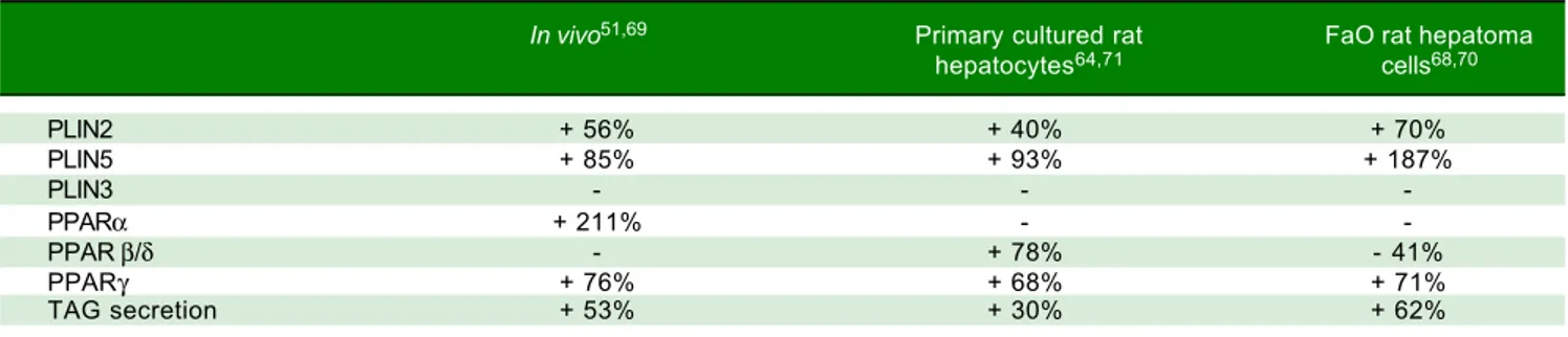
suggest-ed.76 However, due to their collateral dangerous effects, their employment is not recommended. The identifica-tion of TH agonists/analogs retaining anti-obesity and hypolipemic efficacies, while being devoid of thyrotox-ic effects, would represent a potential therapeutthyrotox-ic ad-vance. Several iodothyronines other than T3 and T4 have been proven to display some thyromimetic activities. Among them, 3,5-L-diiodothyronine (T2) mimics sev-eral effects of T3 on energy metabolism without induc-ing thyrotoxic effects.77 The in vivo HFD model of NAFLD described above has been widely utilized to demonstrate T2 action on hepatic lipid metabo-lism.51,69,73,78,79 Rats receiving T
2 along with HFD did not gain weight respect to controls (Figure 5A). In the liver, T2 administration resulted in an almost complete absence of fat accumulation, as revealed by ORO stain-ing (Figure 5B), and HFD-associated lipid
peroxida-tion (Figure 5D); moreover, Pparα and Plin2 mRNA up-regulation in HFD rats was prevented by T2 (Figure 5C). These effects were associated to stimulation of FA oxidation in mitochondria and peroxisomes.72,73
The in vivo studies do not allow to distinguish between the direct anti-steatotic effects of T2 and its indirect effects due to upstream changes in endocrine or metabolic path-ways. Moreover, since in vivo T2 was given to rats simulta-neously to HFD, the observed effects demonstrated that T2 was able to prevent, rather than to ameliorate, hepatic lipid overload. In order to investigate the direct mecha-nism of the lipid-lowering effect of T2 on hepatocytes, as well as its capacity to reverse excess fat accumulation, T2 was administered in vitro after lipid-overloading of cells.
In hepatocyte primary cultures,64 cells were incubat-ed with FA mixture for 24 h; then, the culture mincubat-edium was replaced and supplemented with T2 at different Figure 5.
Figure 5. Figure 5. Figure 5.
Figure 5. In vivo anti-steatotic effects of T2. T2 was administered daily by i.p. injection (25 µg/100 g bw) to rats simultaneously receiving HFD. A.A.A.A.A. Body
weight gain measured throughout the experimental period (30 days) for the 3 experimental groups: C are standard diet-fed rats. Values are expressed as
means ± SD of body-weight gain (%). B.B.B.B.B. ORO staining in liver tissue sections in the 3 experimental groups (magnification 100X). C.C.C.C.C. Relative mRNA expression of
Pparα and Plin2 evaluated by RT-qPCR. Data (mean ± SD, N = 4) are reported as fold induction with respect to controls after normalization for Gapdh
mRNA. Significant differences are indicated (C vs. HFD **p < 0.01, ***p < 0.001; HFD vs. HFD+T2 #p < 0.05, ###p < 0.001). D.D.D.D.D. Liver lipid peroxidation
evalu-ated by TBARs assay. Data (mean ± SD, N = 4) are expressed as nmol MDA/mg protein. Significant differences are indicevalu-ated (C vs. HFD ***p < 0.001; HFD
+ T2 vs. FA ###p < 0.001). C HFD HFD+T2 BBBBB 1.2 1.0 0.8 0.6 0.4 0.2 0.0 AAAAA Bo dy w ei gh t g ai n (% ) 40 35 30 25 20 15 10 5 0 0 5 10 15 20 25 30 Days C HFD HFD + T2 C CC CC Re la tiv e ex pr es sio n 3.5 3.0 2.5 2.0 1.5 1.0 0.5 0.0 Pparα Plin2 ** *** # ### C HFD HFD + T2 D DD DD C HFD HFD + T2 nm ol M D A/ m g pr ot ei n *** ###
Figure 6.
Figure 6.Figure 6.
Figure 6.Figure 6. In vitro lipid-lowering effects of T2. Primary rat hepatocytes or FaO hepatoma cells were lipid-loaded with FA mixture as described in the text.
Af-ter 24 h, the culture medium was replaced and supplemented with T2 at 10-5 M and cells were incubated for further 24 h. A.A.A.A.A. Representative image of
ORO-stained control (C) and FA hepatocytes incubated in the absence or in the presence of T2 (FA + T2). Nuclear staining with hematoxylin is also shown
(magnification 40X and 100X). B.B.B.B.B. Relative mRNA expression of Cpt1 evaluated by RT-qPCR. C.C.C.C.C. Relative mRNA expression of Plin2 evaluated by RT-qPCR.
D.
D.D.
D.D. Relative mRNA expression of Pparγ evaluated by RT-qPCR. B-D.B-D.B-D.B-D. Data (means ± SD, N = 4) are reported as fold induction with respect to controls afterB-D.
normalization for Gapdh mRNA. Significant differences are indicated (C vs. FA *p < 0.05, ***p < 0.001; FA vs. FA+T2 #p < 0.05, ##p < 0.01, ###p < 0.001).
concentrations (from 10-7 to 10-5 M) for further 24 h. A reduction in number and average size of LDs was ob-served after treatment with T2 (Figure 6A), suggesting that the iodothyronine led to dispersion/fragmentation of LDs. A decrease in LD size could make the stored TAGs more accessible to enzymes acting on catabolism/ secretion of FAs and this could explain, at least partial-ly, the ability of T2 to decrease the excess fat by acting directly on the hepatic cell. Actually, in steatotic hepa-tocytes T2 was able to reduce lipid accumulation through the recruitment of adipose triglyceride lipase (ATGL) on LD surface, and to down-regulate Plin3.71 The excess FAs produced by the activity of ATGL were mainly channeled into mitochondria for β-oxidation rather than to secretion, as suggested by both up-regula-tion of Cpt1 expression (Figure 6B) and stimulaup-regula-tion of cytochrome-c oxidase (COX) activity.71 These data
in-dicate a stimulation of the mitochondrial function, in line with results reported in vivo.73 Regarding Ppars and
Plins, T2 was able to reduce the up-regulation of Pparγ
induced by FA exposure, whereas Plin2 expression was unaffected (Figures 6C-6D).
The use of primary rat hepatocytes allowed to verify that the lipid-lowering effect of T2 was a direct action on the hepatocyte, but the involvement of thyroid hor-mone receptors (TRs) in mediating this action re-mained to be elucidated. Therefore, the same experiments were performed in the FaO rat hepatoma cell line, that is defective for functional TRs.68,70 Treat-ment of steatotic FaO cells with T2 for 24 h reduced both TAG content and the number and size of LDs (Figure 6A). As in primary cultured hepatocytes, T2 stimulated mitochondrial FA oxidation by up-regulat-ing the expression of Cpt1 (Figure 6B). The lipid-low-AAAAA C FA FA+T2 Primary culture d ra t hepato cy te s FaO ra t hepatoma cell lin e
Primary cultured rat FaO rat hepatoma hepatocytes cell line BBBBB 6.0 5.0 4.0 3.0 2.0 1.0 0.0 ## *** ### * C FA FA + T2 D DD DD Ppar γ Relative expressio n 2.5 2.0 1.5 1.0 0.5 0.0
Primary cultured rat FaO rat hepatoma hepatocytes cell line
C FA FA + T2 C CC CC Plin 2 Relative expressio n
Primary cultured rat FaO rat hepatoma hepatocytes cell line 3.5 3.0 2.5 2.0 1.5 1.0 0.5 0.0 C FA FA + T2 *** *** ### *** ### * # Cpt 1 Relative expressio n
ering action of T2 was accompanied by a down-regulation of Pparγ expression and an up-regulation of Plin2 (Fig-ure 6C-6D). Moreover, expression of Atgl, Plin5 and un-coupling protein 2 (Ucp2) was also up-regulated by T2.70 The possible role of these proteins in the mechanism underlying the lipid-lowering effects of T2 are schemat-ically depicted in figure 7.
In conclusion, the use of the two in vitro models of fatty hepatocytes allowed to demonstrate that the lipid-lower-ing effects of T2 depend on a direct interaction with the hepatic cell that, at least in part, occurs via “non-receptor-mediated” mechanisms and involves a short-term action by direct stimulation of mitochondrial activity. Altogeth-er, our data support the possible utilization of T2 as a phar-macological tool in the treatment of NAFLD, also in light of its lack of thyrotoxic effects.80
POTENTIAL TRANSLATIONAL VALUE OF SUCH STUDIES
Many efforts have been made in order to introduce reliable and simple models resembling the most im-portant features of NAFLD. In vitro models have the ad-vantage of reducing the use of animals by providing a simpler and highly reproducible system, where the mechanisms can be studied directly at the cellular level, keeping the translational value of the observed results. Therefore, in vitro models offer the possibility to test the therapeutic potential and elucidate the mechanisms of action of a large spectrum of synthetic and natural compounds, without exposing humans to unnecessary
risks.81 All together our studies demonstrated the effica-cy of T2 as anti-steatotic and hepatoprotective com-pound in both in in vivo and in vitro models of NAFLD. These models, especially in vitro model using FaO cell line, may be employed to test the therapeutic potential of other emerging compounds.
CONCLUSIONS AND FUTURE PERSPECTIVES
NAFLD is closely related to obesity and insulin resist-ance and can progress to the necro-inflammatory form of non-alcoholic steatohepatitis. The incidence of NAFLD is rapidly increasing in industrialized countries, and is steer-ing the increased risk of cardiovascular disease and type 2 diabetes. It is therefore mandatory to identify healthier lifestyles and to target NAFLD and its progression by highly active molecules.82 Whereas human studies are of-ten burdened by several ethical, economical and time lim-itations, both animal and cellular models still provide valuable information in this field and will be confirmed at a translational level by clinical studies.47,83 In vitro ap-proaches can be particularly useful to study the molecular mechanisms involved in the onset and progression of NAFLD and to check the effects and mechanisms of ac-tion of endogenous and exogenous compounds, as shown in the case of T2.
COMPETING INTEREST
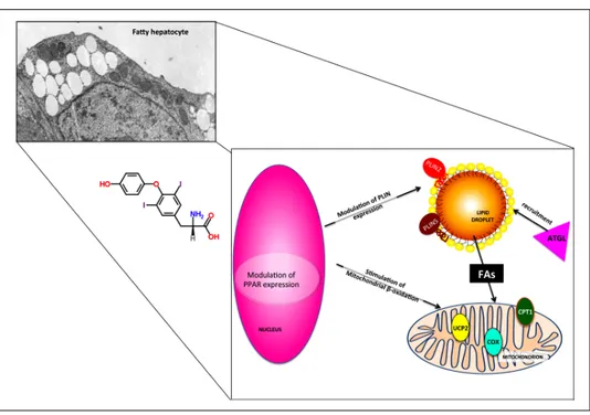
The authors declare that they have no competing interests. Figure 7.
Figure 7. Figure 7. Figure 7.
Figure 7. Possible mechanisms under-lying the direct lipid-lowering effects of
T2. The effects of T2 on fatty
hepato-cytes are partially TR-independent and occur both at the nuclear and cytoplas-mic level. PPAR and PLIN expressions are modulated and adipose triglyceride lipase (ATGL) is recruited on LD sur-face, thus inducing FA release form LD core and their subsequent channeling into mitochondrion. The up-regulation of CPT1 and UCP2 expression and the stimulation of cytochrome-c oxidase (COX) activity indicate a stimulation of mitochondrial function.
ACKNOWLEDGMENTS
We wish to thank for their collaboration Dr. Rita De Matteis and Mr. Valter Capicchioni. Many thanks to Prof. Gabriella Gallo for encouragements and stimulating discussions. Funding support for the studies mentioned here came from MIUR-COFIN 2008 (Prot. 20089SR S2X_002); Compagnia San Paolo, Torino (prat. no. 2009.1824-1067/IT/ pv); Fondi Ateneo Universitá degli Studi di Genova, Area-05, and Fondazione CARIGE (Prot. no. 2010/584-23), and European Union’s Horizon 2020 research and innovation programme under the Marie Sklodowska-Curie grant agreement No. 722619.
REFERENCES
1. Hoyumpa AM, Greene HL, Dunn GD, Schenker S. Fatty liver: biochemical and clinical considerations. Am J Dig Dis 1975; 20: 1142-70.
2. Kleiner DE, Brunt EM, Van Natta M, Behling C, Contos MJ, Cummings OW, Ferrell LD, et al. Design and validation of a histological scoring system for nonalcoholic fatty liver dis-ease. Hepatology 2005; 41: 1313-21.
3. Krawczyk M, Bonfrate L, Portincasa P. Nonalcoholic fatty liver disease. Best Pract Res Clin Gastroenterol 2010; 24: 695-708.
4. Love-Osborne KA, Nadeau KJ, Sheeder J, Fenton LZ, Zeitler P. Presence of the metabolic syndrome in obese ado-lescents predicts impaired glucose tolerance and nonalco-holic fatty liver disease. J Adolesc Health 2008; 42: 543-8. 5. Chalasani N, Younossi Z, Lavine JE, Diehl AM, Brunt EM,
Cusi K, Charlton, et al. The diagnosis and management of non-alcoholic fatty liver disease: practice Guideline by the American Association for the Study of Liver Diseases, American College of Gastroenterology, and the American Gastroenterological Association. Hepatology 2012; 55: 2005-23.
6. Basaranoglu M, Basaranoglu G, Sabuncu T, Senturk H. Fruc-tose as a key player in the development of fatty liver dis-ease. World J Gastroenterol 2013; 19: 1166-72.
7. Abenavoli L, Milic N, Di Renzo L, Preveden T, Mediæ-Sto-janoska M, De Lorenzo A. Metabolic aspects of adult pa-tients with nonalcoholic fatty liver disease. World J
Gastroenterol 2016; 22: 7006-16.
8. Musso G, Gambino R, Cassader M. Recent insights into he-patic lipid metabolism in non-alcoholic fatty liver disease (NAFLD). Prog Lipid Res 2009; 48: 1-26.
9. Calamita G, Portincasa P. Present and future therapeutic strategies in non-alcoholic fatty liver disease. Expert Opin
Ther Targets 2007; 11: 1231-49.
10. Walther TC, Farese RV. The life of lipid droplets. Biochim
Bi-ophys Acta-Mol Cell Biol Lipids 2013; 1791: 459-66.
11. Thiam AR, Farese RV, Walther TC. The biophysics and cell biology of lipid droplets. Nat Rev Mol Cell Biol 2013; 14: 775-86.
12. Pol A, Gross SP, Parton RG. Biogenesis of the multifunction-al lipid droplet: Lipids, proteins, and sites. J Cell Biol 2014; 204: 635-46.
13. Kimmel AR, Brasaemle DL, McAndrews-Hill M, Sztalryd C, Londos C. Adoption of PERILIPIN as a unifying nomenclature for the mammalian PAT-family of intracellular lipid storage droplet proteins. J Lipid Res 2010, 51: 468-71.
14. Bickel PE, Tansey JT, Welte MA. PAT proteins, an ancient family of lipid droplet proteins that regulate cellular lipid stores. Biochim Biophys. Acta - Mol Cell Biol Lipids 2009; 1791: 419-40.
15. Ducharme NA, Bickel PE. Minireview: Lipid Droplets in Lipo-genesis and Lipolysis. Endocrinology 2008; 149: 942-9. 16. Dalen KT. PPAR activators and fasting induce the
expres-sion of adipose differentiation-related protein in liver. J Lipid
Res 2006; 47: 931-43.
17. Grasselli E, Voci A, Pesce C, Canesi L, Fugassa E, Gallo G, Vergani L. PAT protein mRNA expression in primary rat hepatocytes: Effects of exposure to fatty acids. Int J Mol
Med 2010; 25: 505-12.
18. Straub BK, Stoeffel P, Heid H, Zimbelmann R, Schirmacher. Differential pattern of lipid droplet-associated proteins and de novo perilipin expression in hepatocyte steatogenesis.
Hepatology 2008; 47: 1936-46.
19. Varela GM, Antwi DA, Dhir R, Yin X, Singhal NS, Graham MJ, Crooke RM, et al. Inhibition of ADRP prevents diet-induced in-sulin resistance. AJP Gastrointest Liver Physiol 2008; 295: G621-G628.
20. Imai Y, Varela GM, Jackson MB, Graham MJ, Crooke RM, Ahi-ma, RS. Reduction of Hepatosteatosis and Lipid Levels by an Adipose Differentiation-Related Protein Antisense Oligonu-cleotide. Gastroenterology 2007; 132: 1947-54.
21. Magnusson B. Adipocyte Differentiation-Related Protein Pro-motes Fatty Acid Storage in Cytosolic Triglycerides and In-hibits Secretion of Very Low-Density Lipoproteins.
Arterioscler Thromb Vasc Biol 2006; 26: 1566-71.
22. Chang BH-J, Li L, Saha P, Chan L. Absence of adipose dif-ferentiation related protein upregulates hepatic VLDL secre-tion, relieves hepatosteatosis, and improves whole body insulin resistance in leptin-deficient mice. J Lipid Res 2010; 51: 2132-42.
23. Chang BH-J, Li L, Paul A, Taniguchi S, Nannegari V, Heird WC, Chan L. Protection against Fatty Liver but Normal Adipo-genesis in Mice Lacking Adipose Differentiation-Related Pro-tein. Mol Cell Biol 2006; 26: 1063-76
24. Carr RM, Patel RT, Rao V, Dhir R, Graham MJ, Crooke RM, Ahima RS. Reduction of TIP47 improves hepatic steatosis and glucose homeostasis in mice. AJP Regul Integr Comp
Physiol 2012; 302: R996-R1003.
25. Hall AM, Brunt EM, Chen Z, Viswakarma N, Reddy JK, Wolins NE, Finck BN. Dynamic and differential regulation of proteins that coat lipid droplets in fatty liver dystrophic mice. J Lipid
Res 2010; 51: 554-63.
26. Wang C, Zhao Y, Gao X, Li L, Yuan Y, Liu F, Zhang L, et al. Perilipin 5 improves hepatic lipotoxicity by inhibiting lipolysis.
Hepatology 2015; 61: 870-82.
27. Souza-Mello V. Peroxisome proliferator-activated receptors as targets to treat non-alcoholic fatty liver disease. World J
Hepatol 2015; 7: 1012-9.
28. Moras D, Gronemeyer H. The nuclear receptor ligand-binding domain: structure and function. Curr Opin Cell Biol 1998; 10: 384-91.
29. Wahli W, Michalik L. PPARs at the crossroads of lipid signal-ing and inflammation. Trends Endocrinol Metab 2012; 23: 351-63.
30. de Lange P, Lombardi A, Silvestri E, Goglia F, Lanni A, Moreno M. Peroxisome Proliferator-Activated Receptor Delta: A Conserved Director of Lipid Homeostasis through Regula-tion of the Oxidative Capacity of Muscle. PPAR Res 2008; 2008: 172676.
31. Akbiyik F, Cinar K, Demirpence E, Ozsullu T, Tunca R, Hazi-roglu R, Yurdaydin C, et al. Ligand-induced expression of peroxisome proliferator-activated receptor alpha and
activa-tion of fatty acid oxidaactiva-tion enzymes in fatty liver. Eur J Clin
Invest 2004; 34: 429-35.
32. Hsu S-C, Huang C. Changes in liver PPARa mRNA expres-sion in response to two levels of high-safflower-oil diets correlate with changes in adiposity and serum leptin in rats and mice. J Nutr Biochem 2007; 18: 86-96.
33. Patsouris D, Reddy JK, Müller M, Kersten S. Peroxisome Pro-liferator-Activated Receptor á Mediates the Effects of High-Fat Diet on Hepatic Gene Expression. Endocrinology 2006, 147: 1508-16.
34. Nakamuta M, Kohjima M, Morizono S, Kotoh K, Yoshimoto T, Miyagi I, Enjoji M. Evaluation of fatty acid metabolism-related gene expression in nonalcoholic fatty liver disease. Int J Mol
Med 2005; 16: 631-5.
35. Souza-Mello V, Gregório BM, Cardoso-de-Lemos FS, de Car-valho L, Aguila MB, Mandarim-de-Lacerda CA. Comparative effects of telmisartan, sitagliptin and metformin alone or in combination on obesity, insulin resistance, and liver and pan-creas remodelling in C57BL/6 mice fed on a very high-fat diet. Clin Sci 2010; 119: 239-50.
36. Fraulob JC, Souza-Mello V, Aguila MB, Mandarim-de-Lacerda CA. Beneficial effects of rosuvastatin on insulin resistance, adiposity, inflammatory markers and non-alcoholic fatty liver disease in mice fed on a high-fat diet. Clin Sci 2012; 12: 259-70.
37. Rahimian R, Masih-Khan E, Lo M, van Breemen C, McManus BM, Dube GP. Hepatic over-expression of peroxisome prolif-erator activated receptor gamma 2 in the ob/ob mouse model of non-insulin dependent diabetes mellitus. Mol Cell
Bio-chem 2001; 224: 29-37.
38. Pettinelli P, Videla LA. Up-Regulation of PPAR-gamma mRNA Expression in the Liver of Obese Patients: an Additional Re-inforcing Lipogenic Mechanism to SREBP-1c Induction. J Clin
Endocrinol Metab 2011; 96: 1424-30.
39. García-Ruiz I, Rodríguez-Juan C, Díaz-Sanjuán T, Martínez MA, Muñoz-Yagüe T, Solís-Herruzo JA. Effects of rosiglita-zone on the liver histology and mitochondrial function in ob/ ob mice. Hepatology 2007; 46: 414-23.
40. Nagasawa T, Inada Y, Nakano S, Tamura T, Takahashi T, Maruyama K, Yamazaki Y, et al. Effects of bezafibrate, PPAR pan-agonist, and GW501516, PPARdelta agonist, on development of steatohepatitis in mice fed a methionine- and choline-deficient diet. Eur J Pharmacol 2006; 536: 182-91. 41. Dalen KT, Schoonjans K, Ulven SM, Weedon-Fekjaer MS,
Bentzen TG, Koutnikova H, Auwerx J, et al. Adipose tissue expression of the lipid droplet-associating proteins S3-12 and perilipin is controlled by peroxisome proliferator-activat-ed receptor-gamma. Diabetes 2004; 53: 1243-52.
42. Wolins NE, Quaynor BK, Skinner JR, Tzekov A, Croce MA, Gropler MC, Varma V, et al. OXPAT/PAT-1 Is a PPAR-In-duced Lipid Droplet Protein That Promotes Fatty Acid Utiliza-tion. Diabetes 2006; 55: 3418-28.
43. Schadinger SE, Bucher NLR, Schreiber BM, Farmer SR. PPARgamma2 regulates lipogenesis and lipid accumulation in steatotic hepatocytes. Am J Physiol Endocrinol Metab 2005; 288: E1195-E1205.
44. Yamaguchi T, Matsushita S, Motojima K, Hirose F, Osumi T. MLDP, a Novel PAT Family Protein Localized to Lipid Droplets and Enriched in the Heart, Is Regulated by Peroxisome Prolif-erator-activated Receptor. J Biol Chem 2006; 281: 14232-40.
45. Targett-Adams P, McElwee MJ, Ehrenborg E, Gustafsson MC, Palmer CN, McLauchlan J. A PPAR response element regulates transcription of the gene for human adipose differ-entiation-related protein. Biochim Biophys Acta - Gene
Struct Expr 2005; 1728: 95-104.
46. Kanuri G, Bergheim I. In vitro and in vivo models of non-alco-holic fatty liver disease (NAFLD). Int J Mol Sci 2013; 14: 11963-80.
47. Kucera O, Cervinkova Z. Experimental models of non-alco-holic fatty liver disease in rats. World J Gastroenterol 2014; 20: 8364-76.
48. Anstee QM, Goldin RD. Mouse models in non-alcoholic fatty liver disease and steatohepatitis research. Int J Exp Pathol 2006; 87: 1-16.
49. Machado MV, Michelotti GA, Xie G, Almeida Pereira T, de Al-meida TP, Boursier J, Bohnic, B, et al. Mouse models of diet-induced nonalcoholic steatohepatitis reproduce the heterogeneity of the human disease. PLoS One 2015; 10: e0127991.
50. Demori I, Voci A, Fugassa E, Burlando, B. Combined effects of high-fat diet and ethanol induce oxidative stress in rat liv-er. Alcohol 2006; 40: 185-91.
51. Grasselli E, Canesi L, Voci A, De Matteis R, Demori I, Fugas-sa E, Vergani, L. Effects of 3,5-diiodo-L-thyronine adminis-tration on the liver of high fat diet-fed rats. Exp Biol Med
(Maywood) 2008; 233: 549-57.
52. Suh HN, Huong HT, Song CH, Lee JH, Han HJ. Linoleic acid stimulates gluconeogenesis via Ca2+/PLC, cPLA2, and PPAR pathways through GPR40 in primary cultured chicken hepa-tocytes. AJP Cell Physiol 2008; 295: C1518-C1527. 53. Vinciguerra M, Sgroi A, Veyrat-Durebex C, Rubbia-Brandt L,
Buhler LH, Foti M. Unsaturated fatty acids inhibit the expres-sion of tumor suppressor phosphatase and tensin homolog (PTEN) via microRNA-21 up-regulation in hepatocytes.
Hepa-tology 2009; 49: 1176-84.
54. Feldstein AE, Werneburg NW, Canbay A, Guicciardi ME, Bronk SF, Rydzewski R, Burgart LJ, et al. Free fatty acids promote hepatic lipotoxicity by stimulating TNF-α expression via a lysosomal pathway. Hepatology 2004; 40: 185-94. 55. Gómez-Lechón MJ, Donato MT, Martínez-Romero A, Jiménez
N, Castell JV, O’Connor J-E. A human hepatocellular in vitro model to investigate steatosis. Chem Biol Interact 2007; 165: 106-16.
56. Hetherington AM, Sawyez CG, Zilberman E, Stoianov AM, Robson DL, Borradaile NM. Differential Lipotoxic Effects of Palmitate and Oleate in Activated Human Hepatic Stellate Cells and Epithelial Hepatoma Cells. Cell Physiol Biochem 2016; 39: 1648-62.
57. Aaraya J, Rodrigo R, Videla LA, Thielemann L, Orellana M, Pettinelli P, Poniachik J. Increase in long-chain polyunsaturat-ed fatty acid n-6/n-3 ratio in relation to hepatic steatosis in patients with non-alcoholic fatty liver disease. Clin Sci 2004; 106: 635-43.
58. Leamy AK, Egnatchik RA, Shiota M, Ivanova PT, Myers DS, Brown HA, Young JD. Enhanced synthesis of saturated phospholipids is associated with ER stress and lipotoxicity in palmitatetreated hepatic cells. J Lipid Res 2014; 55: 1478-88. 59. Ferrannini E, Barrett EJ, Bevilacqua S, DeFronzo RA. Effect
of fatty acids on glucose production and utilization in man. J
Clin Invest 1983; 72: 1737-47.
60. Shulman M, Nahmias Y. Long-term culture and coculture of primary rat and human hepatocytes. Methods Mol Biol 2013; 945: 287-302.
61. Berry MN, Friend DS. High-yield preparation of isolated rat liver parenchymal cells: a biochemical and fine structural study. J Cell Biol 1969; 43: 506-20.
62. Seglen PO. Preparation of isolated rat liver cells. Methods
Cell Biol 1976; 13: 29-83.
63. Fugassa E, Gallo G, Voci A, Cordone A. RNA synthesis in primary cultures of adult rat hepatocytes. In Vitro 1983; 19: 299-306.
64. Grasselli E, Voci A, Canesi L, De Matteis R, Goglia F, Cioffi F, Fugassa E, et al. Direct effects of iodothyronines on excess fat storage in rat hepatocytes. J Hepatol 2011; 54: 1230-36. 65. Clayton DF, Weiss M, Darnell JE. Liver-specific RNA
metabo-lism in hepatoma cells: variations in transcription rates and mRNA levels. Mol Cell Biol 1985; 5: 2633-41.
66. Spann NJ, Kang S, Li AC, Chen AZ, Newberry EP, Davidson NO, Hui STY, et al. Coordinate transcriptional repression of liver fatty acid-binding protein and microsomal triglyceride transfer protein blocks hepatic very low density lipoprotein secretion without hepatosteatosis. J Biol Chem 2006; 281: 33066-77.
67. König B, Eder K. Differential action of 13-HPODE on PPARa downstream genes in rat Fao and human HepG2 hepatoma cell lines. J Nutr Biochem 2006; 17: 410-8.
68. Grasselli E, Voci A, Canesi L, Goglia F, Ravera S, Panfoli I, Gallo G, et al. Non-receptor-mediated actions are responsi-ble for the lipid-lowering effects of iodothyronines in FaO rat hepatoma cells. J Endocrinol 2011; 210: 59-69.
69. Grasselli E, Voci A, Demori I, Canesi L, De Matteis R, Goglia F, Lanni A, et al. 3,5-Diiodo-L-thyronine modulates the ex-pression of genes of lipid metabolism in a rat model of fatty liver. J Endocrinol 2012; 212: 149-58.
70. Grasselli E, Voci A, Canesi L, Salis A, Damonte G, Compalati AD, Goglia F, et al. 3,5-diiodo-L-thyronine modifies the lipid droplet composition in a model of hepatosteatosis. Cell
Physiol Biochem 2014; 33: 344-56.
71. Grasselli E, Voci A, Demori I, Vecchione G, Compalati AD, Gallo G, Goglia F, et al. Triglyceride Mobilization from Lipid Droplets Sustains the Anti-Steatotic Action of Iodothyronines in Cultured Rat Hepatocytes. Front Physiol 2016; 6: 1-10. 72. Pyper SR, Viswakarma N, Yu S, Reddy JK. PPARalpha:
en-ergy combustion, hypolipidemia, inflammation and cancer.
Nucl Recept Signal 2010; 16: 8:e002.
73. Lanni A, Moreno M, Lombardi A, de Lange P, Silvestri E, Rag-ni M, Farina P, et al. 3,5-diiodo-L-thyroRag-nine powerfully reduc-es adiposity in rats by increasing the burning of fats.
FASEB J 2005; 19: 1552-4.
74. de Lange P, Cioffi F, Senese R, Moreno M, Lombardi A, Sil-vestri E, De Matteis R, et al. Nonthyrotoxic prevention of diet-induced insulin resistance by 3,5-diiodo-L-thyronine in rats.
Diabetes 2011; 60: 2730-9.
75. Mullur R, Liu YY, Brent GA. Thyroid hormone regulation of metabolism. Physiol Rev 2014; 94: 355-82.
76. Pucci E, Chiovato L, Pinchera A. Thyroid and lipid metabolism.
Int J Obes Relat Metab Disord 2000; 24: S109-S112.
77. Cimmino M, Mion F, Goglia F, Minaire Y, Geloen A. Demon-stration of in vivo metabolic effects of 3,5-di-iodothyronine.
J Endocrinol 1996; 149: 319-25.
78. Mollica MP, Lionetti L, Moreno M, Lombardi A, De Lange P, Antonelli A, Lanni A, et al. 3,5-diiodo-l-thyronine, by modulat-ing mitochondrial functions, reverses hepatic fat accumula-tion in rats fed a high-fat diet. J Hepatol 2009; 51: 363-70. 79. Silvestri E, Cioffi F, Glinni D, Ceccarelli M, Lombardi A, de
Lange P, Chambery A, et al. Pathways affected by 3,5-diio-do-l-thyronine in liver of high fat-fed rats: evidence from two-dimensional electrophoresis, blue-native PAGE, and mass spectrometry. Mol Biosyst 2010; 6: 2256-71.
80. Vergani L. Lipid lowering effects of iodothyronines: In vivo and in vitro studies on rat liver. World J Hepatol 2014; 6: 169-77. 81. Fraczek J, Bolleyn J, Vanhaecke T, Rogiers V, Vinken M.
Pri-mary hepatocyte cultures for pharmaco-toxicological stud-ies: at the busy crossroad of various anti-dedifferentiation strategies. Arch Toxicol 2013; 87: 577-610.
82. Markova M, Pivovarova O, Hornemann S, Sucher S, Frahnow T, Wegner K, Machann J, et al. Isocaloric diets high in animal or plant protein reduce liver fat and inflammation in individuals with type 2 diabetes. Gastroenterology 2017; 152: 571-585 e8. 83. Schattenberg JM, Galle PR. Animal models of non-alcoholic
steatohepatitis: Of mice and man. Dig Dis 2010; 28: 247-54. 84. Ikura Y, Caldwell SH. Lipid droplet-associated proteins in
al-coholic liver disease: a potential linkage with hepatocellular damage. Int J Clin Exp Pathol 2015; 8: 8699-708.
85. Grygiel-Górniak B. Peroxisome proliferator-activated recep-tors and their ligands: nutritional and clinical implications-a review. Nutr J 2014; 13: 17.
Correspondence and reprint request:
Elena Grasselli, Ph.D.
DISTAV, Dipartimento di Scienze della Terra, dell’Ambiente e della Vita, Università di Genova.
Corso Europa 26, 16132 Genova, Italy. Tel.: +39 01035338257. Fax: + 39 0103538267