https://doi.org/10.1177/1550762918801070 Nuclear Receptor Signaling Volume 15: 1 –10 © The Author(s) 2018 Article reuse guidelines: sagepub.com/journals-permissions DOI: 10.1177/1550762918801070 journals.sagepub.com/home/nrs
Creative Commons Non Commercial CC BY-NC: This article is distributed under the terms of the Creative Commons Attribution-NonCommercial 4.0 License (http://www.creativecommons.org/licenses/by-nc/4.0/) which permits non-commercial use, reproduction and distribution of the work without further permission provided the original work is attributed as specified on the SAGE and Open Access pages (https://us.sagepub.com/en-us/nam/open-access-at-sage).
Original Article
Abbreviations
ABCA1, ATP-binding cassette A1; ADT, androgen deprivation therapy; AKT/PKB, protein kinase B; AMACR, α-methylacyl CoA racemase; AP-1, activator protein-1; AR, androgen recep-tor; CDKN1A/p21CIP1, cyclin-dependent kinase inhibitor 1; CDKN1B/p27KIP1, cyclin-dependent kinase inhibitor 1B; CRPC, castration-resistant prostate cancer; DHT, dihydrotestos-terone; ERK1/2, extracellular signal–regulated kinases; FXR, farnesoid X receptor; LXR, liver X receptor; MAPK, mitogen-activated protein kinase; MTOR/mTOR, mammalian target of rapamycin; NFκB1/p105, nuclear factor kappa-light-chain-enhancer of activated B cells; OATP, organic-anion-transporting polypeptide; PCa, prostate cancer; PI3K, phosphatidylinositol-4,5-bisphosphate 3-kinase; PSA, prostate-specific antigen; PTEN, Phosphatase and TENsin homolog; SRC/Src, proto-oncogene tyrosine-protein kinase; SHP, small heterodimeric partner; SLCO1B3/OATP1B3, solute carrier organic anion transporter family member 1B3; SLiMs, selective liver X recep-tor modularecep-tors; SOCS3, suppressor of cytokine signaling 3; SREBF/SREBP, sterol regulatory element–binding protein; UGT2, UDP glucuronosyltransferase 2.
Introduction
Prostate Cancer at a Glance
Even though the Greek physician Herophilos (335-280 bc)
was the first to describe the prostate in man according to Galen of Pergamon (129-216 ac), the first surgical case of
prostate cancer (PCa) was documented by George Langstaff
1“Aldo Moro” University of Bari, Italy
2Istituto Nazionale Biostrutture e Biosistemi, Roma, Italy 3Université Clermont Auvergne, Clermont-Ferrand, France 4Centre de Recherche en Nutrition Humaine d’Auvergne,
Clermont-Ferrand, France
5CHU Clermont-Ferrand, France
6IRCCS Istituto Oncologico “Giovanni Paolo II,” Bari, Italy
*M.C. and S.D. are equal first authors **A.M. and J.-M.A.L. are equal last authors Corresponding Author:
Jean-Marc A. Lobaccaro, Université Clermont Auvergne, GReD, Faculté de Médecine, 28 Place Henri-Dunant, 63001 Clermont-Frrand Cedex, France.
Email: [email protected]
LXRs, SHP, and FXR in Prostate Cancer:
Enemies or Ménage à Quatre With AR?
Marica Cariello
1*, Simon Ducheix
2*, Salwan Maqdasy
3,4,5,
Silvère Baron
3,4, Antonio Moschetta
1,2,6**,
and Jean-Marc A. Lobaccaro
1,2,3,4**Abstract
Androgens and androgen receptor (AR, NR3C4) clearly play a crucial role in prostate cancer progression. Besides, the link between metabolic disorders and the risk of developing a prostate cancer has been emerging these last years. Interestingly, “lipid” nuclear receptors such as LXRα/NR1H3 and LXRβ/NR1H2 (as well as FXRα/NR1H4 and SHP/NR0B2) have been described to decrease the lipid metabolism, while AR increases it. Moreover, these former orphan nuclear receptors can regulate androgen levels and modulate AR activity. Thus, it is not surprising to find such receptors involved in the physiology of prostate. This review is focused on the roles of liver X receptors (LXRs), farnesoid X receptor (FXR), and small heterodimeric partner (SHP) in prostate physiology and their capabilities to interfere with the androgen-regulated pathways by modulating the levels of active androgen within the prostate. By the use of prostate cancer cell lines, mice deficient for these nuclear receptors and human tissue libraries, several authors have pointed out the putative possibility to pharmacologically target these receptors. These data open a new field of research for the development of new drugs that could overcome the castration resistance in prostate cancer, a usual phenomenon in patients.
Keywords
prostate cancer, LXR, FXR, SHP, lipid metabolism Received: June 20, 2017; Accepted: January 3, 2018
2 Nuclear Receptor Signaling
in 1817 and histologically defined in 1853.1 As expected, and
despite the urban legend, PCa is not a modern phenomenon. Indeed, Ghabili et al1 nicely reported that radiographic
anal-yses of skeletons and of mummies pointed out that men have been affected by this tumor since stone and bronze ages. However, the reported prevalence was lower compared to the one of modern populations. Interestingly, PCa was also prob-ably present in the Americas centuries prior to European colonization.1
Today, PCa, along with colorectal and breast cancer, dis-plays a higher rate of prevalence in the developed countries, with about 6-fold difference in comparison with low-incidence countries. This increment has been linked to different risk fac-tors and diagnostic practices.2 Indeed, the incidence of PCa is
constantly increasing due in part to new diagnostic methods,3
to the increasing impact of prostate-specific antigen (PSA) testing, to the perceptions of PCa fear,4 and also to the increase
in life expectancy. For example, in high-income countries with a low and gradual increase in the rate of PSA testing, such as Japan or the United Kingdom, the prevalence of PCa contin-ues to slightly increase.5 However, the role of PSA testing in
the reduction of PCa-related mortality rates at the population level is rather controverted.6,7
In contrary to some other cancers, PCa has a relatively slow evolution and about 85% of diagnosed PCa are in patients older than 65 years.8 It is currently admitted that
more men die with PCa rather than from it. Indeed, a study performed by Sakr et al9 pointed out that 50% of the men of
50 years old have a latent PCa on autopsy analyses and that the initiating events leading to a clinically relevant PCa likely occur decades before. Nonetheless, the development and the etiology of the disease are still poorly understood, and various factors such as genetic/ethnical origin, diet, lifestyle, and environmental factors have been suggested to play a role.10
As already stated, great differences in the incidence of PCa have been observed depending on the ethnical origin or the country of the patients.11 A Caucasian American has 30%
less risk to develop a PCa compared with an African American,12,13 but at the same time, Asians develop twice
less PCa than Americans.14 Yet the genetic background
can-not explain everything because the first generation of immi-grants from Asia living in the United States has a more important risk of PCa than those living in Asia.15 Among
various factors putatively identified, higher lipid intake in the United States has been pointed out.16 A comparable
observation had been done years before by Shimizu et al17 in
the Japanese population that moved to America.
PCa, Also a Matter of Metabolic Disorder
As enlightened by previous epidemiological studies describ-ing the link between the high lipid intake in Western coun-tries and the risk of developing a PCa, the high prevalence of obesity and metabolic syndrome is associated with worse oncological outcomes in men with PCa18; the tumor is more
aggressive and the biochemical recurrence rate is higher.19 In
direction with this link, metformin, an antidiabetic drug that leads to significant improvement of metabolic syndrome parameters, has been shown to induce apoptosis in PCa cells.20,21 However, the use of metformin for the treatment of
patients with PCa is still a matter of debate.22
A high lipogenesis has also been associated to PCa, as it supplies the tumor with key membrane components such as phospholipids and cholesterol. Indeed, cancer cells being characterized by a higher rate of multiplication need to abun-dantly build membrane for that.23 Indeed, Swinnen’s group
has been among the first to propose pharmacological inhibi-tion of lipogenesis to induce apoptosis in cancer cell lines24
and to reduce tumor growth in xenograft models, eg, by tar-geting squalene synthase by zaragozic acid25 or by blocking
acetyl-CoA carboxylases26 using soraphen A.
Last but not least, cholesterol imbalance has been pointed out in PCa. Cholesterol accumulation in tumors is not a recent observation. White demonstrated in 1909 an “accu-mulation of crystals of lipid nature in tumors” and suggested that “cholesterol might be associated in some way with the regulation of cell proliferation.”27 Such cholesterol
accumu-lation was also observed later on in skin cancer.28 Then
Swyer showed for the first time an increase of cholesterol content by 2-fold in a zone of the prostate affected by a hypertrophy compared with the surrounding healthy tis-sues.29 Two mechanisms are put forward to explain the
intra-cellular cholesterol accumulation: a higher circulating cholesterol uptake, and an increase in the accumulation of the mevalonate pathway enzymes.30,31
Yue et al identified aberrant accumulation of esterified cholesterol in lipid droplets of high-grade PCa and metasta-ses using imaging data.32 The authors showed that such
cho-lesteryl ester accumulation was a consequence of loss of the tumor suppressor Phosphatase and TENsin homolog (PTEN), one of the most common genetic events in PCa, and thus subsequent activation of the phosphatidylinositol-4,5-bispho-sphate 3-kinase (PI3K)/protein kinase B (PKB/AKT) path-way in PCa cells. In response to the loss of the PTEN, they identified the activation of the mammalian target of rapamy-cin (MTOR/mTOR) pathway and downstream activation of sterol regulatory element–binding protein (SREBF/SREBP), and the upregulation of LDL receptor (LDL-R32). However,
blockade of mTOR by analogs of rapamycin has been shown to be inefficient in castration-resistant prostate cancer (CRPC) so far,33 but more promising in radioresistant PCa.34
Hence, despite a higher accumulation of cholesterol within the tumor has been well demonstrated in PCa, no clear link has been however made between circulating cho-lesterol levels and high Gleason score, positive nodal status, and positive surgical margins.35 Nevertheless, it has been
tempted to test various compounds decreasing the levels of cholesterol in androgen-dependent or -independent cancer cell cultures or in animal models.36-41 Altogether, sufficient
prevention of PCa. Meanwhile, statins have been associated with improved PCa-specific survival, particularly in men undergoing radiotherapy, suggesting usefulness of statins in secondary and tertiary prevention.42 Yet more
epidemiologi-cal and mechanistic studies are needed to eventually use statins in PCa.43
Androgens and Androgen Receptor Control PCa
Progression
Androgens and androgen receptor (AR, NR3C4) play a cru-cial role in PCa. Indeed, since Huggins and Hodges’ princeps article in 1941,44 it has been admitted that PCa is driven by
androgen levels and androgen activity through AR transcrip-tional regulation.
Among the various androgens, dihydrotestosterone (DHT) is the most active on AR to induce cell proliferation. DHT is synthesized from testosterone by 5α-reductase 2 (SRD5A2) (Figure 1), which can be targeted in PCa by dutasteride, a 5α-reductase inhibitor, despite some contro-versies.45 DHT could also be synthesized from androsterone
and 5α-androstane-3α,17β-diol even in small amounts. On the opposite, androsterone, 5α-androstane-3α,17β-diol, and DHT can be transformed in nonactive glucuronides by UDP glucuronosyltransferase 2 (UGT2) B15 and B17.46 Note that
AR downregulates the expression of UGT2B15/17 (Figure 2), thus decreasing the inactivation of androgens.47
Hence, PCa is initially an androgen-dependent disease; unfortunately, about 30% of patients will relapse after pri-mary therapy.48 Androgen deprivation therapy (ADT), by
surgical or biochemical castration, is the main treatment for relapsed patients and provides temporary relief to tumor bur-den.49 This step is performed by downregulating androgen
production by using steroid synthesis inhibitors, as well as antiandrogens, that act at the level of AR.49 However, most of
the PCa will dedifferentiate to CRPC and inevitably will develop a more aggressive and metastatic cancer. Despite approved treatment options for this PCa stage (eg, taxane compounds; abiraterone that inhibits 17alpha-hydroxylase involved in androgen synthesis; enzalutamide that blocks AR; sipuleucel-T, a therapeutic vaccine), drug resistance will eventually develop in few months.
The exact mechanism of transition from castration sensi-tive PCa to castration-resistant disease is still not fully under-stood, but AR definitely plays a key role as described by Bevan’s group50; several mechanisms could be cited, among
them an increased number of the AR encoding gene copies making them more sensitive to lower levels of androgens, development of cellular clones harboring mutations within the ligand-binding domain of AR and hence potentially activated
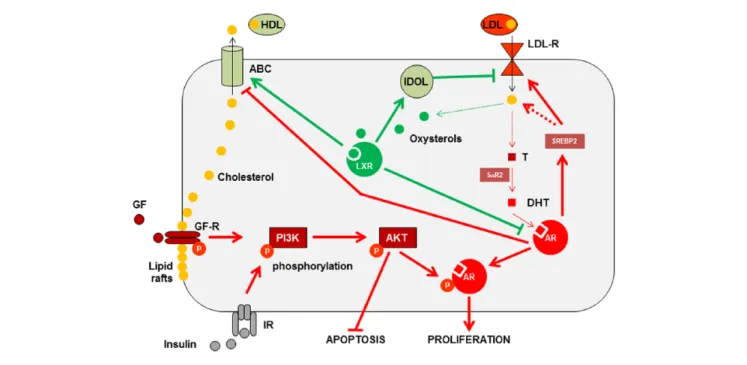
Figure 1. Interconnection between LXRs and AR in prostate cell.
Note. AR and DHT increase the proliferation. When growth factors bind their membrane receptors, they activate phosphorylation cascades, stimulate
PI3K/AKT, and increase AR activity through its phosphorylation. Oxysterol-activated LXR induces ubiquitin ligase IDOL accumulation followed by degradation of LDL-R. ABC proteins increase both export of cholesterol and destructuration of lipid rafts, which in turn will decrease both AKT phosphorylation and inhibition of the apoptotic pathway. Green lines represent favorable effects on PCa management; red lines represent negative effects on PCa management. LXRs = liver X receptors; AR = androgen receptor; DHT = dihydrotestosterone; PI3K = phosphatidylinositol-4,5-bisphosphate 3-kinase; AKT = protein kinase B; LDL-R = LDL receptor; ABC = ATP-binding cassette; PCa = prostate cancer; T = testosterone; GF = growth factors; GF-R = growth factor receptor; P = phosphorylation; IR = insulin receptor; HDL = high density lipoprotein.
4 Nuclear Receptor Signaling
by steroids that usually do not bind AR, and modifications of AR coactivators or corepressors. Hence, despite an extremely low level of circulating androgens, AR remains active and continues to drive PCa progression.
Beside the classical ligand-regulation of the transcrip-tional activity, AR could also rapidly interact with the nonre-ceptor tyrosine kinase SRC/Src increasing cell proliferation through activation of the mitogen-activated protein kinase (MAPK)/extracellular signal–regulated kinases (ERK1/2) cascade,51 or with PI3K/AKT signaling pathway (Figure 1), and controlling cell survival.52 Based on that, innovative therapies will be necessary to counteract this so-called non-genomic signaling of AR during the establishment of meta-static CRPC.53
Finally, “androgens meet lipids” (Figure 1) as AR activa-tion increases fatty acid synthesis 23,54-56 and SREBP2,30 a key-player in de novo synthesis of cholesterol and its cellu-lar uptake,57 and decreases ATP-binding cassette A1 (ABCA1), a cholesterol export pump.58 This last point is crucial because cholesterol is an obligatory precursor for testosterone and DHT synthesis, the only two active andro-gens on AR59; more importantly, tumor cells also have the ability to abnormally synthesize DHT from cholesterol60 or from adrenal androgens.61,62
Together with AR, other nuclear receptors have been involved in PCa (for a review, see Leach et al50); among them the popular “Ménage-à-trois” LXR-FXR-SHP (liver X recep-tor–farnesoid X receptor–small heterodimeric partner) has been described to be the major player in the regulation of both cholesterol and bile acid homeostasis.63 Altogether, targeting this new “Ménage-à-quatre” appears to be of importance to take care of the prostate. Interestingly, the role of these 3 nuclear receptors has been emerging these last years in ex vivo or in vivo experiments, especially with the generation of transgenic animals knock-out for LXRs. Noteworthy, The Cancer Genome Atlas pointed out that these nuclear receptors could present alterations of copy numbers of their respective encoding genes in PCa.50
LXR
α and LXRβ Are Involved in
Prostate Physiology
LXRα/NR1H3 and LXRβ/NR1H2 (as well as FXRα/ NR1H4 and SHP/NR0B2) are members of the nuclear recep-tor superfamily. They are composed of several functional domains, among them a central DNA-binding domain and a C-terminal ligand-biding domain.64 At the end of the 1990s,
Janowski et al demonstrated that LXRs are the bona fide
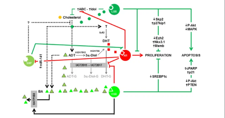
Figure 2. Summary of the interconnections among AR, LXRs, FXR, and SHP in prostate cell.
Note. AR and DHT have a central role in the proliferation of the epithelial cells. LXRs, FXR, and SHP have positive impacts in PCa by blocking AR
transcriptional activity, decreasing proliferation and/or increasing apoptosis. FXR could also play a negative role by decreasing glucuronidation of androgens through the transcriptional regulation of UGT2B15/17 enzymes. Green lines represent favorable effects on PCa management; red lines represent negative effects on PCa management. See the article for more details about the various links. AR = androgen receptor; LXRs = liver X receptors; FXR = farnesoid X receptor; SHP = small heterodimeric partner; DHT = dihydrotestosterone; PCa = prostate cancer; MAPK = mitogen-activated protein kinase; ADT = androgen deprivation therapy; UGT2 = UDP glucuronosyltransferase 2; SREBP = sterol regulatory element–binding protein; PTEN = Phosphatase and TENsin homolog; ABC = ATP-binding cassette; BA = bile acid; cPARP = cleaved Poly (ADP-ribose) polymerase.
receptors for oxysterols,65,66 oxidized derivatives of
choles-terol. Hence, it was suggested that LXRs could regulate cho-lesterol homeostasis in the cell and was demonstrated thanks to the analysis of Lxr-deficient mice.67 Since this seminal
article, others groups have associated LXR roles to numer-ous physiological functions.64,68
In the prostate, Liao’s group was the first to evoke a puta-tive posiputa-tive role of LXRs in PCa.58 The authors showed that ABCA1, a bona fide LXR-target gene which increases
cho-lesterol export, was downregulated by androgens (Figure 2) in LNCaP cells.58 The same group identified that activation
of LXRs by the synthetic agonist T0901317 decreased the percentage of S-phase LNCaP cells in a dose-dependent manner and increased the expression of cyclin-dependent kinase inhibitor CDKN1B/p27KIP158, by decreasing the
S-phase kinase associated protein 2 (SKP2) involved in the degradation of cell cycle inhibitors.69 At last, Chuu et al
dem-onstrated that LXRs and some of their target genes were decreased during the progression of androgen-dependent tumor to androgen-independent relapsed tumors in a xeno-graft model.70 These data thus made a clear link between
LXRs and the proliferative capacities of PCa cells (Figure 2). Likewise, we identified that Lxrα;β-deficient mice fed a high cholesterol diet presented prostatic intraepithelial neo-plasia characterized by an accumulation of the oncogene and histone methyl transferase Enhancer of Zeste Homolog 2 (EZH2) which results in the downregulation of the tumor suppressors microseminoprotein beta (MSMB) and NK3 homeobox 1 (NKX3.1).71 It is noteworthy that
overexpres-sion of EZH2 has been described in patients with an aggres-sive PCa.72 EZH2 controls prostate cell proliferation through
the epigenetic silencing of NKX3.173 and MSMB.74 In
wild-type mice fed a high cholesterol diet, LXRs induce the tran-scription of Inducible Degrader of the LDL receptor MYLIP/ IDOL (Figures 1 and 2), a ubiquitin ligase that targets LDL-R, and of ABC transporters, altogether maintaining a con-trolled level of cholesterol and a low amount of EZH2.71 So
far, it has not been possible to dissociate the exact role of each LXR isoform as they both compensate each other.
In addition to the role of LXRs in the control of cellular cholesterol content and prostate cell proliferation, we showed that the activation of LXRs by various natural or synthetic ligands increases the level of apoptosis in LNCaP cells.75
This phenomenon is linked to the presence of smaller and thinner lipid rafts after LXR stimulation and the downregula-tion of AKT phosphoryladownregula-tion in these lipid rafts. After hav-ing derived new models of epithelial cells from the dorsal prostate (MPECs) of wild-type or Lxr-deficient mice, Dufour et al enlightened that LXRs modulate AKT and MAPK phos-phorylation accumulation, making them potential mediators of LXRs in cell cycle control76 (Figure 2). Altogether, these
results confirm that LXRs are becoming exquisite pharmaco-logical targets for PCa, unless specific modulators, we called SLiMs (selective LXR modulators77,78), could be developed.
On the other side, LXRs also control AR activity. Indeed, Lxr-deficient mice also develop benign prostatic hyperplasia (BPH).79 In man, BPH is clearly due to an excessive activity
of AR and the production of DHT.80 Using transgenic
ani-mals, we were able to demonstrate that LXRα acts as a key modulator of the cross talk between the stromal and epithelial compartments, which is essential for the integration of andro-gen signaling in the prostate and its effect on the epithelium.81
Interestingly, Tsui et al82 pointed out that LXR expression was
higher in androgen-sensitive LNCaP cells than in other PCa cell lines. Moreover, T0901317-activated LXRs decrease AR accumulation and PSA production in LNCaP. Overall, AR and LXRs are definitively interconnected (Figure 1). These data open numerous opportunities to develop new therapeutic concepts especially in CRPC situations where AR activity could become independent of androgen levels. If LXRs nega-tively modulate AR accumulation, a SLiM could increase PCa cell apoptosis, as well as decrease AR activity, hence, bypassing the castration-resistant stage.
One of the most challenging issues in PCa is to slow down the metastatic potential of the tumor.83 Fu et al described an
interesting effect of GW3965, another LXR synthetic ligand, on LNCaP cells. Activation of LXR increases the suppressor of cytokine signaling 3 (SOCS3) accumulation, followed by a decrease of phosphorylated AKT, activator protein-1 (AP-1), and nuclear factor kappa-light-chain-enhancer of acti-vated B cells (NFκB1/p105).84 GW3965 also inhibited PCa
invasion in xenografted mice, suggesting that LXRs could be targetable to prevent metastases.
However, these potential effects of LXRs in PCa treat-ment should be modulated. Indeed, it should be kept in mind that, in prostate, tumor cells are surrounded by immune cells such as dendritic cells that initiate immune responses, includ-ing antitumor activity after their CC chemokine receptor-7 (CCR7)-dependent migration to lymphoid organs. Activation of LXRα inhibits CCR7 expression dampening the antitu-mor immune responses.85 Likewise, drugs inhibiting
choles-terol synthesis (and thus LXR ligands), such as zaragozic acid, increase the efficacy of the treatments in xenografted mice with tumors.86 Once again, these contradictory results
enlighten the necessity to develop tissue- and cell-specific LXR ligands to decipher the exact roles of each LXR in each cell type. It should also be noticed that no variation of LXRα and/or LXRβ expression has been linked to cancer grades. This would imply to stratify patients’ cohorts, which has not been performed so far.
Putative Role of FXR in PCa
FXR/NR1H4 is the nuclear receptor for bile acids.87
However, the inactive androgen androsterone has also been described as a potent activator of FXR.88 This point is
impor-tant because androsterone is present in prostate. Conversely, to LXR and the historical story of cholesterol accumulation
6 Nuclear Receptor Signaling
in prostate tumors, the link between FXR, bile acids, and prostate physiology is less evident.
The first “historical” link came with Wang and Schaffner89
who observed that BIO 87-20 hamsters, developing sponta-neous cystic prostate hypertrophy, had a lower prostate size and weight with much less distended prostatic acini when were treated with colestipol, a bile-acid-sequestering anion-exchange resin. More recently, it has been shown that bile acid content is increased in patients with a PCa treated with an ADT90; in parallel, ADT has been associated with an
increased risk of diverse biliary diseases.91 Alpha-methylacyl
CoA racemase (AMACR) is overexpressed in PCa92 and is a
better diagnostic marker than PSA. AMACR plays a key role in the β oxidation of branched chain fatty acids and the bile acid intermediates dihydroxycholestanoic acid and trihy-droxycholestanoic acid. Furthermore, AMACR is highly expressed in androgen-sensitive PCa cell lines and is required for the proliferation of these cells.93
Even though lithocholic acid selectively induces apopto-sis in androgen-sensitive and -insensitive prostate cell lines,94,95 few studies have focused on the molecular effects
of FXR activation in androgen-sensitive or androgen-insen-sitive prostate cell lines. Indeed, FXR inhibits cell prolifera-tion by decreasing lipid metabolism via targeting SREBP1c (Figure 2).96 Likewise, chenodeoxycholic acid (CDCA, a
natural FXR ligand) and GW4065 (a synthetic FXR agonist) decrease LNCaP cell proliferation by the induction of PTEN accumulation, which in turn decreases the phosphorylation of AKT and the survival pathway.97 Interestingly, new
deriv-atives of ursodeoxycholic acid and CDCA induce the apop-tosis of human prostate androgen-insensitive carcinoma PC-3 cells by increasing cyclin-dependent kinase inhibitor 1 CDKN1A/p21CIP1 and the cleaved form of poly [ADP-ribose] polymerase 1 PARP1.98 However, it has not been
proved yet that FXR mediates the effect of these molecules. Finally, the most important point is that FXR accumulation was found to be significantly lower, at both mRNA and pro-tein levels, in human PCa tissues compared with the pair-matched adjacent normal tissues.97 Again, no correlation was
made with grades/stages of the tumors by the authors. Based on these results, one can suggest that FXR ligands could have some benefit in the treatment of PCa.
Unfortunately, other data showed a negative role of bile acids and FXR in the development of PCa. First of all, andro-gen metabolite androsterone, which is also an activator of FXR, reduces the glucuronidation of androgens catalyzed by UGT2B15/B17 in an FXR-dependent manner in LNCaP cells.99 Such an action would increase the levels of
androste-rone, 5α-androstane-3α,17β-diol, and finally DHT, and thus activate AR and induce cell proliferation. On the contrary, the increase of androsterone could also neutralize the AR-induced proliferation by blocking the proliferation via FXR (Figure 2). This dual paradoxical effect of FXR is also seen in the regulation of the solute carrier organic anion transporter fam-ily member 1B3 SLCO1B3/OATP1B3, which is an export
pump for steroids and bile acids100 and seems to be
upregu-lated by FXR in the prostate,101 but not in the liver.102 If this
regulation is efficient in the prostate, FXR would deplete the cells in steroids (including bile acids) by increasing OATP1B3; bile acid depletion will act as a safety valve and will reduce FXR activity. More investigations are thus needed using ade-quate in vivo models to understand how FXR targeting could be interesting in PCa management.
SHP, a Noncanonical Nuclear Receptor
With Significant Potential in PCa
Short heterodimer partner (SHP/NR0B2) is an atypical orphan nuclear receptor: It lacks the classical DNA- and pos-sesses a ligand-binding domain. Despite its strong repressive activity on other nuclear receptors such as AR, no known ligand for SHP has been identified so far.103
Initially described as downregulating bile acid synthesis in liver by decreasing 7α-cholesterol hydroxylase CYP7A1 in an LXR-dependent manner,104 SHP became a member of
the Triad with LXR and FXR when it appeared that FXR was the nuclear receptor for bile acids105 and SHP was one of its
bona fide target genes.
Indeed, it was the discovery of synthetic SHP ligands that gave the opportunity to link SHP to PCa.106 Some of these
ligands had a strong inhibitory effect on proliferation and inducing effect on apoptosis in the PCa androgen-insensitive DU-145 cells (50% efficacy less than 1µM).
In spite of SHP role in the regulation of androgen synthe-sis directly in the testis107 or indirectly via gonadotropin
hor-mones,108 such role has never been described so far in
prostate, but cannot be excluded. Besides, SHP seems to play an antitumor role in many types of cancers109; unfortunately,
SHP mRNA and/or protein accumulation has never been studied in prostate tumors. The centerpiece pointing out SHP as interesting in PCa comes from the fact that AR negatively regulates the amount of SHP (Figure 2).110 Indeed, miR-141
which targets SHP is induced by AR in LNCaP cells. One could thus hypothesize that increasing concentration of active androgens would block SHP, which antagonizes AR activity and neutralizes the proliferative role of androgens.
Conclusion and Perspectives
PCa incidence is drastically increasing in Westernized coun-tries. Today, the main challenge is to have good diagnostic and prognostic markers that could help in the management of the patients. As androgens have been playing a central role in the progression of the tumors, most of these markers were previously obtained focusing on the screening of AR target genes. The involvement of other transcription factors, mem-bers or not of the nuclear receptor superfamily, has made possible to identify new signaling pathways regulating pro-gression of the tumor until metastasis. LXR, FXR, and SHP have been associated for many years to the regulation of
metabolism. It is finally not surprising to find them as puta-tive pharmacological targets to treat PCa, especially know-ing that they can regulate androgen levels and AR activity (Figure 1). Altogether, developing new specific molecules regulating these nuclear receptors will give the opportunity to offer different therapeutic arsenals. This is probably the most challenging issue, especially in CRPC, which is the fate of almost all PCa.
Author Contributions
M.C., S.D., S.M., S.B., A.M., and J.-M.A.L. collected the information, wrote the manuscript, and drew the figures; all authors read and approved the manuscript.
Declaration of Conflicting Interests
The author(s) declared no potential conflicts of interest with respect to the research, authorship, and/or publication of this article.
Funding
The author(s) disclosed receipt of the following financial support for the research, authorship, and/or publication of this article: Part of this work was financed by grants from Université Blaise Pascal, Region Auvergne Rhône Alpes, Fond Européen de Developpement Régional (FEDER), AAP Plan Cancer Environnement 2016.
References
1. Ghabili K, Tosoian JJ, Schaeffer EM, et al. The history of prostate cancer from antiquity: review of paleopathological studies. Urology. 2016;97:8-12.
2. Jemal A, Bray F, Center MM, Ferlay J, Ward E, Forman D. Global cancer statistics. CA Cancer J Clin. 2011;61:69-90. 3. McDonald ML, Parsons JK. The case for tailored prostate
cancer screening: an NCCN perspective. J Natl Compr Canc
Netw. 2015;13:1576-1583.
4. Cobran EK, Hall JN, Aiken WD. African-American and Caribbean-born men’s perceptions of prostate cancer fear and facilitators for screening behavior: a pilot study. J
Cancer Educ. 2018;33(3):640-648.
5. Baade PD, Youlden DR, Krnjacki LJ. International epidemi-ology of prostate cancer: geographical distribution and secu-lar trends. Mol Nutr Food Res. 2009;53:171-184.
6. Andriole GL, Crawford ED, Grubb RL, et al. Mortality results from a randomized prostate-cancer screening trial. N
Engl J Med. 2009;360:1310-1319.
7. Schröder FH, Hugosson J, Roobol MJ, et al. Screening and prostate-cancer mortality in a randomized European study. N
Engl J Med. 2009;360:1320-1328.
8. Grönberg H. Prostate cancer epidemiology. Lancet. 2003;361: 859-864.
9. Sakr WA, Haas GP, Cassin BF, Pontes JE, Crissman JD. The frequency of carcinoma and intraepithelial neoplasia of the prostate in young male patients. J Urol. 1993;150:379-385. 10. Perdana NR, Mochtar CA, Umbas R, Hamid ARA. The risk
factors of prostate cancer and its prevention: a literature review. Acta Med Indones. 2016;48:228-238.
11. Rebbeck TR. Prostate cancer genetics: variation by race, eth-nicity, and geography. Semin Radiat Oncol. 2017;27:3-10. 12. DeSantis CE, Siegel RL, Sauer AG, et al. Cancer statistics
for African Americans, 2016: progress and opportunities in reducing racial disparities. CA Cancer J Clin. 2016;66:290-308.
13. Pietro GD, Chornokur G, Kumar NB, Davis C, Park JY. Racial differences in the diagnosis and treatment of prostate cancer. Int Neurourol J. 2016;20:S112-S119.
14. Tran HN, Li Y, Udaltsova N, Armstrong MA, Friedman GD, Klatsky AL. Risk of cancer in Asian Americans: a Kaiser Permanente cohort study. Cancer Causes Control. 2016;27:1197-1207.
15. Cook LS, Goldoft M, Schwartz SM, Weiss NS. Incidence of ade-nocarcinoma of the prostate in Asian immigrants to the United States and their descendants. J Urol. 1999;161:152-155. 16. Watanabe M, Nakayama T, Shiraishi T, Stemmermann GN,
Yatani R. Comparative studies of prostate cancer in Japan versus the United States. A review. Urol Oncol. 2000;5:274-283.
17. Shimizu H, Ross RK, Bernstein L, Yatani R, Henderson BE, Mack TM. Cancers of the prostate and breast among Japanese and white immigrants in Los Angeles County. Br J
Cancer. 1991;63:963-966.
18. Taylor RA, Lo J, Ascui N, Watt MJ. Linking obesogenic dys-regulation to prostate cancer progression. Endocr Connect. 2015;4:R68-R80.
19. Gacci M, Russo GI, De Nunzio C, et al. Meta-analysis of metabolic syndrome and prostate cancer. Prostate Cancer
Prostatic Dis. 2017;20(2):146-155.
20. Ben Sahra I, Laurent K, Giuliano S, et al. Targeting cancer cell metabolism: the combination of metformin and 2-deoxy-glucose induces p53-dependent apoptosis in prostate cancer cells. Cancer Res. 2010;70:2465-2475.
21. Loubière C, Goiran T, Laurent K, Djabari Z, Tanti J-F, Bost F. Metformin-induced energy deficiency leads to the inhi-bition of lipogenesis in prostate cancer cells. Oncotarget. 2015;6:15652-15661.
22. Hankinson SJ, Fam M, Patel NN. A review for clinicians: prostate cancer and the antineoplastic properties of metfor-min. Urol Oncol. 2017;35:21-29.
23. Swinnen JV, Heemers H, van de Sande T, et al. Androgens, lipogenesis and prostate cancer. J Steroid Biochem Mol Biol. 2004;92:273-279.
24. Kuemmerle NB, Rysman E, Lombardo PS, et al. Lipoprotein lipase links dietary fat to solid tumor cell proliferation. Mol
Cancer Ther. 2011;10:427-436.
25. Brusselmans K, Timmermans L, Van de Sande T, et al. Squalene synthase, a determinant of Raft-associated choles-terol and modulator of cancer cell proliferation. J Biol Chem. 2007;282:18777-18785.
26. Beckers A, Organe S, Timmermans L, et al. Chemical inhi-bition of acetyl-CoA carboxylase induces growth arrest and cytotoxicity selectively in cancer cells. Cancer Res. 2007;67:8180-8187.
27. White C. The occurrence of crystals in tumours. J Pathol
Bacteriol. 1909;13:3-10.
28. Vargas C. Cholesterine in cutanecous cancer. Urol Cutan
8 Nuclear Receptor Signaling 29. Swyer G. The cholesterol content of normal and enlarged
prostates. Cancer Res. 1942;2:372-375.
30. Chen Y, Hughes-Fulford M. Human prostate cancer cells lack feedback regulation of low-density lipoprotein receptor and its regulator, SREBP2. Int J Cancer. 2001;91:41-45. 31. Thysell E, Surowiec I, Hörnberg E, et al. Metabolomic
char-acterization of human prostate cancer bone metastases reveals increased levels of cholesterol. PLoS ONE. 2010;5:e14175. 32. Yue S, Li J, Lee S-Y, et al. Cholesteryl ester accumulation
induced by PTEN loss and PI3K/AKT activation under-lies human prostate cancer aggressiveness. Cell Metab. 2014;19:393-406.
33. Statz CM, Patterson SE, Mockus SM. mTOR inhibitors in castration-resistant prostate cancer: a systematic review.
Target Oncol. 2017;12:47-59.
34. Chang L, Graham PH, Ni J, et al. Targeting PI3K/Akt/ mTOR signaling pathway in the treatment of prostate cancer radioresistance. Crit Rev Oncol Hematol. 2015;96:507-517. 35. Wettstein MS, Saba K, Umbehr MH, et al. Prognostic role
of preoperative serum lipid levels in patients undergoing radical prostatectomy for clinically localized prostate cancer.
Prostate. 2017;77:549-556.
36. Gordon JA, Midha A, Szeitz A, et al. Oral simvastatin administration delays castration-resistant progression and reduces intratumoral steroidogenesis of LNCaP prostate can-cer xenografts. Prostate Cancan-cer Prostatic Dis. 2016;19:21-27.
37. Hoque A, Chen H, Xu X-C. Statin induces apoptosis and cell growth arrest in prostate cancer cells. Cancer Epidemiol
Biomarkers Prev. 2008;17:88-94.
38. Sekine Y, Furuya Y, Nishii M, Koike H, Matsui H, Suzuki K. Simvastatin inhibits the proliferation of human prostate cancer PC-3 cells via down-regulation of the insulin-like growth factor 1 receptor. Biochem Biophys Res Commun. 2008;372:356-361.
39. Sivaprasad U, Abbas T, Dutta A. Differential efficacy of 3-hydroxy-3-methylglutaryl CoA reductase inhibitors on the cell cycle of prostate cancer cells. Mol Cancer Ther. 2006;5:2310-2316.
40. Zheng X, Cui X-X, Avila GE, et al. Atorvastatin and cele-coxib inhibit prostate PC-3 tumors in immunodeficient mice.
Clin Cancer Res. 2007;13:5480-5487.
41. Zhuang L, Kim J, Adam RM, Solomon KR, Freeman MR. Cholesterol targeting alters lipid raft composition and cell survival in prostate cancer cells and xenografts. J Clin Invest. 2005;115:959-968.
42. Alfaqih MA, Allott EH, Hamilton RJ, Freeman MR, Freedland SJ. The current evidence on statin use and pros-tate cancer prevention: are we there yet? Nat Rev Urol. 2017;14:107-119.
43. Murai T. Cholesterol lowering: role in cancer prevention and treatment. Biol Chem. 2015;396:1-11.
44. Huggins C, Hodges CV. Studies on prostatic cancer. I. The effect of castration, of estrogen and of androgen injection on serum phosphatases in metastatic carcinoma of the prostate.
Cancer Res. 1941;1:293-297.
45. Lacy JM, Kyprianou N. A tale of two trials: the impact of 5α-reductase inhibition on prostate cancer (Review). Oncol
Lett. 2014;8:1391-1396.
46. Gauthier-Landry L, Bélanger A, Barbier O. Multiple roles for UDP-glucuronosyltransferase (UGT)2B15 and UGT2B17 enzymes in androgen metabolism and pros-tate cancer evolution. J Steroid Biochem Mol Biol. 2015 Jan;145:187-92.
47. Bao B-Y, Chuang B-F, Wang Q, et al. Androgen receptor mediates the expression of UDP-glucuronosyltransferase 2 B15 and B17 genes. Prostate. 2008;68:839-848.
48. Cornford P, Bellmunt J, Bolla M, et al. EAU-ESTRO-SIOG guidelines on prostate cancer. Part II: treatment of relapsing, metastatic, and castration-resistant prostate cancer. Eur Urol. 2017;71:630-642.
49. Lowrance WT, Roth BJ, Kirkby E, Murad MH, Cookson MS. Castration-resistant prostate cancer: AUA guideline amendment 2015. J Urol. 2016;195:1444-1452.
50. Leach DA, Powell SM, Bevan CL. Women in cancer the-matic review: new roles for nuclear receptors in prostate can-cer. Endocr Relat Cancan-cer. 2016;23:T85-T108.
51. Migliaccio A, Castoria G, Di Domenico M, et al. Steroid-induced androgen receptor-oestradiol receptor beta-Src complex triggers prostate cancer cell proliferation. EMBO J. 2000;19:5406-5417.
52. Baron S, Manin M, Beaudoin C, et al. Androgen receptor mediates non-genomic activation of phosphatidylinositol 3-OH kinase in androgen-sensitive epithelial cells. J Biol
Chem. 2004;279:14579-14586.
53. Leung JK, Sadar MD. Non-genomic actions of the androgen receptor in prostate cancer. Front Endocrinol. 2017;8:2. 54. Heemers H, Maes B, Foufelle F, Heyns W, Verhoeven G,
Swinnen JV. Androgens stimulate lipogenic gene expres-sion in prostate cancer cells by activation of the sterol regu-latory element-binding protein cleavage activating protein/ sterol regulatory element-binding protein pathway. Mol
Endocrinol. 2001;15:1817-1828.
55. Heemers H, Verrijdt G, Organe S, et al. Identification of an androgen response element in intron 8 of the sterol regula-tory element-binding protein cleavage-activating protein gene allowing direct regulation by the androgen receptor. J
Biol Chem. 2004;279:30880-30887.
56. Swinnen JV, Verhoeven G. Androgens and the control of lipid metabolism in human prostate cancer cells. J Steroid
Biochem Mol Biol. 1998;65:191-198.
57. Brown MS, Goldstein JL. Sterol regulatory element binding proteins (SREBPs): controllers of lipid synthesis and cellular uptake. Nutr Rev. 1998;56:S1-S3; discussion S54-S75. 58. Fukuchi J, Hiipakka RA, Kokontis JM, et al. Androgenic
suppression of ATP-binding cassette transporter A1 expres-sion in LNCaP human prostate cancer cells. Cancer Res. 2004;64:7682-7685.
59. Alioui A, Celhay O, Baron S, Lobaccaro J-MA. Lipids and prostate cancer adenocarcinoma. Clin Lipidol. 2014;9:643-655.
60. Dillard PR, Lin M-F, Khan SA. Androgen-independent pros-tate cancer cells acquire the complete steroidogenic poten-tial of synthesizing testosterone from cholesterol. Mol Cell
Endocrinol. 2008;295:115-120.
61. Luu-The V, Bélanger A, Labrie F. Androgen biosyn-thetic pathways in the human prostate. Best Pract Res Clin
62. Mohler JL, Titus MA, Bai S, et al. Activation of the andro-gen receptor by intratumoral bioconversion of androstane-diol to dihydrotestosterone in prostate cancer. Cancer Res. 2011;71:1486-1496.
63. Kalaany NY, Mangelsdorf DJ. LXRs and FXR: the Yin and Yang of cholesterol and fat metabolism. Annu Rev Physiol. 2006;68:159-191.
64. Maqdasy S, Trousson A, Tauveron I, Volle DH, Baron S, Lobaccaro J-MA. Once and for all, LXRα and LXRβ are gatekeepers of the endocrine system. Mol Aspects Med. 2016;49:31-46.
65. Janowski BA, Willy PJ, Devi TR, Falck JR, Mangelsdorf DJ. An oxysterol signalling pathway mediated by the nuclear receptor LXR alpha. Nature. 1996;383:728-731.
66. Janowski BA, Grogan MJ, Jones SA, et al. Structural require-ments of ligands for the oxysterol liver X receptors LXRalpha and LXRbeta. Proc Natl Acad Sci U S A. 1999;96:266-271. 67. Peet DJ, Turley SD, Ma W, et al. Cholesterol and bile acid
metabolism are impaired in mice lacking the nuclear oxys-terol receptor LXR alpha. Cell. 1998;93:693-704.
68. Hong C, Tontonoz P. Liver X receptors in lipid metabolism: opportunities for drug discovery. Nat Rev Drug Discov. 2014;13:433-444.
69. Chuu C-P, Kokontis JM, Hiipakka RA, Liao S. Modulation of liver X receptor signaling as novel therapy for prostate cancer. J Biomed Sci. 2007;14:543-553.
70. Chuu C, Hiipakka RA, Kokontis JM, Fukuchi J, Chen R-Y, Liao S. Inhibition of tumor growth and progression of LNCaP prostate cancer cells in athymic mice by androgen and liver X receptor agonist. Cancer Res. 2006;66:6482-6486.
71. Pommier AJC, Dufour J, Alves G, et al. Liver x receptors protect from development of prostatic intra-epithelial neo-plasia in mice. PLoS Genet. 2013;9:e1003483.
72. Varambally S, Dhanasekaran SM, Zhou M, et al. The poly-comb group protein EZH2 is involved in progression of pros-tate cancer. Nature. 2002;419:624-629.
73. Kunderfranco P, Mello-Grand M, Cangemi R, et al. ETS transcription factors control transcription of EZH2 and epi-genetic silencing of the tumor suppressor gene Nkx3.1 in prostate cancer. PLoS ONE. 2010;5:e10547.
74. Beke L, Nuytten M, Van Eynde A, Beullens M, Bollen M. The gene encoding the prostatic tumor suppressor PSP94 is a target for repression by the Polycomb group protein EZH2.
Oncogene. 2007;26:4590-4595.
75. Pommier AJC, Alves G, Viennois E, et al. Liver X Receptor activation downregulates AKT survival signaling in lipid rafts and induces apoptosis of prostate cancer cells.
Oncogene. 2010;29:2712-2723.
76. Dufour J, Pommier A, Alves G, et al. Lack of liver x recep-tors leads to cell proliferation in a model of mouse dorsal prostate epithelial cell. PLoS ONE. 2013;8:e58876.
77. Viennois E, Pommier AJC, Mouzat K, et al. Targeting liver X receptors in human health: deadlock or promising trail?
Expert Opin Ther Targets. 2011;15:219-232.
78. Viennois E, Mouzat K, Dufour J, Morel L, Lobaccaro J-M, Baron S. Selective liver X receptor modulators (SLiMs): what use in human health? Mol Cell Endocrinol. 2012;351:129-141.
79. Kim H-J, Andersson LC, Bouton D, Warner M, Gustafsson J-A. Stromal growth and epithelial cell proliferation in ven-tral prostates of liver X receptor knockout mice. Proc Natl
Acad Sci U S A. 2009;106:558-563.
80. Füllhase C, Schneider MP. 5-alpha-reductase inhibitors and combination therapy. Urol Clin North Am. 2016;43:325-336. 81. Viennois E, Esposito T, Dufour J, et al. Lxrα regulates the
androgen response in prostate epithelium. Endocrinology. 2012;153:3211-3223.
82. Tsui K-H, Chung L-C, Feng T-H, et al. Divergent effect of liver X receptor agonists on prostate-specific antigen expres-sion is dependent on androgen receptor in prostate carcinoma cells. Prostate. 2015;75:603-615.
83. Clement JM, Sweeney CJ. Evolving treatment of oligometa-static hormone-sensitive prostate cancer. J Oncol Pract. 2017;13:9-18.
84. Fu W, Yao J, Huang Y, et al. LXR agonist regulates the car-cinogenesis of PCa via the SOCS3 pathway. Cell Physiol
Biochem. 2014;33:195-204.
85. Villablanca EJ, Raccosta L, Zhou D, et al. Tumor-mediated liver X receptor-alpha activation inhibits CC chemokine receptor-7 expression on dendritic cells and dampens antitu-mor responses. Nat Med. 2010;16:98-105.
86. Lanterna C, Musumeci A, Raccosta L, et al. The adminis-tration of drugs inhibiting cholesterol/oxysterol synthesis is safe and increases the efficacy of immunotherapeutic regi-mens in tumor-bearing mice. Cancer Immunol Immunother. 2016;65:1303-1315.
87. Gadaleta RM, Cariello M, Sabbà C, Moschetta A. Tissue-specific actions of FXR in metabolism and cancer. Biochim
Biophys Acta. 2015;1851:30-39.
88. Wang S, Lai K, Moy FJ, Bhat A, Hartman HB, Evans MJ. The nuclear hormone receptor farnesoid X recep-tor (FXR) is activated by androsterone. Endocrinology. 2006;147:4025-4033.
89. Wang GM, Schaffner CP. Effect of candicidin and colesti-pol on the testes and prostate glands of BIO 87.20 hamsters.
Invest Urol. 1976;14:66-71.
90. Saylor PJ, Karoly ED, Smith MR. Prospective study of changes in the metabolomic profiles of men during their first three months of androgen deprivation therapy for prostate cancer. Clin Cancer Res. 2012;18:3677-3685.
91. Saylor PJ, Smith MR, O’Malley AJ, Keating NL. Androgen-deprivation therapy and risk for biliary disease in men with prostate cancer. Eur Urol. 2014;65:642-649.
92. Evans AJ. α-methylacyl CoA racemase (P504S): overview and potential uses in diagnostic pathology as applied to pros-tate needle biopsies. J Clin Pathol. 2003;56:892-897. 93. Zha S, Ferdinandusse S, Denis S, et al. α-methylacyl-CoA
racemase as an androgen-independent growth modifier in prostate cancer. Cancer Res. 2003;63:7365-7376.
94. Gafar AA, Draz HM, Goldberg AA, et al. Lithocholic acid induces endoplasmic reticulum stress, autophagy and mito-chondrial dysfunction in human prostate cancer cells. PeerJ. 2016;4:e2445.
95. Goldberg AA, Titorenko VI, Beach A, Sanderson JT. Bile acids induce apoptosis selectively in androgen-dependent and -inandrogen-dependent prostate cancer cells. PeerJ. 2013;1:e122.
10 Nuclear Receptor Signaling 96. Liu N, Zhao J, Wang J, Teng H, Fu Y, Yuan H. Farnesoid
X receptor ligand CDCA suppresses human prostate cancer cells growth by inhibiting lipid metabolism via targeting sterol response element binding protein 1. Am J Transl Res. 2016;8:5118-5124.
97. Liu J, Tong S-J, Wang X, Qu L-X. Farnesoid X receptor inhibits LNcaP cell proliferation via the upregulation of PTEN. Exp Ther Med. 2014;8:1209-1212.
98. Choi YH, Im EO, Suh H, Jin Y, Yoo YH, Kim ND. Apoptosis and modulation of cell cycle control by syn-thetic derivatives of ursodeoxycholic acid and chenode-oxycholic acid in human prostate cancer cells. Cancer Lett. 2003;199:157-167.
99. Kaeding J, Bouchaert E, Bélanger J, et al. Activators of the farnesoid X receptor negatively regulate androgen glucuron-idation in human prostate cancer LNCAP cells. Biochem J. 2008;410:245-253.
100. Yang M, Xie W, Mostaghel E, et al. SLCO2B1 and SLCO1B3 may determine time to progression for patients receiving androgen deprivation therapy for prostate cancer. J
Clin Oncol. 2011;29:2565-2573.
101. Svoboda M, Riha J, Wlcek K, Jaeger W, Thalhammer T. Organic anion transporting polypeptides (OATPs): regulation of expression and function. Curr Drug Metab. 2011;12:139-153.
102. Jung D, Elferink MGL, Stellaard F, Groothuis GMM. Analysis of bile acid-induced regulation of FXR target genes in human liver slices. Liver Int. 2007;27:137-144.
103. Zhang Y, Hagedorn CH, Wang L. Role of nuclear recep-tor SHP in metabolism and cancer. Biochim Biophys Acta. 2011;1812:893-908.
104. Lu TT, Makishima M, Repa JJ, et al. Molecular basis for feedback regulation of bile acid synthesis by nuclear recep-tors. Mol Cell. 2000;6:507-515.
105. Makishima M, Okamoto AY, Repa JJ, et al. Identification of a nuclear receptor for bile acids. Science. 1999;284:1362-1365. 106. Dawson MI, Xia Z, Liu G, et al. An adamantyl-substituted
retinoid-derived molecule that inhibits cancer cell growth and angiogenesis by inducing apoptosis and binds to small het-erodimer partner nuclear receptor: effects of modifying its car-boxylate group on apoptosis, proliferation, and protein-tyrosine phosphatase activity. J Med Chem. 2007;50:2622-2639. 107. Volle DH, Duggavathi R, Magnier BC, et al. The small
het-erodimer partner is a gonadal gatekeeper of sexual matura-tion in male mice. Genes Dev. 2007;21:303-315.
108. Vega A, Martinot E, Baptissart M, et al. Identification of the link between the hypothalamo-pituitary axis and the tes-ticular orphan nuclear receptor NR0B2 in adult male mice.
Endocrinology. 2015;156:660-669.
109. Zou A, Lehn S, Magee N, Zhang Y. New insights into orphan nuclear receptor SHP in liver cancer. Nucl Receptor
Res. 2015;2:101162.
110. Xiao J, Gong A-Y, Eischeid AN, et al. miR-141 modulates androgen receptor transcriptional activity in human prostate cancer cells through targeting the small heterodimer partner protein. Prostate. 2012;72:1514-1522.