Abstract.
Background: Radio-frequency ablation (RFA) has
recently received much attention as an effective minimally
invasive strategy for the local treatment of tumors. The purpose
of this study was to evaluate the efficacy of single-needle
cool-tip RF breast ablation in terms of temperature distribution and
duration of the procedure as compared to multiprobe RF
breast ablation. Materials and Methods: Two different
commercially available radiofrequency ablation needle
electrodes were compared. Finite-element method (FEM)
models were developed to simulate the thermoablation
procedures. A series of ex vivo radiofrequency thermal lesions
were induced to check the response of the FEM calculations.
Results: Data obtained from FEM models and from ex vivo
procedures showed that cool-tip RF breast ablation assures
better performances than multiprobe RF breast ablation in
terms of temperature distribution and duration of the
procedure. Histopathological analysis of the cool-tip RF
thermoablated specimens showed successful induction of
coagulation necrosis in the thermoablated specimens.
Conclusion: Data obtained from FEM models and from ex
vivo procedures suggest that the proposed cool-tip RF breast
ablation may kill more tumor cells in vivo with a single
application than the multiprobe RF breast ablation.
Surgical breast cancer treatment is effective, but is a highly
invasive procedure. Because of the poor cosmetic result of
such invasive therapy, treatments that would preserve the
greatest amount of normal tissue have great appeal
especially for the treatment of early diagnosed cancers
(small tumors). Thus, the demand for novel minimally
invasive techniques for the treatment of breast cancer has
grown during recent years. Radio-frequency (RF) ablation
has recently received much attention as an effective
minimally invasive strategy for the local treatment of solid
malignancies (1, 2). Although other methods of thermal
ablation such as laser, microwave and High Focused
Ultrasound (HIFU) are being performed, RF is currently
receiving the greatest attention in the light of several
factors: its general availability, the recent technical advances
facilitating its use and the active marketing on the part of
manufacturers.
To accomplish RF tumor ablation, an "RFA probe"
(usually 14 to 21 gauge needle-like electrodes) is placed
directly into the target tissue with the use of ultrasound
(US) imaging guidance (2, 3).
An RF generator produces an RF voltage between the
active electrode and a reference electrode (usually a large
conductive pad in contact with the patient’s skin), thereby
establishing lines of electric field within the patient’s body
between the two electrodes. At the low RF used for this
procedure (460-550 kHz), the electric field pattern is
governed essentially by electrostatic equations. Essentially,
the electric field oscillates with the alternating RF current,
which causes oscillatory movement of ions in the target
tissue in proportion to the field intensity. The mechanism of
tissue heating for RF ablation is frictional. Tissue heating
occurs as a result of ionic agitation surrounding the electrode
as the RF current oscillates while reaching its ground
(reference electrode). It is the generation of tissue heating
that induces cellular death via thermal coagulation necrosis.
Therefore, the volume of RF heat ablation is governed by
the temperature distribution within the tissue (3). Between
60ÆC and 100ÆC, near instantaneous induction of protein
coagulation that irreversibly damages key cytosolic and
Correspondence to: (i) Dr. Eng. Vincenzo Quaranta,Tecnobiomedica S.p.A., Via Vaccareccia 41, I-00040 Pomezia, Rome, Italy. e-mail: [email protected]. (ii) Dr. G. Manenti, Department of Diagnostic and Molecular Imaging, Radiation Therapy and Interventional Radiology, University Hospital Policlinico Tor Vegata (PTV), Viale Oxford 81, 00133 Rome, Italy. e-mail: [email protected]
Key Words: RFA, RF ablation, breast ablation, Finite-element method, FEM, cool-tip electrode, multi-probe.
FEM Analysis of RF Breast Ablation:
Multiprobe versus Cool-tip Electrode
V. QUARANTA
1, G. MANENTI
2, F. BOLACCHI
2, E. COSSU
2, C.A. PISTOLESE
2,
O.C. BUONOMO
2, L. CAROTENUTO
1, C. PICONI
1and G. SIMONETTI
21
Tecnobiomedica S.p.A. Via Vaccareccia 41, 00040 Pomezia, Rome;
2
Department of Diagnostic and Molecular Imaging, Radiation Therapy and Interventional
Radiology, University Hospital Policlinico Tor Vergata (PTV), Rome, Italy
mitochondrial enzymes and nucleic acid-histone complexes
occurs (4, 5).
A key limitation so far for RF ablation has been creating
adequate volumes of tissue destruction. Several methods
have been investigated to increase lesion size.
Multiprobe monopolar RF needles with multiple thin,
curved electrodes that extend from a central cannula have
been successfully used for RF liver ablation (6) and are
currently employed for RF breast ablation (7, 10). Also,
bipolar RF ablation using two active needle electrodes has
been suggested (11). Recently, monopolar single-needle
cool-tip electrodes have been successfully used to induce a
consistent necrosis volume in tumors other than breast
tumors (12, 13).
The purpose of this study was to evaluate the efficacy of
cool-tip electrode RF breast ablation in terms of
temperature distribution and duration of the procedure as
compared with conventional multiprobe RF breast ablation.
Materials and Methods
Finite-element method (FEM) models. Five FEM models have been developed: Model 1: breast tissue, multiprobe LN electrode, power-controlled mode; Model 2: liver tissue, multiprobe LN electrode, power-controlled mode; Model 3: breast tissue, Miras PTV cool-tip electrode, power-controlled mode; Model 4: breast tissue, multiprobe Miras LN electrode, temperature-controlled mode; and Model 5: breast tissue, cool-tip Miras PTV Electrode, temperature-controlled mode.
Solving method. FEM Models were implemented and developed using FEMLAB (Comsol, Burlington MA, USA) on a 3.0 GHz Intel Pentium IV, with 1 Gbyte RAM. The model consisted of 64 034 tetrahedral elements and 12 880 nodes nonuniform mesh; mesh size close to the probe was 0.2 mm and at the model boundaries was 5 mm. Convergence tests had been performed to ensure adequate spatial resolution. The time steps during the solving analysis of the FEM started at 0.05 sec and were subsequently automatically controlled by the solver software so that the maximum temperature change during each step was less than 3ÆC.
Boundary conditions. In all models the boundary conditions were the same.
i. Temperature boundary conditions: For the surface of the cylinder the temperature value was kept costant at 37ÆC. Since the thermal mass of the probe was less than that of the saline, we assumed that heat conduction into the probe itself was minimal. Thus, for all other surfaces, the thermally insulating boundary condition nØ(k∇T)=0 was applied. Only the active surface of the cooled electrode was set to the cooling media temperature, T=5ÆC.
ii. Electrical boundary conditions: For each of the outer surfaces of the model, an electrical boundary condition of V=0 was applied, to simulate the return ground electrode. A source potential (Vo) was applied to the conducting areas of the probe. The non-conducting portions were given an insulating boundary condition such that nØ(Û∇V)=0; where n is the unit vector normal to the surface, Û is the electrical conductivity and V is the voltage at the insulating surface.
Tissue properties. Tissue thermal and electrical properties used for the models have been previously described (14-22) and are reported in Table I. Breast tissue is a mixture of fat, glandular and connective tissues. Since fat has the lowest thermal conductivity of all, due to the low water content, the model was simplified considering all the breast tissue to be adipose tissue.
Model geometry. Three-Dimensional FEM Analyses. FEM models of Miras PTV (Single cooled needle) (Figure 1a), by University Hospital Policlinico Tor Vergata (PTV) (Rome, Italy) in collaboration with INVATEC Srl (Rocadelle, Brescia, Italy) and Miras LN (Multiprobe) (Figure 1b), by INVATEC Srl were developed. The active portion of each probe was embedded into a cylindrical region (height: 12.0 cm/diameter: 10.0 cm) that simulated tissue surrounding the probe. Due to the symmetry of the model, computing time was saved by modelling a quarter of the cylinder only. The symmetric assumption required axial-symmetry of geometry, materials, loadings and boundaries, which also led to an axial-symmetric solution.
A
NTICANCERR
ESEARCH 27: 775-784 (2007)Table I. Thermoelectric properties of tissue.
Tissue Density Thermal Thermal Electrical Permittivity (Kg/m3) capacity conductivity conductivity
[J/(ÆC Kg)] [W/(m ÆC)] (Siemens/ meter)
Liver 1060 3628 0.465 0.148 2957
Fat
(Breast) 940 2600 0.2 0.025 35
Table II. Results from the experimental ex vivo tests.
Test Electrode Tissue Power Time Max T (ÆC)
No. type (W) (sec) External
thermocouple (deployed - 3.0 cm) 1 237 94 2 LN 20 224 96 3 Swine 308 106 4 mammary 444 120 5 PTV 20 384 118 6 603 128 7 Bovine 1165 120 8 LN liver 20 874 120 9 797 120 10 332 95 11 LN Human 20 470 104 12 breast 354 92 13 626 113 14 PTV 20 707 119 15 778 128
The bioheat equation. The potential and temperature distributions in the tissues were monitored using the protocols reported in the literature. Briefly, it was assumed that the ablation probe dissipated the majority of its energy through electrical conduction. In this situation a quasi-static electrical conduction model was assumed, which allowed the use of Laplace’s equation to solve the electric field (16). The expression for the electric potential (V) was solved quickly over the entire volume and the value was implemented into the source term of the heat conduction equation, the so-called bioheat equation (16-19). The energy generated by the metabolic processes was ignored, Qm (W/m) and the blood perfusion contribution considered insignificant.
Energy delivery schemes. Three of the five simulations were performed with the power-controlled ablation mode. The erogation power was set to deliver an average energy of 30 W until a final tissue temperature of 100ÆC (charring temperature) was reached. Two models simulated the temperature-controlled ablation mode. Power value was initially set at 30 W and adjusted to maintain a
temperature of 90ÆC for eight minutes. At the end of the soak time, power modulation was stopped and the last power value applied was maintained until a final temperature of 100ÆC was reached. Temperature distributions were recorded during the procedure. Ex vivo experiments. Tissue samples: three tissue types were tested (Figures 2 and 3). For each tissue type, six samples were obtained (10 cm long x 10 cm wide x 5 cm thick).
Tissues used were swine mammary tissue, from two adult non-nursing female pigs; bovine liver tissue, from a young calf’s liver and human breast tissue from excised breast mastectomy specimens. Radiofrequency system. A 460-kHz generator (Fogazzi, Concesio, Italy) capable of 100 W maximum output power at 200 ø (Figure 4a) was used to perform the tests using two types of electrodes Miras PTV (monopolar cool-tip single needle) (Figure 4c) and Miras LN (monopolar multiprobe needle) (Figure 4d). The Miras LN is a multiprobe stainless steel electrode equipped with four active hooks (0.5 mm diameter). The Miras PTV is a monopolar 200 mm two Figure 1. FemLab FEM models: a) Miras PTV Cool-tip Electrode and b) Miras LN Multiprobe Electrode.
lumens single electrode with active stainless steel tip exposure of 25 mm (Figure 4c). The two lumens were equipped with connectors for inflowing and outflowing of chilled saline solution, which permitted continuous internal cooling. A peristaltic pump (Fogazzi) cooled the electrode internally by delivering a chilled saline perfusion (Na-Cl 38% 2000 ml at 0ÆC) through its cannula sheath, providing optimal cooling of the electrode tip (Figure 4b). Circuitry incorporated in the generator allowed continuous monitoring of the impedance of the active part of the electrode. Constant temperature monitoring was ensured by the thermocouple located three centimeters from the needle insertion (Figure 4c and 4d).
Description of the procedure. A total of fifteen tests were performed: six tests were performed on swine mammary and six on human breast tissue, three of each with the Miras PTV electrode and three with the Miras LN electrode, and three tests were performed on liver tissue with the Miras LN electrode. Tissue samples were positioned
on the ground pad in a container and partially surrounded by saline. The electrodes were inserted in the middle of the tissue sample and power was applied. The end-point of each ablation procedure was the increase of tissue impedance values (>200 ø) due to tissue charring. Time, power, impedance and temperature were monitored throughout the entire procedure. Temperature values, as sensed by the thermistor located 3 cm from the tip of the electrodes, were used as indicators of the effectiveness of the procedure.
Histopathological analysis. To assess whether or not the cool-tip RF breast ablation was able to induce a successful coagulation necrosis, freshly excised tissue samples of thermoablated swine mammary tissue were formalin-fixed and prepared for histology. Four Ìm-thick serial paraffin sections were stained with hematoxylin-eosin and histological analysis performed to identify coagulation necrosis, since previous studies have documented the close correlation between histopathological findings and RF-induced coagulation
A
NTICANCERR
ESEARCH 27: 775-784 (2007)Figure 2. a) Swine mammary sample during Miras PTV cool-tip RFA, b) calf liver sample during Miras LN multiprobe RFA, c) macroscopic appearance of an excised swine mammary specimen after RFA and d) macroscopic appearance of an excised calf liver specimen after RFA.
necrosis (23). Typical signs of the induction of coagulation necrosis, such as the finding of stromal hyalinization with the disappearance of the collagenous fibrillar network, the presence of chromatin migration along the nuclear envelope, nuclear pyknosis and increased cytoplasmic eosinophilia, were recorded (Figure 5).
Results and Discussion
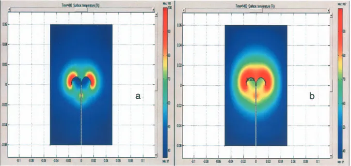
Model 1 versus model 2. In order to assess the efficiency of
the multiprobe electrode RF ablation, FEM models of
multiprobe RF liver tissue ablation were developed and
compared to FEM models of multiprobe RF breast ablation.
Model 1 and model 2 simulate a multiprobe RF breast
ablation and a multiprobe RF liver ablation respectively,
both with power-controlled mode.
In Model 1, a temperature value of 100ÆC was reached in
the tissue directly surrounding the hook tips, while
temperature values recorded in the tissue located close to
the concave areas of the hooks were around 60ÆC.
Temperature distribution values are shown in Figure 6a
(Model 1) and 6b (Model 2).
The ex vivo experiments confirmed data from the FEM
models (Table II). The differences in ablation time and
maximum temperature between mammary and liver tissue
recorded with the Miras LN (multiprobe) electrode were
substantial (Figures 7 and 8).
Our data demonstrated that, due to the low thermal
conductivity of mammary tissue, multiprobe RF breast
ablation rapidly induces the charring threshold in the near
Figure 3. a) Human breast sample during Miras PTV cool-tip RFA, b) macroscopic appearance of an excised human breast specimen after cool-tip RFA showing the ablated area as a firm whitish tissue with a central needle track surrounded by an area of yellowish coagulated adipose tissue, c) longitudinal cut and d) axial cut of the specimen.
proximity of the electrode tip and hooks, thus forcing the
procedure to be stopped prematurely before obtaining an
adequate ablation volume. On the contrary, multiprobe RF
liver ablation, induces heat distribution over larger tissue
volumes, probably due to the high liver heat conductivity
that favors a more efficient distribution of the temperature
within the tissue.
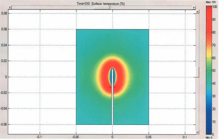
Model 1 versus model 3. Here, model 1 (Figure 6a) is
compared to model 3 (Figure 9), which simulates the
cool-tip needle RF breast ablation with power-controlled mode.
Our data showed that the presence of the cooling moves the
maximum temperature values far away from the electrode
tip (Figure 9). The temperature distribution was correlated
to the tip radius, the power applied and the cooling
temperature. In the absence of cooling, the maximum
temperature value was located at the tissue-electrode
interface, where the resistive heating was usually at its
maximum value (Figure 6a).
The model prediction was confirmed by the ex vivo
procedures performed under the same experimental
conditions. The time period necessary to induce the thermal
lesion was substantially different between the two electrodes
(Table II).
To assess whether or not the cool-tip RF breast ablation
was able to induce a successful coagulation necrosis,
histo-pathological analysis of a thermoablated swine mammary
specimen was performed. Typical signs of the induction of
coagulation necrosis were observed with the Miras PTV
cool-tip RF as shown in Figure 5.
A
NTICANCERR
ESEARCH 27: 775-784 (2007)Figure 4. INVATEC Radiofrequency system: a) TAG100 RF generator, b) cooling Pump, c) Miras PTV cool-tip electrode and d) Miras LN multiprobe electrode.
Our data are in agreement with previous reports (13),
which have shown that the employment of internally cooled
electrodes causes a "heat sink" effect that removes heat
closest to the electrode. The removal of heat closest to the
electrode allows for greater current deposition without
tissue charring or impedance rise, with the consequent
distribution of heat over a larger volume of tissue.
Model 4 versus model 5. To confirm the superior performance
of cool-tip needle over multiprobe needle RF breast ablation,
breast FEM models were also developed with a
temperature-controlled mode.
Model 4 simulating multiprobe RF breast ablation
(Figure 10a) and model 5 simulating cool-tip RF breast
ablation (Figure 10b) correspond to model 1 and 3,
respectively, except for the fact that the
temperature-controlled mode was used instead of the power-temperature-controlled
mode. The remaining parameters were left unchanged. In
model 4, a power output of 40 W was applied and adjusted
to reach (200 sec) and maintain a temperature value of
90ÆC. The time ablation period was 480 sec. The last power
value applied was maintained until 100ÆC was reached
within the tissue (charring point). The overall procedure
lasted twelve minutes. The tissue located under the hooks
reached a temperature below 70ÆC at the end of the
ablation procedure. Tissue located at the external side of
the hooks reached a temperature between 80-90ÆC
(red-yellow) that extended for a radius of 1.5 cm from the hooks.
Figure 5. a) Macroscopic appearance of an excised swine mammary specimen after Miras PTV cool-tip RFA, b) histopathological analysis of a thermoablated swine breast specimen that shows c) chromatin migration along the nuclear envelope, nuclear pyknosis and increased cytoplasmic eosinophilia and d) stromal hyalinization with the disappearance of the collagenous fibrillar network.
In model 5, a power output of 30 W was applied and
adjusted to maintain a temperature value of 90ÆC. The time
ablation period was 480 sec. The last power value applied
was maintained until 100ÆC was reached in the tissue
(charring point).
Our data showed that with the temperature-controlled
mode cool-tip RF breast ablation created a larger
temperature distribution than the multibrobe RF breast
ablation (Figure 10a and 10b).
The FEM models developed in this study have
demonstrated that cool-tip RF breast ablation assured a
higher performance than multiprobe RF breast ablation,
both with the power-controlled and the
temperature-controlled modes (Figure 9 and Figure 10b).
The potential effects of blood perfusion and
temperature dependence of thermal conductivity were not
incorporated into our models since it was assumed that
they would provide only little additional information. In
our study, the FEM breast tissue models were developed
with fat only. Our approach was justified due to the fact
that fat tissue has a lower conductivity than fibrous or
glandular tissue.
A
NTICANCERR
ESEARCH 27: 775-784 (2007)Figure 7. Experimental ex vivo study: Miras LN multiprobe electrode, swine mammary tissue temperature at 3 cm from the electrode insertion point.
Figure 8. Experimental ex vivo study: Miras LN multiprobe electrode, liver tissue temperature at 3 cm from the electrode insertion point.
Figure 6. a) Temperature distribution recorded during power-controlled Miras LN multiprobe RF breast ablation. Power = 30 W; Voltage = 80 V; Time = 400 sec. b) Temperature distribution recorded during power-controlled Miras LN multiprobe RF liver ablation. Power = 30 W; Voltage = 33 V; Time = 1400 sec.
Figure 9. Temperature distribution recorded during power-controlled Miras PTV cool-tip RF breast ablation. Power = 30 W; Voltage = 140 V; Time = 310 sec.
Figure 10. Temperature distribution recorded a) during temperature controlled Miras LN multiprobe RF breast ablation and b) during temperature-controlled Miras PTV cool-tip RF breast ablation.
Conclusion
Data from the FEM models developed in this study and
from ex vivo procedures have shown that cool-tip RF breast
ablation assured better performance than multiprobe RF
breast ablation in terms of temperature distribution and
duration of the procedure.
These differences can probably be attributed to the
cooling effect that moves the maximum temperature
isotherm far away from electrode tip, inducing heat spread
over a larger tissue volume.
Based on our results, it is postulated that in selected
cases, treated by experienced personnel, cool-tip RF breast
ablation would show optimal oncological results. However,
further study of this technique in vivo is warranted to
determine if it is feasible, safe and capable of producing a
sufficient volume of necrosis to be valuable in the treatment
of breast malignancies.
References
1 Mizra A, Fornage B, Sneige N, Kuerer H, Newman L, Ames F and Singletary S: Radiofrequency ablation of solid tumors. Cancer J 7(2): 95-102, 2001.
2 Martin AP, Goldstein RM, Dempster J et al: Radiofrequency thermal ablation of hepatocellular carcinoma before liver transplantation – a clinical and histological examination. Clin Transplant 20(6): 695-705, 2006.
3 Goldberg S: Radiofrequency tumor ablation: principles and techniques. Eur J Ultrasound 13(2): 129-147, 2001
4 Miller MW and Ziskin MC: Biological consequences of hyperthermia. Ultrasound Med Biol 15: 707-722, 1989. 5 Lounsberry W, Goldschmidt V and Linke C: The early
histological changes following electrocoagulation. Gastrointest Endosc 41: 68-70, 1995.
6 Geyik S, Akhan O, Abbasoglu O et al: Radiofrequency ablation of unresectable hepatic tumors. Diagn Interv Radiol 12(4): 195-200, 2006.
7 Izzo F, Thomas R, Delrio P, Rinaldo M, Vallone P, DeChiara A, Botti G, D'Aiuto G, Cortino P and Curley SA: Radiofrequency ablation in patients with primary breast carcinoma. Cancer 92: 2036-2044, 2001.
8 Burak WE Jr, Agnese DM, Povoski SP, Yanssens TL, Bloom KJ, Wakely PE and Spigos DG: Radiofrequency ablation of invasive breast carcinoma followed by delayed surgical excision. Cancer 98: 1369-1376, 2003.
9 Hayashi AH, Silver SF, van der Westhuizen NG, Donald JC, Parker C, Fraser S, Ross AC and Olivotto IA: Treatment of invasive breast carcinoma with ultrasound guided radiofrequency ablation. Am J Surg 185: 429-435, 2003.
10 Fornage BD, Sneige N, Ross MI, Mirza AN, Kuerer HM, Edeiken BS, Ames FC, Newman LA, Babiera GV and Singletary SE: Small (< or = 2- cm) breast cancer treated with US-guided radiofrequency ablation: feasibility study. Radiology 231: 215-224, 2004.
11 McGahan JP, Gu W-Z, Brock JM, Tesluk H and Jones CD: Hepatic ablation using bipolar radiofrequency electrocautery. Acad Radiol 3: 418-422, 1996.
12 Goldberg SN, Gazelle GS, Solbiati L, Rittman WJ and Mueller PR: Radio-frequency tissue ablation: increased lesion diameter with a perfusion electrode. Acad Radiol 3: 636-644, 1996. 13 Lorentzen TA: A cooled needle electrode for radio-frequency
tissue ablation: Thermodynamic aspects of improved performance compared with conventional needle design. Acad Radiol 3: 556-563, 1996.
14 Gabriel C and Gabriel S: Compilation of the dielectric properties of body tissue at RF and Microwave frequencies (www.brooks.af.mil/AFRL/HED/hedr/reports/dielectric/home.ht ml), 2002.
15 Gabriel S, Lau RW and Gabriel C: The dielectric properties of biological tissues: II. Measurements in the frequency range 10 Hz to 20 GHz. Phys Med Biol 41: 2251-2269, 1996.
16 Chang I: Finite element analysis of hepatic radiofrequency ablation probes using temperature-dependent electrical conductivity. Biomed Eng Online 2: 12-30, 2003
17 Ekstrand V, Viksell H, Shultz I, Sandstedt B, Rotstein S and Eriksson A: Influence of electrical and thermal properties on RF ablation of breast cancer: is the tumour preferentially heated? Biomed Eng Online 4: 41-57, 2005.
18 Haemmerich D, Tungjitkusolmun S, Staelin ST, Lee FT Jr, Mahvi DM and Webster JG: Finite-element analysis of hepatic multiple probe radio-frequency ablation. IEEE Trans Biomed Eng 49: 836-842, 2002.
19 Hammerich D and Webster JG: Automatic control of finite element models for temperature controlled radiofrequency ablation. Biomedical Engineering OnLine 4: 42-47, 2005. 20 Duck F: Physical properties of tissue – a comprehensive
reference book. Academic Press, New York, pp. 167-223, 1990. 21 Valvano JW et al: The simultaneous measurement of thermal conductivity, thermal diffusivity and perfusion in small volume of tissue. J Biomech Eng 106: 192-197, 1984.
22 Valvano JW, Cochran JR and Diller KR: Thermal conductivity and diffusivity of biomaterials measured with self-heating thermistors. Int J Themophys 6: 301-311, 1985.
23 Bohem T et al: Radio-frequency tumor ablation: internally cooled electrode versus saline-enhanced technique in an aggressive rabbit tumor model. Radiology 222(3): 805-813, 2002.