Il processo di una scoperta
scientifica è, in effetti, un
continuo conflitto di meraviglie.
UNIVERSITÀ DEGLI STUDI DI ROMA
"TOR VERGATA"
FACOLTA' DI MEDICINA E CHIRURGIA
DOTTORATO DI RICERCA IN BIOLOGIA
MOLECOLARE E SCIENZE BIOCHIMICHE
XX° CICLO
BASEMENT MEMBRANE AND CELLULAR
MIGRATION: ROLE OF GELATINASES
(MMP-2, MMP-9) ON PROTEOLYSIS OF TYPE IV
COLLAGEN
MONACO SUSANNA
Docente Guida/Tutor: Prof. Massimiliano Coletta
Coordinatore: Prof. Alessandro Finazzi Agrò
Table of contents Abstract
Riassunto Introduction I) Basement membranes: role in tumor
angiogenesis 1) basement membrane structure and assembly
a) basement membrane glycoproteins a1) collagens a2) other glycoproteins 2) Basement membrane assembly 3) Angiogenesis and vascular basement
membrane
4) Regulation of angiogenesis by MMPs II) Matrix Metalloproteinases (Matrixin):
Structure and Function
1) Metzincin superfamily 2) Structural complexity of MMPs a) the catalytic domain and the active site b) the hemopexin-like domain c) the hinge region d) the fibronectin-like-domain 3) Activation of proMMPs a) Intracellular activation b) stepwise activation mechanism c) cell surface activation of proMMP2 4) Inhibition of MMP activity a) Endogenous MMP inhibitors b) Exogenous MMP inhibitors 1 8 11 15 16 16 18 19 22 29 30 34 36 36 36 40 46 46 47 47 47 47 48 50 52 52
III) Gelatinases and type IV of collagen degradation
1) Type IV of collagen structure 2) Gelatinases cleavage site on type IV of
collagen degradation
Aim of the study Results I) Degradation of type IV of collagen from
human placenta by MMP2 II) Degradation of type IV of collagen from
human placenta by cdMMP-2 III) Degradation of native collagen type IV
from murine (EHS) sarcoma by whole
MMP-2 IV) Role of MMP2 on the migration of
neutrophils across a type IV of collagen
coating membrane V) Role of the fibronectin like domain of
MMP-2 (CBD) on the processing of type IV of collagen from human placenta by whole
MMP-2 VI) Degradation of type IV of collagen from
human placenta by MMP-9 VII) Degradation of native collagen type IV
from murine EHS sarcoma by wholeMMP-9 VIII) Cntibution of the interaction of
fibronectin-like domain of MMP2 (rCBD) to the processing of collagen type IV from
human placenta by MMP9 IX) Contribution of the interaction of the
fibronectin like domain of MMP-2 (CBD) to the processing of native collagen type IV
from murine EHS sarcoma by MMP9 X) Synergistic effect between MMP-2 and
MMP-9 during neutrophil cells migration across a coating membrane of collagen type IV 54 54 57 63 67 69 71 75 78 80 82 84 87 96 98
Discussion Conclusion Matherials and Methods I) Materials II) Methods 1) Enzymatic activation 2) Activity assay 3) Kinetics of digestion of collagen type IV
from human placenta and of native collagen type IV from murine EHS sarcoma by whole
MMP2 and cdMMP2 4) Human neutrophils isolation
5) chemotaxis assay 6) Role of the fibronectin-like domain of
MMP2 (rCBD) on the processing of collagen type IV from human placenta by
whole MMP2 7) Kinetic analysis
8) kinetics of digestion of collagen type IV from human placenta and of native collagen
IV from murine EHS sarcoma by MMP9 9) Contribution of the fibronectin-like
domain of MMP2 (CBD) on the processing of collagen type IV from human placenta by MMP9
10) Contribution of the fibronectin-like domain of MMP2 (rCBD) on the processing of native collagen type IV from murine EHS
sarcoma by MMP9 11) Kinetic analysis Abbreviations Literature cited Curriculum vitae Articles 101 111 117 118 119 119 119 120 120 121 122 122 123 123 124 124 127 131 147 153
Proteolytic degradation of basement memnbrane influences the cell behaviour during important processes, such as inflammations, tumorigenesis, angiogenesis and allergic diseases. In this study, we have investigated the action of gelatinase A (MMP-2) and B (MMP-9) on collagen IV, the major constituent of the basement membrane. We have compared quantitatively their actions on the soluble forms of collagen IV extracted with or without pepsin (from human placenta and from Engelbreth-Holm-Swarm (EHS) murine sarcoma, respectively). The catalytic efficiency of MMP-2, and also MMP-9, is dramatically reduced in the case of the EHS murine sarcoma with respect to the human placenta, probably due to the much tighter packing of the network which renders very slow the speed of the rate-limiting step. We have also enquired on the role of MMP-2 domains in processing collagen IV. The removal of the hemopexin-like domain, using only the catalytic domain of MMP-2, has only a limited effect on the catalytic efficiency toward collagen IV, indicating that the missing domain has not a great relevance for the overall mechanism. Instead, the addition of the isolated collagen binding domain, corresponding to the fibronectin-like domain of whole MMP-2, greatly inhibits the cleavage process, demonstrating that MMP-2 interacts with collagen type IV preferentially through its fibronectin-like domain. Finally, we have investigated the effect of MMP-2 proteolytic activity ex vivo. MMP-2 action negatively affects the neutrophil cells migration across type IV coated membranes and this is likely related to the production of lower molecular weight fragments which impair the cellular migration.However, for both types of collagen IV the enzymatic processing by MMP-9 is dramatically enhanced in the presence of the Collagen Binding Domain of Gelatinase A (CBD). This effect, clearly indicates that the fibronectin-like domain of MMP-2 and MMP-9 bind to topologically distinct sites on type IV collagen, bringing about a conformational change of the collagen IV molecule. This allows the two enzymes to cooperate with each other through a ligand-linked mechanism, which does not necessarily require the enzymatic action. Therefore, fibronectin-like domains not only increase the affinity between enzyme and substrate to enhance the catalysis, they also act as allosteric third party elements in the MMP action. This synergistic action between MMP-2 and MMP-9 on collagen IV has been tested also with an ex vivo eperiment. The MMP-2 without the catalytic domain, the rCBD and the pro-MMP-2 increase the neutrophil cells migration across collagen IV coated membranesand this is related to the growth of the catalytic activity of MMP-9.
In condizioni patologiche le gelatinasi o meglio la MMP-2 e la MMP-9 vengono espresse dalle cellule in quantità più elevate rispetto alle normali condizioni fisiologiche. Ne sono un esempio i processi infiammatori e la progressione tumorale, dove queste endopeptidasi Ca+2 e Zn+2 dipendenti
sono attivamente coinvolte nel processamento delle membrane basali cellulari; infatti la loro azione permette alle cellule tumorali la loro progressione all’interno di nuovi tessuti, dando origine alla formazione di metastasi. Le gelatinasi influenzano la progressione tumorale in quanto intervengono in un processo che prende il nome di “angiogenic switch”, che consiste nella formazione di una nuova vascolarizzazione in grado di alimentare le cellule in attiva proliferazione. L’ ”angiogenic switch” inizia con la degradazione da parte delle gelatinasi del collagene IV, componente fondamentale della membrana basale vascolare. Esso, una volta processato, libera fattori (quali il VEGF) che stimolano le cellule endoteliali a migrare all’interno di una matrice provvisoria dove si formeranno i nuovi vasi sanguigni. Se da un lato le gelatinasi aumentano l’attività proliferativa delle cellule tumorali, dall’altra sono in grado di inibirla. Sembra infatti che la MMP-2, degradando il collagene di tipo IV, produca dei frammenti con attività anti-angiogenica in grado di bloccare la proliferazione tumorale. Ai fini di una miglior comprensione del ruolo delle Gelatinasi durante la progressione tumorale, abbiamo analizzato l’efficienza delle due metalloproteinasi verso il collagene di tipo IV; e nel caso della MMP-2 abbiamo caratterizzato il ruolo dei suoi diversi domini verso questo substrato naturale. Inoltre è stata effettuata una analisi basata sulla capacità dei neutrofili di migrare attraverso una membrana ricoperta di collagene di tipo IV in presenza di (i) una quantità esogena crescente di MMP-2 (ii) frammenti di arrestina derivanti dal processamento della catene α del collagene IV, (iii) MMP-2 mutata, ossia priva del suo dominio catalitico, (iv) del dominio ricombinante fibronectinico della MMP-2 o rCBD e (v) la MMP-2 inattiva o pro-MMP-2.Per finire abbiamo studiato l’esistenza di una possibile interazione tra le due gelatinasi nel processare il collagene di tipo IV sia in vitro che in condizioni ex vivo.I nostri risultati dimostrano come entrambe le gelatinasi processano il componente fondamentale delle membrane basale vascolare in condizioni fisiologiche, 37°C e pH 7,2. Inoltre l’MMP-2 interagisce con la MMP-9 aumentandone l’efficienza di processamento verso il collagene di tipo IV, per effetto di un controllo allosterico dovuto al semplice legame dell’enzima con il substrato. Quando la MMP-2 si lega al collagene IV per opera del suo dominio fibronectinico
azione quando è prsente da sola. Il fenomeno varia a seconda della concentrazione della MMP-2 che possiede due siti di legame per questo tipo di collagene: uno ad alta affinità ed un secondo sito con una scarsa affinità di legame. A bassa concentrazione di MMP-2 viene legato solo il sito del substrato per cui l’enzima posside una maggior affinità e di conseguenza la MMP-9 legandosi allo stesso substrato lo processa con maggior efficienza. Al contrario, ad alte concentrazioni di MMP-2, l’enzima si lega ad ambedue i siti di attacco che possiede nel collagene IV diminuendo la capacità della MMP-9 di processarlo, a causa di una probabile sovrapposizione del sito di attacco dei due enzimi.L’attività sinergica delle due gelatinasi viene riscontrata anche ex vivo, ossia mediante esperimenti di migrazione cellulare attraverso un coating di collagene IV. Le cellule utilizzate sono neutrofili, cellule in grado di esprimere vari enzimi proteolitici tra cui metalloproteinasi come la MMP-8 e la MMP-9 e proteasi a serina come l’elastasi del neutrofili. Risultano invece non produrre livelli apprezzabili di MMP-2, come dimostrato dalle zimografie delle Figure 10 e 11. I neutrofili, grazie alla MMP-9, sono quindi in grado di oltrepassare la barriera di collagene IV, che viene processato con maggior attività in presenza di quantità esogene di MMP-2 priva del dominio catalitico o in aggiunta del suo dominio di legame al substrato, il dominio ricombinante rCBD.
I) Basement Membranes: role in tumour angiogenesis
In recent yers, the basement membrane (BM), a specialized form of the extracellular matrix (ECM), has been recognized as an important regulator of cell behaviour. The importance of BM becomes relevant when some vascular diseases have been known to be associated with defects on BM components. Diseases such as Alport syndrome and Knoblach syndrome have been associated to mutational defects in type IV of collagen and α chainsof collagen type XVIII.
1) Basement membrane structure and assembly
The basement membrane is an amorph, dense sheet-like structure of 50-100 nm in thickness that generally separates ephitelium from the stroma of any given tissue. It is always in contact with cells, and its function is to provide structural support, todivide tissue into compartments, as well as to regulate cell behaviour.
BMs are highly crosslinked and insoluble material that are composed of several glycoproteins.
Figure 1: Basement membrane and vascular basement membrane localization.
Different structure and localization of the basement membrane (BM) on the top of the figure and of the vascular basement membrane or VBM.
a) Basement membrane glycoproteins
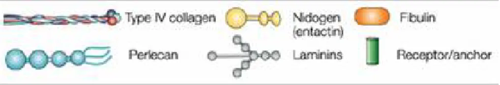
The costituens of BM are usually insoluble molecules that come together to form sheet-like structures via a process known as self-assembly which is driven by cell surface receptors. In general, all cells are known to produce the BM components; some of them are different kinds of collagen, proteoglycans, laminin polymers and nidogen/entactin complexes. Minor components are agrin, SPARC/BM-40/osteopontin, fibulins (Figure 2).
Figure 2: Glycoprotein composition of the BM structure. Type IV of collagen protomers,
a1) Collagen
Collagens, are a class of proteins that are carachterized by a unique sequence stretch in wich every third amino acid is a Glycine (GLY) (Gly-X-Y) motif. The X and Y motifs are usually hydroxyproline and hydroxylysine. Type I of collagen along with type II and type III collagen are classic examples of proteins that have long stretches of the Gly-X-Ymotif and are termed fibrillar collagen for their capacity to form fibres.
Figure 3. Fibrillar collagen. a) b) the molecular structures of the collagenous proteins
depicted with ball-and-stick models c) schematic representation of triple helix d) e) 67 nm D periodic fibrils f) electron microscopy of intact calf skin collagen fibrils.
Collagens have the capacity to self-associate in different forms. At least 15 other types of collagens have been identified, which are not homologous to the fibril forming collagens, and can adopt other supramolecular organizations. These non-fibrillar collagen types include the network assembly of type IV collagen in basement membranes, the antiparallel associations of type VII collagen in anchoring fibrils, and the hexagonal arrays of type VIII collagen in Descemet's membrane (Kramer et al., 2001) The collagen super family is classified into groups according to the polymeric structures they form or to related structural features:
a) fibril forming collagens (types I, II, III, V and XI) b) network forming collagens (type IV, VIII and X)
c) collagens that are found on the surface of fibrils known as fibril-associated collagens with interrupted triple helices (FACIT) (types IX, XII, XVI and XIX)
d) beaded filament-forming collagen (type VI)
e) collagen of anchoring fibrils for basement membranes (type VII) f) collagens with a transmembrane domain (type XIII and XVII) g) family of type XV and XVIII
h) “noncollagen” collagen consists of proteins containing triple-helical domains that have not been defined collagens (subcomponent C1q of complement (Prockop et al., 1995).
The BM contains type IV, XV and XVIII collagen. Type IV of collagen is the most abundant component of vascular basement membrane (VBM) and it is also called network-forming collagen due to its capacity to self assemblate into organized network. It is generally formed by the association of three different α-chains in a primary structure called protomer. Six different type IV of collagen α chains have been identified codified by six different genes.
Type XV collagen is widely distributed in several BM zones of various tissue, it is a non-fibrillar collagen and it is thought to be a homotrimer formed by three α1 chains. It is a highly glycosylated collagen with a central triple helical domain that is interrupted by NC domains and the protein contains large amino terminal and carboxy terminal NC domains. Expression
homologous to type XVIII of collagen for the similarity of the NC terminal domain. This kind of collagen is particulary important for skeletal muscular stability and in mouse deficient for this collagen we show a normal capillary structure.
Type XVIII collagen is a component of several different types of ephitelial and vascular BMs, is constituted by different domains with globular and thriple helical structure and it is expressed as three variants that have differences in their amino-terminal regions. Its carboxy terminal domain (NC) contains a fragment called endostatin with an anti angiogenic activity. Type XVIII collagen is considered important for the normal development of the vasculature in the retina. Infact a splice mutation in human collagen XVIII has been associated with Knobloch syndrome, a disease in which the retinal vasculature fails to develop and atients suffer among other abnormalities retinal degeneration and blindness (Kalluri, 2003).
Figure 4: BM glycoproteins. The basement membrane is carhacterized by the interaction of
a2) other glycoproteins
Laminin was the first noncollagenous glycoprotein purified from tumour material, and it is one of the most potent cell adhesion molecules .
It contains different binding sites specific for cells, proteoglycans and collagens. It is very important for the binding of ephitelial cells to the basement membrane. It has been shown that there are tissue specific isoforms of laminin; and laminins are large disulfide-bonded heterotrimers composed of three genetically distinct polypeptide chains, α, β and γ, whose genes are located on different chromosomes. The best characterized laminin molecule, laminin 1 was isoleted from the Engelbreth-Holm-Swarm tumour. It contains one 400 kDa chain, the α1 chain, and two 200 kDa chains, the β1 and γ1 chains, which associate to form a cross shaped molecule.Variants of the α (α2, α3,α4), β (β2, β3) , and γ (γ2) chains were identified, and there are indications that additional chains exist as well.Association of one α, one β and one g chain into various heterotrimers determines the existence of laminin isoforms.These isoforms differ from lamini 1 by one chain like laminin 2 (α2β1γ2) or laminin 3 (α1β2γ1), two chains like laminin 4 (α2β2γ1), or three chains like laminin 5 (α3β3γ2).
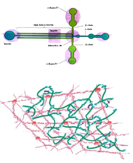
Figure 5: Laminin structure and type IV of collagen network on basement membrane.
Enactin and nidogen are different names for the same compound. This glycoprotein is approximately 150 kDa and consists of three globular domains separeted by two linear segments.It has been linked to a dumb-bell in shape. It is highly susceptible to proteolysis and has both matrix and cell binding propreties.Enactin/nidogen complex binds tightly to laminin via the γ1 chain. Collagen IV also binds to enactin/nidogen. These interaction occur via the carboxyl-terminal globule of enactin/nidogen. It is not clear if this complex has a role other than as a structural stabilizer in the GBM (Levidiotis and A Powe, 2005).
Proteoglycans are found in all basement membranes. They are a class of molecules characterized by a protein core covalently linked to at last one glycosaminoglycan side-chain. The functions of proteoglycans are due to their glycosaminoglycan side-chains. Glycosaminoglycans are unbranched anionic polymers of either glucose-glucose or galactose-glucose derived disaccharides. Heparan sulphates are complex structures belonging to the glycosaminoglycan family. They consist of alternating ialuronic acid and glucosamine residues modified at various positions by sulphation, epimerization or N-acetylation.Proteoglycans are expressed on the surface of most cell types and are important in cell adhesion to the extracellular matrix, cell-cell interactions, and in the interactions of soluble growth factors with their cell surface receptors. Heparan sulphate bound to structural proteoglycans also bind growth factors and other cytokines. Growth factors including platelet-derived growth factor (PDGF), trasforming growth factor-β (TGF-b), fibroblast growth factor (FGF) and vascular endothelial growth factor (VEGF) are bound by heparan sulphate proteoglycans (HSPG).
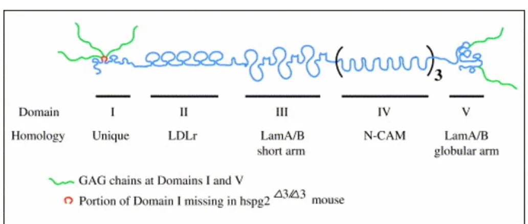
Perlecan is a large multidomain extracellular matrix proteoglycan that has been conserved in organisms. It is expressed in nearly all basement membranes and connetive tissues. The protein core consists of five domains and numerous sites for O-linked glycosylation as well as four potential sites for heparan sulphate chain attachment: three on domain I and one on domain V. These carbohydrate chains, as well as the domains of protein core, are known to interact with a wide range of biological molecules (Knox and Whitelock, 2006).
Figure 6: Perlecan core protein: determined domain structure and GAG attachment.
Domains II–V of perlecan show homology to the LDL receptor (LDLr), the short arm of laminins A and B, the neural cell adhesion molecule (N-CAM), and the globular arm of laminins A and B. The heparan sulfate or chondroitin sulfate GAGs are attached on domains I and V. Also depicted is the region of domain I that is absent when exon 3 is disrupted (Smith and Hassel, 2006 ).
The core protein of perlecan (with its globular domains spaced by rod-like sequences) resembles a string of pearls, hence its name . The protein core is encoded by an approximately 120 kb gene with 97 exons . The gene is well conserved across species, with homologues of mammalian perlecan present in Caenorhabditis elegans , Drosophila and other species. In humans, perlecan's core protein is 466 kDa in size. Mice also have a 369 kDa core protein due to alternative splicing .
Fibulins are a family of five extracellular matrix glycoproteins of a fibulin-type C-terminal domain preceded by tandem calcium binding epidermal growth factor like modules. The 5-member family can be further classified into two subgroups (Figure 6).
Figure 7: Structural features of the fibulin family. The fibulin family currently has six
members, which possess a modular structure and have orthologues in species as divergent as humans and the nematode worm Caenorhabditis elegans. Domain information refers to the human forms of the fibulin family only. The four splice variants of fibulin-1, designated -1A to -1D, are shown. The large immunoglobulin domain of fibulin-6 is contracted for ease of visualisation, as indicated by the use of a dashed line (Gallagher et al., 2005).
Fibulin-1 and fibulin-2, the first subgroup, are substantially larger than the other three members of the family owing to the presence of an extra domain with three anaphylatoxin modules and higher numbers of tandem calcium binding and epidermal growth factor modules.Fibulin-1 with a molecular weight of 90000 kDa has variable C-terminal domain, while fibulin-2 at 200 kDa is the largest of all the fibulins since it possesses an additional N-terminal domain of approximately 400 aminoacids. Members of the second groups: fibulin-3, fibulin-4 and fibulin-5 are similarly small in size (50-60 kDa) and highly homologous to one another in the modular structure. They consist of a modified tandem calcium binding and epidermal growth factor domain at the N-terminus followed by 5 tandem modules and the fibulin C-terminal domain. Fibulin-1 and fibulin-2 are able to bind fibronectin, proteoglycans, tropoelastin and other basement membrane proteins, participating in extracellular supramolecular structures (Kobayashi et al., 2007).
2) Basement membrane assembly
The most interesting feature of the BM is the capacity of its components to self assemble and to form sheet-like structure. Cells at first assemble BM components into functional units (Type IV of collagen protomer, laminin trimers, nidogen/entactan and perlecan) inside the cell, and then they secrete them. Laminin polymerization is believed to initiate the BM scaffold formation at the basolateral surface cells. It is anchored to the cell by receptor proteins such as integrins or dystroglycans. Deposition of this polymer leads to association with type IV of collagen network. Nidogen/entactin bridges the laminin polymer and type IV of collagen network. The other components of the BM interact with the laminin polymer and type IV of collagen network to organize a functional BM on the basolateral aspect of the cell (Kalluri et al., 2003).
Figure 8: Basement membrane formatio. Cells first assemble BM components into
functional units inside the cell (type IV of collagen protomers, laminin trimers, nidogen/entactin complex) and then secrete them. Nidogen/entactin bridges the laminin polymer and type IV of collagen network. The other components of the BM interact with the laminin polymer and the type IV of collagen network to organize a functional BM on the basolateral aspect of the cells
3) Angiogenesis and vascular basement membrane
Angiogenesis is an important biological process considered fundamental to reproduction, development and repair. In the adult, repair and reproductive angiogenesis occurs mainly as brief bursts of capillary blood-vessel growth that usually last only days or weeks. This physiological angiogenesis is tightly regulated by a variety of circulating or sequestred inhibitors that suppress proliferation of vascular endothelium.
The vascular basement membrane (VBM) is composed predominantly of type IV of collagen, laminin, enactin/nidogen and proteoglycans. This complex structure plays a pivotal role in angiogenesis. Infact most of this component sustain the growth and health of vascular endothelium, cryptic domains within these proteins also posses anty angiogenic activity. VBM organization is dependent on the assembly of a type IV of collagen network, wich is believed to occur via the C-terminal globular non collagenous domain (NC1)of collagen IV. Inhibitors of collagen metabolism have anti-angiogenic properties, supporting the notion that basement membrane collagen synthesis and deposition are crucial for blood vessels formation and survival (Maragoudakis et al., 1993, 1995) Figure 9.
Figure 9: Overview of the vascular basement membrane structure and vessels formation..
Blood vessels are made by BMs, pericytes and vascular endothelial cells. During angiogenesis, vascular endothelial cells can proliferate as rapidly as bone-marrow cells. This increase on proliferation is one of the main events that is required for the formation of a new capillary blood vessel. Vascular endothelial cells proliferation is associated to the degradation of the VBM, which leads to sprouting of pre-existing microvessels. These vessels invade the ECM, form tubes and the tips of these tubes eventually connect each other to create loops that are capable of conducting blood flow (Kalluri, 2003). During angiogenesis endothelial cells are dislodged from existing blood vessels, then surrounded by provisional matrix, and eventually come to rest on capillary BMs that are composed of several matrix molecules. The angiogenic response is induced by growth factors such as vascular endothelial growth factor (VEGF), basic FGF (βFGF), platelet-derived growth factor (PDGF), chemokines and others. First, the capillary BM is degraded by several matrix-degrading enzymes such as MMPs , which are produced by stromal cells endothelial cells or by tumour cells themselves. During tumour angiogenesis these factors can also be produced by the immune cells that accumulate around the neoplastic cells even before angiogenesis is initiated. This vascular BM degradation serves multiple purposes, which include the liberation of endothelial cells to migrate and proliferate from their cell-surface integrins, the liberation of sequestred growth factors (VEGF, βFGF) and the detachment of the pericytes that surround and support the blood vessels. The detachment of endothelial cells are now in direct contact with interstitial provisional matrix components, such as vitronectin, fibronectin, type I collagen an thrombin. Pro-angiogenic factors induce endothelial cells to produce many of this matrix molecules. Therefore, provisional matrix provides proliferative cues to endothelial cells, whereas the assembled BM matrix provides growth-arresting cues Figure 10.
Figure 10: The angiogenic switch. Endothelial cells break free from their basement
membrane and surrounding extracellular matrix, migrate, proliferate, and remodel, thus generating new blood vessels or "sprouts" from the parent vessel
4) Regulation of angiogenesis by MMPs
Progression of cancer is dependent on neoangiogenesis. Tumour progression is likely governed by relative levels of pro- and antiangiogenic factors: “the angiogenic balance”. Influenced by oncogenes and tumor suppressor genes, disruption of the “angiogenic check point “can represent an important lethal step in the progression of cancer (Hamano et al., 2003) via the increase in angiogenic factors (such as VEGF) or decrease in the physiological levels of endogenous inhibitors of angiogenesis like tumstatin.
Recent studies have shown that MMP-9 and MMP-2 are required for the mobilization of the sequestred VEGF and the triggering of tumour angiogenesis ( Bergers, et al., 2000).
Gelatinases (MMP-9 and MMP-2) degrade type IV of collagen , probably distrupting the organization of BM, leading to the release of BM-bound VEGF. ( Figure 7). MMPs are predominantly produced by stromal and immune cells. After the matrix is degraded and VEGF is released, angiogenesis is initiated and tumours begin to grow and recruit more immune cells, fibroblasts and other stromal cells, which also produce VEGF and βFGF (Coussens et al., 2001).
MMP-9-mediated release of bound VEGF is potentially important in the very early stages of local tumour progression that are associated with the angiogenic switch.
As the BM undergoes MMP-mediated degradation and structural changes, cryptic domains of partially degraded collagens become exposed (Xu et al., 2001;Xu et al., 2005). These domains have been shown to provide important proangiogenic cues that were sequestred when the BM was fully assembled. Similarly, MMP-mediated degradation of the BM also leads to the generation of fragments with antiangiogenic activity, such as endostatin, arrestin, canstatin, tumstatin and other collagen fragments (Egeblad et al., 2002;Lee et al., 2002; Petitclerc et al., 2000).
Therefore while whereas the initial burst of MMP production releases BM-bound VEGF and other factors that initiate tumour angiogenesis, this need for a VEGF source leads as soluble VEGF is produced in large amounts by the infiltrating immune cells and by the increasing number of cancer cells themselves. As the tumour grows and more
tumour microenviroment and potentially elsewhere. As the BM degradation reaches completion, much of what remains are potential MMP-resistant products, such as endostatin, arrestin, canstatin and tumstatin, all angiogenic inhibitors. MMP-mediated degradation of BM can therefore act as both a positive (early stage) and a negative (middle to late stage) regulator of tumour angiogenesis (Kalluri, 2003; Pozzi et al., 2000; Pozzi et al., 2002) (Figure 11).
Figure 11: degradation of the vascular basement membrane by MMPs.
a) in response to growth factors and matrix metalloproteinases the VBM undergoes degradative and structural changes. The transition from mature and provisional VBM promotes the proliferation and migration of vascular endothelial cells. Growth factors, such as the endothelial growth factor (VEGF), basic fibroblast growth factor (bFGF) and platelet derived growth factor (PDGF) are relased by BM and produced by tumor cells and immune cells. b) Together with the vascular endothelial cells and pericytes, the VBM mediates formation of a new blood vessel.
II) Matrix Metalloproteinases (Matrixin): Structure and Function
1) The Metzincin superfamily
Matrix metalloproteinases belong to a family of zinc and calcium dependent endopeptidases called Metzincin. A quantitative comparison of the three-dimensional structures of the matrixins (MMPs) and the other three protein families (i.e., the astacin, the serralysins, the adamalysins) has revealed striking topological similarities between the catalytic modules of all these proteins. This superfamily is characterized by a catalytically active zinc ion on the active site and is distinguished by a highly conserved motif containing a consensus sequence, HEXXHXXGXXH (single letter code; X is any amino acid residue), which is involved in metal ligation. The three histidines of the conserved consensus sequences serve as ligands for the zinc, the glycine helps the formation of th loop around the metal and the glutamic acid is believed to transfer hydrogen atoms and polarize a zinc-bound water molecule for nucleophilic attack on the scissile peptidic bond of substrate (Stocker, 1995).
2) Structural complexity of MMPs
To date the MMP gene family in humans encodes 25 homologous proteinases (MMPs 1-3, 7-28) and three pseudo-genes. Like many extracellular proteins, MMPs are multi-domain proteins that share similar primary, secondary and tertiary structures.
All MMPs possess a leader domain involved in enzyme secretion, a pro-domain that is auto-inhibitory; and a catalytic pro-domain required for enzyme activity. Most of MMP members possess also a C-terminal hemopexin-domain which is involved in substrate recognition.
The domain composition is different for each MMP and it is listed in table I. Functionally, the most important is obviously the catalytic domain. The prodomain, which is responsible for the enzyme latency, extends from the catalytic domain to the N-terminus of the enzyme. At the C-terminus of the catalytic domain a length-varying linker connects to hemopexin C-terminal domain which is present in most MMPs except for MMP-7 and MMP-26 (Overall et al., 2002).
The structure of domain motifs are illustrated in Figure 11A: prodomain (Figure 11B), catalytic domain (Figure 11C), fibronectin-like domain (Figure 11D), hinge region, hemopexin -like domain (Figure 11E).
The MMPs can be divided into 5 main groups according to their domain composition and their ability to degrade individual component of extracellular matrix:
¾ Matrilysins (MMP-7 and MMP-26) are MMPs that lack the hemopexin C domain;
¾ Collagenases (MMP-1, MMP-8 and MMP-13) are composed of a catalytic domain and hemopexin -like domain;
¾ Stromelysins (MMP-3, MMP-10 and MMP-11) possess the same domains composition of collagenase class;
¾ Gelatinases (MMP-2 and MMP-9) have within the catalytic domain a compact collagen binding domain called fibronectin-like domain;
¾ Membrane Type MMPs (MT-MMP) (MMP-14, MMP-15,
MMP-16, MMP-17, MMP-24 and MMP-25) are inserted in the plasma membrane by a trasmembrane segment or a glycosylphosphatidylisnositol (GPI).
Table I. Schematic representation of the structure of the 24 human matrix metalloproteinases (MMPs), which are classified into four different groups on the basis of domain organization. Archetypal MMPs contain a signal peptide (necessary for
secretion), propeptide, a catalytic domain that binds zinc (Zn2+) and a hemopexin carboxy
(C)-terminal domain. Y, D, and G represent tyrosine, aspartic acid and glycine amino acids that are present in the catalytic domain of all collagenases. Matrilysins contain the minimal domain organization that is required for secretion, latency and catalytic activity. Gelatinases contain fibronectin type II modules that improve collagen and gelatin degradation efficiency. Convertase-activatable MMPs contain a basic insert in the propeptide that is targeted by furin-like proteases (convertase cleavage site). MMPs that belong to this group can be secreted enzymes, or membrane-anchored via GPI (glycosylphosphatidylinositol), type I or type II transmembrane (TM) segments. MMP-23A and MMP-23B contain unique cysteine array (CA) and immunoglobulin (Ig)-like domains in their C-terminal region (Overall et al., 2002).
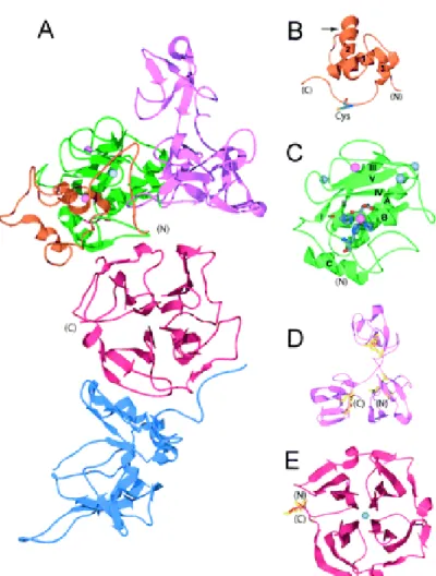
Figure 11. 3D structure of MMPs: ribbon diagram of MMP structures. A)
proMMP-2-TIMP2 complex (1GXD) is shown. Orange indicates propeptide; green, catalytic domain; pink, fibronectin-like domains; red, hemopexin -like domain; and blue TIMP-2. Zinc atoms are pink, and calcium are gray. B) In the MMP-2 propeptide, the cysteine swtch motif is shown. The harrow indicates the position of the initial cleavage resulting in partial activation. C) The catalytic domain of MMP1 is shown. The beta strands are numbered I trough V; the alpha helices are labelled A through C. The N-terminal (N) to C-terminal (C) ordered of the beta strands and alpha helices is I-A-II-III-IV-V-B-C. The histidines coordinating the active site zinc and the active-site glutamic acid are shown. D) The 3 fibronectin-like domains of MMP-2 are shown with their 2 disulfide bonds each. E) The hemopexin -like domain of MMP-1 with 4 beta propeller blades is shown. A disulfide bond is seen between blades I and IV ( Visse et al., 2003).
Many of the MMPs are specifically regulated at the level of gene expression, but their production as inactive proenzymes is another important level of regulation (Sternlicht et al., 2001).
All matrixins are synthesized as pre-pro-enzymes and secreted as inactive pro-MMPs in most cases. The pro-peptide domain (about 80 amino acids) has highly conserved unique PRCG(V/N)PD sequence. The Cys within this sequence (‘the cysteine switch’) coocrdinates the catalytic zinc to maintain the latency of pro-MMPs (Massova et al., 1998).
Structure of the pro-domain is known for MMP-2, MMP-3 and MMP-9; it consists of its three α-helices and connecting loops (Figure 2B). The first loop between helix-1 and helix-2 is a protease-sensitive ‘bait region’. An extended peptide region after helix lies in the substrate binding cleft of the catalytic domain. As already mentioned, this region contains the conserved cysteine switch, which forms a fourth ligand of active site, via its side chain thiol group, keeping the zymogen inactive.
This sequence is missing in stromelysin 11) and MT1-MMP (MMP-14) which were shown to be activated intracellulary by furin, while MMP-23 has a proprotein processing sequence RX(K/R)R at the C-terminal end of the propeptide (Nagase et al., 1999).
It is notable that the orientation of the propeptide backbone is opposite to that one of peptide substrate since it interacts with the active site cleft. However, the hydrogen bonds that it makes with the active site are identical to those of substrate backbone (Visse et al., 2003).
a) The Catalytic Domain and Active Site
The catalytic domains of MMPs are generally very similar with sequence similarities in the range of 50-88% and identities in the range of 33-79 % (Terp et al., 2002). The common structural features includes three α-helix (A, B and C) and a β-sheet consisting of four parallel and one antiparallel strand and connective loops with two zinc ions and between one and three calcium ions.
Their catalytic domains all exhibit an overall spherical shape which can be divided into two subdomains by the substrate-binding cleft with the zinc atom at its bottom. The N-terminal half of the catalytic domain is made up from a twisted β-sheet covering two long α-helices; the central α-helix in the
active site contains the two histidine residues of the consensus motif HEXXHXXGXH (Stocker et al., 1995).
The third histidine zinc ligand of the metzincins is part of the C-terminal half of the catalytic domain being positioned three residues downstream of the conserved glycine. Generally, this C-terminal portion exhibits a long α-helix which packs against the surface of N-terminal domain. The polypeptide chain connecting the third histidine to the C-terminal helix forms a loop made possible by the conserved glycine which interrupse the α-helics, that ends at the catalytic zinc in a unique, tight 1.4 ‘Met turn’ (Bode et al., 1995) (Figure 12).
The very N-terminal region runs parallel to helix C before intruding into the molecular body at a strictly conserved tryptophan residue; furthermore the conformation of the N-terminal portions (consequences of pro enzyme activation) is of extreme importance for the regulation of their proteolytic activity. In fact, two structures of the MMP-8 catalytic domain resulting from the cleavage at two different sites, leaving either Met-80 or Phe-79 as the N-terminal residue display different structures (Reinemer et al., 1994). The latter form is ‘superactivated’, as Phe-79 forms a salt bridge with a Asp-232 thereby preventing the N-terminal sequence from interference with active site. The result is a 3 fold increase in activity compared with activation cleavage at Met 80 (Knauper et al., 1993; Gioia et al., 2002).
Figure 12.The catalytic domain of ligand-free collagenase MMP-1.
Dark spheres calcium atoms; light spheres zinc atoms (Borkakoti et al., 2000).
Figure 13.The typical mode of binding of potent inhibitors to matrix metalloproteinases.
The figures illustrates the interaction of the peptidic inhibitor (Ro 32-4724, ball and stick representation) at the active site of MMP-1 (area identified in figure 3) (Borkakoti et al., 2000).
Catalysis mechanism
As described above, the catalytic zinc is coordinated by the three histidine residues; in the absence of a substrate or inhibitor, a water molecule forms an additional fourth ligand to zinc and is entrapped between the catalytic glutamic acid of the HEXXHXXGXXH motif and the metal. In all MMPs the carbonyl group of scissile peptide bond points towards the catalytic zinc and therefore can be polarized (Figure 13).
The peptide hydrolysis is assisted also by the carboxyl group of the catalytic glutamate, in fact the catalytic water molecule intercalates between this carbonyl group and the glutamate, thereby facilitating the nucleophilic attack of the water molecule on the carboxyl carbon of the peptide scissile bond, and giving rise to a pentacoordinate transition state (Stocker et al.,1995) (see Figure 14).
The tetrahedral intermediate is presumably stabilized by both the zinc and a carbonyl group of the first alanine residue of the edge strand sIV. Simultaneously, one water proton could be transferred to the amino group via the glutamic carboxylate (acting as a proton shuttle); after the cleavage of the peptide bond and the transfer of a second proton this amino group could leave the enzyme-substrate complex (together with the N-terminal substrate fragment) (Bode et al., 1994). In addition, proton transfer to the peptidic nitrogen possibly facilitated by the catalytic glutamate, completes the catalytic reaction, enabling the release of cleavage products and of the free enzyme (Stocker et al., 1995).
Substrate specificity pockets
Although all MMP structures reported to date possess similar core domains, important differences in the side-chains and surface loop alter the size, shape and chemical composition of the specificity subsites.
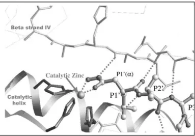
The substrate-binding domain cleft is formed by strand IV and helix B (see Figure 13). The active site is a cavity spanning the entire enzyme and it has been shown that a substrate containing at least six amino acids (three on each side of the scissile bond) is required for the proteolytic activity of MMPs: these six amino acids occupy the subsites S3-S3’(notation according to Schechter and Berger) (Terp, et al.2000) (Figure 14).
Closer examination of the structures of catalytic domains revealed that a conserved aspartic acid is found in the vicinity of the methionine turn, the
side chain of which is buried inside the core of the domain (Massova, et al. 1998). The substrate binds into the catalytic site cleft from the left to the right with respect to its N- and C-termini, and the carboxyl group of the peptide bond coordinates with the active site zinc. A pocket to the right of the active site zinc, called S1’ specificity pocket, accommodates the side chain of the substrate residue, which becomes the new N-terminus after cleavage. The size of S1’ pocket varies among the MMPs and this is one of the major determining factors of substrate specificity (Visse, et al.2003).
Substrate specificity among MMPs is achieved mostly on the recognition pocket S1’, that is of different length and amino acid composition in the individual MMP. The S1’ pockets, which are surrounded by a loop, are generally quite large in all MMPs. However, in the X-ray structure of MMP-1 an arginine defines the bottom of the pocket, whereas in MMP-7 a tyrosine fulfills this purpose, leading to a restriction of the pocket (Terp et al., 2002 and Yaun et al, 1999). In addition, it has been reported that flexibility of this site in some cases could lead to an induced-fit process upon ligand binding; in fact, in MMP-1 the Arginine movements make the pocket able to accommodate larger substituents (Lovejoy et al., 1999). In all other MMPs, this residue is either a leucine or a threonine and the pocket adopts an extended shape. This includes MMP-8, although it resembles MMP-1 by having an arginine defining the bottom of the S1’ pocket (Terp et al., 2002). If we compare the catalytic domain of MMP-2 with that of MMP-8 prominent differences emerge on S1’ loop: in particular, the MMP-2 sequence (YTYTKN--FRL) is two residues shorter than that of MMP-8 and there are seven substitutions including five non-conservative changes (YAFRETSNYSL) (Feng et al., 2002).
Traditionally, S1’ subsite has been labelled as the specificity pocket because of its significant size and shape differences among the rigid x-ray structures of various MMPs. However, modification of P1’ part of the ligands led to both increased selectivity and falures that were explained by observed changes in the structure of the pocket upon inhibitor binding.
The S2’ and S3’ subsites are partly solvent exposed and can accommodate a wide range of functionalities, while differences in the residues surrounding these sites might be used for selectivity purposes.
The primed sites of active site have been described in detail, whereas the unprimed sites have not been examined deeply. However, the importance of other parts of binding site, especially the unprimed side, for the design of more potent and selective inhibitors has been demonstrated as well
Figure 14. Peptide substrate and inhibitor interaction and specificity. Schematic drawing
of the putative encounter complex between a Pro-Leu-Gly-Leu-Ala-Gly-amide hexapeptide substrate and the MMP active site. The substrate polypeptide chain (boldconnections) lies antiparallel to the bulge-edge strand (top) and parallel to the S1' wall-forming segment (bottom) forming up to five and two, respectively, intermain-chain hydrogen bonds (dashed lines) (Bode et al., 1999).
b) The hemopexin -like domain
The C-terminal domain of the MMPs, which is present in all members except the matrilysins, shows strong sequence similarity to members of the hemopexin family, including hemopexin, α-subunit of integrins and vitronectin (Smith et al., 1999). The common functional property possessed by these hemopexin-containing proteins seems to be the participation in quite specific protein-protein and protein-ligand interaction (which differ among various MMPs). All hemopexin-like domains have a β-propeller topology with pseudo four-fold symmetry, with sequence homology to a haem-binding and transporting protein from serum (Bode et al., 1995). The typical hemopexin-like domain comprises ~200 amino acid residues and shows a fourfold internal sequence repeats: the four blades of propeller are made up of antiparallel four β sheets arranged around a tunnel harbouring a number of ions (calcium and chloride). The first and the fourth blades are linked by a disulphide bond which is conserved across all the MMPs (Murphy et al., 1997) (Figure 11E). When present, the hemopexin-like domain also influences TIMP (tissue inhibitor metalloproteinase) binding, the binding of certain substrates, membrane activation and some proteolytic activities (Sternlicht et al., 2001).
c) The hinge region
The N- and C-domains of the MMPs seem to be packed as separate entities in the crystal with connecting flexible linker peptide or ‘hinge’ which is rather loosely arranged. The hinge is composed of 2-72 residues (Lauer-Fields et al., 2002), being short (16 residues) in the collagenases where displays a motif resembling those observed in each α-chain of triple helical collagen (polyproline II) (Knauper et al., 1997). The polyproline II conformation of collagen chains possess backbone φ-γ angles similar to antiparallel and parallel β-sheets (Perona et al., 1997). The residues of the hinge region also influence substrate specificity; the capacity to cleave triple-helical collagen obviously depends on the correct interplay between the catalytic and the hemopexin-like domain. This might be co-determined by
the length and the surface-guiding properties of the linker peptide (Bode et al., 1995).
d) The fibronectin-like domain
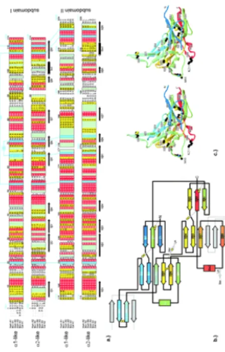
The gelatinases (i.e, MMP-2 and MMP-9) have an additional domain consisting of three tandem copies of 58 amino acid residues forming the fibronectin type II like module, which are inserted between the fifth β-strand and the catalytic helix (adjacent and aminoterminal to S3’ subsite). The structure of each fibronectin-like domain consists of two antiparallel β sheet, connected with a short α-helix and stabilized by two disulfide bonds (Visse et al., 2003). A portion of this domain is called collagen binding domain (CBD), since it appears to be important for binding gelatin and collagen (Lauer-Fields et al., 2002).
3) Activation of proMMPs
a) Intracellular activation
Most proMMPs are secreted from cells and activated extracellulary. Pei, in 1995, first demonstrated that proMMP-11 is activated intracellulary by furin. ProMMP-11 possesses a furin recognition sequence, KX(R/K)R, at the C-terminal end of the propeptide. Several others MMPs, including the six MT-MMPs, MMP-23 and MMP-28, have a similar basic motif in the propeptide. All other MMPs lack a furin-susceptible insert and are thus activated outside the cell following their secretion.
b) Stepwise activation mechanism
MMPs can be activated by proteinases or in vitro by chemical agents such as thiol-modifying agents (4-aminophenylmercuric acetate, HgCl2 and
N-ethylmaleimide), oxidized glutathione, SDS, chaotropic agents and reactive oxygens (Figure 15) (Visse et al., 2003).
Low pH and heat treatment can also lead to activation. These agents most likely work through the disturbance of cysteine-zinc interaction of the cysteine switch. Studies of proMMP-3 activation with a mercurial compound have indicated that the initial cleavage occurs within the propeptide and this
reaction is intramolecular rather than intermolecular. The subsequent removal of the rest of the propeptide is due to intermolecular reaction of generated intermediates. In vivo, it has been shown that NO (nitrix oxide) activates proMMP-9 by interacting with the thiol group of cysteine switch and forming an S-nitrosylated derivative (Visse et al., 2003).
Proteolytic activation of MMPs is stepwise in many cases. The initial proteolytic attack occurs at an exposed loop region between the first and the second helics of propeptide. The cleavage specificity of the bait region is dictated by the sequence found in each MMP. Once a part of the propeptide is removed, this probably destabilizes the rest of the propeptide, including the cysteine switch-zinc interaction, which allows the intermolecular processing by partially activated MMP intermediates or other active MMPs. Thus, the final step in the activation is conducted by a MMP.
The stepwise activation system may have evolved to accommodate finer regulatory mechanisms to control destructive enzymes, in as much as TIMPs (tissue inhibitor of metalloproteinases) may interfere with activation by interacting with the intermediate MMP before it is fully activated.
c) Cell surface activation of proMMP2
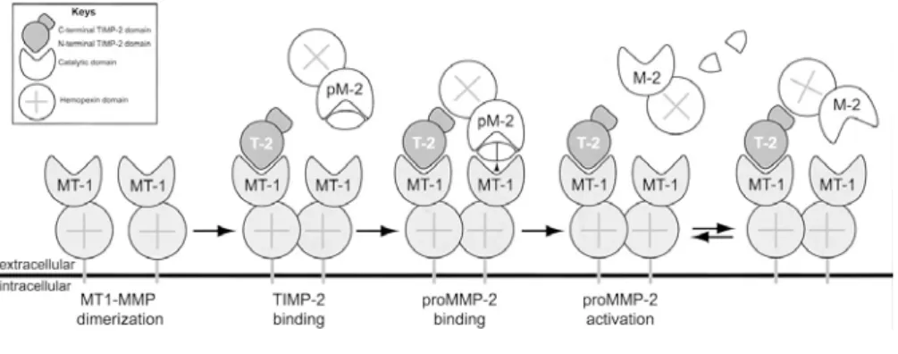
The extracellular activation of most MMPs can be initiated by other already activated MMPs or by several serine proteinases that can cleave peptide bonds within MMP prodomains. However, proMMP-2 is refractory to action of serine proteinases and it is instead activated at the cell surface through a unique multistep pathway involving MT-MMPs (Figure 16).
According to the present knowledge, a cell surface MT1-MMP binds the N-terminal domain of TIMP-2 being inhibited; the C-N-terminal domain of the bound TIMP-2 acts as receptor for the hemopexin-like domain of proMMP-2. Then, an adjacent, MT1-MMP cleaves and activates tethered proMMP-proMMP-2. Following the initial cleavage of proMMP-2 by MT1-MMP, a residual portion of MMP-2 propeptide is removed by another MMP-2 molecule to yield a fully active, mature form of MMP-2 (Sternlicht et al., 2001).
Figure 15. Stepwise activation of proMMPs. ProMMPs secreted as
inactive zymogens can be activated by protease (top pathway) or by non proteolytic agents ( bottom pathway). The catalytic domain is represented as a graycircle, with the active site cleft shown in with ( not in scale), containing the catalytic zin (Zn). The propeptide is schematically shoen as a black line containing the bait region ( black rectangle) and the cysteine switch (C). SH indicates the sulfhydryl of the cysteine. Activation by protease is mediated by cleavage of the bait region; this partly activates the MMP. Full activations achieved by completed removal of the propeptide by intermolecular processing. Chemical activation relies on modification of the cysteine switch sulfhydryl (SX), resulting in partial activation of the MMP and intramolecular cleavage of the propeptide. Full activity results from the removal of the remainder of the propeptide by intermolecular processing (Visse et al., 2002).
Figure 16. Model of proMMP-2 activation by MT1-MMP and TIMP-2. active MT1-MMP
(MT-1) on the membrane binds a molecule of TIMP-2 through its hemopexin-like domain. The second, active, MT1-MMP then cleaves the region of proMMP-2, thereby partly activating it. The MMP-2 (M-2) dissociates from the membrane and is fully activated by intermolecular processing (Visse et al., 2002).
4) Inhibition of MMP activity
Beyond their classical connective-tissue-remodelling functions, MMPs are known to precisely regulate the function of bioactive molecules by proteolytic processing. Under physiological conditions, MMP activity is controlled at least at three levels:
¾ transcription,
¾ proteolytic activation of zymogen form,
¾ inhibition of the active enzyme by endogenous inhibitors. Most MMPs are expressed in adult tissues at low levels or not at all in resting condition. However, several cytokines and growth factors as well as physical cellular interactions provide stimuli that can rapidly induce MMP expression (Stamenkovic et al., 2003). Thus, cell use various strategies to regulate extracellular proteases; in this paragraph, the role of metalloproteinase inhibitors is reviewed (Figure 8 Baker et al., 2002).
A greater understanding of regulatory mechanism that controls MMP activity provides several new avenues for therapeutic intervention, since many drugs are designed to target these key regulatory points (Figure 17).
Figure17. Metalloprotease inhibitors in the pericellular enviroment.
A) tissue inhibitors of matrix metalloprotase (TIMPs). TIMPs- 1-4 are largely MMP inhibitors modulating the activity of soluble, matrix bound and cell associated MMPs. TIMP- 3 is an extracellular matrix protein, probably bound to heparan sulphate proteoglycans and it is a potential inhibitor of the function of some membrane associated ADAMs. TIMP2 acts in junction with MT1-MMP as receptor for the proform of MMP-2 at the cell surface. Allowing an efficient activation and focussing of the active form of this soluble protease. In the same cell types, TIMP-1 and TIMP2 may have receptors directly linked to intracellular signalling pathways regulating cell behaviour. B) other inhibitors. RECK (Reversion inducing cysteine rich protein with Kazal motif) is a GPI-anchored glycoprotein that binds and inhibits a number of MMPs. The pan protease inhibitor alpha 2 macroblobulin, although very large, has some access to the pericellular space in vascularised tissue and may be involved in MMP endocytosis through the low density lipoprotein (LDL-RP). The roles of LDL-RP in MMP-2 removal via a thrombospondin-2 (TFPI-2) has also been described as an MMP binding agents (Baker et al., 2002).
a) Endogenous inhibitors
MMP activity is tightly controlled by several endogenous inhibitors. In tissue fluids, the principal MMP inhibitors is α2macroglobulin, a large serum
protein, which binds MMPs and creates a complex that is itself bound irreversibly (Yang et al., 2001; Woessner et al., 1998).
However, the most throughly studied MMP inhibitors, are TIMPs (Tissue Inhibitors Metallo Proteinases). Four human TIMPs have been characterized thus far; they are small molecules of 21-28 kDa which bind MMPs in a 1:1 stochiometric ratio and reversibly block MMP activity. TIMPs, which are anchored to the extracellular matrix or secreted extracellulary ,differ in their expression pattern.
Among other molecules capable of regulating MMP proteolytic activity we underline thrombospondin-2 and RECK (reversion-inducing cysteine-rich protein with Kazal domain motifs), a GPI-anchored glycoprotein that suppresses angiogenic sprouting (Stamenkovic et al., 2003). Other key regulators of extracellular matrix are TFPI2 (tissue-factor patway-inhibitor-2), a serine protease inhibitor that can act as a MMP inhibitor and PCPE (procollagen C-terminal proteinase enancer), a molecule which possesses a significant inhibitory activity. Other latent MMP inhibitors might be hidden in the NC1 domains of type IV collagen or in the laminin-binding domain of agrin, which are structurally similar to TIMPs (Overall et al., 2002) (Figure 17). However, it may be interesting to outline that endogenous inhibitors may also act as activators. This unexpected function has been quite well described for TIMP2 in the case of MMP2 activation.
b) Exogenous MMP inhibition
In recent years the MMPs have been implicated in a variety of important diseases including cancer (growth and metastasis), atherosclerosis, osteo- and rheumatoid arthritis and emphysema (Palvlaki et al., 2003). However, the evidence has been, consisting largely of correlations such as higher rates of metastasis accompanied by higher level of MMPs. Recent works, based on the development of knock-out mice for several MMPs, suggest more strongly that there is a rational point in developing inhibitors to MMPs with the goal of reversing disease processes. There are several approaches to inhibit MMP
gene transcription based on targeting extracellular factors, signal-transduction pathways or nuclear factors that activate expression of these genes. In addition, ribozymes or antisense constructs down-regulate MMP production by targeting MMP transcripts (Overall et al., 2002).
Inhibition of active MMPs by chelators
Several mechanisms reduced the tissue levels of MMPs; a great deal of attention has been given to chelating agents that bind zinc at the catalytic centre. Most attention has been focussed on improvements of hydroxamates which belong to chelators compound with no specificity and would block any enzyme containing zinc. The hydroxamate moiety offers two oxygen atoms zinc binding as well as nitrogen atom to the carboxyl group of the backbone of protein and results in compounds with nanomolar affinity (Borkakoti et al., 2004).
Several classes of MMP inhibitors have been discovered; the most common is containing a Zn-liganding hydroxamic acid, carboxylic acid or thiol group attached to a small peptide fragment capable of binding to specificity pockets of the MMP enzyme. Ligands coordinate the catalytic zinc atom and interact with either the S1-S3 or S1’-S3’ subsites (Figure 14). The vast majority of inhibitors described to date bind in an extended conformation in the right side (S1’-S3’) of the active site. Recently, the thiazole class of inhibitors was discovered and determined to interact with the left side (S1-S3) of the active site (Yuan et al., 1999).
Natural inhibitors
Unfortunately, most of synthetic compounds produced up to date are quite specific toward the rest of MMPs and at therapeutic doses are toxic even though less toxic than other anticancer agent. However, peptido-mimetic MMP inhibitor developments are yet in-phase-III studies in clinical trial (Pavlaki et al., 2003). On the other hand, among the biologically active components from natural products, green tea polyphenols caused the strong inhibition of MMPs. More particularly, it has been reported that natural extracts components (such as epigallo chatechin gallate EGCG) are able to inhibit the enzymatic activity of various MMPs as well as the activation of proMMP-2 (Demuele et al., 2000).
III) Gelatinases and degradation of type IV collagen
Endothelial cell invasion is an essential event during angiogenesis, a process that consists on new blood vessels formation (Nguyen et al., 2001). Angiogenesis has been identified as a casual or contributing factor in several pathologies, including cancer, where it is a rate-limiting step during tumour progression.
Matrix metalloproteinases (MMPs) are a family of soluble and membrane-anchored proteolytic enzymes that can degrade components of the extracellular matrix (ECM) as well as a growing number of modulators of cell function. Enhanced expression of MMP-2 and MMP-9 has been observed in different kind of cancers, such as breast, colon, lung, skin, ovary and prostate. In particular, gelatinases have been linked to angiogenesis. Potential roles for these proteases during the angiogenic process include degradation of the vascular basement membrane and perivascular ECM components, unmasking of cryptic biologically relevant sites in ECM components, modulation of angiogenic factors and production of endogenous angiogenic inhibitors (Handsley and Edwards, 2005). Gelatinases cleave the main component of the vascular basement membrane, type IV of collagen, leading to VEGF release, necessary to endothelial cells proliferation. (Handsley and Edwards, 2005).
Moreover, vascular basement membrane cleavage gives rise to the formation of fragments with anti angiogenic activity.
1) Type IV of collagen structure
Type IV of Collagen is the most abundant component of the basement membrane, where it is crucial for its stability and assembly. It is termed network-forming collagen to its capability to self assemble into network structure. This proprety makes it different from the fibrillar collagens (type I, II and III). Type IV of collagen is formed by the association of three α chains in a primary structure called protomer. Six different type IV of collagen α chains have been identified codified by six different genes: α1(IV), α2(IV), α3(IV), α4(IV), α5(IV) and α6(IV). The type IV collagen α-chains have similar domain structures and share between 50-70% homology at the amino-acid level. Each α-chain can be separated into three domains: an amino terminal 7S domain, a middle triple helical domain, and a
triple-helical part is the longest domain (about 1,400 amino acids (aa) in length), with 22 interruptions of the classical Gly-X-Y sequence motif that is characteristic of collagens. The NC1 domain of each α-chain is about 230 aa in length. α−chain Usually type IV of collagen of the vascular basement membrane is formed by two α1 chain and 1 α2 chain that coassemble to form a trimer called protomer given by the interaction of the C-terminal domain of each α-chain. Type IV of collagen formation begins with the protomer formation and proceeds with protomer trimerization like a zipper from the carboxy-terminal end, resulting in a fully assembled protomer. The assembled protomer is flexible and can bend at many triple-helical interruption points in the molecule. The next step in the assembly is the type IV collagen dimer formation. Two type IV collagen protomers associate via their carboxy-terminal NC1 trimers to form an NC1 hexamer. Next, four protomers interact at the glycosylated amino-terminal 7S region to form tetramers. These interactions form the nucleus for a type IV collagen scaffold. The scaffold evolves into a type IV collagen suprastructure, with the help of end to end assiciations and also lateral associations between type IV collagen protomers (Kalluri, 2003).
Figure 18: α-chain secondary structure. We can observe: at the N-terminal domain called 7S-domain formed by several sequences repeats; in the middle the triple helical domain or collagenous domain and at the C-terminal domain the so called non collagenous domain or NC1-domain (Kalluri, 2003).
Figure 19: Type IV of collagen suprastructure formation. Type IV of collagen suprastructure begins with the formation of a primary structure called protomer, given by the interaction of the NC1 domain of three a-chains, and proceeds with dimer, hexamer and tetramer constitution to form finally the collagen IV complex structure (Kalluri, 2003).
2) Gelatinases cleavage site for degradation of type IV
collagen
Gelatinases are a particular class of the extracellular degrading enzymes family called matrix metalloproteinase or MMPs. While MMPSs play a critical role in normal and pathological processes involved in the remodeling of the extracellular matix (ECM) gelatinases are involved in the remodeling of BM structures. The BM structure is generally formed by different kind of glycoproteins, such as collagen IV, that is the major component of the vascular basement membrane.In vitro studies have shown that triple helical collagen IV molecules can be hydrolyized by several members of MMP family including gelatinases . Of particular interest is that the contribution of the gelatinases to the degradation of collagen IV seems to be associated to tumour metastasis, a process known to involve alterations in VBM integrity. This is also confirmed by previous studies that have shown that the gelatinases are localized on the surface of tumour cells (Ginestra et al., 1997). The degradation of VBM by gelatinases has been postulated to take place at areas of cell-matrix contacts (Werb, 1997). Searching for potential pro-MMP-9 surface binding molecules, Fridman et al. identified a surface bound α2(IV) chain of collagen IV that specifically formed a high affinity complex with pro-MMP-9 and plays a role in targeting pro-MMP-9 at relevent areas of proteolysis. The proteolysis of the collagen IV network by MMP-2 and MMP-9 seems to bring about also to the formation of fragment with an anti proliferative activity. In particular, MMP-2 has the capability to liberate some cryptic domains from type IV of collagen with anti angiogenic activity . Infact, the non collagenous domain (NC1) of α1 (arrestin) and α2 ( canstatin) and α3 (tumstatin) chains of type IV of collagen were previously shown to inhibit proliferation of endothelial cells in vitro and suppress tumor growth in vivo (Figure 19). Previous studies showed that the C-terminal 185-203 amino acid fragment of tumstatin, the NC1 domain of α3 (IV) collagen, specifically inhibited the proliferation of melanoma cells. (Maeshima et al., 2001; Han et al., 1997), and its antiangiogenic activity was localized at the N-terminal 54-132 amino acids, whereas the full-length tumstatin specifically inhibited endothelial cells proliferation with no effect on other tumor cells, and also supressed tumor growth of human renal and prostate carcinoma ( Maeshima et al., 2000, 2001).Similarly, canstatin, the NC1 domain of a2 (IV) collagen, can also specifically inhibit proliferation of endothelial cells, and
suppress in vivo growth of large and small sizes tumors (Kamphaus et al., 2000).
Figure 19: arretin, constatin, tumstatin and endostatin formation.Arrestin,
constatin and endostatin derive from the NC1 domain of different a(IV) chains, while endostatin is a fragment of type XVIII of collagen.
As canstatin and tumstatin belong to the NC1 domains of type IV of collagen and have similar biological activities, the amino acid sequences of canstatin and tumstatin were compared and found that they share three homologous regions: the N-termianl 1-89 amino acids (N region), the middle 101-135 amino acids (M region), and the C-terminal 157-197 amino acids , and the final 10 amino acids (C region). Angiogenesis is dependent on specific endothelial cell adhesive events mediated by αvβ3
integrin, suggesting that the anti angiogenic activity of endogenous NC1 domain of type IV of collagen (i.e. tumstatin , arrestin and canstatin) takes place through the distruption of the interaction of proliferating endothelial cells with the matrix component such as vitronectin and fibronectin, eventually leading to an important anti-apoptotic signal (Isik et al., 1998, He et al., 2003).
Although, it is well known that gelatinases are involved on VBM degradation during tumor spreading, it’s not clear the function and the mechanism by which these two matrix metalloproteinases are involved.