Original Article
Non-functioning gastroenteropancreatic (GEP) tumors:
a
111In-Pentetreotide SPECT/CT diagnostic study
Angela Spanu1, Orazio Schillaci2, Bastiana Piras1, Diego F Calvisi3, Antonio Falchi1, Roberta Danieli2,
Susanna Nuvoli1, Franca Dore4, Giuseppe Madeddu1
1Unit of Nuclear Medicine, Department of Clinical and Experimental Medicine, University of Sassari, Sassari, Italy; 2Unit of Nuclear Medicine, Department of Biomedicine and Prevention, University of Rome Tor Vergata, Rome,
Italy; 3Department of Clinical and Experimental Medicine, University of Sassari, Sassari, Italy; 4Unit of Nuclear
Medicine, University-Hospital of Trieste, Trieste, Italy
Received April 26, 2017; Accepted August 21, 2017; Epub September 1, 2017; Published September 15, 2017 Abstract: In a retrospective study performed in non-functioning GEP tumor patients we further investigated 111
In-Pentetreotide SPECT/CT usefulness in diagnosis, staging and follow-up also evaluating whether the procedure may give more information than conventional imaging procedures (CIP), such as CT, MRI, US. We enrolled 104 consecu-tive patients with non-functioning GEP tumors, 30 in initial diagnosis and staging phases (IDS) and 74 in follow-up (FU). All patients underwent somatostatin receptor scintigraphy (SRS) whole body scan at 4, 24 and, if necessary, 48 hours followed by abdominal and chest SPECT/CT after 111In-Pentetreotide 148-222 MBq i.v. injection. The patients
previously underwent 2 to 3 CIP. At both CIP and SPECT/CT, 34/104 patients were classified as no evidence of dis-ease (NED); in 70/104 patients, neoplastic lesions were ascertained and 12 IDS and 17 FU were classified as not operable and treated with octeotride or chemotherapy. SPECT/CT and CIP were concordantly positive in 44 patients, while only CIP was positive in 6 cases and only SPECT/CT in 20. Both per-patient sensitivity and accuracy of SPECT/ CT (91.4 and 94.2%, respectively) were higher than CIP (71.4 and 80.8%, respectively), but not significantly. Glob-ally, 292 lesions were ascertained: 141 hepatic, 78 abdominal extra-hepatic and 73 extra-abdominal. CIP detected 191/292 (65.4%) lesions in 50 patients, while SPECT/CT 244/292 (83.6%) in 64, the difference being significant (p<0.0001). No false positive results were found at both SPECT/CT and CIP. Both SPECT/CT sensitivity and accu-racy were higher than CIP in G1, G2, neuroendocrine carcinoma (NEC) and mixed adeno-neuroendocrine carcinoma (MANEC) patients, but significantly only for G1. Globally, SPECT/CT incremental value than CIP was 35.6%. SPECT/ CT correctly modified CIP classification and patient management in 27.9% of cases, while it down-staged the dis-ease than CIP in 9.6% of cases. However, the two procedures combined use could achieve the highest accuracy value. 111In-Pentetreotide SRS, acquired as SPECT/CT, showing high sensitivity and accuracy values, more elevated
than CIP in the present study, can still have a wide employment in the routine diagnostic protocol of non-functioning GEP tumors with significant impact on patient management and therapy planning. The procedure is simple to per-form, has limited cost and wide availability in all Nuclear Medicine Centers.
Keywords: Non-functioning gastroenteropancreatic (GEP) tumors, carcinoid, neuroendocrine carcinoma (NEC), mixed adeno-neuroendocrine carcinoma (MANEC), chromogranin A, 111In-Pentetreotide, SPECT/CT, conventional
imaging procedures (CIP)
Introduction
Gastroenteropancreatic (GEP) neuroendocrine tumors represent a heterogeneous group of rare neoplasms characterized by an overex-pression of somatostatin receptors (SSR) and an increased production of hormones, peptides or other biologically active substances often producing clinical symptoms due to systemic
effects of their uncontrolled production and secretion. The tumors may grow slowly thus per-mitting long survival, but they can be malignant also producing metastases which can repre-sent the first manifestation of the disease, seri-ously affecting patient prognosis [1, 2].
Both clinical symptoms and the various sub-stances secreted by these tumors are often
diagnostic for the tumor type, particularly if the associated tumor secretions are in very high concentration in blood [3, 4].
However, some tumors are non-functioning since, although they may secrete hormones and other substances, such as chromogranin A (CgA), synaptophysin and neuron-specific eno-lase (NSE), these substances can also not be released into the circulation despite their immune-histochemical evidence [5]. Moreover, it is likely that non-functional tumors can secrete one or more hormones that are as yet unidentified; alternatively, they can produce insignificant amounts of biological active hor-mones or inactive forms of horhor-mones. These tumors may also secrete high-molecular-weight precursors of peptide hormones that have dif-ferent biologic activities than those of the mature peptides; consequently, the concentra-tion of immune-active hormones in plasma may not correspond to the level of biologic activity [6]. Therefore, in some patients clinical symp-toms can be minimal or absent, except for those due to mass effect or distant metasta-ses; in the latter cases, the diagnosis of prima-ry tumor and metastases is often ascertained in more advanced stages. Most non-function-ing GEP tumors originate from pancreatic islets of Langerhans, but also from small intestine, and account for 15-52% of neuroendocrine pancreatic tumors [7, 8].
No histologic difference between functioning and non-functioning tumors has been de- scribed, but at surgery the non-functioning tumors are generally larger; moreover, these tumors can also be malignant, like the function-ing forms, developfunction-ing aggressive metastatic lesions which can already be present when diagnosed, seriously affecting patient prog- nosis.
Conventional imaging procedures (CIP), such as CT, ultrasound (US) and MRI, represent the most available diagnostic methods to detect GEP tumors and their metastases, also guiding biopsies. In particular, MRI is considered the most sensitive radiologic method for liver metastases although these, when small in size, are sometimes difficult to be localized [9]. Somatostatin receptor scintigraphy (SRS), us- ing the somatostatin analogue 111In-Pente-
treotide as radiotracer that preferentially binds
to SSR subtypes 2, 3 and 5, especially the for-mer, has proven to be an important diagnostic functional imaging procedure for diagnosis, stage and follow-up of neuroendocrine (NET) tumors, both pulmonary and in particular GEP tumors [10-14]. For over 20 years, the proce-dure has been considered a first line imaging technique. In particular, SPECT has obtained a better performance than planar in the identifi-cation of primary and metastatic lesions derived from both functioning and non-func-tioning GEP tumors [15-17]. In the latter type of tumors some studies have reported a lower sensitivity of planar SRS in respect of CIP but these data have not been confirmed by others [18-20]. In the last years, the utility of SRS has been further augmented with the employment of the hybrid system technologies, such as SPECT/CT; this latter procedure has permitted a better localization and functional character-ization of GEP tumors and a correct identifica-tion of areas of physiologic uptake, reducing false-positive results on planar and SPECT images and correctly classifying lesions. In par-ticular, SPECT/CT has been reported to provide an incremental diagnostic value than both pla-nar and SPECT images, even more using a SPECT/multiphase CT [21-26]. Some soma-tostatin analogues labelled with 99mTecnetium,
such as 99mTc-EDDA/HYNIC-Tyr3-octreotride
and 99mTc-HYNIC-TOC, have also been employed
with SPECT/CT acquisition providing reason-able accuracy, in particular in the evaluation of the pancreatic masses suspected to be neuro-endocrine tumors [27, 28]. Conventional posi-tron emission tomography (PET/CT) with
18F-fluorodeoxyglucose (FDG) appears of
limit-ed value in the diagnosis of GEP tumors, except for the most aggressive forms with high prolif-eration and with less favourable prognosis [29, 30].
More recently, PET/CT with somatostatin ana-logues (DOTATOC, DOTATATE and DOTANOC) labelled with positron emitting radionuclides, as 68Gallium, which have showed a high affinity
for SSR subtypes 2-5, have obtained high sen-sitivity and specificity values in patients with both thoracic NETs and GEP tumors [31-33]. Up to day, 111In-Pentetreotide is still the current
standard technique for SSR imaging and, unlike PET with somatostatin analogues, it has been approved for many years for marketing in the
Europe and USA. Only more recently, USA-FDA has approved 68Ga-DOTATATE injection for
local-ization of NETs in adult and pediatric patients. In the present study, we further investigated the diagnostic usefulness of ¹¹¹In-Pentetreotide hybrid SPECT/CT imaging in a series of patients with non-functioning GEP tumors, also evaluat-ing whether this procedure may give more use-ful information than CIP in the diagnosis, stag-ing and follow-up; we also assessed whether SPECT/CT may have a better clinical impact in the management of patients affected by this type of neuroendocrine tumor whose identifica-tion can often happen late.
Material and methods Patients
One hundred and four consecutive patients were retrospectively studied, 54 males and 50 females, aged 17-86 years (average: 59.4± 16.5), and observed in three different University Centres of Nuclear Medicine. All patients were affected by non-functioning GEP tumors, 30 being in the phase of initial diagnosis and stag-ing (IDS) for a primary tumor and 74 in follow-up (FU) with previous ascertained primary tumor. Twenty-one/30 IDS patients have foregut carci-noid, 5/30 midgut carcicarci-noid, 1/30 hindgut car-cinoid and 3/30 indeterminate neuroendocrine tumor. Eighteen/30 IDS patients were classi-fied as operable and were submitted to surgery after scintigraphy and 12/30 as not operable because of disseminate metastases and seri-ous clinical conditions and underwent medical therapy (octeotride in 9 cases and chemothera-py in 3 cases). Before our observation, 57/74 FU patients had undergone surgery for: foregut carcinoid (29 cases), midgut carcinoid (26 cases), hindgut carcinoid (2 cases). Fifteen/57 patients of these had limited hepatic metasta-ses, 21 had abdominal extrahepatic and 2 extra-abdominal metastases, 10 had both hepatic and abdominal extrahepatic metasta-ses and 9 both abdominal extra-hepatic and extra-abdominal metastases; moreover, 33/57 patients had undergone only surgical proce-dures, while 16/57 had also octreotide therapy and 8/57 chemotherapy. The remaining 17/74 FU patients with GEP tumors were considered not operable for disseminated metastases, with primary tumors being foregut carcinoid (12
cases), midgut carcinoid (4 cases) and indeter-minate neuroendocrine tumors (1 case). Twe- lve/17 not operable patients were on octeotride therapy which could be interrupted in 8 cases in average 10-14 days prior to scintigraphy, while in 4 the therapy was continued because of their serious clinical conditions; the remain-ing 5/17 not operable patients had previously been submitted to chemotherapy.
Furthermore, in presence of hepatic metasta-ses the classification as non-resectable tumors had been performed on the basis of both the number of lesions (numerous and disseminat-ed in both hepatic lobes) and their location (porta hepatis, confluence of hepatic veins entering into inferior vena cava); furthermore, surgery was not considered feasible when the metastatic lesions were too extensive in patients with serious clinical conditions and with other distant metastases, in the latter cases even if hepatic metastases were consid-ered resectable.
In all 104 cases, the definitive diagnosis of the primary tumors was obtained on the basis of histopathologic analysis by haematoxylin and eosin and immune-histochemical method, with positive staining for one or more hormones or peptides, such as CgA and/or NSE, gastrin, vasoactive intestinal peptide (VIP), somatosta-tin, glucagon and pancreatic peptides (PP). The diagnosis of recurrent tumor and/or metas-tases was based on histology of neoplastic sites and/or on imaging features of progressive malignancy, using MRI, contrast-enhanced CT, transabdominal US, endoscopic US, nuclear medicine procedures including bone scintigra-phy and 18F-FDG PET/CT, with a follow-up period
of 6-36 months.
According to WHO 2010 Classification of tumors of the Digestive System, the 104 pri-mary GEP tumors were classified on the basis of proliferative rate assessed as the number of mitoses per unit area of tumor and the percent-age of neoplastic cells immune-labelling for the proliferation marker Ki67. The patients were classified as neuroendocrine tumor grade 1 (G1; 65 cases), neuroendocrine tumor grade 2 (G2; 22 cases), neuroendocrine carcinoma (NEC), large cell (LCNEC) or small cell (SCNEC) type (12 cases), mixed adeno-neuroendocrine carcinoma (MANEC; 5 cases).
At the time of our observation, none of the 104 patients had clinical signs of hormone excess and thus they were classified as affected by non-functioning GEP tumors although a slight increase of CgA serum levels was present in 10 patients, 7 FU and 3 IDS (3 FU and 1 IDS oper-able and 4 FU and 2 IDS inoperoper-able). However, 23 patients referred malaise, dyspepsia, epi-gastric and abdominal pain, 11 also had weight loss and 5 also obstructive jaundice.
Within a month before scintigraphy, the patients had been submitted to at least 2 CIP, such as US, CT, and MRI, all of these centred over abdo-men, thorax and other suspect regions; in 7 cases a conventional whole body scan after i.v. injection of 740 MBq of 99m
Tc-methylendiphos-phonate (MDP) was also performed, as well as in 2 cases of 99mTc-tetrofosmin whole body scan
and in 3 cases whole body 18F-FDG PET/CT.
According to CIP data the patients were initially classified as with no evidence of disease (NED) or with operable or not operable neoplastic lesions on the basis of the aforementioned criteria.
All clinical and instrumental examinations were performed in University Hospitals setting as part of the clinical care of neuroendocrine tumor patients. This retrospective study was performed in accordance with the regulations of the Institutional Review Board and in accor-dance with Helsinki Doctrine. Routinely, written informed consent had been obtained by all patients whose data were treated in
accor-dance with the local privacy rules and regulations.
111In-Pentetreotide SRS and SPECT/CT
In the present study, all patients underwent a low residue diet for 3 days before and 2 days after tracer injection and also took a laxative the day before and daily for 2 days after to bet-ter unsure a bowel cleaning, thus to reduce interfering background radioactivity by intesti-nal content. Scintigraphic images were obtained with 2 hybrid variable-angle dual-head gamma cameras including a low dose x-ray tube, the Millenium VG Hawkeye (GE Medical System) in 49 cases and with Infinia Hawkeye (GE Medical System) in 55 cases, equipped with an integrat-ed x-ray transmission system (low-dose CT) to provide anatomic maps for attenuation correc-tion and image fusion. CT apparatus has a fixed anode oil-cooled x-ray tube installed on the slip-ring gantry of the gamma camera and operates at 140 Kev and up to 2.5 mA. The x-ray tube and the detector array are rotated together in a fixed geometry, at 2.0 rpm for a 90° L-mode scan. Medium energy, parallel-hole collimators were always used in both machines with 20% energy windows centered on the 111In photon
peaks (173 and 247 Kev).
A whole body planar in anterior and posterior views with a speed of 5 cm/min for a total time of 30 min (1024×256 matrix) were always obtained at 4th, 24th and, when necessary, at
48th hours following i.v. injection of 148-222
MBq of 111In-Pentetreotide (Octreoscan, Mal-
linkrodt Medical, Petten, The Netherlands), whose labelling efficacy was carried out accord-ing to the manufacture instructions and always was > 95%. To minimize patient movement dur-ing acquisition, we used a special vacuum cushions to stabilize the position.
The planar acquisitions were first always fol-lowed by SPECT over 360° (180° per head over abdomen, thorax and other suspect regions) using different acquisition and processing parameters according the two different types of gamma cameras. A 128×128 matrix was used with a 30 angular step, an acquisition time of 40 sec. per frame and a zoom factor ranging from 1 to 1.2 according to the individu-al patient. The body contouring system was used to minimize the distance between the Table 1. 111In-Pentetreotide SPECT/CT and
CIP in 104 patients with GEP neuroendocrine non-functioning tumors, 74 in follow-up (FU) and 30 in the phase of initial diagnosis and staging (IDS) CIP SPECT/CT True positive 50 64 True negative 34 34 False negative 20 6 False positive 0 0 Sensitivity % 71.4 91.4 Specificity % 100 100
Positive predictive value % 100 100 Negative predictive value % 63 85
patient and the collimator. SPECT wasfollowed by CT and multiple CT slices were obtained in the helical mode (four 5 mm-thick slices obtained simultaneously with a beam coverage of 2 cm in each gantry rotation and recon-structed online to a 512/512 image matrix). CT scan were acquired within 4.5 min. Cross-sectional attenuation images (128×128 image matrix), in which each pixel represents the attenuation of the imaged tissue, were gener-ated in all cases.
SPECT was first acquired always followed by CT and the images, reconstructed with the itera-tive method (OSEM), were fused with those of CT using a dedicated software package (Xeleris Workstation; GE Medical System), thus obtain-ing a SPECT/CT in all cases.
Data analysis
111In-Pentetreotide SPECT/CT images were in-
dependently interpreted by four experienced nuclear medicine physicians (AS, OS, FD, GM) who were informed of the clinical reason perti-nent to the scintigraphy, but were unaware of the results of any investigations. SPECT/CT data were classified as normal with physiologic tracer distribution or positive with scans evi-denced of neoplastic lesions. Disagreements were resolved by consensus. The results of SPECT/CT were compared with those of CIP, considering that 5 or more hepatic lesions per
lesions. The results were considered significant when p<0.05.
Definitive diagnosis
All SPECT/CT imaging data were related to the definitive diagnosis obtained by surgery, lapa-rotomy, percutaneous CT or by US biopsy. Histopathological and immune-histochemical analyses performed as above, confirmed neu-roendocrine origin from local recurrences or distant metastases in 74 FU patients (23 pan-creas, 10 stomach, 3 duodenum, 3 gallbladder, 9 appendix, 20 small intestine, 3 colon, 2 sig-moid colon and rectum, 1 retro-peritoneum) and diagnosed as primary GEP tumors in 27 IDS (12 pancreas, 6 stomach, 2 duodenum, 2 appendix, 2 small intestine, 1 sigmoid colon, 2 retro-peritoneum), while the remaining 3 IDS were indeterminate GEP tumours.
Results
The overall results of CIP and 111In-pentetreo-
tide SPECT/CT in the 104 GEP tumor patients are reported in Table 1.
As shown in the Table, both per-patient sensi-tivity and accuracy were higher for SPECT/CT in respect of CIP, but not significantly. In particu-lar, in 70/104 patients, 40 FU and 30 IDS, all asymptomatic for neuroendocrine hormone overexpression, notwithstanding a slight in- crease of serum CgA in 7 cases (4 FU and 3
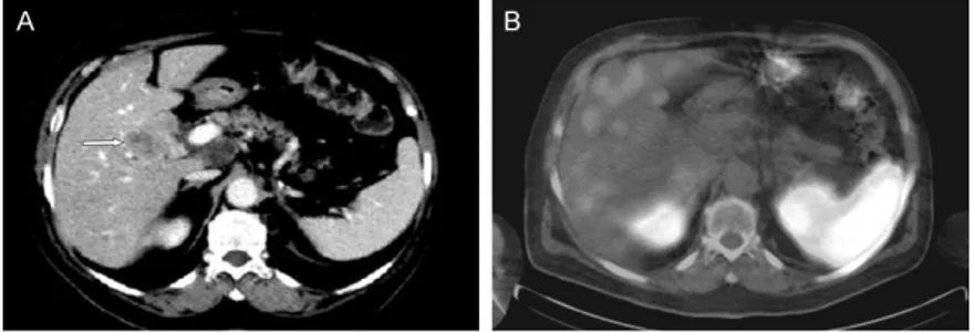
Figure 1. A 73-y-old man with GEP tumor (G1) of duodenum. The patient was asymptomatic for hormone overexpression and negative for characteristic secretory pattern, but he referred malaise, weight loss, abdominal pain and some diarrhoea episodes. Diagnostic CT (A) excludes the presence of any nod-ules or masses in abdomen as it had also been observed at previous ultra-sound. 111In-Pentetreotide SPECT/CT 24 h acquisition image (B) shows a focal
area of somatostatin analogue uptake of 18 mm in size sited in the inferior part of duodenum (arrow). A laparoscopic biopsy evidenced a lesion with a typical trabecular structure, intense immunoreactivity for chromogranin A and synaptophysin, absence of necrosis and rare cells with 1 mitosisx 10 hpf and a Ki 67 index <2%.
patient were counted as 5. SPECT/CT data were con-firmed by pathological find-ings or by clinical and radio-logical follow up for at least a period of 6-36 months in presence of recurrences or/ and metastases when histol-ogy was not available. Statistical analysis
The McNemar test was used to assess the statically sig-nificance of the differences between per-patient sensi-tivity, specificity and accura-cy and per-lesion sensitivity of SPECT/CT and CIP imaging in the detection of GEP tumor
IDS, 6 of whom not operable), neoplastic le- sions were ascertained concordantly at both CIP and SPECT/CT in 44 patients, only at CIP in 6 cases and only at SPECT/CT in 20; CIP were completely false negative in 20 patients (11 FU, 9 IDS), while SPECT/CT was positive; 4 (2 IDS, 2 FU) of these 20 patients were primary tumours: 1 stomach (G1), 1 duodenum (NEC-G3), 1 pan-creas (G1) and 1 sigmoid-colon (G1) with size ≤ 10 mm in G1 patients and ≥ 15 mm in NEC-G3 case; the others 16/20 cases, 13 G1 (≤ 10 mm), 2 NEC G3 (≥ 30 mm) and 1 MANEC (20 mm), had recurrences or metastases in the liver and/or in the abdominal extra-hepatic or extra-abdominal regions (1 mediastinum, 1 bone), being single in 8 cases and multiple in different sites in 6. One of the 20 GEP tumor cases negative at CIP and positive only at SPECT/CT is illustrated in Figure 1.
SPECT/CT was completely false negative in 6 patients (3 FU, 3 IDS), while CIP were positive; 5
No false positive results were observed at both SPECT/CT and CIP.
Globally, 292 neoplastic lesions were ascer-tained, as illustrated in Table 2; 141 were hepatic lesions, 78 abdominal extra-hepatic and 73 extra-abdominal.
All hepatic lesions in 40 patients were metasta-ses from different GEP tumor origin.
Twenty-two/78 abdominal extra-hepatic lesions were primary tumours, 14 of which in 11 IDS patients all operable, including 4 stomach, 1 duodenum, 7 pancreas, 1 small intestine and 1 sigmoid-colon tumors; a case of pancreatic pri-mary tumor evidenced by both CIP and SPECT/ CT, but correctly characterized as GEP only by the latter, is shown in Figure 3. The other 8/22 primary tumors in FU patients, 1 in duodenum, 1 in stomach and 6 in pancreas, were classified as not operable at initial diagnosis. The remain-ing 56/78 lesions in 5 IDS and 14 FU patients Table 2. 111In-Pentetreotide SPECT/CT scintigraphy and CIP data in
70 GEP tumours with ascertained hepatic, abdominal extra-hepatic and extra-abdominal lesions
Lesions Hepatic extra-hepaticAbdominal abdominalExtra- Total
No 141 78 73 292
CIP Positive 113 (80.1%) 40 (51.3%) 38 (52%) 191 (65.4%) SPECT/CT Positive 117 (83%) 67 (85.9%) 60 (82.2%) *244 (83.6%) *p<0.0001 when compared with corresponding CIP.
Figure 2. A 74-y-old women with ileal GEP tumor (G3) previously operated and treated with 3 cycles of chemotherapy with carboplatinum and etoposide. The patient is asymptomatic for hormone over-expression and negative for char-acteristic secretory pattern, but she refers malaise, abdominal pain, vomiting episodes, weight loss and poor condition. Diagnostic CT (A) ascertained an inhomogeneous mass in the hepatic hilum with central areas of hypodensity as colliquative necrosis (arrow). 111In-Pentetreotide SPECT/CT (B) did not
evi-dence foci of pathological uptake. The biopsy of the mass in hepatic hilum showed hepatic infiltration from neoplasia with trabecular structure, abun-dant dysplasia and solid cells, numerous necrosis areas, cellular immunore-activity for CAM 5.2, cytokeratin 19 and CD 56; mitosis index > 20 and Ki-67: 80%. This aspect is indicative for hepatic metastasis from G3 GEP tumor.
of these patients (1 G1, 4 NEC G3) had single metasta-ses (3 hepatic, 2 abdominal extra-hepatic lesions) and the remaining patient (NEC G3) had 2 extra-abdominal metastases in mediastinum; all the lesions were > 10 mm in size, except for the G1 case (4 mm). One of the G3 cases with a liver metastasis negative at SPECT/CT and positive at CIP is illustrated in Figure 2.
The remaining 34/104 pa- tients, all FU after primary non-functioning GEP tumor resection, and with absence of metastases at surgery, did not show focal areas of increased 111In-Pentetreotide
uptake and were also nega-tive at CIP; moreover, these patients, who were appar-ently with no evidence of disease (NED) and without the characteristic serum pa- ttern, except 3 cases with basal slight increase of CgA, remained NED at subse-quent investigations and cli- nical follow-up.
were 43 abdominal lymph node metastases and 13 local recurrences (4 duodenum, 1 stom-ach, 6 pancreas, 2 small intestine).
Moreover, 73 extra-abdominal metastatic lesions were ascertained in 22 of the patients (10 IDS, 12 FU), 5 lesions being sited in brain, 10 in lungs, 39 in lymph nodes (5 in supracla-vicular region, 24 in mediastinum, 10 in pulmo-nary hilum) and 19 in bone; in particular, 3/22 had lesions only in extra-abdominal sites (2 in lung and 1 in mediastinum), 9/22 patients had each more lesions site in extra-abdominal, hepatic and abdominal extra-hepatic regions, 5/22 had both extra-abdominal and hepatic lesions and 5/22 both extra-hepatic lesions and extra-abdominal.
As shown in Table 2, CIP detected 191/292 (65.4%) lesions (113 hepatic, 40 abdominal extra-hepatic and 38 extra-abdominal) in 50 patients and SPECT/CT identified 244/292 (83.6%) lesions (117 hepatic, 67 abdominal extra-hepatic and 60 extra-abdominal) in 64 patients with a better performance of SPECT/ CT in respect of CIP in both abdominal extra-hepatic and extra-abdominal lesions The differ-ence of global sensitivity was significant for SPECT/CT in respect of CIP (P<0.0001).
In particular, both CIP and SPECT/CT concor-dantly identified 89 hepatic lesions in 19
ECT/CT was positive for further 38 lesions (in 7 FU patients and in 10 IDS patients).
Furthermore, SPECT/CT confirmed 25 extra-abdominal neoplastic lesions also ascertained by CIP in 7 patients (4 lesions in brain, 4 in lung, 5 in pulmonary hilum lymph nodes, 10 in medi-astinum, 2 in para-tracheal lymph nodes), and was positive in 35 further lesions: 3 in lung, 12 in lymph nodes (4 in pulmonary hilum, 6 in mediastinum lymph node and 2 in para-trache-al region), 19 in bones and 1 in brain in 5 FU and 4 IDS, all negative at CIP.
CIP was positive in 13 further extra-abdominal neoplastic lesions negative at SPECT/CT in 2 IDS and 2 FU patients (1 FU, 1 IDS completely negative at SPECT/CT): 2 lesions were sited in lungs and 11 in lymph nodes, 2 of these being in pulmonary hilum, 8 in mediastinum and 1 in para-tracheal region.
The smallest lesion visualized by SPECT/CT was a liver metastasis of 5 mm in diameter.
As reported in Table 3, considering the patients according to the WHO 2010 Classification of Tumors of the Digestive System, it was observed that, when mutually comparing the 65 G1 patients, the 22 G2, the 12 NEC-G3 and the 5 MANEC cases, both sensitivity and accuracy of SPECT/CT were higher than CIP in G1, G2, and
Figure 3. A 69-y-old man with pancreatic carcinoid (G1). The patient was as-ymptomatic for hormone overexpression and negative for characteristic secre-tory pattern, but he referred malaise, epigastric pain, weight loss. Diagnos-tic tri-phasic CT (A) showed an inhomogeneous hyper-density nodule of 13 mm with central area of hypo-density as colliquative necrosis in the body-tail passage of the pancreas (arrow) suspect of neoplasia of uncertain origin. At
111In-Pentetreotide SPECT/CT imaging in transaxial view (B), a focal area of
in-creased uptake of somatostatin analogue (arrow) was visualized in the body-tail passage of the pancreas corresponding to the nodule detected by CT. A laparoscopic biopsy ascertained a neuroendocrine G1 tumor of the pancreas with typical trabecular structure with epithelial habitus, intense immunoractiv-ity for chromogranin A, synaptophysin, CD 56 and cytokeratin 19, absence of necrosis, 1 mitosisx10hpf and a Ki-67 index of 0.66%.
patients (12 FU and 7 IDS), while CIP identified 24 lesi- ons undetected by SPECT/CT in 13 patients (9 FU and 4 IDS), 3 of whom were the aforementioned completely negative cases (2 IDS, 1 FU) at SPECT/CT; 28 lesions were evidenced at SPECT/CT while these were not identified by CIP in 11 patients (7 FU and 4 IDS). Moreover, 29 abdominal extra-hepatic le- sions (including 11 FU and 9 IDS patients with primary tumours) were detected by both CIP and SPECT/CT, while only CIP was positive for further 11 lesions (in 3 IDS with primary tumors and in 2 FU patients, one of whom completely negative at SPECT/CT) and only SP-
MANEC patients while both parameters were higher in CIP than SPECT/CT in NEC-G3 patients. The difference was statistically significant (p=0.04) only in the comparison between SPECT/CT and CIP in G1 patients.
Moreover, only for the G1 Group of patients the difference was significant (p=0.02) comparing
the global number of metastatic lesions evi-denced by SPECT/CT in respect of CIP (110 ver-sus 88), while in G2 and MANEC groups the number of lesions evidenced by SPECT/CT, even if more elevated in respect of CIP, the dif-ference was not significantly as well as it was for CIP in respect of SPECT/CT in NEC-G3 patients.
Table 3. 111In-Pentetreotide SPECT/CT and CIP results in 104 patients with GEP neuroendocrine
non-functioning tumors according WHO 2010 Classification of Tumors of the Digestive System (65 G1, 22 G2, 12 NEC, 5 MANEC)
Patients CIP positive SPECT/CT positive
G1 G2 NEC MANEC G1 G2 NEC MANEC
True positive 29 12 6 3 38 17 5 4 True negative 26 5 2 1 26 5 2 1 False negative 10 5 4 1 1 0 5 0 False positive 0 0 0 0 0 0 0 0 Sensitivity % 74.4 70.6 60 75 *97.4 100 50 100 Specificity % 100 100 100 100 100 100 100 100
Positive predictive value % 100 100 100 100 100 100 100 100
Negative predictive value % 72.2 50 33.3 50 96.3 100 28.6 100
Accuracy % 84.6 77.3 66.7 80 98.5 100 58.3 100
*p=0.04 when compared with corresponding CIP.
Figure 4. A 64-y-old female patient, previously operated for differentiated non-functioning ileal GEP tumor (G2) with large trabecular structure, moderate cell atypia, cell immonoreactivity for chromogranine A, synaptophysin, NSE, CD56 and cytokeratin AE1/AE3, 2 mitosisx10hpf and a Ki 67 index of 4%. The patient, treated with octreotide, is as-ymptomatic for hormone over expression and negative for characteristic secretory pattern. Triphasic CT (A-D) shows a nodular lesion of 9 mm in size in the VIII segment of the liver, not evident at basal scan (A), but better evident in arterial (B) portal (C) and equilibrium (D) phases after contrast medium injection, of unclear interpretation. 111
In-Pentetreotide SPECT/CT, performed after octreotide interruption, was negative at 24 h acquisition (E) and showed a focal area of uptake of the somatostatin analogue (arrow) only at 48 h acquisition image (F) corresponding to the liver lesion evidenced at CT. A laparoscopic biopsy confirmed a metastasis from G2 GEP tumor with hepatic infiltra-tion and with morphological and immuno-histological aspects similar to those of the primary tumor.
In the present study, according to the involved structures, SPECT/CT performance was more elevated in duodenum, pancreas, small intes-tine, lymph nodes and bone lesions also of small size in respect of CIP, while the perfor-mance of the latter was higher in stomach lesions.
In most cases SPECT/CT performance was more elevated in the exams acquired at 24 h, but in 13 patients with high suspect of recur-rences or metastases, the exams acquired at 48 h were determinant for the identification of more neoplastic lesions also changing CIP clas-sification and patient management in 6 of these cases, 3 IDS (1 of whom operable) and 3 FU (a case is illustrated in the Figure 4).
Globally, the incremental value for SPECT/CT was 35.6% (37/104), while for CIP was 21.1% (22/104). SPECT/CT correctly modified CIP classification and patient management, also establishing operability or not operability, in 27.9% (29/104) of cases, while it down-staged the disease in 9.6% (10/104) in respect of CIP. The combined use of the two procedures was able to achieve the highest value of sensitivity (100%).
Discussion
Non-functioning GEP tumors represent the most frequent forms of GEP [3]. They grow slow-ly and not present clinical symptoms by over-expression of hormones or other active sub-stances (endocrine syndrome), only secreting peptides or pro-hormones with slight or absent biological activity, thus remaining silent for a long time. However, they can be associated with not specific symptoms such as epigastric and abdominal pains, loss of weight, anorexia, nausea. Therefore, these tumors are of difficult diagnosis in early stage and are often identified late, only when the symptoms appear due to the compression by the mass (jaundice, intesti-nal obstruction) and/or to the invasion of adja-cent organs and when metastases develop. On the other hand, non-functional GEP tumors are characterized by a high percentage of malig-nancy, but in most cases of low grading. Tumor identification and differentiation in pre-operative phase is very important for confirm-ing neuroendocrine nature of the lesion as well as to obtain prognostic data and information on
the grading of the tumor also in association with the assessment of Ki-67 proliferation index [34, 35]. Moreover, a pre-surgery diagno-sis may be crucial for early establishing thera-peutic strategy. In the patients with low risk of malignancy, surgery or clinical and instrumen-tal follow-up can be suggested on the basis of the site and the extension of the tumor other than of its aggressiveness and proliferation index. However, in not operable patients the therapeutic strategy is based on cell prolifera-tion degree and includes somatostatin ana-logues, peptide receptor radionuclide therapy, target therapy, chemotherapy and chemoem-bolization therapy.
Among the instrumental techniques, besides CIP, such as US, endoscopic US, CT and MRI, the radioisotopic procedures often represent the most valuable tools to identify GEP tumors, and, in particular, using somatostatin ana-logues as radiotracers, it is also possible to characterize their neuroendocrine origin. Until some years ago and over two decades,
111In-Pentetreotide SRS has been considered
the radioisotopic procedure of reference and proved useful for revealing the expression of SSR and the degree of differentiation of GEP tumors.
This radioisotopic procedure in the last years, using SPECT/CT technique, has achieved a great popularity in the management of GEP tumor patients since it proved to improve the value of planar SRS and SPECT alone [21-26]. The higher performance of SPECT/CT is due to the identification and anatomic localization of tumors, also of small size, and the characteriza-tion of lesions presenting as unclear focal areas of intense uptake. These lesions can be localized in extra-hepatic abdominal regions and, in particular, in mid upper abdomen as well as in extra-abdominal sites of not easy detection with the other imaging procedures. Thus SPECT/CT can increase sensitivity and accuracy and also give useful information for the correct staging and the evaluation of the response to treatment. Moreover, SPECT/CT is also able to reduce false positive findings of planar SRS in sites of physiologic tracer uptake. In the last years, also PET radiotracers have been employed in the management of GEP tumors. PET with 18F-fluorodihydroxyphenilal-
dopa-mine secretion by GEP tumor cells, has been usefully employed in neoplastic lesion detec-tion but with low sensitivity in pancreatic GEP tumors [36, 37]. Moreover, PET with 11C-5-Hy-
drossitriptofan (11C-5-HTP), that is a precursor
of serotonin, was also used showing high sensi-tivity values in well differentiated GEP tumors, but with less performance in undifferentiated forms, in particular if non-functioning [38]. 18
F-FDG PET/CT has also been proved useful, but only in undifferentiated and more aggressive forms [29, 30].
At present, there is a growing interest on the employment of PET/CT with 68Ga-somatostatin
analogues (DOTATOC, DOTATATE, DOTANOC) that can provide superior detection capacity over 111In-Pentetreotide scintigraphy, even
when acquired with SPECT/CT, due to the high-er spatial resolution of PET scannhigh-er [39, 40]. However, availability and cost represent limiting factors for PET tracers. Moreover, only recently
68Ga-DOTATATE alone has been authorized for
marketing in USA.
Very promising are also the preliminary results obtained in NET patients with new tracers such as somatostatin analogues labelled with 64Cu
(64Cu-DOTATATE, 64Cu-DOTATOC) and 44Sc (44Sc-
DOTATOC), SSR antagonists (68Ga-OPS202)
nd glucagon-like peptide 1 receptor (GLP-1R) labelled with 111Indium (111In-DOTA-exendin-4)
and 68Gallium (68Ga-NOTA-exendin-4) [41]. PET/
CT with 64Cu-DOTATATE, in particular, has sh-
owed advantages not only over 111In-Pentetr-
eotide SRS but also over 68Ga-somatostatin
analogue, detecting a higher number of lesions in NET patients, probably due to the lower posi-tron range of 64Cu in respect of that of 68Ga [42,
43]. GLP-1R scintigraphy, on the other hand, seems to be able to give very high sensitivity values in the detection of benign insulinomas that are frequently missed at scintigraphy with somatostatin analogs [44, 45]. However, up to day, the data reported by different authors on the employment of the most recent afore-mentioned procedures of imaging are few and they need of further confirmation with more elevated number of cases; moreover, none of these radiotracers has been authorized for marketing.
The present study was retrospectively per-formed on a series of patients with
non-func-tioning GEP tumors evaluated in three different University Centers. 111In-Pentetreotide SPECT/
CT was utilized as diagnostic tool in all cases after that they have been submitted to at least two CIP about a month before scintigraphy. SPECT/CT, in our cases, was able to detect non-functioning GEP, also revealing their neuroen-docrine origin, in 91.4% of patients who had neither clinical signs of hormone overexpres-sion nor the characteristic secretion pattern in blood, while CIP was positive in 71.4% of cases. Moreover, SPECT/CT proved very reliable tool for correctly changing patient classification in respect of CIP data in an elevated number of patients (27.9%), including 20 cases complete-ly negative at CIP which down-staged all these cases; 80% of the latter 20 patients had G1 tumors and SPECT/CT ascertained both prima-ry and metastatic lesions, identifying the involved organs and their relationship with adjacent structures. New 53 tumor sites occult at CIP were identified, including 20 sited in extra-abdominal regions as pulmonary, medi-astinal lymph node and bone small size metas-tases. SPECT/CT also contributed to determine resectability of circumscribed lesions, while excluding surgery in presence of extensive met-astatic disease, thus permitting to select the most appropriate therapies.
SPECT/CT was also able to identify unknown primary tumors, that, as is known, cannot be easy to detect in some cases, as well as to stage patients after surgery and to monitor the affected patients in the course of systemic therapy, such as the somatostatin analogue octreotide and/or the chemotherapy, and to early detect recurrences or distant metastases during the follow up.
SPECT/CT was false negative in 6 patients who had metastases from NEC-G3 tumors in 5 cases (83.3%) and from G1 in the remaining case; in 5 patients the metastases were single and in 1 patient the lesions were two, all of these identified by CIP. Thus, SPECT/CT gave an incorrect classification probably for low recep-tor density or other receprecep-tor subtypes not detected by 111In-Pentetreotide, as also
hypoth-esized by other authors [46-49]; on the other hand, in our series SPECT/CT was true positive in 50% of NEC G3 patients, thus suggesting that SSR may also be present in some poor dif-ferentiated GEP tumors. However, in the 6
above mentioned false negative cases, size seems to have little importance in SPECT/CT detection, all lesions being > 10 mm, except for the G1 case in whom also the size could be one of the responsible factors since the lesion was 4 mm.
Thus, SPECT/CT in non-invasive way gave use-ful information for the most therapeutic strate-gy contributing in selecting both the patients to guide towards surgery, and, at the same time, those with diffuse metastases, who can also be not ascertained by CIP and in whom surgical resection is not indicated; these patients, how-ever, could probably benefit from other treat-ments, including octeotride or radiolabeled somatostatin analogue therapy.
Moreover, with regard to grade, SPECT/CT sen-sitivity and accuracy were higher than CIP in all different groups, but significant in G1 group in which 16/20 (80%) patients (3 primary tumors), completely false negative at CIP, were positive at SPECT/CT; this latter result is very important since the early identification of tumor lesions of low grade could permit more correct and preco-cious non-invasive therapeutic procedures which can be decisive with more favorable dis-ease prognosis.
In our series the highest performance of SPECT/ CT has been obtained in most of exams acquired at 24 h and this result could suggest that an acquisition of SPECT/CT at 24 h may be sufficient, in agreement to other authors [26, 50]. However, in our 13 patients the exams at 48 h were determinant, in particular for identi-fying metastatic lesions, and in 6 cases they also changed patient classification and man-agement, thus suggesting that, when the sus-pect of a metastatic disease is elevated, an acquisition over the 24 h could be useful. Moreover, in our cases, SPECT/CT has been more sensitive in the identification of focal lesions than CIP and has also been helpful in differentiating pathological from physiological tracer uptakes with absence of false positive results; therefore, it could suggest that SPECT/ CT may still be the method of choice for staging patients after surgery of primary tumors or for identifying patients with unknown primary tumor as well as for ascertaining recurrences or metastases during the course of therapy with octeotride or chemotherapy. CIP were true
positive in the few cases false negative at SPECT/CT and vice versa the latter was positive in all false negative cases at CIP, thus suggest-ing that the combined use of the two proce-dures can achieve the highest sensitivity val-ues, giving the most correct classification of the patients.
Notwithstanding the recent employment of PET/CT imaging with somatostatin analogous which proved higher sensitivity values in respect of 111In-Pentetreotide, changing
thera-py decision in some cases, but still available in a few Centers, 111In-Pentetreotide SRS can still
represents a very useful diagnostic procedure with elevated accuracy when SPECT/CT is acquired. Despite some limitations due to trac-er relative long decay time and prolonged time commitment by the patients, SPECT/CT gives higher imaging quality in respect of both planar SRS and SPECT with better characterization of focal areas of uptake excluding malignancy in physiologic sites of tracer uptake and with more correct anatomic lesion localization; moreover, the hybrid procedure demonstrates an elevated impact on patient management and therapy planning. SPECT/CT also presents a limited cost and a wide availability in all Nuclear Medicine Centers. Furthermore,
111In-Pentetreotide has represented for many
years the only tracer registered and approved for marketing in both the Europe and USA. However, these suggestions could be reas-sessed when somatostatin analogues marked with positron tracers will be duly authorized and, at the same time, the production systems may be available in all Centers and the radio-tracers employed in an elevated number of cases.
In conclusion, 111In-Pentetreotide SPECT/CT
can still have a wide employment in the routine diagnostic protocol of non-functioning GEP tumors since it has demonstrated to play an important role in the early diagnosis of the neo-plastic lesions, including the identification of unknown primary tumors, when still in a cura-tive phase; this aspect is crucial for a correct choice of treatment. SPECT/CT demonstrated a high performance in the precise definition of tumor anatomic site and in the characterization of unclear lesions. It also achieved a more ele-vated sensitivity than CIP both in the diagnosis and in the disease staging, changing patient
CIP classification and clinical management in 27.9% of cases and also monitoring the response to treatment.
Disclosure of conflict of interest None.
Address correspondence to: Dr. Angela Spanu, Chief of Nuclear Medicine Unit, Department of Clinical and Experimental Medicine, University of Sassari, Viale San Pietro, 8. I-07100 Sassari, Italy. Tel: +39 (0)79 228342; Fax: +39 (0)79 228208; E-mail: [email protected]; Dr. Giuseppe Ma- deddu, Emeritus Professor of Nuclear Medicine, University of Sassari, Viale San Pietro, 8, I-07100 Sassari, Italy. Tel: +39 (0)79 228342; Fax: +39 (0)79 228208; E-mail: [email protected]
References
[1] Jensen RT. Natural history of digestive endo-crine tumors. Recent advances in the patho-physiology and management of inflammatory bowel disease and digestive endocrine tu-mors. Edited by Mignon M, Colombel JF. Paris, France: John Libbey Eurotext; 1999. pp. 192-219.
[2] Fraker DL, Jensen RT. Pancreatic endocrine tu-mors. Cancer: Principles and Practice of Oncol-ogy. Edited by De Vita VT, Hellman S, Rosem-berg SA. Philadelphia, PA, Lippincot-Raven; 1997, pp. 1678-1704 (5th ed.).
[3] Prinz RA, Bermes EW, Kimmel JR. Serum mark-ers for pancreatic islet cell and intestinal carci-noid tumors: a comparison of neuron-specificic enolase, β-human chorionic gonadotropin and pancreatic polypeptide. Surgery 1983; 94: 1019-1023.
[4] O’Connor DT, Leftos LJ. Secretion of Chromo-granin A by peptide-producing endocrine neo-plasias. N Engl J Med 1986; 314: 1145-1151. [5] Kim M, Lee S, Lee J, Park SH, Park JO, Park YS,
Kang WK, Kim ST. The role of plasma chromo-granin A as assessment of treatment respon- se in non-functioning gastroenteropancreatic neuroendocrine tumors. Cancer Res Treat 2016; 48: 153-161.
[6] Bardram L. Progastrin in serum from Zollinger-Ellison patients. Gastroenterology 1990; 98: 1420-1426.
[7] Kent RB, van Heedern JA, Weiland LH. Non-functioning islet cell tumors. Ann Surg 1981; 193: 185-190.
[8] Bartsch DK, Schillin T, Ramaswamy A, Gerdes B, Celik I, Wagner HJ, Simon B, Rothmund M. Management of non-functioning islet cell carci-nomas. World J Surg 2000; 24: 1418-1424.
[9] Debray MP, Geoffroy O, Laissy JP, Lebtahi R, Silbermann-Hoffman O, Henry-Feugeas MC, Cadiot G, Mignon M, Schouman-Claeys E. Im-aging appearances of metastases from neuro-endocrine tumors of the pancreas. Br J Radiol 2001; 74: 1065-1070.
[10] Yellin A, Zwas ST, Rozenman J, Simansky DA, Goshen E. Experience with somatostatin re-ceptor scintigraphy in the management of pul-monary carcinoid tumors. Isr Med Assoc J 2005; 7: 712-716.
[11] Kuyumcu S, Adalet I, Sanli Y, Turkmen C, Ozkan ZG, Yilmazbayhan D. Somatostatin receptor scintigraphy with 111In-octreotide in pulmonary
carcinoid tumours correlated with pathological and 18FDG PET/CT findings. Ann Nucl Med
2012; 26: 689-697.
[12] Lamberts SWJ, Hofland LJ, van Koetsueld PM, Reubi JC, Bruining HA, Bakker WH, Krenning EP. Parallel in vivo and in vitro detection of functional somatostatin receptors in human endocrine pancreatic tumors: consequences with regard to diagnosis, localization, and ther-apy. J Clin Endocrinol Metab 1990; 71: 566-574.
[13] Krenning EP, Kwekkeboom DJ, Bakker WH, Breeman WAP, Kooij PPM, Oei HY, van Hagen M, Postema PTE, de Jong M, Reubi JC, Visser TJ, AEM Reijs, LJ Hofland, JW Koper, SWJ Lam-berts. Somatostatin receptor scintigraphy with [111In-DTPA-d-Phe1]- and [123I-Tyr3
]-octreo-tide: the Rotterdam experience with more than 1000 patients. Eur J Nucl Med 1993; 20: 716-731.
[14] Krausz Y, Bar-Ziv J, de Jong RB, Ish-Shalom S, Chisin R, Shibley N, Glaser B. Somatostatin-re-ceptor scintigraphy in the management of gas-troenteropancreatic tumors. Am J Gastroen-terol 1998; 93: 66-70.
[15] Schillaci O, Scopinaro F, Angeletti S, Tavolaro R, Danieli R, Annibale B, Gualdi G, Delle Fave G. SPECT improves accuracy of somatostatin receptor scintigraphy in abdominal carcinoid tumors. J Nucl Med 1996; 37: 1452-1456. [16] Schillaci O, Spanu A, Scopinaro F, Falchi A,
Danieli R, Marongiu P, Pisu N, Madeddu G, Delle Fave G, Madeddu G. Somatostatin recep-tor scintigraphy in liver metastasis detection from gastroenteropancreatic neuroendocrine tumors. J Nucl Med 2003; 44: 359-368. [17] Schillaci O, Spanu A, Scopinaro F, Falchi A,
Cor-leto V, Danieli R, Marongiu P, Pisu N, Madeddu G, Delle Fave G, Madeddu G. Somastostatin receptor scintigraphy with 111In-pentetreotide
in non-functioning gastroenteropancreatic neuroendocrine tumors. Int J Oncol 2003; 23: 1687-1695.
[18] King CM, Reznek RH, Bomanji J, Ur E, Britton KE, Grossman AB, Besser GM. Imaging
neuro-endocrine tumours with radiolabelled soma-tostatin analogues and X-ray computed tomog-raphy: a comparative study. Clin Radiol 1993; 48: 386-391.
[19] Kisker O, Bartsch D, Weinel RJ, Joseph K, Welcke UH, Zaraca F, Rothmund M. The value of somatostatin-receptor scintigraphy in newly diagnosed endocrine gastroenteropancreatic tumors. J Am Coll Surg 1997; 184: 487-492. [20] Bartsch DK, Langer P, Wild A, Schilling T, Celik
I, Rothmund M, Nies C. Pancreaticoduodenal endocrine tumors in multiple endocrine neo-plasia type 1: surgery or surveillance? Surgery 2000; 128: 958-966.
[21] Moreira AP, Duarte LH, Vieira F, João F, Pedro-so de Lima J. Value of SPECT/CT image fusion in the assessment of neuroendocrine tumours with 111In-pentetreotide scintigraphy. Rev Esp Med Nucl 2005; 24: 14-18.
[22] Wong KK, Cahill JM, Frey KA, Avram AM. Incre-mental value of 111-In-pentetreotide SPECT/ CT fusion imaging of neuroendocrine tumors. Acad Radiol 2010; 17: 291-297.
[23] Bural GG, Muthukrishnan A, Oborski MJ, Mountz JM. Improved benefit of SPECT/CT compared to SPECT alone for the accurate lo-calization of endocrine and neuroendocrine tumors. Mol Imaging Radionucl Ther 2012; 21: 91-96.
[24] Schillaci O, Spanu A, Paumbo B, Danieli R. SPECT/CT in neuroendocrine tumours. Clin Transl Imaging 2014; 2: 477-489.
[25] Chiaravalloti A, Spanu A, Danieli R, Dore F, Pi-ras B, Falchi A, Tavolozza M, Madeddu G, Schil-laci O.111In-Pentetreotide SPECT/CT in pul-monary carcinoid. Anticancer Res 2015; 35: 4265-4270.
[26] Ruf J, von Wedel F, Furth C, Denecke T, Stelter L, Steffen IG, Schüttle K, Arend J, Ulrich G, Klose S, Bornschein J, Apostolova I, Amthauer H. Significance of a single-time-point soma-tostatin receptor SPECT/Multiphase CT Proto-col in the diagnostic work-up of gastroentero-pancreatic neuroendocrine neoplasm. J Nucl Med 2016; 57: 180-185.
[27] Sepúlveda-Méndez J, de Murphy CA, Pedraza-López M, Murphy-Stack E, Rojas-Bautista JC, González-Treviño O. Specificity and sensitivity of 99mTc-EDDA/HYNIC-Tyr³-octreotide (99mTc-TOC) for imaging neuroendocrine tumors. Nucl Med Commun 2012; 33: 69-79.
[28] Qiao Z, Zhang J, Jin X, Huo L, Zhu Z, Xing H, Li F. 99mTc-HYNIC-TOC imaging in the evaluation of pancreatic masses which are potential neu-roendocrine tumors. Clin Nucl Med 2015; 40: 397-400.
[29] Kayani I, Bomanji JB, Groves A, Conway G, Gac-inovic S, Win T, Dickson J, Caplin M, Ell PJ.
Functional imaging of neuroendocrine tumors with combined PET/CT using 68Ga-DOTATATE (DOTA-DPhe1, Tyr3-octreotate) and 18F-FDG. Cancer 2008; 112: 2447-2455.
[30] Panagiotidis E, Alshammari A, Michopoulou S, Skoura E, Naik K, Maragkoudakis E, Mohmad-uvesh M, Al-Harbi M, Belda M, Caplin ME, Toumpanakis C, Bomanji J. Comparison of the impact of 68Ga-DOTATATE and 18F-FDG PET/ CT on clinical management in patients with neuroendocrine tumors. J Nucl Med 2017; 58: 91-96.
[31] Kayani I, Conry BG, Groves AM, Win T, Dickson J, Caplin M, Bomanji JB. A comparison of 68Ga-DOTATATE and 18F-FDG PET/CT in pul-monary neuroendocrine tumors. J Nucl Med 2009; 50: 1927-1932.
[32] Haug AR, Cindea-Drmus R, Auernhammer CJ, Reincke M, Wangler B, Uebleis C, Schmidt GP, Goke B, Bartenstein P, Hacker M. The role of 68Ga-DOTATATE PET/CT in suspected neuroen-docrine tumors. J Nucl Med 2012; 53: 1686-1692.
[33] Wild D, Bomanji JB, Benkert P, Maecke H, Ell PJ, Reubi JC, Caplin ME. Comparison of 68Ga-DOTANOC and 68Ga-DOTATATE PET/CT within patients with gastroenteropancreatic neuroen-docrine tumors. J Nucl Med 2013; 54: 364-372.
[34] Chatzipantelis P, Konstantinou P, Kaklamanos M, Apostolou G, Salla C. The role of cyto-mor-phology and proliferative activity in predicting biologic behaviour of pancreatic neuroendocri-ne tumors: a study by endoscopic ultrasound--guided fine-needle aspiration cytology. Cancer 2009; 117: 211-216.
[35] Alexiev BA, Darwin PE, Goloubeva O, Ioffe OB. Proliferative rate in endoscopic ultrasound fine-needle aspiration of pancreatic endocrine tumors: correlation with clinical behaviour. Cancer 2009; 117: 40-45.
[36] Jager PL, Chirakal R, Marriott CJ, Brouwers AH, Koopmans KP, Gulenchyn KY. 6-L-18F-fluorodi-hydroxyphenylalanine PET in neuroendocrine tumors: basic aspects and emerging clinical applications. J Nucl Med 2008; 49: 573-586. [37] Montravers F, Kerrou K, Nataf V, Huchet V, Lotz
JP, Ruszniewski P, Rougier P, Duron F, Boucha-rd P, Grangé JD, Houry S, Talbot JN. Impact of Fluorodihydroxyphenylalanine-(18F) positron emission tomography on management of adult patients with documented or occult digestive endocrine tumors. J Clin Endocrinol Metab 2009; 94: 1295-1301.
[38] Orlefors H, Sundin A, Eriksson B, Skogseid B, Oberg K, Akerström G, Hellman P. PET-guided surgery-high correlation between positron emission tomography with 11C-5-Hydroxytryp-tophane (5-HTP) and surgical findings in
ab-dominal neuroendocrine tumours. Cancers (Basel) 2012; 4: 100-112.
[39] Srirajaskanthan R, Kayani I, Quigley AM, Soh J, Caplin ME, Bomanji J. The role of 68Ga-DOT-ATATE PET in patients with neuroendocrine tu-mors and negative or equivocal findings on 111In-DTPA-octreotide scintigraphy. J Nucl Med 2010; 51: 875-882.
[40] Sadowski SM, Millo C, Cottle-Delisle C, Merkel R, Yang LA, Herscovitch P, Pacak K, Simonds WF, Marx SJ, Kebebew E. Results of (68) Galli-um-DOTATATE PET/CT scanning in patients with multiple endocrine neoplasia type 1. J Am Coll Surg 2015; 221: 509-517.
[41] Kulkarni HR, Singh A, Baum RP. Advances in the diagnosis of neuroendocrine neoplasms. Semin Nucl Med 2016; 46: 395-404.
[42] Pfeifer A, Knigge U, Binderup T, Mortensen J, Oturai P, Loft A, Berthelsen AK, Langer SW, Rasmussen P, Elema D, von Benzon E, Højgaard L, Kjaer A. 64Cu-DOTATATE PET for neuroendocrine tumors: a prospective head-to-head comparison with 111In-DTPA-Octreo-tide in 112 patients. J Nucl Med 2015; 56: 847-854.
[43] Johnbeck CB, Knigge U, Loft A, Berthelsen AK, Mortensen J, Oturai P,Langer SW, Elema DR, Kjaer A. Head-to-head comparison of 64
Cu-DOTATATE and 68Ga-DOTATOC PET/CT: a
pro-spective study of 59 patients with neuroendo-crine tumors. J Nucl Med 2017; 58: 451-457. [44] Christ E, Wild D, Ederer S, Béhé M, Nicolas G,
Caplin ME, Brändle M, Clerici T, Fischli S, Stet-tler C, Ell PJ, Seufert J, Gloor B, Perren A, Reubi JC, Forrer F. Glucagon-like peptide-1 receptor imaging for the localisation of insulinomas: a prospective multicentre imaging study. Lancet Diabetes Endocrinol 2013; 1: 115-122.
[45] Luo Y, Pan Q, Yao S, Yu M, Wu W, Xue H, Kie-sewetter DO, Zhu Z, Li F, Zhao Y, Chen X. Gluca-gon-Like Peptide-1 Receptor PET/CT with 68Ga-NOTA-Exendin-4 for detecting localized insulinoma: a prospective cohort study. J Nucl Med 2016; 57: 715-720.
[46] O’Byrne KJ, Carney DN. Somatostatin and the lung. Lung Cancer 1993; 10: 151-172. [47] Kirsch CM, von Pawel J, Grau I, Tatsch K.
Indi-um-111-pentetreotide in the diagnostic work-up of patients with bronchogenic carcinoma. Eur J Nucl Med 1994; 21: 1318-1325. [48] Fujita T, Yamaji Y, Sato M, Murao K, Takahara J.
Gene expression of somatostatin receptor sub-types, SSTR1 and SSTR2, in human lung can-cer cell lines. Life Sci 1994; 55: 1797-1806. [49] Reisinger I, Bohuslavitzki KH, Brenner W,
Braune S, Dittrich I, Geide A, Kettner B, Otto HJ, Schmidt S, Munz DL. Somatostatin recep-tor scintigraphy in small-cell lung cancer: re-sults of a multicenter study. J Nucl Med 1998; 39: 224-227.
[50] Attili F, Capurso G, Vanella G, Fuccio L, Delle Fave G, Costamagna G, Larghi A. Diagnostic and therapeutic role of endoscopy in gastroen-teropancreatic neuroendocrine neoplasms. Dig Liver Dis 2014; 46: 9-17.