DOTTORATO DI RICERCA IN BIOCHIMICA CICLO XXXII (A.A. 2016-2019)
“Endocrine disrupting effects of the pollutant β-hexachlorocyclohexane in cancer cells: possible chemopreventing role of the natural
sesquiterpene β-caryophyllene”
Tutor Ph.D. Coordinator
Prof. Margherita Eufemi Prof. Stefano Gianni
Ph.D. Student Stefania Carissimi
“Non temete i momenti difficili. Il meglio viene da lì”
CHAPTER 1: INTRODUCTION ... 7
1.1 Endocrin Disruptors compounds ... 7
1.2 Organochlorine pollutants ... 12
1.2.1 OCPs as endocrine and metabolic disruptors ... 16
1.2.2 OCPs and oncogenic modulation ... 18
1.3 β-Hexachlorocyclohexane ... 19
1.4 STAT3 protein ... 20
1.4.1 STAT3 Activation and Regulation ... 22
1.5 STAT3 Post-Translational Modifications (PTMs) ... 24
1.5.1 Phosphorylation ... 24
1.5.2 Acetylation ... 25
1.5.3 Methylation ... 25
1.5.4 Oxidation and Glutathionylation ... 26
1.6 STAT3 in tumorigenesis ... 27
1.6.1 STAT3 Inhibitors ... 28
1.7 The sesquiterpene β-caryophyllene ... 29
1.7.1 β-CRY as anticancer agent ... 30
CHAPTER 2: AIM OF THE RESEARCH ... 33
CHAPTER 3: MATERIALS AND METHODS ... 37
3.1 Reagents ... 37
3.2 Cell cultures ... 37
3.3 Cell viability assay ... 38
3.4 Cell lysate preparation ... 38
3.5 Western blotting ... 39
3.6 Total RNA Extraction ... 40
3.7 cDNA synthesis and Quantitative Real Time-PCR ... 40
3.9 Scratch test Assay ... 41
3.10 Cell Proliferation Assay ... 42
3.11 Statistical analyses ... 42
4. RESULTS ... 43
4.1 β-Hexachlorocyclohexane triggers STAT3 pathways ... 43
4.2 Investigation of caryophyllene protective role against β-hexachlorocyclohexane carcinogenic property ... 48
4.2.1 β-caryophyllene inhibition of STAT 3 activation ... 48
4.2.2 β-caryophyllene inhibition of proliferation and migration ... 49
4.3 β-hexachlorocyclohexane effect on placenta ... 55
CHAPTER 5: DISCUSSION ... 59
REFERENCES ... 61
CHAPTER 1: INTRODUCTION
1.1 Endocrin Disruptors compounds
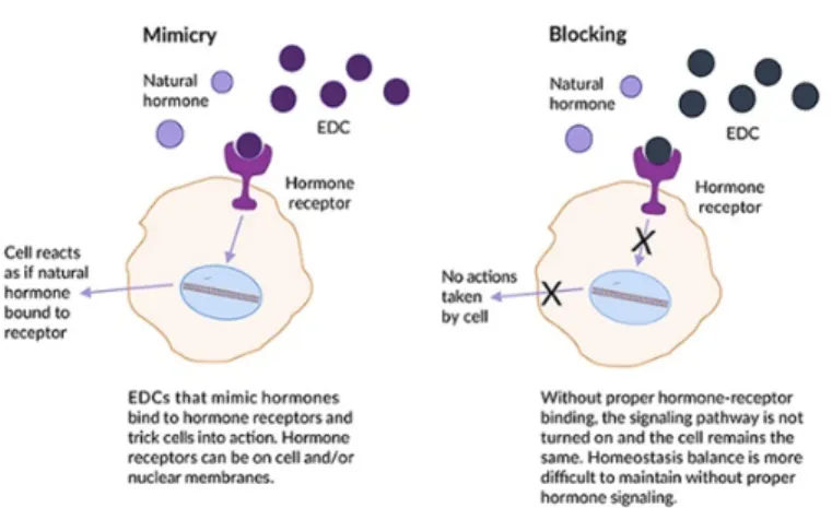
The U.S. Environmental Protection Agency (EPA) defines an endocrine-disrupting compound (EDC) as “an exogenous agent that interferes with synthesis, secretion, transport, metabolism, binding action, or elimination of natural blood-borne hormones that are present in the body and are responsible for homeostasis, reproduction, and developmental process.” From a physiological perspective, an endocrine-disrupting substance is a compound, either natural or synthetic, which, through environmental or inappropriate developmental exposures, alters the homeostasis of hormonal system that enable the organism to communicate with and respond to its environment (Figure 1).
Fig. 1: Schematic representation of EDCs interference with physiological
Today, there are nearly 1000 chemicals reported to have endocrine effects (“Home,” n.d.). The prevalence of EDCs in our environment and their persistence in our bodies represents a significant health challenge since the endocrine system plays a central role in all vertebrates and regulates such critical biological functions as metabolism, development, reproduction, and behaviour.
Epidemiological studies link EDCs with alterations of the reproductive system, such as malformations, infertility and increased risk of endometriosis, neurobehavioral and neurodevelopmental changes, metabolic syndrome, bone disorders, immune disorders, and cancers in humans (“WHO | State of the science of endocrine disrupting chemicals - 2012,” n.d.).
The group of molecules identified as endocrine disruptors is highly heterogeneous and includes synthetic chemicals used as industrial solvents and their by-products (polychlorinated biphenyls, polybrominated biphenyls, dioxins), plastics (bisphenol A), plasticizers (phthalates), pesticides (methoxychlor, chlorpyrifos, dichlorodiphenyltrichloroethane, hexachlorocyclohexane), fungicides (vinclozolin), and pharmaceutical agents (diethylstilbestrol).
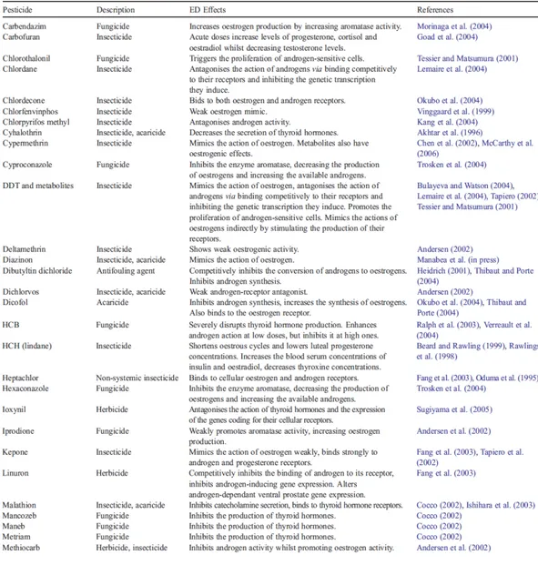
Listed below, the main EDCs, their mechanism of action, hormones affected (Table 1)and effects on the physiological endocrine activity (Table 2).
Table 1: List of common pesticides with Endocrine Disruptors effects,
hormones affected and their mechanism of action.
The sources of exposure to EDCs are diverse and vary widely around the world. People who work with pesticides, fungicides, and industrial chemicals are at particularly high risk for exposure and thus for developing a reproductive or endocrine abnormality (McKinlay et al., 2008). Some EDCs were banned decades ago and others more recently, with significant differences between countries (Table 3). These complex mixtures enter the food chain and accumulate in animals and humans.
Table3: It is reported, for each EDC, the type of source, the NR involved and
the legal status.
Research conducted so far allow to identify some key issues necessary to a full understanding of mechanism of action and consequences of exposure to EDCs: 1) Age of exposure – The field of endocrine disruption has embraced the terminology “the fetal basis of adult disease” (Barker, 2003), since developing organisms are extremely sensitive to perturbation and fetal development is commonly known to be a period of increased susceptibility to chemical insult. Thus, it is crucial to determine whether or what kind of consequence the exposure to EDCs will lead to, taking into account when the exposure occurs;
2) Latency from Exposure - Effects of EDCs may not be immediately apparent early in life but may be manifested in adulthood or during aging;
3) Importance of mixtures – Contamination of environments are rarely due to a single compounds and effects of different classes of EDCs may be additive or even synergistic (Crews et al., 2003).
4) Non traditional dose-response dynamics - even infinitesimally low levels of
exposure may cause endocrine or reproductive abnormalities, particularly if exposure occurs during a critical developmental window. Moreover, low doses may even exert more potent effects than higher doses.
5) Transgenerational, epigenetic effects - Recent evidence suggests that the
mechanism of transmission may involve the germ line and may be non genomic. One of the hypothesis leans toward epigenetic inheritance patterns, which involve chemical modifications to the DNA (DNA methylation), histone modifications, and non coding RNAs, rather than mutations of the DNA sequence itself.
1.2 Organochlorine pollutants
Organochlorine pesticides (OCPs) are a structurally heterogeneous class of organic compounds composed primarily of carbon, hydrogen and several chlorine atoms per molecule. They have been employed in agriculture and in public health as insecticides and biocides for several decades but were mostly banned between 1970 and 1990. Only a few active substances are still used in tropical countries. So, most exposures derive from past uses and, in certain cases, are still significant because of the long persistence and
biomagnifications of these compounds. Once released in the environment, they break down very slowly in air, water, soil and in living organisms and are thus subject to long trans-boundary air pollution transport where they are carried
over long distances via the atmospheric transport and human activities (Jones and de Voogt, 1999).
The main OCPs are:
- Dichlorodiphenyl-trichloroethane (DDT), classified as “probably carcinogenic to humans (group 2A)” was widely used in agriculture and to eradicate malaria. It is readily absorbed and distributed in the body by lymphatic and blood circulation, with a preference for lipid tissue. Associations between cancer and exposure to DDT have been investigated in more than 100 cohort and case-control studies. A few of these, conducted on Chinese population, reported dose related association between liver cancer and blood DDT level (McGlynn et al., 2006), (Persson et al., 2012);
- Hexachlorobenzene (HCB), at first produced as a fungicide, is still formed as a major by-product and contaminant in the manufacture of chemicals such as solvents, chlorine-containing compounds and pesticides (ATSDR 1996)It has a strong impact on the human androgen axis and it is shown to cause liver tumors in animals (Randi et al., 2003);
- Polychlorinated biphenyls (PCBs), industrial chemicals consisting of paired phenyl rings with various degrees of chlorination which are able to bind to TRs. Some authors showed that PCBs can reduce circulating levels of T4 in animals (Goldey et al., 1995), others proposed they can exert neurotoxic effects on the developing brain by causing a state of hypothyroidism (Crofton, 2004);
- Endosulfan, mainly used to control pests in vegetables, fruits, cereal grains and cotton, ornamental shrubs, trees, vines and ornamental plants. In vitro studies show that Endosulfan acts as antagonist of androgen receptor (Li et al., 2008) and significantly increases cell
proliferation and estrogen receptor (ER) transactivation gene response in MCF-7 cells (Andersen et al., 2002). A cohort study conducted in 2003, among male school children (10–19 years old) showed that endosulfan exposure was associated with delayed male sexual maturity and interfered with the male sex-hormone synthesis (Saiyed Habibullah et al., 2003);
- Hexachlorocyclohexane, (HCH) belongs to a family of eight isomers of 1,2,3,4,5,6-hexachlorocyclohexane that differ in their equatorial substitution pattern around the ring. They are denoted by Greek letters (α, β, γ, δ, ε, η and θ) (Figure 2) (Willett et al., 1998).The physical and chemical properties of the HCH isomers are quite different from one another as illustrated in Table 4.
Table 4: Selected Physical Properties of HCH isomers.
The γ-isomer (γ-HCH), called lindane, has been widely used as a pesticide, due to its effectiveness, low cost and acute toxicity. The estimation of its global production between 1950 and 2000 was 600,000 tonnes and the majority was used in agriculture, as an insecticide on fruits and vegetables, rice paddies, Christmas trees, and animals and as a seed treatment. In medicine, γ-HCH has been applied topically to people for the treatment of lice and scabies. Although its employment has been restricted since the 1970’s, because of its toxicity, the problem of residues of all isomers of HCH remains because of the high persistence and interconversion of these isomers in soil (Chessells et al., 1988), (Concha-Graña et al., 2006).
During the production process of γ-HCH others three isomers, α, β, and δ, are formed, however only the γ-HCH is known to be effective as an insecticide, despite its content ranges between 12% and 15% of the isomer mixture. Consequently, the α, β and δ-isomer mixture represents a big amount of by-products, which are often dumped under unsuitable circumstances.
As OCPs bio-magnify through the food chain, they accumulate in the body. e.g., DDT can remain in the body for 50 years, lifelong sequestrated into the soft tissue compartments (mainly the adipose tissues), from which they partition to the bloodstream into plasma or serum lipids and are actively secreted into breast milk as the main elimination pathway in mammals (Mrema et al., 2013). Human exposure to OCPs begins during early prenatal
and continues during the breast-feeding neonatal periods, which are critical stages for the development and differentiation of sensitive body organs and systems. In fact, these chemicals cross the placenta to the fetus and are secreted into breast milk (Perera et al., 2005).In adults the dietary exposure route accounts for more than 90% of total organochlorine compounds burden for the general population, while workers are exposed mainly through inhalation and skin contact. OCPs are rapidly absorbed in the small intestine and enter circulatory system where they are distributed throughout the body and accumulate in body tissues with high lipid contents, with a continuous exchange between blood and tissues. As such these chemicals have been detected in human tissues such as blood (whole blood, cord blood, serum and plasma), adipose tissues (in autopsies and living subjects), breast milk, muscles and hair.
1.2.1 OCPs as endocrine and metabolic disruptors
There is growing concern in the scientific community that OCPs may be contributing to the rapid increased rates of diabetes and metabolic syndrome and a lot of literature links this pathological conditions to the exposure to compounds such as BPA, dioxins, organochlorine and organophosphate pesticides. The chlorine atoms are poorly reactive towards nucleophilic elimination reactions and thus their biodegradation reactions are limited to anaerobic environment of sludges. As a consequence, their interaction with biological systems is mostly limited to agonistic or antagonistic binding to the intracellular receptors for which natural hydrophobic substances, such as steroid derivatives, are the endogenous ligands. Agonistic binding leads to recruitment of coactivators and increase of transcriptional activity while antagonistic binding prevents coactivator recruitment and/or attracts corepressors, leading to decreased transcriptional activity of the receptors.
There are three main pathways through which EDCs cause metabolic disruption (Casals-Casas and Desvergne, 2011), shown in Figure 3:
Fig. 3: Schematic representation of EDCs interactions with Nuclear
Receptors, Xenosensors and PPARs and consequent activation and cross-talk between involved pathways.
1) Interaction with hormone receptors, such as ER (Estrogen Receptors), TR (thyroid Hormone receptors) and GR (Glucocorticoid Receptors): These receptors are important for the control of adipogenesis, weight gain, and insulin levels, although the underlying mechanisms are not yet well understood
2) Interaction with Xenosensors, such as PXR (pregnane X receptor), CAR (constitutive androstane receptor) and AhR (aryl hydrocarbon receptor): these receptors are mainly involved in regulating the metabolism of xenobiotics, yet their contribution to fatty acid, lipid, and glucose metabolism has been only recently appreciated
3) Interaction with PPARs (Peroxisome Proliferator–Activated Receptors): PPARγ is a member of the nuclear receptor superfamily and it is one of the major regulators of adipogenesis. It is primarily expressed in adipose tissue, and its activation promotes adipocyte differentiation as well as the induction of lipogenic enzymes. Additionally, it contributes to maintenance of metabolic homeostasis through transcriptional activation of genes implicated in energy balance.
Altogether, these many examples of EDCs interaction with receptors
highlight the fact that a given compound can interfere with different NRs and different pathways. The final result of these interactions is given by cross-talk between these pathways, rather than to a linear causation chain and are much more complex to decipher in vivo.
1.2.2 OCPs and oncogenic modulation
It has been demonstrated that several OCPs exposure cause cancer in humans. Taking into account DNA toxicity OCPs fall into two classes of carcinogens: genotoxic and non-genotoxic. The latter one includes compounds which are able to affect the expression of epigenetic enzymes, in turn regulating the transcription of genes involved in pathological processes.
Hexachlorobenzene, an environmental pollutant, is known to cause liver tumours in animals through activation of c-myc, c-fos, c-jun proto-oncogenic proteins and PKC activity induction (Randi et al., 2003); the β isomer of Lindane is able to increase the mRNA expression level of metalloproteinase MMP-13 and the expression of a number of proto-oncogenes such as c-Neu, cyclin D1 and p27 (Wong and Matsumura, 2007).
Genome hypomethylation has been found in tumours (Das and Singal, 2004), whereas a recent study reported a strong correlation between increasing levels
of POPs, such as DDT, and global DNA hypomethylation (an aberrant epigenetic pattern of malignant cells) in a sample size of 70 subjects in the Greenlandic Innuit (Rusiecki et al., 2008). In a recent review it has been examined the association between work-related exposures and prostate cancer and the evidence for a possible relationship has been assessed (Krstev and Knutsson, 2019). The highest risk estimates for specific organochlorine pesticides were estimated for chlordane in a Swedish study (Hardell et al., 2006) and Lindane in the USA studies (Mills and Yang, 2003).
1.3 β-Hexachlorocyclohexane
The β-isomer (β-HCH) of Hexachlorocyclohexane is characterized by high lipid solubility, great stability and high persistence in the environment, due to its chemical structure. As it is shown in Figure 4 this compound has all chlorine atoms in an equatorial position, and it lacks aromatic character and any axial chlorine atom, which can be the site for 1,2-elimination.
Direct, non/genomic mechanisms of toxicity involve perturbation of the homeostatic redox levels of biological compartments and consequent cellular death through apoptosis. Indirect, genomic mechanisms of toxicity involve permanent modification of the transcription of gene elements by interference at the epigenetic level of DNA functioning.
Coosen et al. showed that 1μM β-HCH has mitogenic activity in human breast cancer cell line MCF-7 (Coosen and van Velsen, 1989) while the same phenomenon is not evident in human triple negative breast cancer cell line MDA-MB231. Moreover, β-HCH increased, in MCF-7 cells, pS2 gene mRNA level. The pS2 gene product is a polypeptide that is expressed by one-half of all breast tumors, and although its function has not been determined, its presence identifies tumors that are sensitive to anti-hormonal therapy (Steinmetz et al., 1996). In vivo assay with mouse xenograft model showed that 10-8M β-HCH significantly increased MCF-7 cell number achieving maximal responses at 10-5M (Steinmetz et al., 1996). β-HCH has been shown to induce activation of caspase-8, that plays the role in transduction of death signal (Said et al., 2004)and caspase-3, that initiates cell apoptosis(Khan et al., 2000). Moreover, Shi et al. demonstrated that β-HCH enhances reactive oxygen species (ROS) production in rat Sertoli cells and induces activation of JNKs and NFK-B and expression of FasL. Upon engagement of FasL to Fas receptor, the intrinsic program of apoptotic death is stimulated in cells leading to the activation of caspase-8. Finally, apoptosis of Sertoli cells is mediated by caspase-3, thereby disturbing thespermatogenic process(Shi et al., 2011). 1.4 STAT3 protein
Signal Transducer and Activator of Transcription 3 (STAT) is a member of the STAT family. STAT3 is a transcriptional factor that transmits signals from
cells surface into the nucleus by binding to specific DNA promoter sequences and regulating gene expression involved in many cellular processes.
STAT3 exists in two isoforms originating from alternative splicing: the full-length, called STAT3 α and the C-terminal truncated form, called STAT3β (Figure 5) (Dewilde et al., 2008), (Ng et al., 2012). The β isoform lacks the C-terminal Transactivation domain, including the S727 residue. For this reason it has long been considered as a dominant negative regulator of STAT3α transcriptional activity (Caldenhoven et al., 1996). However, several recent studies showed that STAT3β plays unique functions, activating sets of specific genes. Its expression levels are lower compared to STAT3α but when STAT3β is over expressed can play different and opposite roles: it induces apoptosis and cell-cycle arrest, in several STAT3-dependent tumor cell lines with persis-tent STAT3 tyrosine (705) phosphorylation and plays an oncogenes function by triggering aggressive T cell leukemia in transplanted bone marrow cells (Ecker et al., 2009). In contrast however, STAT3β seems to have a potent anti-inflammatory role in the initiation phases of skin and colon tumorogenesis (Marino et al., 2014).
Fig 5: Schematic structure of STAT3 proteins. Comparison between Stat3α
Despite STAT3 transcriptional activity is mainly regulated by Y705 phosphorilation, there is an increasing consensus that unphosphorylated STAT3 (U-STAT3) is also transcriptionally active and continuously shuttling between the cytoplasm and the nucleus (Cimica et al., 2011) and it is shown to be able to interact with other factors and to directly or indirectly bind to DNA on binding sites distinct from the canonical gamma interferon activation site (GAS) sequences, thus exerting its control on a different subset of target genes, including several oncogenes (Yang et al., 2007), (Timofeeva et al., 2012), (Nishimoto et al., 2013). Ng D.C. et al showed that U-STAT3 can inhibit the microtubule-destabilizing protein stathmin, resulting in enhanced microtubules polymerization and cell migration (Ng et al., 2006).
STAT3 can be also found in the mitochondrion, phosphorylated on S727 residue. Activated Mito-STAT3 is shown to interact with Electron Transporter Chain (ETC) I and II, to preserve optimal ETC activity, increasing membrane polarization and ATP production, and enhancing the activity of lactate dehydrogenase. As a consequence, aerobic glycolysis is induced and ROS production decreased (Wegrzyn et al., 2009), (Szczepanek et al., 2011), (Tammineni et al., 2013).
1.4.1 STAT3 Activation and Regulation
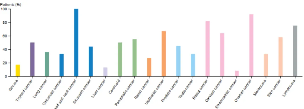
In physiological conditions, the STAT3 protein is temporary activated and usually this activation lasts from a few minutes to several hours. However, numerous studies demonstrated the constitutive activation of STAT3 in a large number of human tumor cell lines. As reported in the Human Protein ATLAS, (Figure 6), the majority of cancer tissues display weak to moderate cytoplasmic and nuclear positivity to STAT3 protein.
Fig. 6: Color-coded bars indicate the percentage of patients (maximum 12
patients) with high and medium STAT3 protein expression level (ATLAS).
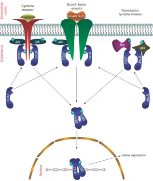
This protein is canonically activated by Y705 phosphorilation downstream of cytokines (IL-6 family, leptin, IL-12, IL-17, and IL-10, which activate gp130/JAK/STAT3 pathway) growth factors (granulocyte colony-stimulating factor G-CSF-, epidermal growth factor EGF) and oncogenes (Src, Abl, Sis, Fps, Ros, Met and ErbB2 (Bowman et al., 2000)), (Figure 7) to mediate their functions under both physiological and pathological conditions (Yu et al., 2014), (Yuan et al., 2015). Under physiological conditions, STAT3 activation is tightly controlled by negative regulators, which mainly fall into three groups: Phosphatases (SHP-1, SHP-2, PTP1B) (Xu and Qu, 2008); Suppressor Of Cytokine Signaling (SOCS) proteins (SOCS3 act as a negative feedback regulator to inhibit JAK activity) (Krebs and Hilton, 2001); Protein Inhibitor of Activated STAT (PIAS) proteins (PIAS3, prevents the binding of STAT3 to its target DNA sequence) (C. D. Chung et al., 1997).
Fig 7: Main pathways responsible of STAT3 activation.
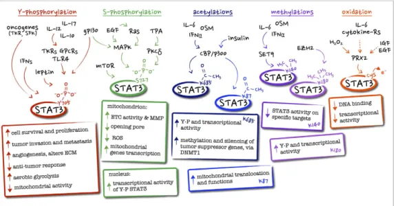
1.5 STAT3 Post-Translational Modifications (PTMs)
STAT3 can be post-translationally modified in a number of alternative ways, variably affecting its activity and cellular localization.
The main PTMs of STAT3 are Phosphorylation, Acetylation, Methylation, and Glutathionylation (Figure 8).
1.5.1 Phosphorylation
As already mentioned, STAT3 canonical activity mainly occurs due to Y705 phosphorylation, which enable STAT3 to form functional dimers, translocate in the nucleus and bind DNA. This is positively regulated by many activatory
signals such as cytokines (IL-6 family, leptin, IL-12, IL-17, and IL-10, as well as interferons) growth factors (granulocyte colony-stimulating factor -G-CSF-, epidermal growth factor -EGF-) and a number of oncogenes-G-CSF-, the prototype of which are Src family kinases but also including Abl, Sis, Fps, Ros, Met and ErbB2. Phosphorylation can also occur on S727 residue and be carried out by kinases including MAPK and mTOR. While S727 phosphorylation is required for the optimal transcriptional induction of a subset of target genes (Aznar et al., 2001), it can also modify STAT3 activation dynamics by inhibiting the subsequent Y705 phosphorylation (J. Chung et al., 1997). It is now clear that Serine phosphorylation is responsible of STAT3 mitochondrial localization. 1.5.2 Acetylation
Under citokynes and grow factors stimuli STAT3 can also be acetylated on Lysine residues ( K685 and K87) by the CBP/p300 histone acetyltransferase while its deacetylation is carried out by the nicotinamide adenine dinucleotide (NAD)-dependent silent information regulator protein (SIRT)1 (Nie et al., 2009). K685 acetylation leads to an enhanced STAT3 transcriptional activity and increased Y705 phosphorylation and dimer stability (Yuan et al., 2005), while K87 acetylation, promoted by insulin stimulation, can increase mitochondrial translocation and function (Xu et al., 2016).
1.5.3 Methylation
STAT3 methylation can occur in the nucleus on Lysine residues (K140 or K180). K140 is methylated by the histone methyltransferase SET9 when STAT3 is bound to its promoter, leading to impaired transcriptional activation of the target genes (Yang et al., 2010). In contrast, K180 is methylated by the lysine methyltransferase of the polycomb complex 2 (EZH2) operates. Kim et al. demonstrated that EZH2 methylation of STAT3 K180 residue is essential,
in glioblastoma cells, to enhance STAT3 activation and promote its transcriptional activity (Kim et al., 2013).
1.5.4 Oxidation and Glutathionylation
Finally, STAT3 can be both oxidized and glutathionylated on multiple cysteine residues, under conditions of oxidative stress and cytokine signalling, impairing its transcriptional activity (Xie et al., 2009), (Li et al., 2010). STAT3 oxidation can occur downstream of the cytoplasmic thiol peroxidase peroxiredoxin-2 (Prx2), one of the major H2O2 scavengers within the cell
(Sobotta et al., 2015). This achieves ROS detoxification while at the same time impairing IL-6-induced, STAT3-mediated transcription, suggesting that STAT3 is part of a redox relay controlling redox homeostasis, and ROS and cytokine signalling. Therefore, this cross-talk between oxidative and non-oxidative STAT3 modifications can affect the activities of YP-STAT3 as well as cell proliferation and survival.
Fig 8: main STAT3 post-translational modifications, together with their known
biological effects and upstream regulator.
1.6 STAT3 in tumorigenesis
STAT3 is constitutively activated in many tumours and most of its activators/regulator are deregulated in cancer. As a consequence it is mostly considered as an oncogenes (Yuan et al., 2015), (Yu et al., 2014), (Avalle et al., 2017).
Different mechanisms are correlated to aberrant STAT3 activation: 1) loss of the negative regulation of STAT3;
2) excessive stimulation of STAT3;
3) positive feedback loops that sustain persistent STAT3 activation; 4) somatic mutations that confer a hyperactive property to STAT3.
The direct effect of STAT3 activation consists in an increased expression of its target genes which are able to mediate all the crucial steps of cancer progression such as proliferation and survival of cells, their migration and invasion properties and their potential to induce angiogenesis and to evade the host immune response. In particular, STAT3 regulates:
a) cell cycle, promotion of G1/S and G2/M transitions through cyclin D1, cyclin B and CDK1 stimulation (Ma et al., 2015);
b) cellular survival, by controlling the anti-apoptotic genes Mcl-1, 2, Bcl-xl, Survivin (Dokduang et al., 2016) and by down-regulating the apoptotic gene p53 (Nicholas and B, 2011);
c) tumor invasion and metastasis, by the expression of matrix metalloproteinase (MMPs);
d) Angiogenesis, by promoting vascular endothelial growth factor (VEGF) (Zhu et al., 2011).
1.6.1 STAT3 Inhibitors
STAT3 pivotal role in cancer initiation, progression and metastasis makes it a great target for anticancer therapy. A lot of STAT3 pharmacological inhibitors are available and they mainly act at two levels: 1) indirectly blocking abnormally activated upstream proteins (Cetuximab and Panitumumab, monoclonal antibodies against EGFR; Gefitinib, Erlotinib, Lapatinib and PD153035, small molecules designed to block EGFR tyrosine kinase activity; AG490, and AZD1480, JAK inhibitors; Dasatinib and Indirubin, Src inhibitors); 2) directly suppressing the STAT3 activation (STA-21, S3I-201 and Stattic, small non peptidic molecules which are able to directly bind to STAT3 SH2 domain, preventing its dimerization; novel platinum (IV) compounds, CPA-1, CPA-7 able to cross link DNA and inhibit STAT3 DNA binding (Turkson et al., 2004)).
Besides synthetic molecules, several natural compounds display a STAT3 inhibitory activity. Among these we can find polyphenols such as Quercitin, Silibinin, and Epigallocatechin gallate; Curcumin, Caffeic acid and Celastrol, and the Sesquisterpene β-Caryophyllene.
1.7 The sesquiterpene β-caryophyllene
β-caryophyllene (CRY) is a plant compound which belongs to the bicycle sesquiterpene family. In nature, it mainly occurs as trans-βcaryophyllene ((E)-CRY) mixed with small amounts of its isomers, cis-βcaryophyllene (Z)-β-caryophyllene) and α-caryophyllene (α-humulene), together with its oxidation derivative β-caryophyllene oxide (CRYO) (Figure 9)
Fig. 9: Structures of trans-CRY, its isomers and oxidative product. β-CRY has a low water solubility but is able to interact with artificial lipid bilayer, which strongly suggests its high affinity to the cell membranes. It is one of the major active components of essential oils derived from large number of spice and food plants. It is commonly found in basil, cinnamon, black pepper, cloves, cannabis, lavender, oregano and rosemary and it is used as a flavouring agent (approved by the Food and Drug Administration (FDA) and by the European Food Safety Authority (EFSA) with identification number FL no: 01.007).
Its biological effects include anti-inflammatory (Medeiros et al., 2007), anticarcinogenic (Langhasova et al., 2014), antimicrobial (Sabulal et al., 2006), antioxidative (Singh et al., 2006), and analgesic activities (Klauke et al., 2014). The latter is typical of β-CRY which belongs to a class of cannabinoids (CBs),
specifically phyto-cannabinoids (pCBs), that were identified as plant derivatives of Cannabis sativa L.
Gertsch et al. demonstrated that the (E)-CRY is the first Cannabis-derived functional CB receptor ligand which displays higher biding affinity to CB2 than its isomer Z-CRY, whereas CRYO and α-humulene possess no CB2 binding properties (Gertsch et al., 2008).
1.7.1 β-CRY as anticancer agent
Dahham et al. reported the antiproliferative effect of β-CRY on several cancer cell lines (Dahham et al., 2015). β-CRY isolated from essential oils of Aquilaria crassna stem bark led to strong growth inhibition in two colon cancer cell lines, HCT-116 and HT-29, as well as in pancreatic cancer cells, PANC-1, whereas it had no effects on other tested cancer cell lines. In contrast, Ambrož et al. studies revealed that β-CRY obtained from Myrica rubra did not affect CaCo-2 intestinal cancer cell viability (Ambrož et al., 2015). Jung et al. described the anticancer effects of β-CRY in in vivo model of high-fat diet-induced obese C57BL/6N mice. (Jung et al., 2015). They observed that a high-fat diet (HFD) induces larger and more aggressive tumors in animals injected with B16F10 melanoma cells. β-CRY treatment abolished the HFD pro-cancer effects. The anticancer activity of β-CRY in vivo was also presented at the Euro Global Summit on Cancer Therapy in Valencia, 2015. In this report, a growth and vascularization of tumors developed from orthotopically grafted colon cancer cells into nude mice were reduced significantly after administration of β-CRY isolated from agar wood.
Furthermore β-CRY is able to enhance the efficacy of classical anticancer drugs, such as paclitaxel or doxorubicin (Ambrož et al., 2015), (Legault and Pichette, 2007), (Kim et al., 2014).
In the last few years, Di Sotto et al. deeply investigated the role of β-CRY, focusing their attention on the protective effects against DNA-damage caused by different toxic compounds. These authors demonstrated that β-CRY was able to protect cultured human lymphocytes from the genotoxic damage induced by ethyl methanesulfonate and colcemid (Di Sotto et al., 2010). Another study shows the capability of β-CRY to inhibit the genotoxicity of a condensate of cigarette smoke both in bacterial and mammalian cells (Giacomo et al., 2016). At last, Giacomo et al. demonstrated that β-CRY protective role, against damage caused by Condensed Smoke Cigarette in mammalian cells, is exerted by inhibition of CSC-mediated STAT3 phosphorylation (Di Giacomo et al., 2018).
CHAPTER 2: AIM OF THE RESEARCH
OCPs have been deeply studied in the last decades and cytotoxic mechanisms of compounds such as dioxin and polychlorinated biphenyls have been widely described in literature (Yang et al., 2015). Still, the β-isomer of hexaclorocyclohexane has not been largely investigated, despite its relatively common geographic distribution (Fagnocchi et al., 2018) and its physicochemical properties make it extremely dangerous for human health. During the first part of my research we started investigating the cellular mechanisms triggered by β-HCH, as it has been found to be a major contaminant of the “Sacco River Valley” area, in the south of Rome. In 2005, an environmental emergency state was declared in the aforementioned area and a bio-monitoring study started in 2006 (Porta et al., 2013). Results of the epidemiological study, conducted on 1000 people residing in this area (Sorveglianza Sanitaria Ed Epidemiologica Della Popolazione Residente In Prossimità Del Fiume Sacco Rapporto tecnico attività 2013-2015 Dipartimento di Epidemiologia del Servizio Sanitario Regionale - Regione Lazio) showed above the average emetic concentrations of β-HCH; thus we decided to test, in
vitro, a concentration that was extrapolated by those results. Taking into
account the pleiotropic role of the protein signal transducer and activator of transcription 3 (STAT3), its function as a hub protein in cellular signalling pathways triggered by β-HCH was investigated in different cancer cell lines corresponding to tissues that are especially vulnerable to damage caused by environmental pollutants. Data obtained were recently published (Rubini et al., 2018) and suggest that β-HCH is able to exert its toxicity triggering STAT3 cellular pathways.
The second part of my research project was founded by the Sovena Foundation which gave me the chance to carry on a collaboration with Antonella Di Sotto
research group, at the department of Physiology and Pharmacology of La Sapienza, Rome. In the context of this collaboration it has been previously demonstrated that the sesquiterpene β-Caryophyllene is able to exert anticancer activities decrising Y705-STAT3 phosphorilation induced by Condensed Smoke Cigarette (Di Giacomo et al., 2018). Therefore we decided to evaluate β-CRY protective role against the deregulation of STAT3 pathways induced by β-HCH in human breast cancer cell lines MCF-7 and MDA-MB-468. The obtained data confirm that β-CRY exerts a chemopreventing effect by inhibiting β-HCH-induced STAT3 activation.
The last part of my PhD project has been carried out at University of Copenhagen, Department of Biomedical Science, where I worked in the laboratory of the Professor Ole Hartvig Mortensen. Here, I started investigating the effect of β–HCH on placental cells. We selected this model since there are increasing evidences that the placenta acts as a partial barrier for OCPs (Perera et al., 2005). A recent publication shows that β–HCH, together with many others OCPs, is ubiquitous in umbilical cord sera and placentas, indicating that the adverse effects of OCPs on foetus and infants should be of concern. Moreover, the concentrations of OCPs in umbilical cord sera and placentas are significantly associated with those in maternal sera, which suggests a direct influence of the maternal burden(Zhang et al., 2018). Moreover, a study published this year on Nature shows that black carbon particles, derived from particulate matter combustion, accumulate on the fetal side of the placenta, probably affecting the foetus from early life onwards (Bové et al., 2019).
For this last set of experiments, we selected a trophoblast cancer model, therefore using BeWo cell line. Since optimal and appropriate biomarkers of placental toxicity are still being researched, we mainly focused our attention on STAT3 pathways, as this protein seems to be a good marker for this model.
We investigated whether β-HCH molecular mechanism is exerted through STAT3 pathways activation. Moreover, we measured the mRNA expression level of several genes encoding transport proteins which are necessary for nutrients transport across placenta and their absorption. Preliminary data show that STAT3 is ones more involved in the cellular response to β-HCH and that the organochlorine compound also affects the expression level of nutrient transports such as GLUT1 and TauT.
CHAPTER 3: MATERIALS AND METHODS
3.1 Reagents
β-caryophyllene (CRY; ≥ 98.5% purity, Sigma-Aldrich Co, St. Louis, MO, USA), β-hexaclorocyclohexane (β-HCH) (Sigma-Aldrich), protease inhibitors cocktail (Sigma-Aldrich); BSA (Sigma-Aldrich); I-block (Thermo Fisher Scientific, Rodano, Italy); RPMI-1640 and DMEM (InVitrogen, MI); fetal bovine serum (FBS- Sigma-Aldirich); MTT (3-(4,5-dimethylthiazol-2-yl)-2,5-diphenyl-2H-tetrazolium bromide) (Sigma-Aldrich,); Peroxidase and Alkaline phosphatase-conjugated secondary antibody (Jackson ImmunoResearch, Pero, Italy); ECL Fast Femto reagent (Immunological Science, Roma, Italy); BCIP/NBT reagents (Carl Roth, Milano, Italy, CAS No. 298-83-9 and 6578-06-9); TRIzol® Reagent (Immunological Science); Super Script II
R-Transcriptase (FS-RT-3022, Fisher Molecular Biology, Rodano, Italy). β-hexaclorocyclohexane (β-HCH) (Sigma-Aldrich, 33376), at a final concentration of 10 μM, was tested on each cell line pre-treated or not with specific inhibitors: 6 μM AZD1480 (Sigma-Aldrich, SML1505), 100_μM S3I-201 (Sigma-Aldrich, SML0330), 70 nM Dasatinib (Selleckchem, Roma,Italy, Cat. No. S1021), 0.8 μM Lapatinib (Sigma-Aldrich, CDS022971), and 15 μM Gefitinib (Sigma-Aldrich, SLM1657).
hexaclorocyclohexane was dissolved in sterile 100% DMSO while β-caryophyllene in sterile 100% Ethanol and stored at -20°C.
3.2 Cell cultures
Cell culture was carried out according to standard procedures in a humidified 5% CO2 incubator at 37°C. Human HER2+ breast cancer cell line MCF-7 and
human prostate cancer cell line LNCaP, were maintained in RPMI-1640 (Roswell Park Memorial Institute) medium; human triple negative breast
cancer cell line MDA-MB-468 and human hepatoma cell line HepG2 were grown in DMEM (InVitrogen, MI). All media were supplemented with 10% v/v fetal bovine serum (FBS- Sigma-Aldrich), 100 units/mL penicillin and 100 μg/mL streptomycin, 1% w/v sodium pyruvate, 2mM glutamine.
Human choriocarcinoma cell line BeWo, was grown in DMEM High Glucose (Thermo Fisher Scientific). The medium was supplemented with 10% v/v fetal bovine serum (FBS- Sigma-Aldrich), 1% Penicillin-Streptomycin 1000 units/mL (Invitrogen) and 2mM glutamine.
β-hexaclorocyclohexane (β-HCH), at a final concentration of 10 µM, was tested on each cell line, overnight pre-treated or not with 10 μg/mLβ-caryophyllene (β-CRY).
3.3 Cell viability assay
The β-HCH cytotoxicity was evaluated by seeding cells in 96-well plates and measuring cell viability after 24 and 48 hours of incubation in the presence of different concentrations of β-HCH (5, 10, 25, 50, 75, 100, 125, 150, 175, and 200 μM). Cell viability was measured using MTT (3-(4,5-dimethylthiazol-2-yl)-2,5-diphenyl-2H-tetrazolium bromide) (Sigma-Aldrich M2128). Briefly, the culture medium was removed and 125 µL of MTT solution (0.5 mg/mL MTT in culture medium) was added to each well. After 2 hours of incubation, the solution was removed and the insoluble formazan dye resulting from the conversion of tetrazolium salt by metabolically active cells was dissolved by adding 125 µL/well of DMSO and measured at 570 nm using the Multiskan™ FC Microplate Photometer (ThermoFisher).
3.4 Cell lysate preparation
Adherent cells were washed and scraped with cold PSB. Total protein extracts were obtained using a lysis buffer containing 2% SDS, 20 mM
Tris-hydrocloride Ph = 7.4, 2 M urea, 10% glycerol added with 2 mM sodium orthovanadate, 10 mM DTT, and a protease inhibitors cocktail diluted 1:100 (Sigma-Aldrich). After a 30’’ sonication with Labsonic M (Sartorius), and centrifugation (20min, 14.000 RPM, 4°C), the total protein extracted were quantified by measuring spectrophotometrically the absorbance at 260 nm, and re-suspended in 4X Laemmli Buffer ( 2% SDS, 62,5 mM Tris-HCl pH 6.8, 10% v/v glycerol, 5 mM DTT, 0.02% bromophenol blue) for the electrophoresis.
3.5 Western blotting
The lysate samples in 4X Laemmli buffer proteins were resolved by SDS-PAGE 10% TGX FastCast™ Acrylamide gel (BioRad, Segrate, Italy) (200V, 45 min) and transferred on PVDF membranes (BioRad) using Trans-Blot® Turbo™ Transfer System (BioRad). Protein bands were electrotransferred to a PVDF membrane. The membranes were blocked with 3% w/v non-fat dried milk or 0.2% w/v I-block (Thermo Fisher Scientific, Rodano, Italy) in Tris-buffered saline containing 0.05% Tween-20 (TBS-T) and incubated with a specific primary antibody for 1 h. Subsequently, membranes were washed three times in TBS-T, and then incubated for an additional hour with appropriate horseradish peroxidase- or alkaline phosphatase-conjugated secondary antibody (Jackson ImmunoResearch, Pero, Italy). The peroxidase signal was detected with ECL Fast Femto reagent (Immunological Science, Roma, Italy), acquired by Molecular Imager® ChemiDoc™ MP System (Bio-Rad), and the intensity of protein bands was quantified using the ImageLab Software. The alkaline phosphatase signal was detected with BCIP/NBT reagents(Carl Roth, Milano, Italy). β-actin was used as normalization protein. Immunodetection was carried out using primary anti-pY705-STAT3 (Cell Signaling D3A7, antibody dilution 1:2000), anti Tot-STAT3 (Cell Signaling
D3Z2G, antibody dilution 1:1000), anti-JAK2 (Cell Signaling D2E12, antibody dilution 1:1000), anti-PY1007/1008JAK2 (Cell Signaling C80C3, antibody dilution 1:1000), anti-EGFR (Cell Signaling D38B1, antibody dilution 1:1000), anti-pY1173EGFR (Cell Signaling 53A5, antibody dilution 1:1000), Src (Cell Signaling 32G6, antibody dilution 1:1000), and anti-pY416Src (Cell Signaling 6943S, antibody dilution 1:1000) primary antibodies.
At least three experimental replicates were performed for each biological sample. All results are expressed as mean ± SD. Differences between experimental groups were determined by Student’s t-test. p-value of <0.01 was considered statistically significant
3.6 Total RNA Extraction
Cells were harvested and total RNA was isolated with TRIzol® Reagent
(Immunological Science) following the manufacturer’s instructions. RNA was precipitated by adding isopropanol and subsequently centrifuged at 12,500 g for 10 minutes at 4°C. The precipitated RNA was washed with 75% ethanol and air dried. Total RNA was than resuspended in RNase-free water and quantified spectrophotometrically. Its quality was assessed by 1% agarose gel electrophoresis and staining with ethidium bromide.
3.7 cDNA synthesis and Quantitative Real Time-PCR
The reverse transcription was carried out with Super Script II R-Transcriptase (FS-RT-3022, Fisher Molecular Biology, Rodano, Italy). Gene expression was evaluated with specific primers for BIRC-5, c-MYC, CRP, p21, CCND1, MMP2 and S18 (housekeeping) (all from Qiagen S.r.l., Milano, Italy), Taut, GLUT1 and GLUT3 (TAG Copenhagen A/S), using CFX Connect™ Real-Time PCR Detection System (BioRad) with a SYBR green fluorophore based
real-time reaction (Brilliant SYBR Green QPCR Master Mix, Thermo Fisher Scientific). Expression data were analyzed using CFX Manager™ Real Time PCR Detection System Software, Version 3.1 (BioRad).
3.8 Cell Cycle Assay
Cell cycle analysis was performed by flow cytometry using Propidium Iodide (PI) staining. MCF-7 cells were seeded on 6 well plates (150x103/well), and
grown for 24 hours. Then, cells were treated for 48 hours with 10 μM β-HCH alone of after an over-night pre-treatment with 10μg/ml β-CRY. Cells were scraped, collected and washed with PBS. Subsequently, cells were fixed with 70% ethanol solution, drop-wise added during vortex agitation and incubated for 24 hours at 4°C. Cells were collected by centrifugation, washed in PBS, resuspended in 200 μl of PBS containing PI (30 μg/ml), Triton-X100 (0,1 % w/v), RNase (0.2 mg/ml) and then incubated for 30 minutes at room temperature. Fluorescence intensity of PI (FL2) was recorded by a cytoflorimeter (Accuri C6, Becton Dickinson) and cell cycle analysis was performed using a specific software (Modfit LT, Verity Software House). Results were expressed, according to PI intensity, as the percentage of cells in each cell cycle phase.
3.9 Scratch test Assay
The effects of β-HCH and β-CRY on cell migration was evaluated. MCF7 and MDA-MB-468 cells (1 × 10⁴/well) were plated in 6 well plates (VWR®, tissue
culture plate), and incubated for 24 hours to allow them to adhere. Afterwards, the cell monolayer was scraped with a pipette tip in order to create a "scratch", dividing the well in two distinctive chambers. Cells were washed with PBS to remove the debris, then fresh medium was added. Cells were incubated with 10 µM β-HCH for 12, 24 and 48 hours, with or without an over-night
pre-treatment with 10 μg/ml β-CRY. Cells that had invaded the scratch were detected via microscopy (Leica Microsystems).
3.10 Cell Proliferation Assay
A cell proliferation assay was performed to investigate the antiproliferative effect of β-CRY. Briefly, 10 x 105 cells/well of both MCF-7 and
MDA-MB-468 cell lines were seeded into 6-well plates and treated for 12, 24 and 48 hours with 10 µM β-HCH alone or after an over-night pre-treatment with β-CRY 10 μg/ml. Then, at each time point, cells were trypsinized, resuspended in fresh medium and viable cells number was evaluated using Trypan Blu dye and the Thoma cell counting chamber. Counts were carried out from 3 to 5 times. 3.11 Statistical analyses
The repeatability of results was confirmed by performing all experiments at least three times.
The obtained values are presented as mean and standard deviation. Statistical analysis was performer with GraphPad Prisma software using Student’s t-test.
4. RESULTS
4.1 β-Hexachlorocyclohexane triggers STAT3 pathways
Data we recently published suggest that β-HCH is able to exert its toxicity triggering STAT3 cellular pathways. We chose a panel of cell lines representing different human tumor types associated with the expression and activation of specific receptors potentially involved in STAT3 activation: membrane and membrane associated tyrosine kinase receptors (EGFR in MDA-MB 468, JAK2 in HepG2, and HER2 in MCF-7 cells) and cytoplasmic non-receptor tyrosine kinases (SRC in LNCaP). The β-HCH tested concentration (10 μM) was extrapolated both from environmental– epidemiological studies carried out on the exposed population living throughout the “Valle del Sacco” area (Sorveglianza Sanitaria Ed Epidemiologica Della Popolazione Residente In Prossimità Del Fiume Sacco Rapporto tecnico attività 2013-2015 Dipartimento di Epidemiologia del Servizio Sanitario Regionale - Regione Lazio) and from previous in vitro studies (Sharma et al., 2010), (Briz et al., 2011).
A time course assay was at first performed. The activation of STAT3 upon β-HCH treatment was followed by immunoblotting analysis, together with the activation of the specific receptors taken into account. Results indicate that STAT3 phosphorylation occurs within the same time as the receptors activation with the exception of MCF‐7 cells (Figure 10 A). The delayed activation of pY705–STAT3 observed in MCF‐7 cells is not directly related to the HER2 receptor but seems to be mediated by the JAK2 receptor (Figure 10 B)
A
B
Int. J. Mol. Sci 19 (7), 2108. “STAT3, a Hub Protein of Cellular Signaling Pathways, Is Triggered by β-Hexaclorocyclohexane”
Fig. 10: (A) Immunoblot analysis of the time course assay performed on
different cell lines treated with 10 μM β‐HCH. Samples were analyzed for STAT3 and each cell line for a specific membrane or membrane associated tyrosine kinase receptor. Both unmodified and phosphorylated form were detected for each protein using specific antibodies. (B) Evaluation of JAK2 and STAT3 phosphorylation level in MCF-7 cell line treated with 10 μM β-HCH. Cells were incubated for 15 minutes and 4 hours in the absence or presence of JAK2 inhibitor (AZD1480), then subjected to Immunoblot analysis.
In order to verify if the cell-specific action of β-HCH is dependent on STAT3-mediated pathways, all cell lines were exposed to 10 μM β -HCH, pre-treated or not with specific inhibitors of the different receptors/cytoplasmic tyrosine kinases analysed (Dasatinib as Src inhibitor, AZD1480 as JAK2 inhibitor, Gefitinib as EGFR inhibitor, and Lapatinib as HER2 inhibitor). For this experiment, a single incubation time with β-HCH was selected for each cell line, according to results of Y705–STAT3 phosphorylation obtained from the time course assay. In particular, we chose 15 minutes of β-HCH treatment for MDA-MB 468, HepG2, and LNCaP cells, and 2 hours for MCF-7 cells. Western blot assay was performed to analyze both unphosphorylated and phosphorylated receptors and STAT3. Immunoblot results show the absence of STAT3 and receptors phosphorylation in cells pre-treated with the specific inhibitors (Figure 11).
To further confirm the role of STAT3 in the cellular response to β-HCH, the expression profile of STAT3 specific target genes, which represents a different phase of the carcinogenesis process (P21 for cell cycle, CRP for inflammation, BIRC5-Survivin for apoptosis, and c-MYC for proliferation) was evaluated. The same analysis was performed in the presence of a specific STAT3 inhibitor, S3I-201. All cell lines were treated with β-HCH in the presence or absence of S3I-201 and the time points of treatment were selected taking into account the rapid or delayed STAT3 activation, previously observed. Results shown in Figure 12 B seem to confirm that STAT3 is involved in the cellular response to β-HCH and can probably mediate its potential tumor activity. Indeed, in all cell lines tested, β-HCH treatment leads to an increase in the expression level of STAT3-specific genes analyzed.
Int. J. Mol. Sci 19 (7), 2108. “STAT3, a Hub Protein of Cellular Signaling Pathways, Is Triggered by β-Hexaclorocyclohexane”
Fig. 11: Inhibition of specific signalling pathways activated by β-HCH. Each
cell line was treated with 10 μM β-HCH in the presence or absence of specific inhibitors: Gefitinib as EGFR inhibitor in MDA-MB 468 cells; AZD1480 as JAK2 inhibitor in HepG2 cells; Dasatinib as SRC inhibitor in LNCaP cells; and Lapatinib as HER2 inhibitor in MCF-7 cells. Cells were incubated (15 min for MDA-MB 468, HepG2, and LNCaP cells and 2 h for MCF-7 cells) and cellular extracts were subjected to Immunoblot analysis. Detection of the unmodified and corresponding phosphorylated form was carried out using specific antibodies
As expected, this increase was not observed when cells were co-treated with the STAT3 inhibitor. As control, STAT3 activation was checked by Western Blotting in all cell lines and the pre-treatment with S3I-201 significantly
reduced STAT3 phosphorylation (Figure 12 A). The overall results support the hypothesis that STAT3 is involved in pathways triggered by β-HCH and that it can mediate the inflammatory, anti-apoptotic and proliferative activity of this organochlorine compound.
Int. J. Mol. Sci 19 (7), 2108. “STAT3, a Hub Protein of Cellular Signaling Pathways, Is Triggered by β-Hexaclorocyclohexane”
Fig. 12: (A) Immunoblot analysis of pY705–STAT3 in cells treated for 4 hours
with 10 μM β-HCH, in the presence or absence of a specific STAT3 inhibitor (S3I-201). Detection of phosphorylated and unphosphorylated STAT3 was carried out using specific antibodies. (B) RT-qPCR analysis of STAT3 target genes (CRP, BIRC5, p21, and c-MYC) performed on β-HCH treated or untreated cells, as well as on cells pre-incubated with a specific STAT3 inhibitor (S3I-201). Statistically significant differences (p < 0.05) between β-HCH treated and β-β-HCH untreated cells are marked by *, while statistically significant differences (p < 0.05) between β-HCH treated and pre-incubated or not with a specific STAT3 inhibitor are marked by §.
4.2 Investigation of caryophyllene protective role against β-hexachlorocyclohexane carcinogenic property
4.2.1 β-caryophyllene inhibition of STAT 3 activation
We decided to focus our attention on breast cancer cell lines MCF-7 and MDA-MB-468 and we investigated the protective role of the sesquiterpene β-caryophyllene against the β-HCH-induced phosphorylation of Y705-STAT3. Cells were over-night pre-treated with 10 μg/mL of the natural compound. Afterwards, MDA-MB468 and MCF7 cells were incubated with 10 μM β-HCH for 15 minutes or 2 hours, respectively. As shown in Figure 13, β-CRY was able to down-modulate phosphorylation of Y705-STAT3 in both cell lines.
Fig. 13: Immunoblot analysis of human breast cancer cell lines (MCF‐7 and
MDA‐MB 468) treated with 10 μM β‐HCH, in presence or absence of β-Caryophyllene 10 μg/mL (over-night pre-treatment). Cells were incubated (15 min and 2h of β‐HCH treatment for MDA-MB 468 and MCF-7 respectively), then cellular extracts were subjected to Immunoblot analysis of unmodified-STAT3, pY705 STAT3 and Actin.
4.2.2 β-caryophyllene inhibition of proliferation and migration
A cell proliferation and a scratch test assays were performed on both MCF-7 and MDA-MB-468 cell lines, to investigate the protective role of β-caryophyllene against β-HCH potential carcinogenic effect. The same experimental conditions were used for the two assays: cells were exposed for 12, 24 and 48 hours to 10 μM β-HCH, alone or after an over-night pre-treatment with 10 μg/mL β-CRY.
As shown in Figure 14,β-HCH is able to induce, compared to untreated cells, a 2-fold increase of cells number while β-CRY is able to decrease the cell proliferation. This down-modulation is much more evident in cells over-night pre-treated with β-CRY and then exposed to β-HCH.The Scratch Test Assay confirmed data obtained from the proliferation assay. It is clear that cell invasion is greatly enhanced by β-HCH and decreased by β-CRY, especially when cells are pre-treated with the sesquiterpene and afterwards exposed to β-HCH (Figure 15). These results show that β-Caryophyllene is able to protect cells against β-HCH effects.
Fig. 14: Proliferation assay performed on MCF-7 and MDA-MB-468 cell
lines. Cells were treated for 12h, 24h and 48h with 10 μM β-HCH, alone or after an over-night pre-treatment with 10 μg/mL β-CRY. Trypan Blue was used to detect viable cells after 12h, 24h and 48h. Data represent the means of three independent experiments
Fig 15 Cells migration was detected performing a Scratch Test Assay on A)
MCF-7 cell line and B) MDA-MB-468 cell line. Cells were treated for 12h, 24h and 48h with 10 μM β-HCH, alone or after over-night pre-treatment with 10 μg/mL β-CRY. Images are representative of three different experiments.
To further investigate if β-CRY protective role against carcinogenic effect of β-HCH is due to the modulation of STAT3 protein activity, the expression profile of two genes under STAT3 control (CCND1, for cell cycle and MMP2, for cell invasion) was analyzed. Cells were over-night pre-treated with 10 μg/mL β-CRY and then exposed to 10 μM β-HCH for 48 hours. After RNA extraction, RT-PCR was performed.
β-HCH treatment was able to increase the expression of CCND1 and MMP2 in both cell lines. β-CRY pre-treatment down-modulated the expression of these genes (Figure 16 A and B). These data are in line with previous results, shown in Figure 10 and Figure 11. Indeed, CCND1 positive modulation can explain increased cells proliferation while MMP2 level increase confirm that cells ability to close the scratch depends on the effect on cell motility, rather than cell proliferation.
B
Fig 16: RT‐qPCR analysis of STAT3 target genes (CCND1 and MMP2).
Analysis was performed on A) MCF-7 cell line and B) MDA-MB-468 cell line. Cells were treated for 48 hours with 10 μM β‐HCH, alone or after over-night pre-treatment with 10 μg/mL β‐CRY. Statistically significant differences (p < 0.05) between β‐HCH treated cells and control are marked with stars, while statistically significant differences (p < 0.05) between β‐CRY pre-treated cells and β‐HCH treated cell are marked with rhombus.
The mechanism underlying this inhibitory activity was further investigated with cell cycle analysis. MCF-7 cells were incubated for 48 hours with 10 μM β-HCH, alone or after over-night pre-treatment with 10 μg/mL β -CRY, then analysed by flow cytometer. Figure 17 indicates a significant block of cell
cycle in G2/M phase when cells are exposed to both β-HCH and β‐CRY. This checkpoint blocks the entry into mitosis when DNA is damaged. There is evidence in literature of another natural compound able to inhibit cancer cells proliferation arresting the cell cycle at this checkpoint. (Luk et al., 2005). Our results could explain the capability of β‐CRY to contain β-Hexachlorocyclohexane caused damage, inducing both cell cycle arrest and decrease of cell cycle protein (CCND1) expression levels.
Fig 17: Analysis of cell cycle progression. MCF-7 cells were treated with 10
μM β‐HCH, alone or after over-night pre-treatment with 10 μg/mL β‐CRY. Cells were harvested after 48 hours, then they were fixed, stained and analyzed for DNA content. The percentage off cell distribution in G1, S and G2/M phase of the cell cycle are indicated.
4.3 β-hexachlorocyclohexane effect on placenta
Despite the evidence that β–HCH can accumulate in the placental tissue and be related to many foetal and infant diseases, we lack information about the molecular mechanism involved. The most interesting trophoblast cells feature is their physiological capability of invading the surrounding tissues. This phenomenon is temporally and locally controlled and it starts immediately after embryo implantation into the endometrium; still, this invasive capability is typically correlated to tumor progression and must be strictly regulated to avoid the cancerogenic development of placenta cells. This process is strictly regulated by numerous signalling mediators and the JAK/STAT3 is one of the most important pathways involved during the development of placenta. (Fitzgerald et al., 2008). We decided to investiagte, using an in vitro model of cariocarcinoma (BeWo cell line), β-HCH activity on STAT3 pathways. First of all, the effect of β-HCH on cell viability was evaluated. As is it shown in Figure 18, after 24 hours of exposure β-HCH induced null cytotoxic effects up to the concentration of 125 μM, while it is evident, after 48 hours of exposure and only at the higher concentrations, up to 50% of cytotoxicity. No appreciable reduction in cell viability was observed when exposing cells to 10 μM β-HCH so we used this concentration further on.
Fig. 18: BeWo cells were treated with increasing concentrations of β-HCH,
for 24 and 48 hours. Cell viability was assessed by MTT assay. Cell viability is expressed as percentage of measured data of treated cells compared to control. Data represent the mean of at least three independent measurements and error bars indicate SEM. Data were subjected to statistical analysis and significant differences (p < 0.05) are marked with asterisks.
A time course assay on BeWo cell line treated with 10 μM β-HCH was at first performed. The activation of STAT3 pathways, upon β-HCH treatment was followed by immunoblotting analysis. Preliminary results show that STAT3 phosphorylation occurs within the same time as the pY416-Src activation, already after 30 minutes of exposure to β-HCH, indicating that in this model the activation of pY705–STAT3 could be mediated by the cytoplasmatic non-receptor tyrosine kinase Src (Figure 19)
A
B
Fig 19: (A) Immunoblot analysis of the time course assay performed on BeWo
cell line treated with 10 μM β‐HCH. Samples were analysed for pJAK2, pY705-STAT3, pY416-Src and Actin proteins using specific antibodies (B) Densitometry of pY416-Src and pY705-STAT3 proteins, normalized to Actin, is shown. Statistically significant differences (p < 0.05) between β-HCH treated and β-HCH untreated cells are marked with asterisks.
The last experiment performed is a Real Time qPCR of genes encoding transport proteins expressed in the placental tissue, such as GLUT1, GLUT3 and TauT. Figure 20 shows that 10 μM β-HCH treatment is able to decrease the mRNA expression level of both GLUT1 and TauT genes, after 12 and 24 hours of treatment respectively. The correct expression of these genes is fundamental to guarantee the foetus nutrients supply during the pregnancy; so these preliminary results let us suppose that in uterus β-HCH exposure could lead to severe adverse effects on fetal development.
Fig 20: RT‐qPCR analysis was performed on 10 μM β‐HCH treated or
untreated BeWo cells. The expression level of genes encoding glucose and Taurine transport (GLUT1/3 and TauT) was analyzed at different time points (6h, 12h, 18h, 48h). Statistically significant differences (p < 0.05) are marked with asterisks.
CHAPTER 5: DISCUSSION
My PhD project firstly aimed to clarify some of the molecular mechanisms involved in the alteration of key signalling pathways triggered by high levels of β-HCH, such as those revealed in human blood of people residing in the Valle del Sacco area, chronically exposed to this and many other organochlorine compounds. The obtained results support the hypothesis that STAT3 is involved in several cellular pathways affected by β-HCH.
STAT3 phosphorylation on residue Y705 was observed in all evaluated cell lines and it can mediate both a rapid and a delayed cellular response to β‐HCH through different cellular pathways, distinctive of each cell line.
RT-PCR results show that this protein can also mediate the inflammatory, anti‐ apoptotic and proliferative activity of β–HCH (Rubini et al., 2018).
The natural sesquiterpene β-caryophyllene was evaluated for its beneficial properties against the cell injury induced by β-HCH. Particularly, the ability of the test substance to inhibit the STAT3 pathway, which is known to be involved in the β-HCH tumorigenic mechanism and in the neoplastic progression of damaged cells, has been demonstrated. β-CRY was also shown to be active against β-HCH-induced proliferation and migration. This inhibitory property seems to be related to β-CRY ability to interfere with STAT3 transcriptional activity. Indeed, gene such as CCDN1 and MMP2, under STAT3 control, were analyzed and their mRNA expression level was significally decreased in cells pre-treated with the sesquiterpene β-CRY, compared to those exposed to β-HCH alone.
Even though results we obtained are extremely interesting and promising, nothing is known yet about the nature of the interaction between β-CRY and STAT3 protein. It would be interesting to deeply investigate the molecular mechanism underlying the inhibition of STAT3 activation exerted by this
sesquiterpene. Moreover, the effect of β-HCH on placenta needs to be deeper investigated, together with the molecular mechanism involved. The very preliminary data shown here would need to be validated and the effect of this organochlorine in uterus exposure could be studied in vivo, to truly understand the effect of this compound on the fetal development. Finally the protective role of β-Caryophyllene could be investigated in placenta and in vivo too, in order to understand if this natural compound could be one day used as a chemopreventive agent.