Research Article
Cross-Modal Audiovisual Modulation of Corticospinal Motor
Synergies in Professional Piano Players: A TMS Study during
Motor Imagery
Simone Rossi
,
1Danilo Spada,
2Marco Emanuele,
3Monica Ulivelli,
1Emiliano Santarnecchi
,
1,4Luciano Fadiga,
3,5Domenico Prattichizzo,
6Alessandro Rossi,
1and Daniela Perani
71Department of Medicine, Surgery and Neuroscience, Unit of Neurology and Clinical Neurophysiology, Siena Brain Investigation and Neuromodulation Lab (Si-BIN Lab), University of Siena, Italy
2Department of Brain and Behavioral Sciences, University of Pavia, Italy 3Section of Human Physiology, University of Ferrara, Italy
4Berenson-Allen Center for Noninvasive Brain Stimulation, Department of Neurology, Division of Cognitive Neurology, Beth Israel Deaconess Medical Center, Harvard Medical School, Boston, MA, USA
5Center for Translational Neurophysiology, Istituto Italiano di Tecnologia, Italy
6Human Centered Robotics Group, SIRSLab, Department of Information Engineering and Mathematics, University of Siena, Italy 7Vita Salute San Raffaele University, Division of Neuroscience, Scientific Institute San Raffaele, Milan, Italy
Correspondence should be addressed to Simone Rossi; [email protected]
Received 8 November 2018; Revised 25 February 2019; Accepted 12 March 2019; Published 4 April 2019 Academic Editor: Eckart Altenmüller
Copyright © 2019 Simone Rossi et al. This is an open access article distributed under the Creative Commons Attribution License, which permits unrestricted use, distribution, and reproduction in any medium, provided the original work is properly cited. Transcranial magnetic stimulation was used to investigate corticospinal output changes in 10 professional piano players during motor imagery of triad chords in C major to be“mentally”performed with three fingers of the right hand (thumb, index, and little finger). Five triads were employed in the task; each composed by a stable 3rd interval (C4-E4) and a varying third note that could generate a 5th (G4), a 6th (A4), a 7th (B4), a 9th (D5), or a 10th (E5) interval. The 10th interval chord was thought to be impossible in actual execution for biomechanical reasons, as long as the thumb and the indexfinger remained fixed on the 3rd interval. Chords could be listened from loudspeakers, read on a staff, or listened and read at the same time while performing the imagery task. The corticospinal output progressively increased along with task demands in terms of mental representation of hand extension. The effects of audio, visual, or audiovisual musical stimuli were generally similar, unless motor imagery of kinetically impossible triads was required. A specific three-effector motor synergy was detected, governing the representation of the progressive mental extension of the hand. Results demonstrate that corticospinal facilitation in professional piano players can be modulated according to the motor plan, even if simply“dispatched” without actual execution. Moreover, specific muscle synergies, usually encoded in the motor cortex, emerge along the cross-modal elaboration of musical stimuli and in motor imagery of musical performances.
1. Introduction
Since the musculoskeletal system is highly redundant, the motor system is thought to employ a restricted set of modular commands, or synergies, to accomplish both automatic and goal-directed actions [1]. For example, using principal com-ponent analysis (PCA), it has been demonstrated that few
principal components account for a great amount of variance
in the hand’s degrees of freedom (i.e., joint angles) during
maintenance of static hand postures [2]. This strategy aims to reduce the dimensionality of motor commands with valu-able computational advantages. Several lines of evidence both in humans and nonhuman primates suggest that motor syn-ergies are implemented in the corticospinal outputs. In
Volume 2019, Article ID 1328453, 11 pages https://doi.org/10.1155/2019/1328453
rhesus macaques, a motor cortical region containing neurons that specify functional synergies of distal and proximal mus-cles has been identified [3, 4]. In addition, electrical
microsti-mulation of monkey’s motor cortex evokes complex and
highly coordinated movements across multiple joints,
match-ing common gestures in the monkey’s natural repertoire [5].
Moreover, in a kinematic study addressing hand’s movements
evoked by transcranial magnetic stimulation (TMS), PCA revealed few synergies resembling those extracted from voli-tional motion of the hand [6]. Recently, in a study combining kinematic, electromyographic and neuroimaging recordings, synergies involved in several hand postures were successfully predicted by neural activation pattern in the motor cortex
[7]. Thesefindings suggest that neural assemblies in the motor
cortex are connected in a complex way to the periphery and might contribute to arm movements that require the coordi-nated activation of some muscles and relaxation of others. Therefore, the control of movements in the motor cortex might be organized in terms of behaviorally useful actions.
By TMS of the motor cortex, it is possible to dissect the engagement of the motor system in planned [8] or executed actions [9] in relation to several physiological properties. Therefore, TMS is the most appropriate tool to investigate changes of corticospinal output during a variety of cognitive and motor tasks involving the primary motor cortex and the connected brain regions [10, 11]. Motor plans
dis-patched, but not executed, towards the “prime mover” of
the imagined movement involving either wrist or intrinsic hand muscles can be disclosed by TMS in healthy human
subjects [12–14]. Together with neuroimaging
investiga-tions, these studies converge on the conclusion that neural networks underpinning imagined and executed actions largely overlap and functionally engage the primary motor
cortex as a final effector area, although to a lesser extent
for motor imagery than for execution [15–17].
In the musical domain, neuroimaging investigations comparing piano execution with imagery showed overlap-ping activations in a widespread frontoparietal network [18] including the premotor areas, the precuneus, and the medial part of the left intraparietal sulcus [19, 20], but sur-prisingly, the involvement of the sensorimotor cortex during imagery is still controversial [19, 21]. Although TMS can disclose the causal involvement of brain regions in cognitive and behavioral tasks, studies investigating motor imagery employing this technique in piano players, and in musicians in general, have been seldom performed [22]. Moreover, the involvement of the sensorimotor cortex during the imagina-tion of musical performance execuimagina-tion would be expected in skilled musicians, given their natural ability in translating audiovisual musical stimuli into motor commands [23].
Here, we asked whether a cross-modal modulation of the corticospinal output occurs in professional piano players during motor imagery of triad chords by manipulating the sensory modality through which chords are prompted (i.e., visual, auditory, or audiovisual). Triad chords were
"per-formed" with three fingers: the thumb (controlled by the
Abductor Pollicis Brevis (APB) muscle), the index finger
(controlled by the Flexor Digitorum Superficialis (FDS)
mus-cle, beyond thefirst dorsal interosseous muscle), and the little
finger (controlled by the Abductor Digiti Minimi (ADM) muscle and by the synergic wrist extensor muscles Extensor Communis Digitorum (ECD) muscle).
We reasoned that TMS-evoked responses could reflect the recruitment of motor synergies involving the ADM muscle (i.e., the prime mover of the experimental task, therefore the one that should better differentiate the various experimental conditions) and the other muscles included into the mental representation of the progressive hand extension, as required
by the progressively larger musical intervals (from the 5thto
the extremely demanding for most subjects 10th interval).
Moreover, we added a condition in which the actual execution of the chord to be mentally imagined was kinetically
impossi-ble for the tested subjects (i.e., a 10thinterval chord, while
keep-ing the thumb and the indexfinger on the 3rdinterval keys).
2. Subjects and Methods
2.1. Participants. The sample was composed of 12 fully right-handed professional piano players (7 males, age range: 22-41 years) with more than 12 years of 4-hour daily piano practice
and master degree at the Conservatory “Luigi Cherubini”
(Florence, Italy) and at the Istituto Superiore di Studi
Musi-cali “Rinaldo Franci” (Siena, Italy). Due to data corruption,
2 subjects (1 male) were discarded from analysis. The proto-col was approved by the Local Ethics Committee, and the subjects gave their written informed consent to participate. None had contraindications to TMS [24].
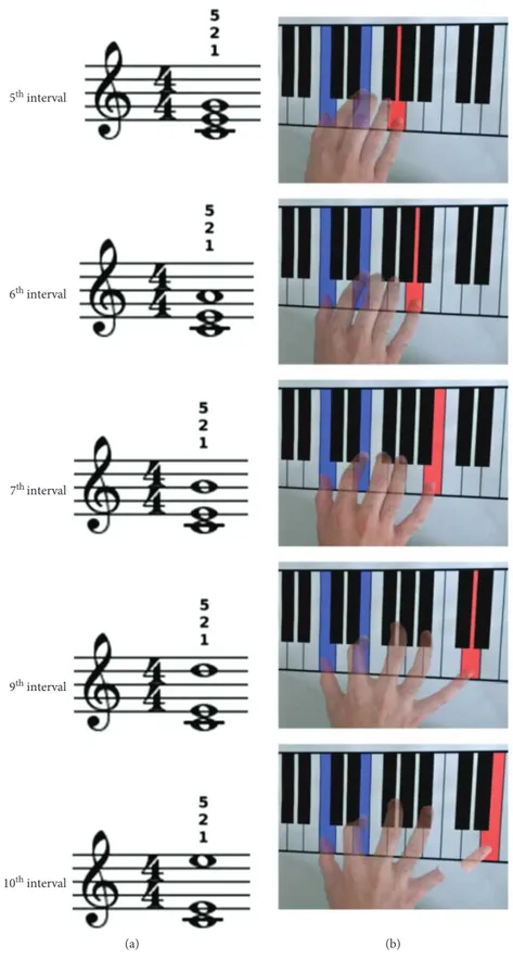
2.2. Paradigm and Stimuli. Participants sat in front of a table where a piano keyboard was depicted (Figure 1) with their arms fully relaxed. The task consisted into performing the mental execution of 5 chords in C major with the right hand
on the presented keyboard. Chords were formed by afixed
3rdinterval (C4-E4) topped by a varying third note, resulting
in a 5th(G4), 6th(A4), 7th(B4), 9th(D5), or 10th(E5) interval
(Figure 1, column 1). The actual execution of the 10thinterval
chord was thought to be kinetically impossible due to biome-chanical constraints. An auditory, visual, or bimodal audiovi-sual stimulus prompted the imagery of 1 of the 5 chords at each trial. In particular, 5 auditory stimuli were used, corre-sponding to the 5 chords digitally recorded through a MIDI-controlled sampler playing real piano sounds. Short staves with the chord to be imagined written on it served as visual stimuli. In the audiovisual condition, the two stimuli were delivered together at the same time. Stereo loudspeakers and a PC monitor were used for auditory and visual stimuli administration, respectively. Each stimulus lasted 3 s and was preceded by a warning acoustic signal lasting from 1 to 2 s. An experimental constraint was that subjects were always required to mentally execute the triads using the thumb and
the indexfinger for C4 and E4, respectively, and the fifth
fin-ger for the third note (Figure 1). All subjects were given the opportunity to train with the task before starting the experi-ment, until they were able to perform the imagery without showing any electromyographic activity in the recorded mus-cles. Participants were instructed to initiate motor imagery of the visually, auditory, or audiovisually prompted chords immediately after the appearance of the corresponding
5th interval 6th interval 7th interval 9th interval 10th interval (a) (b)
Figure 1: Experimental sketch. Triads were prompted through a visual, auditory, or audiovisual stimulus. The visual stimulus was the chord written on a stave (a); small numbers over each stave denote the requiredfingering for each chord (i.e., 1 stands for the thumb, 2 for the index finger, and 5 for the little finger). Participants were instructed on the fingering to employ before initiating the experiment; therefore, no further indication on thefingers to be used were administered as chords were prompted. The imagined extension of the hand increased across chords (b); the brokenfinger in the 10th interval chord denotes that the actual execution was impossible due to biomechanical constraints.
stimulus. The appearance of an empty stave served as a con-trol condition during which no motor imagery was required. All conditions and musical intervals were repeated 12 times in a fully randomized order. The duration of the experiment ranged from 45 to 60 minutes.
2.3. Neurophysiological Procedures. A circular nonfocal coil connected with a Magstim 200 monophasic stimulator (Magstim, Whiteland, Dyfed, UK) was positioned on the vertex with its handle pointing backwards. The cortical rep-resentations of the right Extensor Communis Digitorum (ECD), Flexor Digitorum Superficialis (FDS), Abductor Pollicis Brevis (APB), and Abductor Digiti Minimi (ADM) were targeted within the left motor cortex. Motor-evoked potentials (MEPs) were recorded by means of surface elec-trodes placed with a tendon-belly montage and connected to a four-channel Neuropack electromyograph (Nihon Koh-den, Tokyo, Japan) sampling at 20 kHz with a bandpass
20 Hz-5 kHzfilter. The choice of a nonfocal coil guaranteed
stable simultaneous responses from all the considered fore-arm and hand muscles, even if positioned outside the hot spot of each muscle [25]. To the same aim, the intensity of the TMS pulse was adjusted to obtain fairly stable motor-evoked potentials (MEPs) simultaneously from the right ECD, FDS, APB, and ADM muscles. The intensity of TMS
was set at 110-120% of the resting motor threshold, defined
as the minimal intensity to produce MEPs of less than
100μV in the target muscles with 50% probability.
In order to minimize habituation, each TMS pulse was delivered following a jittered time interval ranging from 1 s to 2 s from the deployment of the prompting stimulus, there-fore well outside a simple reaction time that could have biased the resulting MEPs’ amplitude [12, 13, 26, 27]. This time served also to monitor the EMG silence in the target muscles in the time preceding the brain stimulation. The EMG silence preceding the TMS pulse was also monitored using an acoustic feedback provided by the EMG recorder. Trials contaminated by EMG or other artifacts were less than 18%, so that 8-10 MEPs per muscle were available for the statistical analyses in each condition.
2.4. Data Analysis. For each muscle, the peak-to-peak ampli-tudes of the MEPs obtained in each experimental condition were averaged and expressed as a percentage of the average MEP amplitude recorded during the control condition.
Since the assumption of normality was violated as assessed by the Shapiro-Wilk test, nonparametric test statis-tics were adopted. A paired sample permutation test [28] based on a t-statistic was used to perform pairwise compari-sons of MEP amplitude for the factor muscle, condition, and chord; the condition by muscle, chord by muscle, and chord by condition by muscle interactions were also evaluated. At least 5000 permutations were run for each comparison. This is considered an appropriate number of permutations for a significance level of 0.05 [29]. p values were adjusted using the false discovery rate (FDR) method [30] in order to con-trol familywise error rate.
Nonnegative matrix factorization (NNMF) [31] was used to extract synergies from muscle activity elicited by TMS
during motor imagery. The algorithm was fed with input matrices containing 4 columns (i.e., 1 for each recorded muscle) and 10 rows storing the average MEPs’ amplitude in each subject during motor imagery of a given chord or under one particular condition. The procedure was repeated for all chords and conditions. Given the number of synergies to extract, NNMF generates two output matrices whose product approximates the original matrix, i.e., a synergy
matrix and a matrix of coefficients. An iterative method
start-ing from initial random values of synergy and coefficients is
used to minimize the root-mean-squared residual error between the original matrix and the product of the two out-put matrices. In our case, the algorithm used the multiplicative update rule [32] and ran 50000 times using different random initial values and selected the solution corresponding to the lowest error in reconstruction. The goodness of factorization was assessed by means of the variance account for (VAF) method [33], i.e., by evaluating the ratio of the sum of squared residuals and the sum of squared residual from mean activa-tion. Pairwise comparisons of the extracted synergies were performed for the factor condition and chord. To this end,
the similarity between paired synergies wasfirst assessed by
means of the dot product of their matrices of coefficients.
Then, the measured dot products were compared to the 99th
percentile of the distribution of shuffled dot products
com-puted by random labeling of the matrix of coefficients
(p < 0 01) [34].
3. Results
3.1. Pairwise Permutation Tests. For the factor muscle, pair-wise comparisons of MEP amplitude of ECD vs. APB
(p < 0 001), ECD vs. ADM (p < 0 001), and FDS vs. APB
(p < 0 001) reached significance. A trend toward significance
was found for the comparison of FDS vs. ADM (p = 0 090)
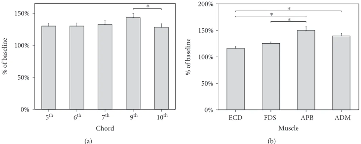
(Figure 2). Therefore, corticospinal excitability assessed dur-ing motor imagery of chords was greater at the APB com-pared to the ECD and FDS and at the ADM comcom-pared to the ECD. No significant differences in the corticospinal out-put emerged between the APB and the ADM nor between the ECD and the FDS.
The comparison of MEP amplitude recorded during
motor imagery of the 9thinterval chord vs. the 10thinterval
chord yielded a significant result (p = 0 01). Additionally, a
trend toward significance was observed with respect to the
comparison between the 6thinterval chord and the 9th
inter-val chord (p = 0 09). Overall, an increase of MEP amplitude
was observed from the 5thto the 9thchord, although not
sig-nificantly. Conversely, corticospinal excitability was
signifi-cantly reduced during motor imagery of the 10th chord
compared to the 9thchord (Figure 2).
3.2. Nonnegative Matrix Factorization and Synergy
Extraction. As shown in Figure 3, following the extraction of 2 synergies, a VAF of 83.16%, 87.47%, 91.34%, and
94.17% in the 5th, 6th, 7th, 9th, and 10th interval chords,
respectively, emerged. Thefirst component showed a greater
coefficient for the APB than for the other muscles. Con-versely, the ECD, FDS, and ADM were mainly represented
in the second component. Pairwise comparisons of the syner-gies extracted across all chords never yielded significance
using the 99th percentile of the distribution of the shuffled
dot products. However, all the dot products of the synergy’s
pairs in the first component and almost all in the second
component fell within the 95thpercentile; the remaining ones
distributed below the 90thpercentile.
VAF exceeded 90% after extraction of two synergies in all the three conditions (i.e., 92.61%, 90.80%, and 93.72% for the auditory, visual, and audiovisual condition, respec-tively). In the auditory condition, the APB was mainly
represented in the first component, whereas ECD, FDS,
and ADM in thefirst. The opposite situation was observed
in the visual and audiovisual condition. Therefore, a
sig-nificant difference emerged in pairwise comparisons of
au-ditory vs. audiovisual and visual vs. audiovisual condition
(Figure 4). Conversely, the dot products of coefficients
dis-tributed below the 95th percentile of the shuffled dot
prod-uct distribution with respect to the comparison of the visual vs. audiovisual condition. Despite the difference observed, it is worth to note that a similar segregation in different component was observed for the APB compare to ECD, FDS, and ADM.
4. Discussion
It is well known that some of the most worldwide famous piano players, as Horowitz, Schoenberg, and Rubinstein, successfully used motor imagery as musical training or immediately prior to a concert exhibition to improve profi-ciency in musical performance [35]. However, neuroimaging research has provided conflicting results on the functional involvement of the primary motor cortex in pianists per-forming musical imagery tasks [19, 21]. Taking into consid-eration that hand motor synergies (i.e., the patterns of muscle activity whose timing and amplitude modulation enable the correct production of movements [33, 36]) are encoded in the human primary motor cortex for hand ges-tures [7], such lack of motor cortex involvement during
imagery is an unexpected finding that could depend upon
several, not mutually exclusive, aspects: playing or imag-ining music require the allocation of most of functional resources for a dynamic integration of perceptual, cogni-tive, and emotional operations [37], so that activations of sensorimotor areas may remain hidden at a certain statistical mapping level. Notably, the motor executive aspects in expert musicians are obviously overlearned and somewhat automatized, so that they may require less
functional activation of final common effector cortices
[38, 39]: this would allow players to better concentrate on expressivity, emotions, or online control of the produced sounds. Obviously, not only the motor cortex controls motor synergies for hand gestures [7]: intracortical connec-tions [40] as well as propriospinal branching of corticosp-inal axons are also regarded as the neural substrates of muscle synergies involved in coordinated multijoint
move-ments [3, 40–42].
Nevertheless, converging neuroimaging and neurophys-iological studies showed that the activity of the hand area of the motor cortex in piano players is increased during lis-tening of familiar musical pieces [22, 43, 44] or during the
observation offingering errors on a keyboard [45]. However,
there are no studies investigating by electrophysiological techniques the online modulation of corticospinal output
for different muscle groups during motor imagery of chords
in professional piano players and in musicians in general. This is precluding any step of knowledge regarding motor synergies used by musicians during musical planning/execu-tion, as well as how these are modulated by audio and visual musical stimuli. This, despite the evidence that musical training promotes the emergence of audio- [43] or visuomo-tor [46–48] cross-modal activations in a frontoparietal net-work including the primary motor cortex [49]. Moreover, action sound and action observation of everyday hand ges-tures, congruent with the perceived action, have been already proven to produce selective corticospinal facilitation in normal subjects [50]. Hence, it is reasonable that
profes-sional piano players, in which a sort “musical grammar”
⁎ 5th 6th 7th 9th 10th Chord 0% 50% 100% 150% % of baseline (a) ⁎ ⁎ ⁎
ECD FDS APB ADM
Muscle 0% 50% 100% 150% 200% % of baseline (b)
Figure 2: Mean MEPs’ peak-to-peak amplitude for each chord (a) and muscle (b) expressed as percent change from baseline. (a) Motor imagery of a 9thinterval triad chord produced a significant increase in MEPs’ amplitude with respect to motor imagery of a 10thinterval chord. (b) APB muscle showed greater facilitation than ECD and FDS. Similarly, ADM showed increased corticospinal excitability when compared to ECD. Bars denote standard errors. Asterisks indicate significant differences.
Component 1 −0.5 0 0.5 Component 2 5th ECD FDS APB ADM Component 1 −0.5 0 0.5 Component 2 6th ECD FDS APB ADM Component 1 −0.5 0 0.5 Component 2 7th ECD FDS APB ADM Component 1 −0.5 0 0.5 Component 2 9th ECD FDS APB ADM −0.8 −0.6 −0.4 −0.2 0 0.2 0.4 0.6 0.8 −0.8 −0.6 −0.4 −0.2 0 0.2 0.4 0.6 0.8 −0.8 −0.6 −0.4 −0.2 0 0.2 0.4 0.6 0.8 −0.8 −0.6 −0.4 −0.2 0 0.2 0.4 0.6 0.8 −0.8 −0.6 −0.4 −0.2 0 0.2 0.4 0.6 0.8 Component 1 −0.5 0 0.5 Component 2 10th ECD FDS APB ADM (a) Component 1 −0.8 −0.6 −0.4 −0.2 0 0.2 0.4 0.6 0.8 Component 2 Auditory ECD FDS APB ADM Component 1 −0.8 −0.6 −0.4 −0.2 0 0.2 0.4 0.6 0.8 Component 2 Visual ECD FDS APB ADM −0.8 −0.6 −0.4 −0.2 0 0.2 0.4 0.6 0.8 −0.8 −0.6 −0.4 −0.2 0 0.2 0.4 0.6 0.8 −0.8 −0.6 −0.4 −0.2 0 0.2 0.4 0.6 0.8 Component 1 −0.8 −0.6 −0.4 −0.2 0 0.2 0.4 0.6 0.8 Component 2 Audiovisual ECD FDS APB ADM (b)
Figure 3: Synergies extracted by means of NNMF across different chords (a) and conditions (b). Line vectors indicate coefficients, whereas dots correspond to the values estimated in the synergy matrix (see text for further details).
for musical-related hand gestures is operating at cortical level [51, 52], might well capitalize from cross-modal per-ceptions to tune at the best of their motor output towards
efficient motor synergy production, such as during very fine
action representations required by progressively increasing triad chords intervals.
Current results show that professional pianists are able to cross-modally modulate their corticospinal output during the mental imagery of triad chords: motor imagery produced
the highest corticospinal facilitation in the hand muscles rather than in the forearm ones. This is not surprising, as either the ABP or ADM was always engaged in the mental execution of each triad chord, although with different demands: the former remained stable on the C note and the
latter was required to be“mentally extended.”
However, such fine tuning is sustained by a definite
three-effector (at least) motor synergy including the FDS
and ADM muscles (that represent the prime movers for the
Component 1: 5th vs 6th Component 1: 5th vs 7th Component 1: 5th vs 9th Component 1: 5th vs 10th
Component 2: 5th vs 6th Component 2: 5th vs 7th Component 2: 5th vs 9th Component 2: 5th vs 10th Component 1: 9th vs 10th Component 2: 9th vs 10th Component 1: 7th vs 9th Component 2: 7th vs 9th Component 2: 7th vs 10th Component 1: 7th vs 10th Component 1: 6th vs 7th Component 2: 6th vs 7th Component 1: 6th vs 9th Component 2: 6th vs 9th Component 1: 6th vs 10th Component 2: 6th vs 10th ECDFDS APB ADM ECDFDS APB ADM ECDFDS APB ADM -0.5 -1 0 0.5 1 -1 -0.5 0 0.5 1 -0.5 -1 0 0.5 1 -1 -0.5 0 0.5 1 -1 -0.5 0 0.5 1 -0.5 -1 0 0.5 1 -0.5 -1 0 0.5 1 -0.5 -1 0 0.5 1 -0.5 -1 0 0.5 1 -0.5 -1 0 0.5 1 -0.5 -1 0 0.5 1 -0.5 -1 0 0.5 1 -0.5 -1 0 0.5 1 -0.5 -1 0 0.5 1 -0.5 -1 0 0.5 1 -0.5 -1 0 0.5 1 -1 -0.5 0 0.5 1 -0.5 -1 0 0.5 1 -0.5 -1 0 0.5 1 -1 -0.5 0 0.5 1 ECDFDS APB ADM ECDFDS APB ADM ECDFDS APB ADM ECDFDS APB ADM ECDFDS APB ADM ECDFDS APB ADM ECDFDS APB ADM ECDFDS APB ADM ECDFDS APB ADM ECDFDS APB ADM ECDFDS APB ADM ECDFDS APB ADM ECDFDS APB ADM ECDFDS APB ADM ECDFDS APB ADM ECDFDS APB ADM ECDFDS APB ADM (a)
Component 1: auditory vs visual
ECD FDS APB ADM
Component 1: auditory vs audiovisual
ECD FDS APB ADM
Component 1: visual vs audiovisual
ECD FDS APB ADM
Component 2: auditory vs visual
ECD FDS APB ADM Component 2: auditory vs audiovisual
ECD FDS APB ADM
Component 2: visual vs audiovisual
-1 -0.5 0 0. 5 1 -1 -0.5 0 0. 5 1 -1 -0.5 0 0. 5 1 -1 -0.5 0 0. 5 1 -1 -0.5 0 0. 5 1 -1 -0.5 0 0. 5 1 ECD FDS APB ADM (b)
required progressive hand extension) as well as the ECD muscle that is coacting with the ADM muscle for the little finger abduction. A gradual increase, although not signifi-cant, in corticospinal excitability was observed moving from
the 5thinterval chord to the 9thinterval chord across
progres-sively wider imagined extensions of the hand. Since a trend
toward significance was detected comparing the 6thinterval
chord with the 9thinterval chord, the lack of significant
dif-ferences between the other chords might reflect the fact that
such intervals are the easiest to be recognized and“executed”
for musicians, that the task did not engage sufficiently the primary motor cortex, or that the sample size was too small to make small changes statistically significant.
Not surprisingly, prompting chords using auditory,
visual, or audiovisual musical stimuli produced similar effects
on corticospinal excitability towards the prime mover mus-cles: this may indicate that professional musicians are able to translate and capitalize the musical information indepen-dently by the sensory channel (i.e., auditory or visual) used to acquire them (see [43, 48, 53]). Adding a cross-modal rein-forcement (as in the audiovisual condition) did not result in additional modulation of the corticospinal system or motor synergy variations, suggesting that the corticospinal tuning was likely already working at its best in professional musi-cians even during monomodal presentation. However, since a control group of nonmusicians was not included in the study, this conclusion remains highly probable but specula-tive. Further studies are needed to disentangle the role of musical skillfulness in motor imagery.
Even if not investigated here, besides the primary motor cortex, the premotor cortex (PMC) might also be a candidate for the observed cross-modal tuning of corticospinal output in piano players. Indeed, electrophysiological studies in ani-mals have shown that neurons in the PMC respond to audi-tory and visual stimuli that are linked to known actions [54] and, in humans, perturbation of PMC by repetitive TMS dis-rupts learning of listened melodies [55] or rhythmic entrain-ment [56]. PMC also plays a relevant role in visuomotor transformation in humans [57]. The PMC is closely con-nected with the primary motor cortex that according to recent neuroimaging evidence [7] encodes motor synergies for human hand gestures.
Language [43], music [58], and actions [59] share a com-mon syntactic-like overlapping structure. Effects of audiovi-sual feedback on corticospinal output in piano players may be regarded as the analogue of the many physiologically demonstrable multisensory cross-modal interactions on the
motor system: action observation [26, 27, 60–63] or action
listening [64], speech listening [65–67], especially in the case
of action-related words [68, 69], or smelling food [70], induce corticospinal facilitation in the muscles the actor would use to actually execute congruent actions. Moreover, it is known that neural representations of action-related sounds depend on motor familiarity [71], as chords for musicians. These facilitatory effects induced by audio- and visuomotor trans-formations could be particularly amplified in professional musicians [72], thanks to their enhanced functional [73] and structural [74] adaptive plastic capabilities in the senso-rimotor brain areas.
It might be argued that an involuntary motor cortical activity may be elicited in piano players by music listening, especially for chords requiring the action of the thumb and
of the littlefinger, and that these activities might have biased
MEP amplitude. This effect is ruled out by the initial practice carried out by all subjects with the task and by continuous visual and acoustic monitoring of electromyographic activity in seconds preceding the TMS pulse throughout the
experi-ment. Moreover, the musical-related specificity of the
observed cross-modal effects on the engagement of specific
motor synergies, but even on corticospinal output in general, rules out the possibility that corticospinal changes might be solely due to the peculiar pianists’ skillfulness in finger motor abilities: it is unlikely, but it is a matter to be verified experi-mentally, that music-naïve typewriters or braille readers might undergo to similar music-related corticospinal effects, beyond the facilitation induced by motor practice alone.
Data Availability
All data used to support the findings of this study are
included within the article. Additional data can be requested to the corresponding author.
Conflicts of Interest
All the authors declare that there is no conflict of interest
regarding the publication of this paper.
Authors’ Contributions
Simone Rossi, Danilo Spada, and Marco Emanuele contrib-uted equally to the work.
Acknowledgments
The authors thank Drs. Raffaele Spidalieri, Alberto De Capua, Matteo Feurra, Riccardo Mazzocchio, Giovanni Bianco, and Federica Felici for their experimental help in an early phase of the study. The research was partly granted by the EU Project BrainTuning FP6-2004
NEST-PATH-028570 and by “Piano di Sostegno alla Ricerca
2018”, University of Siena.
References
[1] N. Bernstein, The Co-ordination and Regulation of Move-ments, Pergamon Press, Oxford, 1967.
[2] M. Santello, M. Flanders, and J. F. Soechting,“Postural hand synergies for tool use,” The Journal of neuroscience, vol. 18, no. 23, pp. 10105–10115, 1998.
[3] H. Devanne, L. G. Cohen, N. Kouchtir-Devanne, and C. Capaday, “Integrated motor cortical control of task-related muscles during pointing in humans,” Journal of Neuro-physiology, vol. 87, no. 6, pp. 3006–3017, 2002.
[4] M. C. Park, A. Belhaj-Saïf, M. Gordon, and P. D. Cheney, “Consistent features in the forelimb representation of primary motor cortex in rhesus macaques,” The Journal of Neurosci-ence, vol. 21, no. 8, pp. 2784–2792, 2001.
[5] M. S. A. Graziano, C. S. R. Taylor, T. Moore, and D. F. Cooke, “The cortical control of movement revisited,” Neuron, vol. 36, no. 3, pp. 349–362, 2002.
[6] R. Gentner and J. Classen,“Modular Organization of finger movements by the human central nervous system,” Neuron, vol. 52, no. 4, pp. 731–742, 2006.
[7] A. Leo, G. Handjaras, M. Bianchi et al.,“A synergy-based hand control is encoded in human motor cortical areas,” eLife, vol. 5, article e13420, 2016.
[8] L. Cattaneo, F. Caruana, A. Jezzini, and G. Rizzolatti, “Repre-sentation of goal and movements without overt motor behav-ior in the human motor cortex: a transcranial magnetic stimulation study,” Journal of Neuroscience, vol. 29, no. 36, pp. 11134–11138, 2009.
[9] R. N. Lemon, R. S. Johansson, and G. Westling, “Corticosp-inal control during reach, grasp, and precision lift in man,” The Journal of Neuroscience, vol. 15, no. 9, pp. 6145–6156, 1995.
[10] S. Rossi and P. M. Rossini,“TMS in cognitive plasticity and the potential for rehabilitation,” Trends in Cognitive Sciences, vol. 8, no. 6, pp. 273–279, 2004.
[11] L. Fadiga, L. Craighero, and E. Olivier,“Human motor cor-tex excitability during the perception of others’ action,” Current Opinion in Neurobiology, vol. 15, no. 2, pp. 213–218, 2005.
[12] S. Rossi, P. Pasqualetti, F. Tecchio, F. Pauri, and P. M. Rossini, “Corticospinal excitability modulation during mental simula-tion of wrist movements in human subjects,” Neuroscience Let-ters, vol. 243, no. 1–3, pp. 147–151, 1998.
[13] P. M. Rossini, S. Rossi, P. Pasqualetti, and F. Tecchio, “Corti-cospinal excitability modulation to hand muscles during movement imagery,” Cerebral Cortex, vol. 9, no. 2, pp. 161– 167, 1999.
[14] L. Fadiga, G. Buccino, L. Craighero, L. Fogassi, V. Gallese, and G. Pavesi,“Corticospinal excitability is specifically modulated by motor imagery: a magnetic stimulation study,” Neuropsy-chologia, vol. 37, no. 2, pp. 147–158, 1998.
[15] M. Lotze, P. Montoya, M. Erb et al.,“Activation of cortical and cerebellar motor areas during executed and imagined hand movements: an fMRI study,” Journal of Cognitive Neurosci-ence, vol. 11, no. 5, pp. 491–501, 1999.
[16] E. Gerardin, “Partially overlapping neural networks for real and imagined hand movements,” Cerebral Cortex, vol. 10, no. 11, pp. 1093–1104, 2000.
[17] C. A. Porro, M. P. Francescato, V. Cettolo et al.,“Primary motor and sensory cortex activation during motor perfor-mance and motor imagery: a functional magnetic resonance imaging study,” The Journal of Neuroscience, vol. 16, no. 23, pp. 7688–7698, 1996.
[18] R. J. Zatorre, A. R. Halpern, and M. Bouffard, “Mental reversal of imagined melodies: a role for the posterior parietal cortex,” Journal of Cognitive Neuroscience, vol. 22, no. 4, pp. 775–789, 2010.
[19] I. G. Meister, T. Krings, H. Foltys et al., “Playing piano in the mind - an fMRI study on music imagery and perfor-mance in pianists,” Cognitive Brain Research, vol. 19, no. 3, pp. 219–228, 2004.
[20] R. S. Schaefer, A. M. Morcom, N. Roberts, and K. Overy, “Moving to music: effects of heard and imagined musical cues on movement-related brain activity,” Frontiers in Human Neuroscience, vol. 8, p. 774, 2014.
[21] M. Lotze, G. Scheler, H. R. M. Tan, C. Braun, and N. Birbaumer, “The musician’s brain: functional imaging of amateurs and professionals during performance and imagery,” NeuroImage, vol. 20, no. 3, pp. 1817–1829, 2003.
[22] A. D’Ausilio, E. Altenmüller, M. Olivetti Belardinelli, and M. Lotze,“Cross-modal plasticity of the motor cortex while listening to a rehearsed musical piece,” European Journal of Neuroscience, vol. 24, no. 3, pp. 955–958, 2006.
[23] R. J. Zatorre, J. L. Chen, and V. B. Penhune,“When the brain plays music: auditory-motor interactions in music perception and production,” Nature Reviews Neuroscience, vol. 8, no. 7, pp. 547–558, 2007.
[24] S. Rossi, M. Hallett, P. M. Rossini, A. Pascual-Leone, and Safety of TMS Consensus Group, “Safety, ethical consider-ations, and application guidelines for the use of transcranial magnetic stimulation in clinical practice and research,” Clinical Neurophysiology, vol. 120, no. 12, pp. 2008–2039, 2009.
[25] P. M. Rossini, D. Burke, R. Chen et al.,“Non-invasive electrical and magnetic stimulation of the brain, spinal cord, roots and peripheral nerves: basic principles and procedures for routine clinical and research application: an updated report from an I.F.C.N. Committee,” Clinical Neurophysiology, vol. 126, no. 6, pp. 1071–1107, 2015.
[26] M. Feurra, G. Bianco, N. R. Polizzotto, I. Innocenti, A. Rossi, and S. Rossi,“Cortico-cortical connectivity between right pari-etal and bilateral primary motor cortices during imagined and observed actions: a combined TMS/tDCS study,” Frontiers in Neural Circuits, vol. 5, p. 10, 2011.
[27] G. Bianco, M. Feurra, L. Fadiga, A. Rossi, and S. Rossi, “Bi-hemispheric effects on corticospinal excitability induced by repeated sessions of imagery versus observation of actions,” Restorative Neurology and Neuroscience, vol. 30, no. 6, pp. 481–489, 2012.
[28] R. C. Blair and W. Karniski,“An alternative method for signif-icance testing of waveform difference potentials,” Psychophys-iology, vol. 30, no. 5, pp. 518–524, 1993.
[29] B. F. Manly, Randomization and Monte Carlo Methods in Biology, Chapman & Hall, New York, NY, USA, 1997. [30] Y. Benjamini and Y. Hochberg, “Controlling the false
dis-covery rate: a practical and powerful approach to multiple testing,” Journal of the Royal Statistical Society: Series B (Methodological), vol. 57, pp. 289–300, 1995.
[31] D. D. Lee and H. S. Seung,“Learning the parts of objects by non-negative matrix factorization,” Nature, vol. 401, no. 6755, pp. 788–791, 1999.
[32] D. Lee and H. Seung, Algorithms for Non-Negative Matrix Factorization, pp. 535–541, 2001, http://papers.nips.cc. [33] A. d’Avella, A. Portone, L. Fernandez, and F. Lacquaniti,
“Control of fast-reaching movements by muscle synergy combinations,” Journal of Neuroscience, vol. 26, no. 30, pp. 7791–7810, 2006.
[34] S. A. Overduin, A. d’Avella, J. M. Carmena, and E. Bizzi, “Microstimulation activates a handful of muscle synergies,” Neuron, vol. 76, no. 6, pp. 1071–1077, 2012.
[35] H. Schoenberg, Great Pianists, Fireside Books, St. Louis, MO, USA, 1987.
[36] M. Santello, M. Bianchi, M. Gabiccini et al.,“Hand synergies: integration of robotics and neuroscience for understanding the control of biological and artificial hands,” Physics of Life Reviews, vol. 17, pp. 1–23, 2016.
[37] S. Koelsch and W. A. Siebel,“Towards a neural basis of music perception,” Trends in Cognitive Sciences, vol. 9, no. 12, pp. 578–584, 2005.
[38] V. Puttemans, N. Wenderoth, and S. P. Swinnen,“Changes in brain activation during the acquisition of a multifrequency bimanual coordination task: from the cognitive stage to advanced levels of automaticity,” Journal of Neuroscience, vol. 25, no. 17, pp. 4270–4278, 2005.
[39] K. Petrini, F. E. Pollick, S. Dahl et al.,“Action expertise reduces brain activity for audiovisual matching actions: an fMRI study with expert drummers,” NeuroImage, vol. 56, no. 3, pp. 1480– 1492, 2011.
[40] C. Capaday, H. Devanne, L. Bertrand, and B. A. Lavoie, “Intra-cortical connections between motor “Intra-cortical zones controlling antagonistic muscles in the cat: a combined anatomical and physiological study,” Experimental Brain Research, vol. 120, no. 2, pp. 223–232, 1998.
[41] B. J. McKiernan, J. K. Marcario, J. H. Karrer, and P. D. Cheney, “Corticomotoneuronal postspike effects in shoulder, elbow, wrist, digit, and intrinsic hand muscles during a reach and prehension task,” Journal of Neurophysiology, vol. 80, no. 4, pp. 1961–1980, 1998.
[42] B. Tantisira, B. Alstermark, T. Isa, H. Kummel, and M. Pinter, “Motoneuronal projection pattern of single C3-C4 propriosp-inal neurones,” Canadian Journal of Physiology and Pharma-cology, vol. 74, no. 4, pp. 518–530, 1996.
[43] M. Bangert, T. Peschel, G. Schlaug et al.,“Shared networks for auditory and motor processing in professional pianists: evi-dence from fMRI conjunction,” NeuroImage, vol. 30, no. 3, pp. 917–926, 2006.
[44] J. Haueisen and T. R. Knösche,“Involuntary motor activity in pianists evoked by music perception,” Journal of Cognitive Neuroscience, vol. 13, no. 6, pp. 786–792, 2001.
[45] M. Candidi, L. M. Sacheli, I. Mega, and S. M. Aglioti, “Somato-topic mapping of piano fingering errors in sensorimotor experts: TMS studies in pianists and visually trained musically naïves,” Cerebral Cortex, vol. 24, no. 2, pp. 435–443, 2014. [46] M. Bangert and E. O. Altenmüller, “Mapping perception to
action in piano practice: a longitudinal DC-EEG study,” BMC Neuroscience, vol. 4, no. 1, p. 26, 2003.
[47] T. Hasegawa, K. I. Matsuki, T. Ueno et al.,“Learned audio-visual cross-modal associations in observed piano playing acti-vate the left planum temporale. An fMRI study,” Cognitive Brain Research, vol. 20, no. 3, pp. 510–518, 2004.
[48] B. Haslinger, P. Erhard, E. Altenmüller, U. Schroeder, H. Boecker, and A. O. Ceballos-Baumann,“Transmodal sen-sorimotor networks during action observation in professional pianists,” Journal of Cognitive Neuroscience, vol. 17, no. 2, pp. 282–293, 2005.
[49] K. Alaerts, S. P. Swinnen, and N. Wenderoth,“Interaction of sound and sight during action perception: evidence for shared modality-dependent action representations,” Neuropsycholo-gia, vol. 47, no. 12, pp. 2593–2599, 2009.
[50] D. Sammler, G. Novembre, S. Koelsch, and P. E. Keller, “Syn-tax in a pianist’s hand: ERP signatures of embodied syntax processing in music,” Cortex, vol. 49, no. 5, pp. 1325–1339, 2013.
[51] R. Bianco, G. Novembre, P. E. Keller et al.,“Syntax in action has priority over movement selection in piano playing: an erp study,” Journal of Cognitive Neuroscience, vol. 28, no. 1, pp. 41–54, 2016.
[52] I. Bufalari, A. Sforza, P. Cesari, S. M. Aglioti, and A. D. Fourkas, “Motor imagery beyond the joint limits: a trans-cranial magnetic stimulation study,” Biological Psychology, vol. 85, no. 2, pp. 283–290, 2010.
[53] J. Buchmann, W. Gierow, S. Weber et al., “Restoration of disturbed intracortical motor inhibition and facilitation in attention deficit hyperactivity disorder children by methyl-phenidate,” Biological Psychiatry, vol. 62, no. 9, pp. 963– 969, 2007.
[54] C. Lega, M. A. Stephan, R. J. Zatorre, and V. Penhune,“Testing the role of dorsal premotor cortex in auditory-motor associa-tion learning using transcranical magnetic stimulaassocia-tion (TMS),” PLoS One, vol. 11, no. 9, article e0163380, 2016. [55] F. Giovannelli, I. Innocenti, S. Rossi et al.,“Role of the dorsal
premotor cortex in rhythmic auditory-motor entrainment: a perturbational approach by rTMS,” Cerebral Cortex, vol. 24, no. 4, pp. 1009–1016, 2014.
[56] E. Hoshi and J. Tanji,“Differential involvement of neurons in the dorsal and ventral premotor cortex during processing of visual signals for action planning,” Journal of Neurophysiology, vol. 95, no. 6, pp. 3596–3616, 2006.
[57] A. D. Patel,“Language, music, syntax and the brain,” Nature Neuroscience, vol. 6, no. 7, pp. 674–681, 2003.
[58] L. Fadiga, L. Craighero, and A. D’Ausilio, “Broca’s area in lan-guage, action, and music,” Annals of the New York Academy of Sciences, vol. 1169, no. 1, pp. 448–458, 2009.
[59] L. Fadiga, L. Fogassi, G. Pavesi, and G. Rizzolatti,“Motor facil-itation during action observation: a magnetic stimulation study,” Journal of Neurophysiology, vol. 73, no. 6, pp. 2608– 2611, 1995.
[60] M. Gangitano, F. M. Mottaghy, and A. Pascual-Leone, “Phase-specific modulation of cortical motor output during movement observation,” Neuroreport, vol. 12, no. 7, pp. 1489– 1492, 2001.
[61] L. Aziz-Zadeh, F. Maeda, E. Zaidel, J. Mazziotta, and M. Iacoboni, “Lateralization in motor facilitation during action observation: a TMS study,” Experimental Brain Research, vol. 144, no. 1, pp. 127–131, 2002.
[62] C. Urgesi, M. Candidi, F. Fabbro, M. Romani, and S. M. Aglioti,“Motor facilitation during action observation: topo-graphic mapping of the target muscle and influence of the onlooker’s posture,” European Journal of Neuroscience, vol. 23, no. 9, pp. 2522–2530, 2006.
[63] E. Ricciardi, G. Handjaras, D. Bonino, T. Vecchi, L. Fadiga, and P. Pietrini,“Beyond motor scheme: a supramodal distrib-uted representation in the action-observation network,” PLoS One, vol. 8, no. 3, article e58632, 2013.
[64] A. Lahav, E. Saltzman, and G. Schlaug,“Action representation of sound: audiomotor recognition network while listening to newly acquired actions,” Journal of Neuroscience, vol. 27, no. 2, pp. 308–314, 2007.
[65] L. Fadiga, L. Craighero, G. Buccino, and G. Rizzolatti,“Speech listening specifically modulates the excitability of tongue mus-cles: a TMS study,” European Journal of Neuroscience, vol. 15, no. 2, pp. 399–402, 2002.
[66] A. Flöel, T. Ellger, C. Breitenstein, and S. Knecht,“Language perception activates the hand motor cortex: implications for motor theories of speech perception,” European Journal of Neuroscience, vol. 18, no. 3, pp. 704–708, 2003.
[67] L. Bracco, F. Giovannelli, V. Bessi et al., “Mild cognitive impairment: loss of linguistic task-induced changes in motor
cortex excitability,” Neurology, vol. 72, no. 10, pp. 928–934, 2009.
[68] G. Buccino, L. Riggio, G. Melli, F. Binkofski, V. Gallese, and G. Rizzolatti,“Listening to action-related sentences modulates the activity of the motor system: a combined TMS and behavioral study,” Cognitive Brain Research, vol. 24, no. 3, pp. 355–363, 2005.
[69] B. Tomasino, G. R. Fink, R. Sparing, M. Dafotakis, and P. H. Weiss,“Action verbs and the primary motor cortex: a compar-ative TMS study of silent reading, frequency judgments, and motor imagery,” Neuropsychologia, vol. 46, no. 7, pp. 1915– 1926, 2008.
[70] S. Rossi, A. de Capua, P. Pasqualetti et al.,“Distinct olfactory cross-modal effects on the human motor system,” PLoS One, vol. 3, no. 2, article e1702, 2008.
[71] G. Novembre and P. E. Keller,“A conceptual review on action-perception coupling in the musicians’ brain: what is it good for?,” Frontiers in Human Neuroscience, vol. 8, p. 603, 2014. [72] T. F. Münte, E. Altenmüller, and L. Jäncke,“The musician’s
brain as a model of neuroplasticity,” Nature Reviews Neurosci-ence, vol. 3, no. 6, pp. 473–478, 2002.
[73] K. Rosenkranz, A. Williamon, and J. C. Rothwell, “Motorcor-tical excitability and synaptic plasticity is enhanced in profes-sional musicians,” Journal of Neuroscience, vol. 27, no. 19, pp. 5200–5206, 2007.
[74] C. Gaser and G. Schlaug, “Brain structures differ between musicians and non-musicians,” The Journal of Neuroscience, vol. 23, no. 27, pp. 9240–9245, 2003.
Hindawi
www.hindawi.com Volume 2018
Research and Treatment
Autism
Depression Research and Treatment Hindawi www.hindawi.com Volume 2018 Neurology Research International Hindawi www.hindawi.com Volume 2018 Alzheimer’s Disease Hindawi www.hindawi.com Volume 2018 International Journal of Hindawi www.hindawi.com Volume 2018 BioMed Research International Hindawi www.hindawi.com Volume 2018 Research and TreatmentSchizophrenia
Hindawi Publishing Corporation
http://www.hindawi.com Volume 2013
Hindawi www.hindawi.com
World Journal
Volume 2018 Hindawiwww.hindawi.com Volume 2018
Neural Plasticity
Scientifica
Hindawi www.hindawi.com Volume 2018 Hindawi www.hindawi.com Volume 2018Parkinson’s
Disease
Sleep Disorders
Hindawiwww.hindawi.com Volume 2018 Hindawiwww.hindawi.com Volume 2018
Neuroscience
Journal
Medicine
Advances in Hindawi www.hindawi.com Volume 2018 Hindawi www.hindawi.com Volume 2018Psychiatry
Journal
Hindawi www.hindawi.com Volume 2018 Computational and Mathematical Methods in Medicine International Hindawi www.hindawi.com Volume 2018Stroke
Research and Treatment Hindawi www.hindawi.com Volume 2018 Hindawi www.hindawi.com Volume 2018