Upper limb motor rehabilitation impacts white matter
microstructure in multiple sclerosis
☆
Laura Bonzano
a,b,⁎
, Andrea Tacchino
c, Giampaolo Brichetto
c, Luca Roccatagliata
b,d, Adriano Dessypris
a,e,
Paola Feraco
a, Maria L. Lopes De Carvalho
f, Mario A. Battaglia
g, Giovanni L. Mancardi
a,b, Marco Bove
e,⁎⁎
a
Department of Neuroscience, Rehabilitation, Ophthalmology, Genetics, Maternal and Child Health, University of Genoa, Genoa, Italy
b
Magnetic Resonance Research Centre on Nervous System Diseases, University of Genoa, Genoa, Italy
cScientific Research Area, Italian Multiple Sclerosis Foundation (FISM), Genoa, Italy d
Department of Health Sciences, Biostatistics Unit, University of Genoa, Genoa, Italy
eDepartment of Experimental Medicine, Section of Human Physiology and Centro Polifunzionale di Scienze Motorie, University of Genoa, Genoa, Italy f
AISM Rehabilitation Service, Italian Multiple Sclerosis Society, Genoa, Italy
g
Department of Physiopathology, Experimental Medicine and Public Health, University of Siena, Siena, Italy
a b s t r a c t
a r t i c l e i n f o
Article history:
Accepted 14 December 2013 Available online 25 December 2013 Keywords:
Diffusion tensor imaging Motor rehabilitation Multiple sclerosis Upper limb Voluntary movements White matter
Upper limb impairments can occur in patients with multiple sclerosis, affecting daily living activities; however there is at present no definite agreement on the best rehabilitation treatment strategy to pursue. Moreover, motor training has been shown to induce changes in white matter architecture in healthy subjects.
This study aimed at evaluating the motor behavioral and white matter microstructural changes following a 2-month upper limb motor rehabilitation treatment based on task-oriented exercises in patients with multiple sclerosis. Thirty patients (18 females and 12 males; age = 43.3 ± 8.7 years) in a stable phase of the disease presenting with mild or moderate upper limb sensorimotor deficits were randomized into two groups of 15 patients each. Both groups underwent twenty 1-hour treatment sessions, three times a week. The“treatment group” received an active motor rehabilitation treatment, based on voluntary exercises including task-oriented exercises, while the“control group” underwent passive mobilization of the shoulder, elbow, wrist and fingers.
Before and after the rehabilitation protocols, motor performance was evaluated in all patients with standard tests. Additionally,finger motor performance accuracy was assessed by an engineered glove.
In the same sessions, every patient underwent diffusion tensor imaging to obtain parametric maps of fractional anisotropy, mean diffusivity, axial diffusivity, and radial diffusivity. The mean value of each parameter was sep-arately calculated within regions of interest including thefiber bundles connecting brain areas involved in volun-tary movement control: the corpus callosum, the corticospinal tracts and the superior longitudinal fasciculi. The two rehabilitation protocols induced similar effects on unimanual motor performance, but the bimanual coor-dination task revealed that the residual coorcoor-dination abilities were maintained in the treated patients while they sig-nificantly worsened in the control group (p = 0.002). Further, in the treatment group white matter integrity in the corpus callosum and corticospinal tracts was preserved while a microstructural integrity worsening was found in the control group (fractional anisotropy of the corpus callosum and corticospinal tracts: p = 0.033 and p = 0.022; radial diffusivity of the corpus callosum and corticospinal tracts: p = 0.004 and p = 0.008). Conversely, a significant increase of radial diffusivity was observed in the superior longitudinal fasciculi in both groups (p = 0.02), indicating lack of treatment effects on this structure, showing damage progression likely due to a demy-elination process.
All thesefindings indicate the importance of administering, when possible, a rehabilitation treatment consisting of voluntary movements. We also demonstrated that the beneficial effects of a rehabilitation treatment are task-dependent and selective in their target; this becomes crucial towards the implementation of tailored rehabilitative approaches.
© 2013 The Authors. Published by Elsevier Inc. All rights reserved.
☆ This is an open-access article distributed under the terms of the Creative Commons Attribution-NonCommercial-No Derivative Works License, which permits non-commercial use, dis-tribution, and reproduction in any medium, provided the original author and source are credited.
⁎ Correspondence to: L. Bonzano, Department of Neuroscience, Rehabilitation, Ophthalmology, Genetics, Maternal and Child Health, Largo Daneo 3 (ex via De Toni 5), 16132 Genoa, Italy. Fax: +39 0103538639.
⁎⁎ Correspondence to: M. Bove, Department of Experimental Medicine, Section of Human Physiology, Viale Benedetto XV 3, 16132 Genoa, Italy. Fax: +39 0103538194. E-mail addresses:[email protected](L. Bonzano),[email protected](M. Bove).
1053-8119/$– see front matter © 2013 The Authors. Published by Elsevier Inc. All rights reserved.
http://dx.doi.org/10.1016/j.neuroimage.2013.12.025
Contents lists available atScienceDirect
NeuroImage
Introduction
Impaired sensorimotor function is frequent in multiple sclerosis (MS). Sensorimotor impairments of the lower limbs affecting mobility are reported in 75% of patients with MS (PwMS), whereas dysfunctions of the upper limbs occur in 66% of PwMS (Johansson et al., 2007; Spooren et al., 2012). The level of arm and hand functioning greatly de-fines the ability to perform daily living activities like eating, dressing, and grooming (Yozbatiran et al., 2006).
Neurorehabilitation is targeted at maintaining and possibly im-proving the residual capacities of neurological patients with the aim to preserve their personal and social activities, and it constitutes an important part of quality health care in PwMS. There is at present no definite agreement on which specific exercise therapy program can be considered the most successful in improving activities and participation. Different training programs have been employed for upper limb neurorehabilitation, ranging from more traditional strat-egies to newer techniques emphasizing the learning and practice of functional motor skills within a “task-specific” context (Solari et al., 1999; Spooren et al., 2012). In addition, it has been proposed that a training based on the performance of voluntary movements showed significant improvements in motor performance in healthy subjects with respect to passive training (Bayona et al., 2005; Lotze
et al., 2003). Further, active training has been found to induce
more prominent increases in fMRI activation of the contralateral pri-mary motor cortex (M1), corticospinal excitability and intracortical facilitation than passive training (Lotze et al., 2003). All these find-ings suggest the important role for voluntary drive in motor learning and neurorehabilitation. In agreement with this notion, voluntary exercise has been convincingly shown to attenuate the clinical de fi-cits and the underlying neuropathological process in animal models of neurodegenerative disorders (Ang and Gomez-Pinilla, 2007; Cotman and Berchtold, 2002; Cotman et al., 2007; Kramer and Erickson, 2007; Rossi et al., 2009).
Recently, changes in white matter (WM) architecture have been ob-served in healthy subjects after motor training (Draganski and May, 2008; Scholz et al., 2009; Taubert et al., 2010). WMfiber pathways form the brain communication network; thus, the physical condition of a given pathway can determine the efficiency of signal transmissions between brain regions and might thereby influence behaviors relying on that pathway (Fields, 2008; Johansen-Berg, 2010; Johansen-Berg et al., 2010; Scholz et al., 2009). In this framework, the increasing senso-rimotor impairment observed in PwMS over the disease course could be mainly due to the progression of WM damage, that is present in these pa-tients since the early stages (Evangelou et al., 2000; Ferguson et al., 1997; Ge et al., 2005; van Waesberghe et al., 1999). In particular, reductions in the microstructural integrity of the corpus callosum (CC) have been shown to be associated with decreased sensorimotor performance, impairment in visuomotor learning and deficit in bimanual coordination (Bonzano et al., 2008, 2011a,b; Larson et al., 2002; Pelletier et al., 1992). The present study was designed to evaluate the motor behavioral and WM microstructural architecture changes, with a focus on the WMfiber bundles connecting brain areas involved in voluntary movement control, following a 2-month upper limb motor rehabilita-tion treatment including task-oriented exercises in PwMS.
Material and methods Patients
Thirty right-handed PwMS in a stable phase of the disease presenting with mild or moderate sensorimotor deficit in one or both upper limbs were recruited for this study. The Medical Research Council (MRC) scale (0 to 5 grades) was adopted for testing muscle strength at the proximal (i.e., shoulder and elbow) and distal (i.e., wrist andfingers) segments (Compston, 2010). Inclusion criteria were the following
MRC scores of patient's effort: grade 4 in all muscle groups or grade 3 in no more than two joints (mild deficit), or grade 3 in all muscle groups (moderate deficit). We excluded patients with relapses and steroid-use or a worsening of the Expanded Disability Status Scale (EDSS) score (Kurtzke, 1983) in the last three months, psychiatric disorders and se-vere cognitive impairment.
Among the included patients (18 females and 12 males; mean age = 43.3 ± 8.7 years) 22 were affected by a relapsing–remitting and 8 by a secondary progressive form of MS. Demographic and clinical characteristics of the patients are reported inTable 1.
The study was approved by the ethical committee of our institution and the patients' consent was obtained according to the Declaration of Helsinki.
Rehabilitative protocols
We were interested in investigating the effects of an active upper limb rehabilitation treatment based on volitional tasks on motor performance and white matter microstructure. To this aim, we de-fined a “control treatment”, as strongly suggested in a recent critical review of studies assessing structural plasticity following training
(Thomas and Baker, 2013). In fact, comparing two groups who have
been trained on different tasks allows showing that potential changes are specific to a given task and not a general effect of any training. Therefore, the 30 recruited PwMS were randomly assigned to two groups, with the use of a computer-generated schedule: one receiving an active motor rehabilitation treatment (“treatment group”—15 patients) and one receiving a passive motor rehabilitation treatment (“control group”—15 patients) (Table 1).
The two rehabilitative protocols were designed with the intention that all the patients were similarly invested in the study by equating pa-tients' overall experience thus limiting possible biases (Thomas and Baker, 2013); both groups of patients underwent twenty 1-hour treat-ment sessions, three times a week, at AISM Rehabilitation Centre, Italian Multiple Sclerosis Society, Genoa, Italy.
In details, the patients assigned to the treatment group were re-habilitated with an active protocol based on voluntary exercises for neuromuscular control to improve proprioceptive sensibility, mus-cle strength, stability and coordination of the upper limbs, mainly including task-oriented exercises with the goal to improve activities of daily living (Nelson, 1996). Thefirst 5 sessions of the rehabilita-tive protocol were focused on voluntary exercises executed unilater-ally with the right and left upper limbs (60% of treatment time). This part of the treatment dealt with both non task-oriented exercises, such as grasping wooden cubes of different sizes, pinching, reaching a target displayed in front of the patient, and task-oriented exercises such as ironing a shirt and putting a dish in a draining board. In the last 40% of the treatment, bimanual task-oriented exercises, such as sewing, doing patchwork and paper mandala, cooking, sweeping, and screwing a cap on a bottle, were administered to the patients. Grad-ually, from the 6th to the 12th sessions, the percentage of bimanual task-oriented exercises increased to reach 100% in the last 5 sessions. Thus, unimanual and bimanual voluntary exercises were differently weighted in each session along the rehabilitative program (sessions 1–5: 60%–40%, respectively; sessions 6–10: 40%–60%, respectively; sessions 11–15: 20%–80%, respectively; and sessions 16–20: 0%–100%, respectively).
The control group only performed tasks without detectable muscle activity, through passive mobilization of the shoulder, elbow, wrist andfingers delivered by a physical therapist. Analogously, in the passive rehabilitation protocol the percentage of unimanual and bimanual pas-sive mobilizations delivered by the therapist followed the scheme used for the“treatment group” (i.e., sessions 1–5: 60%–40%, respectively; sessions 6–10: 40%–60%, respectively; sessions 11–15: 20%–80%, respectively; and sessions 16–20: 0%–100%, respectively).
Motor performance evaluation
Before (“PRE session”, i.e., baseline) and after (“POST session”) the re-habilitation treatment, motor performance was evaluated in all the pa-tients for both arms with the following standard measures of global disability and sensorimotor dysfunction: upper limb motor functions by the Action Research Arm Test (ARAT) (Lyle, 1981), hand dexterity by the nine Hole Peg Test (9-HPT) (Fischer et al., 1999), and grip strength by a dynamometer (GRIP).
In addition, an engineered glove was used to quantifyfinger motor performance accuracy; this simple and objective method has been recently demonstrated to be able to discriminate healthy con-trols and PwMS even with very low disability (Bonzano et al., 2013). Specifically, patients were asked to perform with their eyes closed repetitivefinger opposition movements of thumb to index, medium, ring and littlefingers, with the dominant hand (right for all the patients) at their spontaneous and maximal velocity. The fin-ger motor sequence was repeated with both hands simultaneously and paced with a metronome tone set at a rate of 2 Hz, to assess bi-manual coordination. From the raw data recorded by the glove system, different parameters were then extracted: the movement rate at spon-taneous (RATE-SV) and maximum velocity (RATE-MV) conditions. When the task was performed with the two hands, the inter hand inter-val (IHI) was calculated as index of bimanual coordination: the larger the IHI value, the more severe the impairment in bimanual coordination (Bonzano et al., 2008).
Conventional MRI
Axial dual-echo proton density (PD)/T2-weighted images (slice thickness: 4 mm; TR: 2500 ms; TE: 25.5/127.4 ms;flip angle: 90°;
FOV: 250 mm; matrix: 256 × 256) were acquired to detect T2 lesions. Particularly, two observers, blinded to the clinical data and rehabilitative protocol, identified hyperintense lesions on PD/T2-weighted scans and checked for each patient whether he/she developed new T2 lesions dur-ing the study period, by compardur-ing the POST scan with the PRE scan.
Diffusion tensor imaging
Before (“PRE session”, i.e., baseline) and after (“POST session”) the rehabilitation protocol, every patient underwent a magnetic resonance imaging examination on a 1.5-Tesla scanner (Signa Excite General Electric, WI), including the acquisition of axial single-shot spin-echo echo-planar diffusion tensor imaging (DTI) (slice thickness: 2 mm; TR: 16,000 ms; TE: 105 ms; flip angle: 90°; field of view (FOV): 240 mm; matrix: 128 × 128 interpolated during reconstruction to 256 × 256; number of excitations (NEX): 2), with diffusion gradients applied in 15 noncollinear directions (b = 1000 s/mm2) and two
base-line acquisitions without diffusion gradients (b0 images).
DTI data were processed by using the FMRIB's Diffusion Toolbox, FDT (Smith et al., 2004). After correction for eddy current distortions and motion artifacts, a diffusion tensor model wasfitted at each voxel and the three eigenvalues (λ1,λ2, andλ3) were calculated; hence,
DTI-derived parametric maps were obtained (Basser, 1995; Basser and Pierpaoli, 1996). Particularly, for each patient and for each study session, in order to investigate white matter microstructural integrity we ana-lyzed fractional anisotropy (FA), axial diffusivity (λ∥), i.e., the water
diffusivity parallel to the axonalfibers, represented by λ1, radial
diffu-sivity (λ⊥), i.e., the water diffusivity perpendicular to the axonalfibers,
obtained as the average ofλ2andλ3(Song et al., 2002), and mean
dif-fusivity (MD).
Table 1
Demographic and clinical characteristics of the patients included in the two groups: the“treatment group” received an active motor rehabilitation treatment including task-oriented ex-ercises, the“control group” received a passive motor rehabilitation treatment, based on upper limb mobilization techniques performed by a physical therapist.
Group ID Age (years) Gender MS phenotype EDSS at baseline Disease duration (months)
Time from last relapse before treatment (months) Disease-modifying therapy Affected upper limb Severity of motor deficit
Treatment 1 35 F RR 4 62 5 Immunosuppressant Left Mild
2 56 M SP 4 148 11 Immunosuppressant Right Mild
3 35 F RR 5.5 115 4 Immunosuppressant Right Mild
4 47 M SP 4.5 84 N12 None Bilateral Moderate
5 39 F RR 2 52 N12 Immunomodulant Right Mild
6 33 F RR 4.5 88 N12 None Left Mild
7 31 F RR 3 64 11 Immunosuppressant Bilateral Mild
8 51 M SP 6 88 N12 Immunosuppressant Left Mild
9 49 M SP 6 100 N12 None Left Mild
10 47 M RR 3 110 N12 none Right Mild
11 59 F RR 4 160 N12 immunomodulant Bilateral Mild
12 47 F RR 4.5 188 N12 None Bilateral Mild
13 43 F RR 6.5 22 N12 Immunomodulant Left Mild
14 30 F RR 5 120 6 Immunosuppressant Bilateral Mild
15 49 F RR 3 234 9 None Bilateral Mild
Mean (SD) 43.4 (9.1) 4.4 (1.3) 109.0 (55.3)
Control 1 33 F RR 5.5 28 N12 None Left Mild
2 35 F RR 4.5 60 6 Immunosuppressant Right Mild
3 56 M SP 5 139 N12 Immunosuppressant Left Mild
4 31 F RR 3 49 N12 Immunosuppressant Right Mild
5 35 F RR 4.5 112 5 Immunomodulant Right Mild
6 33 F RR 4 81 10 None Left Mild
7 49 M SP 6 78 N12 Immunosuppressant Right Mild
8 49 M SP 5.5 91 N12 None Bilateral Mild
9 38 M RR 4.5 60 N12 Immunomodulant Right Mild
10 47 M RR 3.5 102 11 None Right Mild
11 55 F RR 4 354 N12 None Left Mild
12 50 F RR 3.5 241 5 Immunomodulant Left Mild
13 39 F RR 2.5 42 10 Immunomodulant Right Mild
14 47 M RR 3 79 N12 Immunosuppressant Bilateral Mild
15 51 M SP 6 81 7 Immunosuppressant Bilateral Mild
Mean (SD) 43.2 (8.6) 4.3 (1.1) 106.5 (85.2) RR = relapsing–remitting; SP = secondary progressive.
All the obtained parametric maps were nonlinearly transformed and aligned to 1 × 1 × 1 mm standard space according to the TBSS routines (Smith et al., 2006). We created some regions of interest (ROIs) from the JHU ICBM 81 white matter labels atlas included in FSL (Mori et al., 2005), visually checked the location of each ROI on each map and calculated the mean value of the different DTI metrics in each ROI. In details, different masks were selected, including the WMfiber bundles connecting brain areas involved in voluntary movement control, i.e., the corpus callosum (CC), the left and right corticospinal tract (CST) and the left and right su-perior longitudinal fasciculus (SLF) (Fig. 1). Indeed, CC pathology occurs in MS since the early disease phase (Evangelou et al., 2000; Ge et al., 2004), and CC abnormalities have been related to decreased sensorimo-tor performance, impairment in visuomosensorimo-tor learning and deficit in bi-manual coordination (Bonzano et al., 2008, 2011a,b; Larson et al., 2002; Pelletier et al., 1993). CST abnormalities can be associated with weakness in MS (Reich et al., 2008), spasticity, deficits in executing fine movements and in motor control of the limbs. The SLF allows the integration of motor and decision-making centers with visual and sen-sory ones; it can be damaged in MS (Bonzano et al., 2009) and could af-fect grasping actions, movement preparation and planning (Jang and Hong, 2012; Koch et al., 2010).
Furthermore, we calculated the white matter signal-to-noise ratio (SNR) by measuring the SNR within each ROI, for each scan of each pa-tient, with the method proposed byPrice et al. (1990), which estimates the noise from the subtraction of two sequentially acquired images. In details, we subtracted the second b0 image from thefirst one obtaining a measure of the random noise introduced by the scanner itself (“noise image”) and we calculated the SNR by the formula:
SNRROI¼
ffiffiffi 2
p SROI
σROIð ÞN
where SROIis the mean signal intensity of thefirst b0 image within the
selected ROI andσROI(N) is the standard deviation of the voxel values
of the noise image in the same ROI. Statistics
First, to evaluate differences in the initial motor performance and white matter microstructural integrity between the two groups of PwMS (treatment group vs. control group), ANOVA was separately per-formed on all the motor performance and DTI-derived parameters col-lected at baseline (PRE session).
To evaluate a possible change in SNR between the two sessions (POST session vs. PRE session) a paired t-test was performed for each analyzed ROI. Then, to assess the effects of the active rehabilitation treatment and the passive mobilization protocol, the obtained measure-ments were compared between the two sessions (POST session vs. PRE session) and the two groups (treatment group vs. control group) by means of factorial ANOVA with repeated measures (RM-ANOVA), with TIME (PRE and POST) as within-subject factor and GROUP (treatment and control) as between-subject factor. When the task had to be
performed with the two hands separately (ARAT, 9-HPT and GRIP) the factor HAND (left and right) was considered as within-subject factor.
In addition, for the DTI-derived parameters (FA,λ∥,λ⊥and MD) the
factor HEMISPHERE (left and right) was taken into account as a within-subject factor when analyzing the left and right CST and SLF fiber bundles.
Significant main effects were explored with the Newman–Keuls post-hoc test.
Results
Motor performance
At baseline (PRE session), motor performance standard tests showed no difference between the treatment and the control group for both hands (ARAT: F(1,56) = 0.23, p = 0.63; 9-HPT: F(1,56) = 0.30, p = 0.58; GRIP: F(1,56) = 1.02, p = 0.32). Also,finger opposition movement performance with the right hand and bimanual coordina-tion did not differ between the two groups (RATE-SV: F(1,28) = 0.13, p = 0.72; RATE-MV: F(1,28) = 0.08, p = 0.78; IHI: F(1,28) = 0.97, p = 0.33).
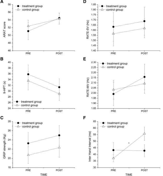
The active rehabilitation treatment and the passive mobilization protocol induced similar effects on unimanual motor performance. In-deed, on average, a statistically significant improvement as effect of TIME was found in ARAT score (F(1,56) = 5.53, p = 0.022), average time to complete the 9-HPT (F(1,56) = 27.59, pb 0.000001), GRIP strength (F(1,56) = 11.40, p = 0.0013), and RATE-MV (F(1,28) = 6.32, p = 0.018), while no change was observed in RATE-SV (F(1,28) = 0.56, p = 0.46). The similar trend between the groups was underlined by the lack of TIME × GROUP interaction (ARAT: F(1,56) = 0.44, p = 0.51; 9-HPT: F(1,56) = 0.0008, p = 0.98; GRIP: F(1,56) = 0.01, p = 0.92, RATE-SV: F(1,28) = 0.0003, p = 0.98; RATE-MV: F(1,28) = 1.39, p = 0.25). As we found no significant differ-ence in treatment-induced motor performance improvements between the two hands, the results are reported as an average on the data collect-ed for the two hands (Figs. 2A–E).
A significant change, as effect of TIME, was found in IHI after treat-ment (F(1,28) = 7.66, p = 0.001). However, differently from the other motor performance parameters, in the bimanual coordination task there was a significant difference between the treatment and the control group, as indicated by the significant interaction TIME × GROUP (IHI: F(1,28) = 8.40, p = 0.007) (Fig. 2F). In fact, IHI remained stable after the rehabilitation program in the treatment group, indicating a maintenance of the coordination abilities in the treated patients, but sig-nificantly increased in the control group (p = 0.002), demonstrating a worsening in bimanual coordination in these patients.
On the other hand, no patient showed any change in EDSS score after treatment. No patient had a relapse during the study.
Conventional MRI
We found that one patient belonging to the control group showed a new T2 lesion in the right cerebral peduncle (included in our mask of
Fig. 1. Regions of interest (ROIs) selected to investigate the microstructural integrity of the white matterfiber bundles connecting brain areas involved in voluntary movement control. (A) Corpus callosum (CC). (B) Left and right corticospinal tract (CST). (C) Left and right superior longitudinal fasciculus (SLF).
the corticospinal tract), while one patient belonging to the treatment group showed an enlarging T2 lesion in the anterior corpus callosum. DTI—signal-to-noise ratio
No significant change in SNR was observed at the POST session scan with respect to baseline (CC, PRE: 18.42 ± 4.02, POST: 18.75 ± 4.06; df = 29, t = 0.38, p = 0.71. CST, PRE: 18.56 ± 4.02, POST: 18.80 ± 4.90; df = 29, t = 0.31, p = 0.76. SLF, PRE: 18.63 ± 3.43, POST: 18.56 ± 3.28; df = 29, t = 0.14, p = 0.89). DTI-parameter maps of diffusion direction color-encoded FA, axial and radial diffusiv-ities for a representative subject are shown inFig. 3.
DTI—fractional anisotropy
At baseline, white matter structural integrity, evaluated by FA, was similar in the two groups for all the investigated ROIs (CC: F(1,28) = 0.051, p = 0.82; CST_left: F(1,28) = 0.62, p = 0.44; CST_right: F(1,28) = 0.81, p = 0.38; SLF_left: F(1,28) = 0.53, p = 0.47; SLF_right: F(1,28) = 2.20, p = 0.15).
After 2 months, FA values were found to be slightly but not signi fi-cantly different with respect to baseline (effect of TIME) in the CC and bi-laterally in the CST in the two groups of patients (CC: F(1,28) = 2.24, p = 0.15; CST: F(1,56) = 2.86, p = 0.096). However, the significant interaction TIME × GROUP (CC: F(1,28) = 5.12, p = 0.03; CST:
Fig. 2. Motor performance parameters (mean ± s. e.) measured at the different tests for the two groups (treatment and control) in the two sessions, before (PRE) and after (POST) the rehabilitation treatment. (A) Score obtained at the Action Research Arm Test (ARAT score). (B) Time to complete the nine Hole Peg Test (9-HPT). (C) Hand grip strength assessed with a dynamometer (GRIP strength). (D) Movement rate (i.e., number offinger taps per second) in the spontaneous velocity condition (RATE-SV). (E) Movement rate in the maximum velocity condition (RATE-MV). (F) Inter hand interval (IHI). Higher values indicate greater impairment in bimanual coordination. (A–C) The reported values are the average of the two hands. * indicates statistical significance.
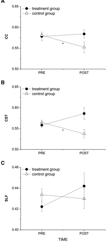
F(1,56) = 10.66, p = 0.0019) indicated that this trend was different be-tween the two groups. Post-hoc analysis showed that FA values in both the CC and CST after passive mobilization were significantly lower than those measured at baseline in the same group, indicating damage pro-gression in the control group (CC: p = 0.033; CST: p = 0.022). Con-versely, no significant change in the CC FA was observed in the treatment group; only a slight but not significant increase in the CST FA was found after the treatment (Figs. 4A and B). It should be noted that a similar effect of the treatment was observed on the left and right CST as shown by the lack of interaction TIME × GROUP × HEMISPHERE (F(1,56) = 0.025, p = 0.88). For this reason, data in the graph repre-sent the average on the left and right CST (Fig. 4B). Finally, no significant FA change with respect to baseline was observed in the SLF of both hemispheres in both groups indicating no effect of either passive mobi-lization or treatment on this brain structure (F(1,56) = 2.51, p = 0.12) (Fig. 4C).
DTI—axial diffusivity
At baseline,λ∥was similar in the two groups for all the investigated
ROIs (CC: F(1,28) = 0.58, p = 0.45; CST_left: F(1,28) = 0.93, p = 0.34; CST_right: F(1,28) = 0.26, p = 0.61; SLF_left: F(1,28) = 0.11, p = 0.74; SLF_right: F(1,28) = 0.02, p = 0.88).
No significant change in λ∥was observed in the investigated ROIs
after both the treatment and passive mobilization, as indicated by the lack of TIME effect (CC: F(1,28) = 0.09, p = 0.77; CST: F(1,56) = 0.07, p = 0.79; SLF: F(1,56) = 1.41, p = 0.24) and of interaction TIME × GROUP (CC: F(1,28) = 0.57, p = 0.46; CST: F(1,56) = 2.31, p = 0.13; SLF: F(1,56) = 0.005, p = 0.94) (Figs. 5A–C).
DTI—radial diffusivity
At baseline,λ⊥was similar in the two groups for all the investigated
ROIs (CC: F(1,28) = 0.23, p = 0.63; CST_left: F(1,28) = 0.01, p = 0.90; CST_right: F(1,28) = 0.14, p = 0.71; SLF_left: F(1,28) = 0.13, p = 0.72; SLF_right: F(1,28) = 0.02, p = 0.90).
On average, a significant effect of TIME on λ⊥was observed in the CC
(F(1,28) = 7.02, p = 0.01) while only a slight but not significant effect was observed in the CST (F(1,56) = 3.64, p = 0.06). However, a differ-ent trend between the two groups in the CC and CSTfiber bundles was indicated by the TIME × GROUP interaction (CC: F(1,28) = 7.33, p = 0.01; CST: F(1,56) = 6.19, p = 0.01). Post-hoc analysis showed a significant increase in λ⊥in the CC and CST in the control group after
2 months of passive mobilization (CC: p = 0.004; CST: p = 0.008) while no change was observed at POST in the treatment group with re-spect to baseline (CC: p = 0.97; CST: p = 0.91) (Figs. 5D and E).
Concerning the SLF, on average, a significant increase of λ⊥was
observed with respect to baseline (effect of TIME) (F(1,56) = 5.85, p = 0.02). Further, we found no significant TIME × GROUP interaction indicating a similar trend forλ⊥change between the two groups
(F(1,56) = 1.41, p = 0.24) (Fig. 5F).
DTI—mean diffusivity
At baseline, MD was similar in the two groups for all the investigated ROIs (CC: F(1,28) = 0.4, p = 0.53; CST_left: F(1,28) = 0.36, p = 0.56; CST_right: F(1,28) = 0.0026, p = 0.96; SLF_left: F(1,28) = 0.15, p = 0.70; SLF_right: F(1,28) = 0.26, p = 0.87).
Fig. 3. DTI-parameter maps of an axial slice at the level of the corpus callosum for a repre-sentative subject. (A) Diffusion direction color-encoded fractional anisotropy. (B) Axial diffusivity. (C) Radial diffusivity.
Fig. 4. Fractional anisotropy (FA) values (mean ± s. e.) within the selected ROIs in the two groups of patients (treatment and control) before (PRE) and after (POST) the rehabil-itation treatment. (A) Corpus callosum (CC). (B) Corticospinal tract (average on the left and right CST). (C) Superior longitudinal fasciculus (average on the left and right SLF). * indicates statistical significance.
No significant change in MD was observed after treatment in both groups (effect of TIME) (CC: F(1,28) = 2.60, p = 0.12; CST: F(1,56) = 2.33, p = 0.13; SLF: F(1,56) = 3.59, p = 0.07). This trend was similar in the two groups as indicated by the lack of TIME × GROUP interaction (CC: F(1,28) = 0.49, p = 0.49; CST: F(1,56) = 0.56, p = 0.46; SLF: F(1,56) = 0.32, p = 0.58).
Discussion
In this work we showed that a 2-month upper limb rehabilitation treatment including task-oriented exercises in patients with multiple sclerosis positively influenced motor behavior and impacted white mat-ter architecture. Indeed, following this treatment white matmat-ter integrity
Fig. 5. Axial (λ∥) and radial (λ⊥) diffusivity values (mean ± s. e.) within the selected ROIs in the two groups of patients (treatment and control) before (PRE) and after (POST) the
reha-bilitation treatment. (A and D) Corpus callosum (CC). (B and E) Corticospinal tract (average on the left and right CST). (C and F) Superior longitudinal fasciculus (average on the left and right SLF). * indicates statistical significance. Please note that * in F refers to a significant effect of TIME in both groups.
in the corpus callosum and corticospinal tracts was preserved, while it is generally affected by the disease course. In fact, in a control group of pa-tients receiving only passive limb mobilization we observed a micro-structural integrity worsening in thesefiber bundles, although these patients significantly improved some aspects of their motor behavior. Motor behavior
Both active and passive motor rehabilitation protocols induced an improvement in unimanual motor tasks. In details, the ARAT score sig-nificantly increased in both groups of PwMS; similarly, a significant im-provement was observed in hand dexterity (i.e., reduction of the time occurring to perform the 9-HPT) and in grip strength in both groups. Further,finger movement opposition rate at maximal velocity signifi-cantly increased in all patients.
However, it should be noted that the positive effects on motor be-havior obtained with both the rehabilitation protocols may have differ-ent explanations. One can refer to the stimulation of the upper limb proprioceptors and cutaneous receptors during both active and passive movements inducing a continuous updating and enhancement of the sensorimotor cortical representation of the treated limb. Indeed, even though passive mobilization is usually considered less robust than active motor tasks in rehabilitation, the involvement of M1 has been recently discussed also in passive protocols (Blatow et al., 2011). Fur-thermore, M1 has been shown to play an important role in the somatic perception of limb movements (Naito et al., 2002). Recently, it has been demonstrated that there is a contralateral M1 activation in a similar lo-cation with active and passive motor stimulation, but the passive task is sometimes associated with lower signals than the active one (Francis et al., 2009; Guzzetta et al., 2007; Reddy et al., 2001). Also, a more prom-inent increase in the activation of contralateral M1, corticospinal excit-ability and intracortical facilitation was found after training based on the performance of voluntary movements compared with passive train-ing (Lotze et al., 2003). From all thesefindings, we cannot exclude that although passive movements have a lower influence on the sensorimo-tor areas than the active ones they can, in the same way, induce a posi-tive training effect on motor behavior. Another explanation may deal with the effects of rehabilitation on muscle properties. At rest, the human muscle undergoes a significant progressive increase in stiffness (Hagbarth et al., 1985): in patients with upper limb motor impairment this increased stiffness can reduce the ability to correctly perform the motor tasks increasing the time occurring to accomplish the goal. It has been demonstrated that this stiffening can be reversed by active or passive movements (Lakie and Robson, 1988). The biophysical basis of this thixotropic process is likely to involve a long-term rearrange-ment of bonds between actin and myosin molecules and to be related to the presence of the short-range elastic component in the muscle (Hill, 1968). Therefore, thixotropy may explain the beneficial effects of limbering up before exercise and the efficacy of certain forms of physio-therapy, based on the repetition of passive or active movements, in the treatment of muscle stiffness (Lakie and Robson, 1988).
However, these two explanations are not contradictory and we could hypothesize that the observed effects can be due to a combination of the discussed processes.
Nevertheless, it should be considered that bimanual coordination was found to be preserved only in the treatment group while it wors-ened in the control group. In a previous work, we showed that IHI, an index of bimanual coordination, is significantly higher in PwMS than in healthy subjects indicating a deficit in bimanual coordination in these patients (Bonzano et al., 2008). In the present study, similar IHI values were observed in the two groups of patients at the time of enroll-ment revealing altered bimanual coordination in both the treatenroll-ment and the control groups. Yet, in the treatment group IHI remained stable after the 2 months of treatment while significantly increased in the con-trol group. Thisfinding indicates that when patients are asked to per-form high complexity tasks requiring the coordination of both limbs
the typology of the rehabilitation treatment becomes important. We can hypothesize that performing task-oriented exercises with one or two limbs, as those administered to the treatment group, can have pos-itive effects on motor behavior because they induce a higher activity of the brain areas involved in voluntary movements than passive move-ments, with a continuous exchange of sensorimotor information be-tween homologous areas of the two cortical hemispheres. This last process has been demonstrated to be crucial in both bimanual coordina-tion and the interhemispheric transfer of sensorimotor informacoordina-tion dur-ing motor task performance or motor traindur-ing (Bonzano et al., 2008, 2011a,b; Lenzi et al., 2007).
Regardless of treatment-induced changes in the upper limb motor behavior in both groups, no patient changed the baseline EDSS score after treatment. The lack of change in this score can be explained by the fact that EDSS is weighted toward the lower limb function (Kurtzke, 1983) and, independently of treatment, by the short observa-tion period (i.e., 2 months).
White matter integrity
After 2 months of upper limb passive mobilization (i.e., control group), FA values in both the CC and CST (left and right tracts) were sig-nificantly lower than those measured at baseline and λ⊥significantly
increased in both the CC and CST, indicating a WM damage progression in this group. On the other hand, no significant change in FA and λ⊥was
observed in the treatment group in both the CC and CST, indicating a preservation of these WMfiber bundles. Further, no significant change inλ∥and MD was observed in the investigated ROIs in the treatment
and the control groups.
The changes observed in FA andλ⊥, and not inλ∥and MD, in the
con-trol group might suggest that the WM damage progression in these pa-tients might be due to increased diffusivity of water molecules across WMfibers likely related to demyelination processes rather than to altered impedance of diffusivity along the tract as a consequence of ax-onal injury or loss (Budde et al., 2007; Nair et al., 2005; Song et al., 2003).
The bases of thesefindings can be derived from recent studies con-sidering the effects of motor training in healthy subjects and animal models. In healthy subjects, learning a novel skill may be mediated by a multi-stage process (Dayan and Cohen, 2011): a rapid skill learning, which is facilitated by an increase in spine density, and consolidation and slow learning phases over long periods of training, which can be mediated by changes in other cellular processes such as angiogenesis, myelination or axonal remodeling (Thomas and Baker, 2013). However, the nature of the structural changes may be strongly influenced by the type of training task and the neuroanatomical substrate. In PwMS, we showed that a rehabilitation treatment including task-oriented exer-cises can preserve WM microstructure and potentially induce a slight but not significant trend to improvement (i.e., FA increase in the CST, seeFig. 4B). We might assume that the same treatment in a group of healthy subjects can have a stronger impact on WM as it has been shown to occur after motor training (Scholz et al., 2009). However, Morgen et al. (2004)found that when training the motor functions in PwMS cortical reorganization of sensorimotor networks can occur, but on a lesser scale than in healthy subjects. The reduced cortical reorgani-zation and the progression of the disease can explain why we observed only a preservation of WM integrity and not a considerable improve-ment. Interestingly,Rossi et al. (2009)found that in mice with myelin oligodendrocyte glycoprotein-induced experimental autoimmune en-cephalomyelitis (EAE), a model of MS, exercise was able to contrast den-dritic spine loss induced by EAE in striatal neurons.
In general, the DTI measurements could reflect changes also within plaques (we did not create a lesion mask to exclude these areas from the DTI analysis, thus the DTI measurements can include both plaques and normal-appearing white matter). However, the only two patients developing an enlarging or a new T2 lesion in the analyzed ROIs after
treatment showed a trend in DTI parameters similar to the other patients belonging to the same group (treatment and control group, respectively). Indeed, it should be considered that T2 hyperintensities are not specific for the underlying pathological process, since inflamma-tion, demyelinainflamma-tion, gliosis, edema, and axonal loss may increase the signal intensity, without any specific pattern (Bruck et al., 1997).
Recently, an interesting longitudinal study based on DTI in MS (Harrison et al., 2011) showed significant changes in white matter structures over time and in particular in the corpus callosum. The FA changes found in this work are lower than those observed in our control group; however, the authors underlined, as possible confound in their analysis, that some patients taking disease-modifying drugs changed the therapy during the course of the study. Also, their inclusion criteria allowed MRI scans in all the patients who did not take corticosteroid within 30 days from the DTI evaluation. All these conditions might have affected the results by reducing the absolute change over time. Conversely, in our work, the two groups were matched for MS pheno-type, EDSS, disease duration, disease-modifying therapy and time from last relapse (seeTable 1). Further, although some patients were taking disease-modifying drugs all of them did not change therapy in the 3 months preceding the enrollment and there was no therapeutic change during the study. All the patients did not use corticosteroid since their last relapse, and this occurred more than 3 months before the study, as from inclusion criteria (indeed, 8 out of 15 patients of the control group had the last relapse more than 12 months before the beginning of the study). In addition, we should take into account that in the control group, as in the treatment group, there were secondary progressive PwMS who can have a progressive deterioration (Cassol et al., 2004) augmenting the averaged damage progression of the con-trol group.
Finally, we cannot exclude that passive limb mobilization could accelerate disease progression or negatively impact white matter in-tegrity. This might have important consequences in thefield of neurorehabilitation. Indeed, it has been already demonstrated that passive limb mobilizations and task-specific exercises have different effects on functional plasticity in the sensorimotor cortex (Hubbard et al., 2009) and it should be very interesting to better understand if this might differently impact the neural structures. Particularly, repetition alone, without usefulness or meaning in terms of function, could be not enough to produce increased motor cortical representa-tions; on the other hand task-specific training regimens could pro-duce cortical reorganization and associated, meaningful functional improvements (Bayona et al., 2005). Recently, in an elegant review Doron and Gazzaniga (2008)proposed this question:“Is the callosal microstructure shaped by the strategies of the brain, vice-versa, or does it result from interplay of the two?” We might assume that dur-ing a traindur-ing based on task-oriented exercises different non-motor neural pathways located in the frontal, parietal and posterior cortical areas are active. Therefore, in this condition the majority of the callosalfibers, and not only the sensorimotor ones as in a passive mobilization treatment, could have a role in allowing the interhemi-spheric communication and at the same time undergo structural plasticity processes.
Further, we demonstrated that the type of training task cannot have a general impact on brain architecture, as it is able to influence only spe-cific structures. Indeed, we did not find a significant positive effect of training on the SLF. On the other hand, we found a significant increase ofλ⊥in the SLF with respect to baseline in both groups, likely due to a
demyelination process as effect of the disease. The main reason for the lack of treatment effects on the SLF might deal with its involvement also in other functions related to cognitive processes (Bonzano et al., 2009; Genova et al., 2013), thus a combined rehabilitation approach in-cluding also cognitive domains might be more efficient on these fiber bundles. Thesefindings strongly support the idea that the beneficial ef-fects of a rehabilitation treatment are task-dependent and selective in their target. This last suggestion assumes relevant significance towards
the implementation of tailored rehabilitative approaches, according to which a personalized treatment should be defined for the single patient on the bases of the specific functional aspects to be rehabilitated and the brain structures damaged by the disease to be preserved.
Conclusions
The commonly adopted tests showed an improvement in motor per-formance in both the treatment and the control group. Conversely, bi-manual coordination and WM integrity in the corpus callosum and corticospinalfiber bundles, generally affected by the disease, were pre-served only in the treatment group. This result points out to the need to administer, when possible, a rehabilitation treatment based on volun-tary movements since it seems to be more efficient than passive mobi-lization. Further, we can make the hypothesis that life style and experiences might influence the clinical course of inflammatory neuro-degenerative diseases with effects on WM architecture, as occurs when PwMS undergo aerobic exercise training (Prakash et al., 2010).
Finally, we can also suggest that the choice of the outcomes to eval-uate the efficacy of a rehabilitation treatment is crucial. Indeed, diverse treatments can influence the neuromuscular system at different levels also activating, in some cases, compensatory mechanisms but showing similar changes in the evaluated outcomes. Therefore, we can propose that in neurorehabilitation, where a successful treatment has to in flu-ence both behavior and neural structures, it should be desirable to com-bine the analysis of behavioral data with the analysis of brain structure and function to assess more completely the effects of a treatment.
Acknowledgments
This work was supported by the Italian Multiple Sclerosis Foundation—FISM (project n. 2011/R/8).
Conflict of interest
The authors have no conflict of interest to disclose.
References
Ang, E.T., Gomez-Pinilla, F., 2007.Potential therapeutic effects of exercise to the brain. Curr. Med. Chem. 14, 2564–2571.
Basser, P.J., 1995.Inferring microstructural features and the physiological state of tissues from diffusion-weighted images. NMR Biomed. 8, 333–344.
Basser, P.J., Pierpaoli, C., 1996.Microstructural and physiological features of tissues eluci-dated by quantitative-diffusion-tensor MRI. J. Magn. Reson. B 111, 209–219.
Bayona, N.A., Bitensky, J., Salter, K., Teasell, R., 2005.The role of task-specific training in rehabilitation therapies. Top. Stroke Rehabil. 12, 58–65.
Blatow, M., Reinhardt, J., Riffel, K., Nennig, E., Wengenroth, M., Stippich, C., 2011.Clinical functional MRI of sensorimotor cortex using passive motor and sensory stimulation at 3 Tesla. J. Magn. Reson. Imaging 34, 429–437.
Bonzano, L., Tacchino, A., Roccatagliata, L., Abbruzzese, G., Mancardi, G.L., Bove, M., 2008.
Callosal contributions to simultaneous bimanualfinger movements. J. Neurosci. 28, 3227–3233.
Bonzano, L., Pardini, M., Mancardi, G.L., Pizzorno, M., Roccatagliata, L., 2009.Structural connectivity influences brain activation during PVSAT in multiple sclerosis. Neuroimage 44, 9–15.
Bonzano, L., Tacchino, A., Roccatagliata, L., Mancardi, G.L., Abbruzzese, G., Bove, M., 2011a.
Structural integrity of callosal midbody influences intermanual transfer in a motor reaction-time task. Hum. Brain Mapp. 32, 218–228.
Bonzano, L., Tacchino, A., Roccatagliata, L., Sormani, M.P., Mancardi, G.L., Bove, M., 2011b.
Impairment in explicit visuomotor sequence learning is related to loss of microstruc-tural integrity of the corpus callosum in multiple sclerosis patients with minimal dis-ability. Neuroimage 57, 495–501.
Bonzano, L., Sormani, M.P., Tacchino, A., Abate, L., Lapucci, C., Mancardi, G.L., Uccelli, A., Bove, M., 2013.Quantitative assessment offinger motor impairment in multiple scle-rosis. PLoS One 8, e65225.
Bruck, W., Bitsch, A., Kolenda, H., Bruck, Y., Stiefel, M., Lassmann, H., 1997.Inflammatory central nervous system demyelination: correlation of magnetic resonance imaging findings with lesion pathology. Ann. Neurol. 42, 783–793.
Budde, M.D., Kim, J.H., Liang, H.F., Schmidt, R.E., Russell, J.H., Cross, A.H., Song, S.K., 2007.
Toward accurate diagnosis of white matter pathology using diffusion tensor imaging. Magn. Reson. Med. 57, 688–695.
Cassol, E., Ranjeva, J.P., Ibarrola, D., Mekies, C., Manelfe, C., Clanet, M., Berry, I., 2004. Dif-fusion tensor imaging in multiple sclerosis: a tool for monitoring changes in normal-appearing white matter. Mult. Scler. 10, 188–196.
Compston, A., 2010. Aids to the investigation of peripheral nerve injuries. Medical Re-search Council: Nerve Injuries ReRe-search Committee. His Majesty's Stationery Office: 1942; pp. 48 (iii) and 74figures and 7 diagrams; with aids to the examination of the peripheral nervous system. By Michael O'Brien for the Guarantors of Brain. Saunders Elsevier: 2010; pp. [8] 64 and 94 Figures. Brain 133, 2838–2844. Cotman, C.W., Berchtold, N.C., 2002.Exercise: a behavioral intervention to enhance brain
health and plasticity. Trends Neurosci. 25, 295–301.
Cotman, C.W., Berchtold, N.C., Christie, L.A., 2007.Exercise builds brain health: key roles of growth factor cascades and inflammation. Trends Neurosci. 30, 464–472.
Dayan, E., Cohen, L.G., 2011.Neuroplasticity subserving motor skill learning. Neuron 72, 443–454.
Doron, K.W., Gazzaniga, M.S., 2008.Neuroimaging techniques offer new perspectives on callosal transfer and interhemispheric communication. Cortex 44, 1023–1029.
Draganski, B., May, A., 2008.Training-induced structural changes in the adult human brain. Behav. Brain Res. 192, 137–142.
Evangelou, N., Esiri, M.M., Smith, S., Palace, J., Matthews, P.M., 2000.Quantitative patho-logical evidence for axonal loss in normal appearing white matter in multiple sclero-sis. Ann. Neurol. 47, 391–395.
Ferguson, B., Matyszak, M.K., Esiri, M.M., Perry, V.H., 1997.Axonal damage in acute mul-tiple sclerosis lesions. Brain 120 (Pt 3), 393–399.
Fields, R.D., 2008.White matter in learning, cognition and psychiatric disorders. Trends Neurosci. 31, 361–370.
Fischer, J.S., Rudick, R.A., Cutter, G.R., Reingold, S.C., 1999.The Multiple Sclerosis Function-al Composite Measure (MSFC): an integrated approach to MS clinicFunction-al outcome as-sessment. National MS Society Clinical Outcomes Assessment Task Force. Mult. Scler. 5, 244–250.
Francis, S., Lin, X., Aboushoushah, S., White, T.P., Phillips, M., Bowtell, R., Constantinescu, C.S., 2009.fMRI analysis of active, passive and electrically stimulated ankle dorsiflexion. Neuroimage 44, 469–479.
Ge, Y., Law, M., Johnson, G., Herbert, J., Babb, J.S., Mannon, L.J., Grossman, R.I., 2004. Pref-erential occult injury of corpus callosum in multiple sclerosis measured by diffusion tensor imaging. J. Magn. Reson. Imaging 20, 1–7.
Ge, Y., Law, M., Grossman, R.I., 2005.Applications of diffusion tensor MR imaging in mul-tiple sclerosis. Ann. N. Y. Acad. Sci. 1064, 202–219.
Genova, H.M., DeLuca, J., Chiaravalloti, N., Wylie, G., 2013.The relationship between exec-utive functioning, processing speed, and white matter integrity in multiple sclerosis. J. Clin. Exp. Neuropsychol. 35, 631–641.
Guzzetta, A., Bonanni, P., Biagi, L., Tosetti, M., Montanaro, D., Guerrini, R., Cioni, G., 2007.
Reorganisation of the somatosensory system after early brain damage. Clin. Neurophysiol. 118, 1110–1121.
Hagbarth, K.E., Hagglund, J.V., Nordin, M., Wallin, E.U., 1985.Thixotropic behaviour of humanfinger flexor muscles with accompanying changes in spindle and reflex re-sponses to stretch. J. Physiol. 368, 323–342.
Harrison, D.M., Caffo, B.S., Shiee, N., Farrell, J.A., Bazin, P.L., Farrell, S.K., Ratchford, J.N., Calabresi, P.A., Reich, D.S., 2011.Longitudinal changes in diffusion tensor-based quan-titative MRI in multiple sclerosis. Neurology 76, 179–186.
Hill, D.K., 1968.Tension due to interaction between the slidingfilaments in resting striat-ed muscle. The effect of stimulation. J. Physiol. 199, 637–684.
Hubbard, I.J., Parsons, M.W., Neilson, C., Carey, L.M., 2009.Task-specific training: evidence for and translation to clinical practice. Occup. Ther. Int. 16, 175–189.
Jang, S.H., Hong, J.H., 2012.The anatomical characteristics of superior longitudinal fascicu-lus I in human brain: diffusion tensor tractography study. Neurosci. Lett. 506, 146–148.
Johansen-Berg, H., 2010.Behavioural relevance of variation in white matter microstruc-ture. Curr. Opin. Neurol. 23, 351–358.
Johansen-Berg, H., Scholz, J., Stagg, C.J., 2010.Relevance of structural brain connectivity to learning and recovery from stroke. Front. Syst. Neurosci. 4, 146.
Johansson, S., Ytterberg, C., Claesson, I.M., Lindberg, J., Hillert, J., Andersson, M., Widen Holmqvist, L., von Koch, L., 2007.High concurrent presence of disability in multiple sclerosis. Associations with perceived health. J. Neurol. 254, 767–773.
Koch, G., Cercignani, M., Pecchioli, C., Versace, V., Oliveri, M., Caltagirone, C., Rothwell, J., Bozzali, M., 2010.In vivo definition of parieto-motor connections involved in plan-ning of grasping movements. Neuroimage 51, 300–312.
Kramer, A.F., Erickson, K.I., 2007.Capitalizing on cortical plasticity: influence of physical activity on cognition and brain function. Trends Cogn. Sci. 11, 342–348.
Kurtzke, J.F., 1983.Rating neurologic impairment in multiple sclerosis: an expanded dis-ability status scale (EDSS). Neurology 33, 1444–1452.
Lakie, M., Robson, L.G., 1988.Thixotropic changes in human muscle stiffness and the ef-fects of fatigue. Q. J. Exp. Physiol. 73, 487–500.
Larson, E.B., Burnison, D.S., Brown, W.S., 2002.Callosal function in multiple sclerosis: bi-manual motor coordination. Cortex 38, 201–214.
Lenzi, D., Conte, A., Mainero, C., Frasca, V., Fubelli, F., Totaro, P., Caramia, F., Inghilleri, M., Pozzilli, C., Pantano, P., 2007.Effect of corpus callosum damage on ipsilateral motor
activation in patients with multiple sclerosis: a functional and anatomical study. Hum. Brain Mapp. 28, 636–644.
Lotze, M., Braun, C., Birbaumer, N., Anders, S., Cohen, L.G., 2003.Motor learning elicited by voluntary drive. Brain 126, 866–872.
Lyle, R.C., 1981.A performance test for assessment of upper limb function in physical re-habilitation treatment and research. Int. J. Rehabil. Res. 4, 483–492.
Morgen, K., Kadom, N., Sawaki, L., Tessitore, A., Ohayon, J., McFarland, H., Frank, J., Martin, R., Cohen, L.G., 2004.Training-dependent plasticity in patients with multiple sclero-sis. Brain 127, 2506–2517.
Mori, S., Wakana, S., Nagae-Poetscher, L.M., van Zijl, P.C.M., 2005.MRI Atlas of Human White Matter. Elsevier, Amsterdam.
Nair, G., Tanahashi, Y., Low, H.P., Billings-Gagliardi, S., Schwartz, W.J., Duong, T.Q., 2005.
Myelination and long diffusion times alter diffusion-tensor-imaging contrast in myelin-deficient shiverer mice. Neuroimage 28, 165–174.
Naito, E., Roland, P.E., Ehrsson, H.H., 2002.I feel my hand moving: a new role of the pri-mary motor cortex in somatic perception of limb movement. Neuron 36, 979–988.
Nelson, D.L., 1996.Therapeutic occupation: a definition. Am. J. Occup. Ther. 50, 775–782.
Pelletier, J., Habib, M., Brouchon, M., Poncet, M., Lyon-Caen, O., Salamon, G., Khalil, R., 1992.Interhemispheric transfer in multiple sclerosis. Morphofunctional correlations. Rev. Neurol. (Paris) 148, 672–679.
Pelletier, J., Habib, M., Lyon-Caen, O., Salamon, G., Poncet, M., Khalil, R., 1993.Functional and magnetic resonance imaging correlates of callosal involvement in multiple scle-rosis. Arch. Neurol. 50, 1077–1082.
Prakash, R.S., Snook, E.M., Motl, R.W., Kramer, A.F., 2010.Aerobicfitness is associated with gray matter volume and white matter integrity in multiple sclerosis. Brain Res. 1341, 41–51.
Price, R.R., Axel, L., Morgan, T., Newman, R., Perman, W., Schneiders, N., Selikson, M., Wood, M., Thomas, S.R., 1990.Quality assurance methods and phantoms for magnetic resonance imaging: report of AAPM nuclear magnetic resonance Task Group No. 1. Med. Phys. 17, 287–295.
Reddy, H., Floyer, A., Donaghy, M., Matthews, P.M., 2001.Altered cortical activation with finger movement after peripheral denervation: comparison of active and passive tasks. Exp. Brain Res. 138, 484–491.
Reich, D.S., Zackowski, K.M., Gordon-Lipkin, E.M., Smith, S.A., Chodkowski, B.A., Cutter, G.R., Calabresi, P.A., 2008.Corticospinal tract abnormalities are associated with weak-ness in multiple sclerosis. AJNR Am. J. Neuroradiol. 29, 333–339.
Rossi, S., Furlan, R., De Chiara, V., Musella, A., Lo Giudice, T., Mataluni, G., Cavasinni, F., Cantarella, C., Bernardi, G., Muzio, L., Martorana, A., Martino, G., Centonze, D., 2009.
Exercise attenuates the clinical, synaptic and dendritic abnormalities of experimental autoimmune encephalomyelitis. Neurobiol. Dis. 36, 51–59.
Scholz, J., Klein, M.C., Behrens, T.E., Johansen-Berg, H., 2009.Training induces changes in white-matter architecture. Nat. Neurosci. 12, 1370–1371.
Smith, S.M., Jenkinson, M., Woolrich, M.W., Beckmann, C.F., Behrens, T.E., Johansen-Berg, H., Bannister, P.R., De Luca, M., Drobnjak, I., Flitney, D.E., Niazy, R.K., Saunders, J., Vickers, J., Zhang, Y., De Stefano, N., Brady, J.M., Matthews, P.M., 2004.Advances in functional and structural MR image analysis and implementation as FSL. Neuroimage 23 (Suppl. 1), S208–S219.
Smith, S.M., Jenkinson, M., Johansen-Berg, H., Rueckert, D., Nichols, T.E., Mackay, C.E., Watkins, K.E., Ciccarelli, O., Cader, M.Z., Matthews, P.M., Behrens, T.E., 2006. Tract-based spatial statistics: voxelwise analysis of multi-subject diffusion data. Neuroimage 31, 1487–1505.
Solari, A., Filippini, G., Gasco, P., Colla, L., Salmaggi, A., La Mantia, L., Farinotti, M., Eoli, M., Mendozzi, L., 1999.Physical rehabilitation has a positive effect on disability in multi-ple sclerosis patients. Neurology 52, 57–62.
Song, S.K., Sun, S.W., Ramsbottom, M.J., Chang, C., Russell, J., Cross, A.H., 2002.
Dysmyelination revealed through MRI as increased radial (but unchanged axial) dif-fusion of water. Neuroimage 17, 1429–1436.
Song, S.K., Sun, S.W., Ju, W.K., Lin, S.J., Cross, A.H., Neufeld, A.H., 2003.Diffusion tensor im-aging detects and differentiates axon and myelin degeneration in mouse optic nerve after retinal ischemia. Neuroimage 20, 1714–1722.
Spooren, A.I., Timmermans, A.A., Seelen, H.A., 2012.Motor training programs of arm and hand in patients with MS according to different levels of the ICF: a systematic review. BMC Neurol. 12, 49.
Taubert, M., Draganski, B., Anwander, A., Muller, K., Horstmann, A., Villringer, A., Ragert, P., 2010.Dynamic properties of human brain structure: learning-related changes in cortical areas and associatedfiber connections. J. Neurosci. 30, 11670–11677.
Thomas, C., Baker, C.I., 2013.Teaching an adult brain new tricks: a critical review of evidence for training-dependent structural plasticity in humans. Neuroimage 73, 225–236.
van Waesberghe, J.H., Kamphorst, W., De Groot, C.J., van Walderveen, M.A., Castelijns, J.A., Ravid, R., Lycklama a Nijeholt, G.J., van der Valk, P., Polman, C.H., Thompson, A.J., Barkhof, F., 1999.Axonal loss in multiple sclerosis lesions: magnetic resonance imag-ing insights into substrates of disability. Ann. Neurol. 46, 747–754.
Yozbatiran, N., Baskurt, F., Baskurt, Z., Ozakbas, S., Idiman, E., 2006.Motor assessment of upper extremity function and its relation with fatigue, cognitive function and quality of life in multiple sclerosis patients. J. Neurol. Sci. 246, 117–122.