Department of Science and Technology for Agriculture, Forestry, Nature
and Energy (DAFNE)
XXVI Cycle of Doctorate in Plant Protection
In collaboration with INRA Institute of Avignon, France, Department of
Plant Pathology
Thesis:
Importance of environmental habitats as a reservoir of phytopathogenic
bacteria and their role in the evolution of pathogenicity traits
AGR/12
By:
Dott. Claudia Bartoli
Coordinator and Supervisor: Assistant supervisor
Prof. Leonardo Varvaro Dott. Cindy E. Morris
Signature Signature
Dott. Giorgio M. Balestra
Signature
Defended on: Monday 30 June 2014
Thesis Committee
Prof. Laura Mugnai
Dr. Elena Di Mattia
Prof. Kubilay Kurtulus Bastas
Referee
Prof. Matthieu Arlat
Prof. Gonçalo Almeida
Importance of environmental habitats as a reservoir of phytopathogenic
bacteria and their role in the evolution of pathogenicity traits
Claudia Bartoli
Thesis
Submitted in partial fulfilment of requirements for the degree of Doctor of Philosophy in Plant Protection at Tuscia University by the authority of the Rector Magnificus
Prof. Alessandro Ruggieri In the presence of the
Thesis committed appointed by the Doctorate Board To be defended in public
Fidelity is the faithful driver
Microbes depend on good housekeeping
They thrive, survive and flourish
Yet new forms appear
Confounding the scientist
Woe to the status quo
.
Chapter 2 - Methyl-directed mismatch repair: a mechanism underlying
emergence of pathogenic bacteria...7
Chapter 3 - A user’s guide to a data base of the diversity of Pseudomonas syringae and its application to classifying strains in this phylogenetic complex...25
Chapter 4 - The Pseudomonas viridiflava phylogroups in the P. syringae species complex are characterized by genetic variability and phenotypic plasticity of pathogenicity-related traits...59
Chapter 5 -A framework to gage the epidemic potential of plant pathogens in environmental reservoirs: the example of kiwifruit canker...95
Chapter 6 –Hypermutability in Pseudomonas viridiflava: programmed bet-hedging via antibiotic resistance, exopolysaccharide production and pathogenicity...144
Chapter 7 - General Conclusions………...175
Chapter 8 -English Summary……….181
Italian Summary………...183
Chapter
1
General Introduction
1
Pseudomonas syringae is a ubiquitous phyotopathogenic bacterium well adapted both to
plants and environmental habitats linked with the water cycle (Hirano and Upper, 2000; Morris et al., 2007, 2008). Since only the beginning of this century, 55 reports of disease outbreaks in 25 countries have been associated with P. syringae in 25 woody hosts, for example (Lamichhane et al., 2014). All these new disease are caused by new genomic P.
syringae lineages that after their occurrence rapidly spread worldwide causing plant diseases.
Examples of new emerging disease caused by P. syringae are the bacterial canker of kiwifruit and the bleeding canker of horse chestnut for what concern the woody plants. The kiwifruit bacterial canker is caused by what is currently called P. syringae pv. actinidiae (Psa). It was demonstrated that the current kiwifruit epidemic is caused by a clonal Psa population which occurred in all countries where kiwifruit is intensively grown (Chapman et al., 2012; McCann et al., 2013; Vanneste et al., 2013). Likewise, horse chestnut bleeding canker was reported to be caused by a clonal population of P. syringae pv. aesculi (Green et al., 2010). By contrast, there are examples of P. syringae diseases whereby more than one genetic lineage is associated. An example is the bacterial canker of hazelnut caused by two distantly related lineages of P. syringae pv. avelllanae (O’Brien et al., 2012). The authors reported that these lineages convergent the virulence phenotype but dramatically differ in terms of Type Three Secretion System effectors. It is likely that in P. syringae more than one evolutionary strategies exist whereby both clonal and convergent populations are able to colonize and cause diseases on plants.
The P. syringae genomic lineages responsible of the several diseases on both herbaceous and woody plants, that in the past were attributed to the pathovar name, are all grouped in the P.
syringae complex. The terminology complex emerged because of the wide diversity that
composes P. syringae. In particular, genomic analysis based on housekeeping genes of strains isolated from diseased crops demonstrated the existence of 7 phylogenetic groups (phylogroups) which comprise the so-called pathovars (Parkinson et al., 2011). Further studies on the ecology of P. syringae in different environmental habitats demonstrated that the diversity estimated in the agricultural context (in particular diseased crops) is only a small fraction of the whole diversity that characterize this bacterial species (Morris et al., 2008) In addition strains from environmental substrates not only form new genomic lineages but they are also characterized by pathogenic strains (Morris et al., 2010).
2
evolutionary mechanisms that drive the emergence of new pathogenic lines. Only recently, it has been demonstrated that P. syringae strains isolated from the environment are pathogenic to several plants (Morris et al., 2008, 2010). In addition, a recent study on strains isolated from environmental substrates and pathogenic to tomato, demonstrates that the environment could represent a reservoir of new pathogenic tomato strains (Montail et al., 2013). The authors showed that strains isolated from the environment share some of the effectors with the tomato pathogen, P. syringae pv. tomato. Because P. syringae has been isolated from all habitats related to the water cycle (Morris et al., 2007; 2008) suggests the role of the environment in the evolution of P. syringae strains that could represent new emerging pathogens. The frequency of emergence of diseases caused by P. syringae and the high diversity of this bacterium, armed with traits for pathogenicity, in the environment at large begs questions about the processes underlying pathogenic diversification. In this light, the objectives of my thesis were: i) to investigate the evolutionary mechanisms (at the molecular level) that can drive the emergence of P. syringae pathogenic lineages and, ii) to explore the role of the environment as a reservoir of pathogenic P. syringae strains. In particular, I focused on strains isolated from different environmental habitats (rain, snow, fresh water, litter, epilithic biofilms and wild and cultivated plants) that were genetically close to strains isolated from diseased kiwifruit. In Chapter 2, I reviewed the molecular mechanisms responsible for the evolution of bacterial pathogens in both human and plant pathogens. From the literature review it became apparent that a defective methyl-directed mismatch repair system (involved in DNA repair) drives the occurrence of horizontal gene transfer, pathoadaptive mutations and prophage integration in bacteria (Meyers and Bull, 2002). These molecular mechanisms are responsible for the evolution of pathogenicity determinants in bacteria. In Chapter 3, I present the results of a thorough study on the diversity (both at phenotypic and genetic level) of the P. syringae complex, based on a collection of 7000 strains of P. syringae. I participated in this research that was initiated in the laboratory at INRA well before my arrival. The collection was established by the MISTRAL team of INRA at Avignon, and includes strains isolated both from agricultural and non-agricultural habitats. This study revealed that the diversity of the P. syringae species complex was underestimated by only considering strains isolated from crops. In light of this, we asked how this diversity could influence the evolution of pathogenic strains.
3
syringae complex, my work focused mainly on the kiwifruit pathogens within several P. syringae phylogroups. I first performed an in-depth study on P. viridiflava (Chapter 4), the
causal agent of kiwifruit blossom blight (Balestra et al., 2008), which was previously reported to belong to phylogroup 7. However, here we showed that P. viridiflava is composed of two different phylogroups. In addition, two different types of Type Three Secretion System are present in P. viridiflava but they do not apparently affect the ability of strains to induce disease. Finally, we observed and characterized phase variation in P. viridiflava which influences phenotypes related to pathogenicity. The molecular basis of this phase variation is addressed in a subsequent chapter.
In Chaper 5, I present work on the investigation of the capacity of P. syringae strains from environmental habitats to cause canker of kiwifruit To date, three different kiwifruit outbreaks associated with different genetic lineages have occurred. Work has been conducted to examine the origin of some of these different outbreaks, but we wondered about the potential for new future outbreaks. Here, we tested the pathogenicity of environmental strains to cause disease to kiwifruit. The first step in addressing the question was to choose candidate strains. This was accomplished by identifying a marker linked to the pathogenicity of P.
syringae to woody plant species. We found several strains in the collection with this marker
and characterized their pathogenic potential for kiwifruit as well as for other woody plants and herbaceous plants as well. In addition to being pathogenic on a range of plant species, environmental strains could co-exist endophytically with epidemic strains thereby suggesting that they could be in favorable conditions to acquire genes for virulence via horizontal gene transfer. This allowed us to gage the potential for emergence of environmental strains and to contribute to the perspective of strategies for surveying for pre-emergent strains before they cause major epidemics.
Finally in Chapter 6, I present work on the role in pathogenecity of hypermutable variants produced by P. viridiflava strains. The formation of different phase variants in P. viridiflava seems to be a strategy for the regulation of pathogenicity and antibiotic resistance. In human pathogens, however, it was reported that hypermutable variants were due to the mutation of genes related to methyl-directed mismatch repair system. Surprisingly, in P. viridiflava the methyl-directed mismatch repair system does not seem to be involved in phase variation. This
4
mechanisms that could be targeted as a means of disease control.
References
Balestra, G. M., Mazzaglia, A., Quattrucci, A., Renzi, M., & Rossetti, A. (2009). Current status of bacterial canker spread on kiwifruit in Italy. Australasian Plant Disease Notes,
4, 34–36.
Balestra, G. M., Mazzaglia, A., & Rossetti, A. (2008). Outbreak of bacterial blossom blight caused by Pseudomonas viridiflava on Actinidia chinensis kiwifruit plants in Italy. Plant
Desease, 92, 707.
Chapman, J. R., Taylor, R. K., Weir, B. S., Romberg, M. K., Vanneste, J. L., Luck, J., & Alexander, B. J. R. (2012). Phylogenetic relationships among global populations of
Pseudomonas syringae pv. actinidiae. Phytopathology, 102, 1034–1044.
Green, S., Studholme, D. J., Laue, B. E., Dorati, F., Lovell, H., Arnold, D., Kamoun, S. (2010). Comparative genome analysis provides insights into the evolution and adaptation of Pseudomonas syringae pv. aesculi on Aesculus hippocastanum. PloS One, 5, e10224.
Hirano, S. S., & Upper, C. D. (2000). Bacteria in the leaf ecosystem with emphasis on
Pseudomonas syringae a pathogen, ice nucleus, and epiphyte. Microbiol Mol Biol Rev, 64, 624–653.
Koh, Y. J., & Nou, I. S. (2002). DNA markers for identification of Pseudomonas syringae pv.
actinidiae. Mol Cells, 13, 309–314.
Lamichhane, J. R., Varvaro, L., Parisi, L., Audergon, J.-M., & Morris, C. E. (2014). Disease and frost damage of woody plants caused by Pseudomonas syringae: seeing the forest for the trees. In Adv Agron, 126, 231-291.
McCann, H. C., Rikkerink, E. H. A., Bertels, F., Fiers, M., & Lu, A. et al. (2013). Genomic analysis of the kiwifruit pathogen Pseudomonas syringae pv. actinidiae provides insight into the origins of an emergent plant disease. PLoS Pathogens, 9, e100.
5
uncertain world. Trends Ecol Evol. doi:10.1016/S0169-5347(02)02633-2
Monteil, C. L., Cai, R., Liu, H., Mechan Llontop, M. E., Leman, S., Studholme, D. J.,
Vinatzer, B. A. (2013). Nonagricultural reservoirs contribute to emergence and evolution of Pseudomonas syringae crop pathogens. New Phytol, 199, 800–811.
Morris, C. E., Kinkel, L. L., Xiao, K., Prior, P., & Sands, D. C. (2007). Surprising niche for the plant pathogen Pseudomonas syringae. Infect Gen Evol, 7, 84–92.
Morris, C. E., Sands, D. C., Vinatzer, B. A., Glaux, C., Guilbaud, C., Buffière, A., Thompson, B. M. (2008). The life history of the plant pathogen Pseudomonas syringae is linked to the water cycle. The ISME J, 2, 321–34.
Morris, C. E., Sands, D. C., Vanneste, J. L., Montarry, J.Oakley, B., Guilbaud, C., Glaux, C (2010). Inferring the evolutionary history of the plant pathogen Pseudomonas syringae from its biogeography in headwaters of rivers in North America, Europe and New Zealand. mBio ,1, 00107-10.
Parkinson, N., Bryant, R., Bew, J., & Elphinstone, J. (2011). Rapid phylogenetic
identification of members of the Pseudomonas syringae species complex using the rpoD locus. Plant Pathology, 60, 338–344.
Scortichini, M. (1994). Occurrence of Pseudomonas syringe pv. actinidiae on kiwifruit in Italy. Plant Pathology, 43, 1035–1038.
Takikawa, Y., Serizawa Setsuo, I., Takeshi Tsuyumo, S., & Goto, M. (1989). Pseudomonas
syringae pv. actinidiae pv. nov.: the casual bacterium of canker of kiwifruit in japan. Ann Phytopathol Soc Japan, 55, 437–444.
Vanneste, J. L. (2013). Recent progress on detecting , understanding and controlling
Pseudomonas syringae pv . actinidiae: a short review. New Zeal Plant Prot, 177, 170–
6
(2013). Identification, virulence, and distribution of two biovars of Pseudomonas
Chapter
2
Methyl-directed mismatch repair: a
mechanism underlying emergence of
pathogenic bacteria
7
bacteria
Claudia Bartoli1,2 Odile Berge2, Benoît Moury2, Leonardo Varvaro1 & Cindy E. Morris2
1
Departement of Science and Technology for Agriculture, Forestry, Nature and Energy (DAFNE), Tuscia University, 01100 Viterbo, Italy
2
INRA, UR0407 Pathologie Végétale, F-84143 Montfavet cedex, France
Abstract
The evolution of non-pathogenic bacteria toward a pathogenic form is a process that involves the acquisition, loss or rearrangement of genomic determinants though different molecular mechanisms. A defective methyl-directed mismatch repair system (MMR) leads to increased mutation rate and makes bacteria more competent for horizontal gene transfer, genomic rearrangements and prophage integration. These molecular mechanisms regulate the acquisition of pathogenic determinants leading to emerging diseases. Environmental stresses enhance the number of DNA mismatches and contribute to emergence of new variants including new pathotypes. Understanding the molecular mechanisms responsible of the occurrence on new pathogenic bacteria could lead to better control of disease emergence.
Key words:
8
Emerging diseases, defined as infections that have newly appeared in a population or have existed but are rapidly increasing in incidence or geographic range [1], are serious threats to the economy of food production systems, medical systems and to human well-being in general. The emergence of new diseases is caused by a combination of ecological or environmental factors and socio-economic factors [2]. For example, several emerging bacterial pathogens come from human-modified environments such as the Shiga-toxin producing Escherichia coli O157:H7 that gained access to the food chain because of food production practices. The emergence of Legionella pneumophila was facilitated by air-conditioning systems [2]. With the oncoming climate changes, a large number of new infectious diseases have been reported. The occurrence of these diseases rose in correspondence to the climate anomalies that occurred during the 1990s [3]. The latter is especially true for diseases carried by vectors such as mosquitos that are particularly sensitive to meteorological conditions [3]. It was reported that in the last 40 years, 335 new infectious diseases of humans emerged and that the majority of these diseases are caused by multi-drug resistant bacteria [4]. On the other hand, the peak of new infectious diseases caused by bacteria occurred between 1980 and 1990 as a result of the influence of HIV/AIDS on susceptibility to infections [4]. New pathotypes also emerge among plant pathogens. For example, the phytopathogenic bacterium Pseudomonas syringae has been reported to be responsible for more than 55 new diseases of fruit, ornamental and forest trees in the last 10 years, most caused by different genetic lines of the bacterium [5]. The seeming continuous emergence of new epidemics raises questions about the origin of new genetic lines of pathogens and specifically about the evolutionary processes involved.
Among the five evolutionary forces, two of them (mutation and recombination or sexual reproduction) lead directly to changes in DNA sequences and provide the substance on which gene flow, genetic drift and natural selection act to permit tangible emergence [6]. Concerning evolution of traits underlying pathogenicity, the origin of the variation in DNA has been attributed to point mutations, horizontal gene transfer (HGT), pathoadaptive mutations and integration of bacteriophages into genomes (Table. 1). Here we summarize the role of these different mechanisms in emergence of bacteria causing diseases to plants, animals and humans and we highlight a point in common for the ensemble of these mechanisms – the need for error in the mechanisms of DNA repair. In particular, a defective methyl-directed mismatch repair system (MMR) that itself produces bacterial variability by facilitating the
9
mutations and for prophage integrations. All these molecular mechanisms can take place in the same bacterium and drive the evolution of bacterial pathogens (Fig. 1). Finally we also point out how the environmental context can modulate the occurrence of these molecular mechanisms and in particular how it can influence the expression of genes involved in MMR.
The role of defective methyl-directed mismatch repair in hypermutability and pathoadaptive mutation
Methyl-directed mismatch repair is a conserved system, from prokaryotes to eukaryotes, responsible for post-replicative repair by correcting base-base and insertion/deletion mismatches that escaped the proofreading of DNA polymerases [7] (Box 1). When MMR is defective, mutations are incorporated into the DNA of the replicating cell leading to variability. Hypermutability of bacterial cells, that is associated with the emergence of antibiotic resistant lines, is due to a defective MMR that abolishes their capacity to correct the replication mistakes leading to an increase of the mutation rate [8]. Hence, hypermutable bacteria control poorly DNA repair and they can accumulate mutations that could eventually be favourable or unfavourable for the emergence of new allelic forms of genes associated with pathogenicity. In E. coli, it has been demonstrated that mutations in over 20 genes can confer the hypermutable phenotype [9]. Alterations in mutS, mutL, mutY and mutH genes in the MMR system are known to be responsible for the hypermutable phenotype in natural, clinical and experimental conditions [10,11]. Mutants in mut genes are associated with antibiotic resistance in several bacterial species such as P. aeruginosa [11,12],
Staphylococcus aureus [13], E. coli [14], Neisseria meningitidis [15], Salmonella enterica
[16], Haemophilus influenza [17] and Streptococcus pneumoniae [18]. The resulting antibiotic resistant mutants are often less adapted in normal conditions compared to their non-mutant counterparts. But in presence of antibiotics they present a fitness advantage. Furthermore, resistant strains can acquire secondary mutations that help them to compensate the fitness cost even in antibiotic-free environments [19].
Hypermutable strains have been found more frequently in pathogenic bacterial populations than in commensal populations [14]. This observation could be explained by the fact that the hypermutable phenotype might accelerate the evolution of pathogenic strains by the acquisition of pathogenicity factors as well as antibiotic resistance that is reinforced by natural selection in medical environments [20]. However, Matic et al. [14] demonstrated that in 504
10
same as in the pathogenic strains, suggesting that further study on the mechanisms of hypermutability is needed.
Because of its role in the rapid occurrence of new mutations, a defective MMR is related to what is called pathoadaptive mutation comprising all the molecular mechanisms that lead to the evolution of virulence traits without horizontal gene transfer (HGT). In general, pathoadaptive mutation is a genetic mechanism that enhances bacterial virulence without HGT but by loss or modification of functions of pre-existing pathogenicity genes toward optimal fitness in new hosts [21]. Several authors proved that pathoadaptive mutation is strongly related to a defective MMR system. For example in E. coli, down-regulation of mut genes occurred during stationary phase and long-term periods of starvation leading to a greater rate of production of mutants under conditions of starvation [22]. In this light, the MMR apparatus could be down-regulated in stressful conditions thereby favouring the bacterium to adapt to a new environment or host.
Pathoadaptive bacterial lines could therefore show a disadvantage in the ancestral habitat since the mutations they carry might be favourable only in the new host. To overcome this, the pathoadapted pathogens could directly pass to a new host with the same niche characteristics as the former host and progressively become an obligate pathogen [21]. Pathoadaptive mutation is important for obligate pathogens in which the main molecular mechanisms to evolve their virulence determinants are mutation, recombination and transpositions that occur within the host during the infection [23].
An example of pathoadaptive mutation via point mutations is illustrated by P. aeruginosa. It was proposed that chronic cystic fibrosis is caused by strains of P. aeruginosa that over-express genes for production of alginate. The alginate exopolysaccharide allows P.
aeruginosa to evade the pulmonary clearance mechanisms thereby fostering lung infections.
Knockout mutations in mucA, a repressor of alginate biosynthesis, leads to the over-expression of the algU gene involved in alginate biosynthesis fostering P. aeruginosa lung colonization and biofilm formation [24]. Another recent study conducted over 38 years on cystic fibrosis patients demonstrated that P. aeruginosa accumulated mutations in genes involved in antibiotic resistance such as gyrA/B and rpoB, in which the amino acid changes confer resistance against fluoroquinolones and rifampicin [25]. The same authors found mutations in genes involved in synthesis of the cell envelope and gene regulation, and they also showed that the pathoadaptive mutation of the clonal DK2 strain was correlated with a
11 the infection time.
The occurrence of pathoadaptive mutations and other molecular evolutionary mechanisms such as HGT are not mutually exclusive and both mechanisms might occur in the same bacterial cell (Fig. 1). As discussed below, Y. pestis evolved from a less pathogenic Yersinia species by acquiring different genomic islands, but the pathogenicity of this bacterium was enhanced also by a point-mutation in the yopA gene. The knockout mutation of yopA, involved in neutrophil adhesion, led to Y. pestis being able to successfully evade host phagocytes. When the mutation was restored the pathogenicity of this bacterium was strongly reduced [26].
Another example of how pathoadaptive mutations, coupled to HGT, can drive evolution comes from the plant pathogen P. syringae. This bacterium acquired the Type Three Secretion System (T3SS) hopZ2 and hopZ3 genes via HGT while the hopZ1 effector gene, having a cysteine-protease activity, evolved via deletions and insertions into three functional forms (hopZ1a, hopZ1b and hopZc) and two non-functional forms [27]. The evolution of the three
hopZ1 homologous genes was driven by bacterial-host interactions in which the ancient hopZ1a form under selective pressure evolved toward a more effective form [27].
Pathoadaptive mutations can also involve deletion of genes or operons that are unfavourable for host colonization and thus for optimal fitness of the pathogen. A clear example is the complete deletion of the cadA gene, encoding the lysine decarboxylase enzyme in shiga-toxin producing E. coli strains that favoured the adherence of the bacterium to the human intestinal epithelium [28,29].
A defective MMR favours horizontal gene transfer: the rapid acquisition of new pathogenic determinants
In plants, animal and human bacterial pathogens MMR seems to be a major barrier to chromosomal gene transfer among different bacterial genomic lineages or species [30–33]. In fact, HGT is highly inhibited by mutS and mutL genes in E. coli under experimental conditions and those genes appeared to be acquired and lost different times during the evolution of this bacterial species enhancing the acquisition of foreign DNA [34]. One of the more emblematic examples of how the MMR system can dramatically influence genomic exchange between different species was described for E. coli and S. typhimurium. These bacterial species can not normally exchange DNA fragments, but mutations in mutS and mutL
12
we can suggest that the MMR system is responsible for the “species barrier” in bacteria. The role of mutS and recA genes in HGT was also recently demonstrated in the plant pathogen
Ralstonia solanacearum [35] in which the exchange of genetic elements has been
demonstrated during tomato infection [36]. It is also important to specify that after the acquisition of new genomic elements via HGT, rearrangements of the novel genes by mutation are an important requisite for aiding the integration of the element into the new host genome [37]. This observation highlights that defective MMR could play an important role not only in the first phases of HGT but also after the acquisition of the new genomic determinants.
Recently the metabolic cost of HGT has been assessed [37] leading to the suggestion that only a clear fitness advantage could compensate the metabolic cost linked to HGT. The rapid acquisition of virulence determinants that lead to higher reproduction rates is the main advantage of HGT in bacterial pathogens. Pathogenicity islands (PAIs) are one of the most impressive examples of HGT of virulence traits both in animals and plant pathogens [38]. The presence of cryptic genes, bacteriophage attachment sites and plasmids confirm that PAIs have spread among bacteria via HGT [39]. For example, the hrp/hrc cluster, that encodes for the T3SS in the genera Pseudomonas, Erwinia, Xanthomonas and Ralstonia showed the existence of two different hrp/hrc groups. The discrepancy between the distributions of these two groups in relationship with the phylogeny of the different bacterial species is strong evidence that the hrp/hrc cluster has been horizontally acquired [40].
HGT may lead to the formation of new emerging pathogens in a very short period compared with other mechanisms such as point mutations that could require thousands of years before the emergence of a new pathogen [41]. One impressive example of how a commensal bacterium became a pathogen through HGT comes from Pantoea agglomerans. The acquisition of a plasmid harbouring a hrp/hrc gene cluster, T3SS effectors and a gene cluster encoding for biosynthesis of indole-3-acetic acid, transformed the epiphytic P. agglomerans into a host-specific gall-forming pathogen of two different hosts, gypsophila and sugar beet [42]. Recently, it has been demonstrated that strains in the P. syringae phylogroup 10 horizontally acquired pathways for the production of syringomicin-like toxins. Given the few Type Three Secretion effectors present in this phylogroup, it is tempting to speculate that the toxin is involved in the capacity of strains to be pathogenic on Nicotiana benthamiana [43].
13
by acquiring virulence determinants. A recent example comes from the epidemic methicillin-resistant Staphylococcus aureus in which its emergence coincided with the acquisition, via HGT, of an arginine catabolic mobile element harbouring spermidine genes coding for proteins degrading the toxic polyamines produced in human skin. The early acquisition of this genomic element from S. epidermis provided an advantage to S. aureus during skin colonization, leading to its emergence and spread [44].
HGT can contribute to the evolution of a non-pathogenic environmental strain into a pathogenic one adapted to a given host. This is what occurred within the Yersinia genus in which environmental non-pathogenic strains, by acquiring the pCD1 plasmid carrying T3SS genes, became aggressive human pathogens. Subsequently, acquisition of several pathogenic traits led to the divergence of the three human Yersinia pathogens (Y. enterocolitica, Y.
pseudotuberculosis and Y. pestis) [45]. Y. pestis then further bifurcated from Y. pseudoturberculosis by acquiring different plasmids and a genomic island carrying
pathogenicity determinants that enhanced its virulence [46].
The role of phages in the evolution of bacterial pathogens and their link with MMR
Bacteriophages are viruses that infect almost all bacterial species including human bacterial pathogens [47] and phytopathogenic bacteria [48]. When a bacteriophage invades a host, two different cycles are possible: the lytic cycle or the lysogenic cycle that leads to the prophage state. The prophage state can cause bacteria can acquire new genomic elements (parts of or all of the prophage) some of which can be pathogenicity determinants [49]. A prophage can also provide a benefit to the bacterial host by increasing its fitness. For example, lambda E. coli lysogens are more fit than the non-lysogenic strains when grown under low carbon source conditions [50]. Also P1, P2 and Mu lysogen E. coli strains reproduce more rapidly than their non-lysogenic counterparts when they are growth in glucose-limited chemostas [51].
The demonstration that Corynebacterium diphtheriae contains bacteriophages encoding diphtheria toxin is the first study in which the role of phages in bacterial pathogenicity was demonstrated [52]. After that, several studies focusing on bacteriophages showed their role in driving the evolution of pathogenic determinants in bacteria.
The role of phages in the evolution of bacterial pathogens may involve acquisition of toxins that enhance the aggressiveness of a given bacterium, passage of PAIs or single T3SS genes and passage of genes different from T3SS effector or toxin genes that play an important role
14
of toxins include the bacteriophage CTXФ carrying the cholera toxin, a potent A-B-type enterotoxin, that has been shown to increase the aggressiveness of the current O1 and O139
Vibrio cholerae strains [53] and also the Shiga-like toxin carried by the 933J phage and
responsible for aggressiveness of E. coli strains causing food borne toxi-infections [54]. The passage of T3SS genes via temperate phage to pathogenic bacteria was reported in
Salmonella typhimurium, the casual agent of human and animal salmonellosis. S. typhimurin
required the Pathogenicity Island 1 SopE effector protein for efficient entry into cell hosts. Mirold et al; [55] demonstrated that the SopE effector was transferred in the genome of S.
typhimurin from a P2 temperate phage after lysogenic conversion of the phage.
Lipopolysaccharides (LPS) are known to be important virulence and colonization factors in bacterial pathogens. In Shigella flexneri the temperate bacteriophage Sf6 harbors genes that encode for LPS that are indispensable virulence and colonization factors in S. flexneri [56]. The S. flexneri/Sf6 model is a clear example of how phages can also harbour genes not related to T3SS or toxin production but are fundamental in the conversion of non-pathogenic into highly virulent strains responsible for global epidemics.
Integration of prophages into host DNA could be regulated by a “permissive” MMR system.
Streptococcus pyogenes controls the expression of the mutL gene by excision and
re-integration of a prophage in response to growth. During stationary growth, S. pyogenes integrates the prophage that blocks the expression of the mutL gene leading to a mutator phenotype [57].We could consider this as a regulatory system that S. pyogens uses for regulation of mutL expression, or we could also consider that mutL expression is down-regulated every time that the prophage integrates into the genome of its host. This could occur for several bacteria-prophage systems. However, the role of MMR in prophage integration needs to be further investigated.
Environmental stress exacerbates defective MMR
The emergence of new pathogenic variants can result from processes that do not necessarily involve direct interaction with the host. Abiotic and indirect biotic selection pressures can be important factors in the evolution of new pathogenic lines [58]. Furthermore, the environment is a reservoir of pathogens of both humans and plants [58]. However the environment can also enhance the occurrence of the genetic processes underlying changes in genomic traits. For example, reactive oxygen species (ROS) are a by-product of bacterial metabolism and they
15
stages of infection, ROS rapidly increase in the host and they contribute to pathogen clearance and to signalling cascades linked to inflammation and immune responses [59]. In humans it is known that several bacteria can also limit the production of ROS in the host thereby increasing the persistence of infections. The latter has been reported for Francisella
tularensis, Anaplasma phagocytophilum and Chlamydia trachimatis [59]. Also during the
early phase of plant infections, ROS are produced as a defence response against the microbes suggesting the universality of this defence system in both animals and plants [60]. During host infection reactive oxygen species can influence the expression of MMR genes and the conversion to a hypermutable phenotype, as described in P. aeruginosa [61]. In general, the efficiency of MMR can decrease under particular environmental conditions as a result of the decrease in the amount of MMR proteins. In E. coli for example, a treatment with DNA-damaging agents can saturate the MMR of the bacterium leading to the incapacity to correct mismatches [62]. In addition, high level nutritional conditions, starvation and nutrient limitations can also induce mutatgenesis leading to antibiotic resistance. In E. coli, stress-induced mutations have led to resistance to fluoroquinolone ofloxacin and β-lactame [63]. HGT is also induced under particular environmental conditions; in Bacillus subtilis, activation of genes for cell competence occurs only in high nutritional conditions while in Pseudomonas
stutzeri it occurs only in low nutritional environments as an adaptive strategy based on
acquisition of genomic determinants triggered by poor food supplies [64]. Recently, it has also been demonstrated that marine α-Proteobacteria isolated in a subtropical ocean are rich in genes that favour HGT in marine environments. High salinity seems to be an important requisite for HGT development in this group of bacteria [65] suggesting that marine habitats may represent a reservoir of genomic element exchanges. This could be of fundamental importance for bacterial pathogens, such as Vibrio cholerae, that have part of their cycle in sea waters. If the acquired genetic elements confer traits that are dual use factors for adaptations to general survival and for pathogenicity, then environmental selective pressures can inadvertently contribute to positive selection for pathogens [58].
Concluding remarks
The rapid emergence of new bacterial pathogens raises questions about the molecular mechanisms that regulate this emergence and about the reservoirs in which the new emerging pathogens reside. Here we describe how the molecular mechanisms leading to diversification
16
hence on the MMR system. The behaviour of the MMR system can be influenced by rather generic environmental conditions. Therefore, mutations, HGT and prophage integration could occur at different stages of bacterial life history in response to a range of habitats (Fig. 1). Knowing these molecular evolutionary mechanisms and the environmental forces that drive them could help in evaluating the probability of occurrence of new pathogenic bacteria. Furthermore, full characterization of the MMR system and other mechanisms directly involved in DNA repair could open new perspectives for mitigating emergence by inhibiting the molecular mechanisms that underlie diversification.
Acknowledgements
We thank the graphic artist Mr. Louis Wallet for drawing the schematic picture in this review. We also thank Prof. David C. Sands, Montana State University, for the fruitful discussions and for reading and editing the manuscript and Prof. David Baltrus, University of Arizona, for providing crucial references.
17
Evolutionary mechanism Brief description Consequences Examples References
Defective mismatch repair system
The bacterium is not able to repair the mistakes linked to DNA replication This state is called hypermutable • Fitness reduction • Antibiotic resistance • Flexibility in HGT and acquisition of foreign DNA •Rearrangement of new genes Pseudomonas aeruginosa, Staphylococcus aureus, Escherichia coli, Neisseria meningitidis, Salmonella enterica, Haemophilus influenza, Streptococcus pneumoniae, Ralstonia solanacearum [11,13,15– 18,20,35]
Horizontal gene transfer (HGT)
Transfer of genomic elements from a donor organism to a host organism. HGT is usually intra-specific but it can occur also between more distant taxa Acquisition of pathogenicity determinants or entire pathogenicity islands Pseudomonas spp, Ralstonia spp, Erwinia spp, Xanthomonas spp, Pantoea spp, Staphylococcus spp, Salmonella spp and Streptococcus spp [33,42,44,46 ,40,43] Pathoadaptive mutation A complex of mutations, loss and modification of pre-existing genes that leads to the emergence of a new pathogen without HGT • Fitness increase in a new host • Fitness penalty in the ancestral host/habitat • Enhancement of the functionality of the pathogenicity determinants Pseudomonas aeruginosa, Yersinia pestis, Pseudomonas syringae, Escherichia coli, [24–26,29] Bacteriophage infection Infection of phage followed by a lysogenic phase that leads to the formation of the prophage state. The prophage is integrated into the genome of the host leading to the acquisition of new genomic elements by the bacterium Acqusition of toxins, Type Three Secretion System effectors or other genes related to pathogenicity Escherichia coli, Corynebacterium diphtheriae, Vibrio cholerae, Salmonella typhimurium, Shigella flexneri [51,52,56,66 ,67]
18
The methyl-directed mismatch repair system
During DNA replication the proofreading exonuclease associated with the DNA polymerase is able to edit the mistakes made by the DNA polymerase and then to make a second attempt at correct synthesis. However mistakes can escape from the proofreading and they can be corrected just after the DNA strand duplication by the methyl-mismatch repair system (MMR). The latter increase the replication fidelity 50-1000-fold [68]. In
Escherichia coli the MMR system is composed by MutS, MutL and MutH proteins that can recognize the
hemi-methylated DNA. In fact the parent DNA strand can be distinguished from the daughter strand because the methylation of newly synthesized DNA does not occur immediately after the replication [68]. When the methyl directed mismatch is functional, MutS and MutL incise the unmethylated strand at a hemi-methylated GATC site (the signal for the mismatch repair proteins) and then the excision of that portion is made by MutH [68]. Then the DNA is repaired and the DNA polymerase III holoenzyme fills the gap created by the excision. Every time that the MMR system is defective because of spontaneous mutations in the genes that codify for the Mut proteins or because of DNA damages caused by external factors, the replication mistakes are undetected leading to mutations and DNA instability that is also favourable for a high level of DNA recombination.
MMR is considered to be a conserved system; in fact, genes encoding homologous of MutS and MutL have been identified in several eukaryotes, but no homologous of the MutH excision protein have been found in different organisms other than bacteria.
In bacteria, DNA instability caused by a defective MMR is correlated with mutations and recombination, while in humans; there is a direct link between mutant MMR genes and tumour cells. The latter is well demonstrated for colorectal cancer in which an increase in mutation rate was observed in the tumor cells [69]. A defective human MMR gives similar phenotypes as observed for bacteria but with some exceptions in cell behaviour. For example, in humans a defective MMR is related to tolerance to DNA damaging agents as it is for bacteria; however, in human cells tolerance to damage is also related to the apoptotic response of the cell. However, the common phenotype for both prokaryotic and eukaryotic cells carrying a defective MMR is the increased rate of mutation.
19
Figure 1. A putative evolutionary scenario regulated by a defective methyl-directed mismatch repair system.
1= A defective methyl-mismatch repair system (MMR), induced by environmental stresses such as reactive oxygen species or UV light, or spontaneously defective, induces mutations in a commensal bacterium, 2= the bacterial cell then become competent and can acquire pathogenicity determinants via horizontal gene transfer (HGT) or prophage integration, 3= the bacterial cell carrying the new pathogenicity determinant(s) can infect a new host and become a newly emerging pathogen 4= the new bacterial pathogen can have two different evolutionary destinies: 5= to become an obligate pathogen (an evolutionary dead end) or 6= to maintain a facultative life style, to pass into a new environment and acquire or lose pathogenicity determinants (mechanisms driven by a defective MMR system) and be transformed into a new emerging pathogen or in a more aggressive pathogen that infects the same host species (7). In each step in which the bacterium acquires, rearranges or loses pathogenicity determinants, a defective MMR is the main driver and it can be influenced by environmental stresses. In addition acquisition, or loss of genes related to pathogenicity can be under the selection of environmental pressures.
20
1 Morse, S.S. (1995) Factors in the emergence of infectious diseases. Emerg. Infect. Dis. 1, 7–15
2 Morens, D.M. et al. (2004) The challenge of emerging and re-emerging infectious diseases. Nature 430, 242–249
3 Epstein, P.R. (2001) Climate change and emerging infectious diseases. Microbes Infect. 3, 747–754
4 Jones, K.E. et al. (2008) Global trends in emerging infectious diseases. Nature 451, 990–993
5 Lamichhane, J.R. et al. (2014) Disease and frost damage of woody plants caused by
Pseudomonas syringae: seeing the forest for the trees. In Adv Agron 126, 231-291
6 McDonald, B.A. and Linde, C. (2002) Pathogen population genetics, evolutionary potential, and durable resistance. Annu. Rev. Phytopathol. 40, 349–379
7 Schofield, M.J. and Hsieh, P. (2003) DNA mismatch repair: molecular mechanisms and biological function. Annu. Rev. Microbiol. 57, 579–608
8 Meyers, L.A. and Bull, J.J. (2002)Fighting change with change: adaptive variation in an uncertain world. Trends in Ecol Evol, 17, 551–557
9 Horst, J.P. et al. (1999) Escherichia coli mutator genes. Trends Microbiol. 7, 29–36
10 Moxon, E.R. et al. (1994) Adaptive evolution of highly mutable loci in pathogenic bacteria. Curr. Biol. 4, 24–33
11 Oliver, A. et al. (2000) High frequency of hypermutable Pseudomonas aeruginosa in cystic fibrosis lung infection. Science. 288, 1251–1253
12 Ciofu, O. et al. (2010) Genetic adaptation of Pseudomonas aeruginosa during chronic lung infection of patients with cystic fibrosis: strong and weak mutators with
heterogeneous genetic backgrounds emerge in mucA and/or lasR mutants.
Microbiology 156, 1108–1119
13 Wang, S. et al. (2013) Hypermutable Staphylococcus aureus strains present at high frequency in subclinical bovine mastitis isolates are associated with the development of antibiotic resistance. Vet. Microbiol. 165, 410–415
14 Matic, I. et al. (1997) Highly variable mutation rates in commensal and pathogenic
Escherichia coli. Science. 277, 1833–1834
15 Richardson, A.R. et al. (2002) Mutator clones of Neisseria meningitidis in epidemic serogroup A disease. Proc. Natl. Acad. Sci. U. S. A. 99, 6103–6107
21
typhimurium LT2 by reversion of his alleles. Mutat. Res. 400, 89–97
17 Watson, M.E. et al. (2004) Hypermutable Haemophilus influenzae with mutations in mutS are found in cystic fibrosis sputum. Microbiology 150, 2947–2958
18 Del Campo, R. et al. (2005) Population structure, antimicrobial resistance, and mutation frequencies of Streptococcus pneumoniae isolates from cystic fibrosis patients. J. Clin. Microbiol. 43, 2207–2214
19 Lenski, R.E. (1998) Bacterial evolution and the cost of antibiotic resistance. Int.
Microbiol. 1, 265–270
20 Taddei, F. et al. (1997) To be a mutator, or how pathogenic and commensal bacteria can evolve rapidly. Trends Microbiol. 5, 427–428
21 Sokurenko, E. V et al. (1999) Pathoadaptive mutations: gene loss and variation in bacterial pathogens. Trends Microbiol. 7, 191–195
22 Feng, G. et al. (1996) Depletion of the cellular amounts of the MutS and MutH methyl-directed mismatch repair proteins in stationary-phase Escherichia coli K-12 cells. J.
Bacteriol. 178, 2388–2396
23 Levin, B.R. and Bull, J.J. (1994) Short-sighted evolution and the virulence of pathogenic microorganisms. Trends Microbiol. 2, 76–81
24 Boucher, J.C. et al. (1997) Mucoid Pseudomonas aeruginosa in cystic fibrosis: characterization of muc mutations in clinical isolates and analysis of clearance in a mouse model of respiratory infection. Infect. Immun. 65, 3838–3846
25 Marvig, R.L. et al. (2013) Genome analysis of a transmissible lineage of Pseudomonas
aeruginosa reveals pathoadaptive mutations and distinct evolutionary paths of
hypermutators. PLoS Genet. 9, e1003741
26 Rosqvist, R. et al. (1988) Increased virulence of Yersinia pseudotuberculosis by two independent mutations. Nature 334, 522–524
27 Ma, W. et al. (2006) Type III effector diversification via both pathoadaptation and horizontal transfer in response to a coevolutionary arms race. PLoS Genet. 2, e209
28 Torres, A.G. et al. (2005) Pathoadaptive mutation that mediates adherence of shiga toxin-producing Escherichia coli O111. Infect. Immun. 73, 4766–4776
29 Maurelli, A.T. et al. (1998) “Black holes” and bacterial pathogenicity: a large genomic deletion that enhances the virulence of Shigella spp. and enteroinvasive Escherichia
coli. Proc. Natl. Acad. Sci. 95, 3943–3948
22
Salmonella typhimurium is disrupted in mismatch-repair mutants. Nature 342, 396–401
32 Townsend, J.P. et al. (2003) Horizontal acquisition of divergent chromosomal DNA in bacteria: effects of mutator phenotypes. Genetics 164, 13–21
33 Bertolla, F. and Simonet, P. (1999) Horizontal gene transfers in the environment: natural transformation as a putative process for gene transfers between transgenic plants and microorganisms. Res Microbiol 150, 375–384
34 Denamur, E. et al. (2000) Evolutionary implications of the frequent horizontal transfer of mismatch repair genes. Cell 103, 711–721
35 Fall, S. et al. (2007) Horizontal gene transfer regulation in bacteria as a “spandrel” of DNA repair mechanisms. PLoS One 2, e1055
36 Bertolla, F. et al. (1999) During infection of its host, the plant pathogen Ralstonia
solanacearum naturally develops a state of competence and exchanges genetic
material. Mol. Plant-Microbe Interact. 12, 467–472
37 Baltrus, D. a (2013) Exploring the costs of horizontal gene transfer. Trends Ecol. Evol. 28, 489–495
38 Schmidt, H. and Hensel, M. (2004) Pathogenicity islands in bacterial pathogenesis.
Clin. Microbiol. Rev. 17, 14–56
39 Hacker, J. et al. (1997) Pathogenicity islands of virulent bacteria: structure, function and impact on microbial evolution. Mol. Microbiol. 23, 1089–1097
40 Alfano, J.R. and Collmer, A. (1997) The type III (Hrp) secretion pathway of plant pathogenic bacteria: trafficking harpins, Avr proteins, and death. J. Bacteriol. 179, 5655–5662
41 Stukenbrock, E.H. and McDonald, B.A. (2008) The origins of plant pathogens in agro-ecosystems. Annu. Rev. Phytopathol. 46, 75–100
42 Barash, I. and Manulis-Sasson, S. (2009) Recent evolution of bacterial pathogens: the gall-forming Pantoea agglomerans case. Annu. Rev. Phytopathol. 47, 133–152
43 Hockett, K.L. et al. (2013) Interactions between genome architecture and virulence genes in Pseudomonas syringae, strain CC1557 as a model. bioRxiv DOI:
10.1101/000869
44 Planet, P.J. et al. (2013) Emergence of the epidemic methicillin-resistant
Staphylococcus aureus strain USA300 coincides with horizontal transfer of the
arginine catabolic mobile element and speG-mediated adaptations for survival on skin.
23 bacterial pathogens. Nat. Rev. Microbiol. 1, 55–64
46 Zhou, D. et al. (2004) DNA microarray analysis of genome dynamics in Yersinia
pestis: insights into bacterial genome microevolution and niche adaptation. J. Bacteriol.
186, 5138–5146
47 Wommack, K.E. and Colwell, R.R. (2000) Virioplankton: viruses in aquatic ecosystems. Microbiol. Mol. Biol. Rev. 64, 69–114
48 Kidambi, S.P. et al. (1994) Evidence for phage-mediated gene transfer among
Pseudomonas aeruginosa strains on the phylloplane. Appl. Environ. Microbiol. 60,
496–500
49 Brüssow, H. et al. (2004) Phages and the evolution of bacterial pathogens: from genomic rearrangements to lysogenic conversion. Microbiol. Mol. Biol. Rev. 68, 560– 602
50 Lin, L. et al. (1977) Increased reproductive fitness of Escherichia coli lambda lysogens. J. Virol. 21, 554–559
51 Edlin, G. et al. (1977) Reproductive fitness of P1, P2, and Mu lysogens of Escherichia
coli. J. Virol. 21, 560–564
52 Freeman, V.J. (1951) Studies on the virulence of bacteriophage-infected strains of
Corynebacterium diphtheriae. J. Bacteriol 61, 675–688
53 Waldor, M.K. and Mekalanos, J.J. (1996) Lysogenic conversion by a filamentous phage encoding cholera toxin. Science 272, 1910–1914
54 Newland, J.W. et al. (1985) Cloning of Shiga-like toxin structural genes from a toxin converting phage of Escherichia coli. Science 230, 179–181
55 Mirold, S. et al. (1999) Isolation of a temperate bacteriophage encoding the type III effector protein SopE from an epidemic Salmonella typhimurium strain. Proc. Natl.
Acad. Sci. U. S. A. 96, 9845–9850
56 Clark, C.A. et al. (1991) The oac gene encoding a lipopolysaccharide O-antigen acetylase maps adjacent to the integrase-encoding gene on the genome of Shigella
flexneri bacteriophage Sf6. Gene 107, 43–52
57 Scott, J. et al. (2008) Phage-associated mutator phenotype in group A streptococcus. J.
Bacteriol. 190, 6290–6301
58 Morris, C.E. et al. (2009) Expanding the paradigms of plant pathogen life history and evolution of parasitic fitness beyond agricultural boundaries. PLoS Pathog. 5,
24
persistence and inflammation. Int. J. Mol. Sci. 12, 334–352
60 Torres, M.A. et al. (2006) Reactive oxygen species signaling in response to pathogens.
Plant Physiol. 141, 373–378
61 Torres-barceló, C. et al. (2013) A trade-off between oxidative stress resistance and DNA repair plays a role in the evolution of elevated mutation rates in bacteria . Proc.
R. Soc. B 280, 20130007
62 Cupples, C.G. and Miller, J.H. (1989) A set of lacZ mutations in Escherichia coli that allow rapid detection of each of the six base substitutions. Proc. Natl. Acad. Sci. U. S.
A. 86, 5345–5349
63 Poole, K. (2012) Stress responses as determinants of antimicrobial resistance in Gram-negative bacteria. Cell 20, 227–234
64 Bertolla, F. and Simonet, P. (1999) Horizontal gene transfers in the environment: natural transformation as a putative process for gene transfers between transgenic plants and microorganisms. Res Microbiol 150, 375–384
65 McDaniel, L.D. et al. (2012) Environmental factors influencing gene transfer agent (GTA) mediated transduction in the Subtropical Ocean. PLoS One 7, e43506
66 Waldor, M.K. and Mekalanos, J.J. (1996) Lysogenic conversion by a filamentous phage encoding cholera toxin. Science 272, 1910–1914
67 Mirold, S. et al. (1999) Isolation of a temperate bacteriophage encoding the type III effector protein SopE from an epidemic Salmonella typhimurium strain. Proc. Natl.
Acad. Sci. U. S. A. 96, 9845–9850
68 Li, G.-M. (2008) Mechanisms and functions of DNA mismatch repair. Cell Res. 18, 85–98
69 Fishel, R. et al. (1993) The human mutator gene homolog MSH2 and its association with hereditary nonpolyposis colon cancer. Cell 75, 1027–1038
Chapter
3
A user’s guide to a data base of the
diversity of Pseudomonas syringae and its
application to classifying strains in this
phylogenetic complex
25
A user’s guide to a data base of the diversity of Pseudomonas syringae and its application to classifying strains in this phylogenetic complex.
Odile Berge1, Caroline L. Monteil1, Claudia Bartoli1,2, Charlotte Chandeysson1, Caroline Guilbaud1, David C. Sands3. & Cindy E. Morris1,3*
1
INRA, UR0407 Pathologie Végétale, 84143 Montfavet, France 2
Department of Science and Technology for Agriculture, Forestry, Nature and Energy (DAFNE), Tuscia University, Viterbo, Italy
3
Department of Plant Sciences and Plant Pathology, Montana State University, Bozeman, MT 59717-3150, USA
*For correspondence: E-mail [email protected]; (+33) (0) 432722886; Fax (+33) (0) 432722841
26
ABSTRACT
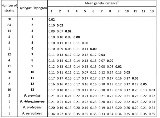
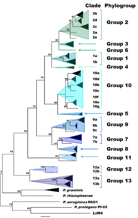
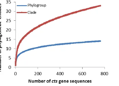
The Pseudomonas syringae complex is composed of numerous genetic lineages of strains from both agricultural and environmental habitats including habitats closely linked to the water cycle. The new insights from the discovery of this bacterial species in habitats outside of agricultural contexts per se have led to the revelation of a wide diversity of strains in this complex beyond what was known from agricultural contexts. Here, through Multi Locus Sequence Typing (MLST) of over 216 strains, we identified 23 clades within 13 phylogroups among which the seven previously described phylogroups were included. Robustness of phylogroups was shown by using core genome phylogeny on 29 strains representative of nine phylogroups. We show that phenotypic traits almost never provide a satisfactory means for classification of strains. We demonstrate that the citrate synthase (cts) housekeeping gene can accurately predict the phylogenetic affiliation for more than 97 % of strains tested and we propose a list of cts sequences to be used as a simple tool for quickly and precisely classifying new strains. Finally, our analysis leads to predictions about the diversity of P. syringae that is yet to be discovered. Nonetheless, we present here an expandable framework mainly based on cts genetic analysis into which more diversity can be integrated.
27
INTRODUCTION
Pseudomonas syringae was first reported as a plant pathogen of lilac by van Hall in 1902 [1].
Since its first description, P. syringae has become recognized as a phylogenetic complex of strains from terrestrial and aquatic habitats [2]. The classification of strains into the various sub-groups that constitute this complex has mirrored the historical trends in bacterial classification that were initially based on phenotypes (physiological and ecological characteristics) and then progressively were based on genotypes (DNA-DNA hybridization, phylogenetic analysis of housekeeping genes sequences) [3]. Commonly, seven phylogroups based on housekeeping gene phylogeny are recognized in the P. syringae complex [4] and some authors also include Pseudomonas cichorii a closely related phytopathogenic species [5, 6]. These seven groups are more or less consistent with the species or genomospecies described based on DNA-DNA hybridization [7, 8] such as P. savastanoi [7], P. viridiflava [9] and P. avellanae [10] the latter recently re-defined with more accurate genomic analysis [11]. As for many bacterial pathogens, the allocation of strains into pathovars is very common for the P. syringae group. Although the concept of pathovar is not related to phylogeny, these pathovars are frequently used as an analytical framework for classifications based on physiological phenotypes [12, 13], MLST (Multi Locus Sequence Typing) phylogeny [14–16] or DNA-DNA hybridization [7]. More recently, strains of P. syringae were isolated from contexts where they were saprophytes in a range of environmental substrates. For these strains, the concept of pathovar had no apparent relevance, especially as they sometimes represented phylogroups not previously described among the strains isolated from diseased plants [2, 17]. These discoveries raise questions about how to classify these strains that have not been resolved in a standardized way.
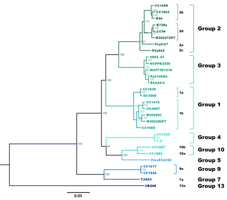
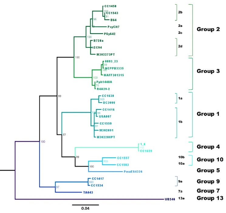
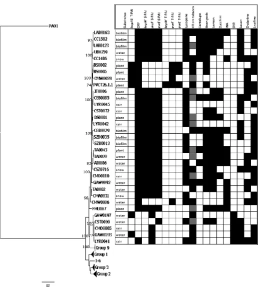
In light of the growing diversity of what is being called P. syringae and of the lack of a guide for homogenous classification and naming of strains, we were led to examine the validity of the biochemical indicators and to attempt to clarify the situation. Here we present the results of genotypic and phenotypic characterization of 764 strains of P. syringae collected from a wide range of habitats in which this bacterium has been described up to date. These strains were selected to represent the full breadth of the genetic diversity in a collection of over 1600 strains of P. syringae for which some phylogenetic information was available. Through phylogenetic analyses based on 4 housekeeping genes we defined 23 clades within 13 phylogroups. Robustness of phylogroups was shown through core genome phylogeny on 29
28
strains representative of 9 of the 13 phylogroups. Phenotypic characterization on 764 strains illustrated that phenotypic traits do not provide a satisfactory means for identification of strains at the clade or phylogroup level. We illustrate that the cts housekeeping gene alone can accurately predict the phylogenetic situation for most strains at the phylogroup and clade level. Overall, we describe the diversity of P. syringae and the utility of the data-base as a tool for classifying strains. Our analysis permits predictions about the diversity of P. syringae beyond what has been discovered and hence it provides a framework for future studies of the ecology of this bacterium.
MATERIALS AND METHODS
Bacterial strains
Most strains used in this study were taken from a collection of over 7000 strains of P.
syringae maintained at INRA in Montfavet. This collection was initiated in about 1995 and
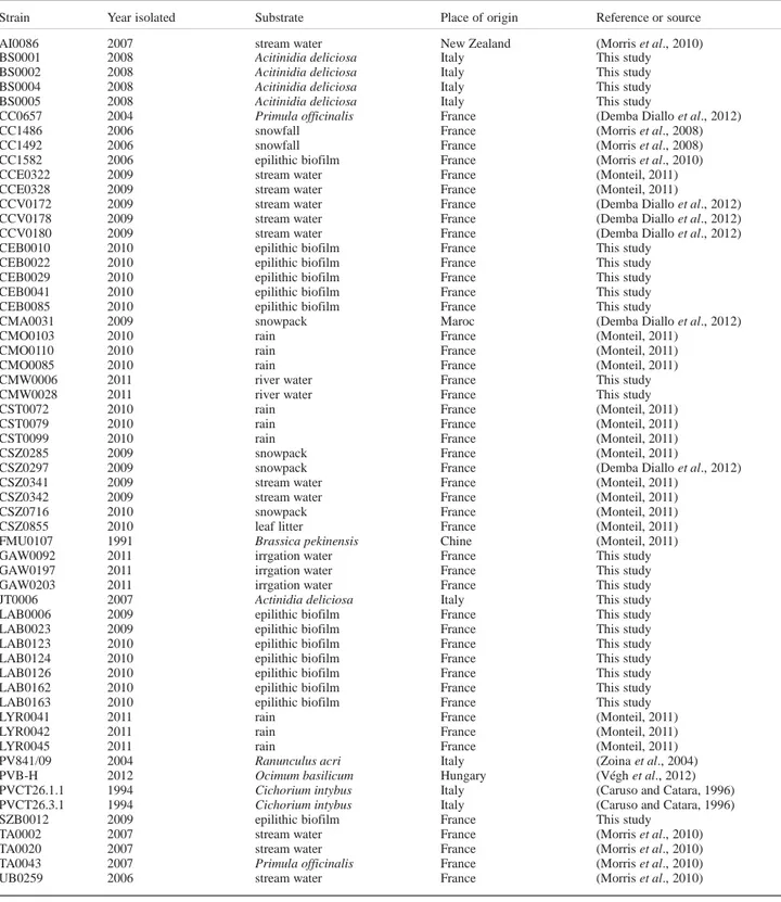
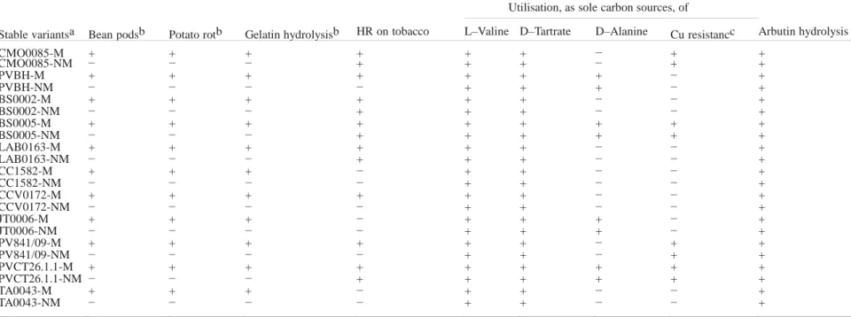
consists of strains collected from crops as well as different environmental habitats (rain, snow, epilithic biofilms, litter and water) via isolation on modified medium B of King (KBC) [18]. Some strains from crops were kindly provided by colleagues or obtained from public collections. All strains were purified before being stored at – 80° C in 40 % glycerol and they all lacked arginine dihydrolase and cytochrome c oxidase activity except for the P. cichorii reference strains that are oxidase positive. In addition, they varied in their production of fluorescent pigment on King’s medium B (KB) [19] and in induction of a hypersensitive reaction (HR) on tobacco. Over the past several years the housekeeping gene encoding for citrate synthase, cts (also named gltA) was sequenced for a subset of 1630 strains isolated from environmental habitats and crops [2, 20] during the exploratory work for our studies. Construction of a phylogenetic tree based on the 1630 cts sequences allowed us to select strains that represented the range of genetic diversity within each of the phylogroups and clades that could be delimited in the first-approximation analysis (unpublished data). This led us to select the 764 strains isolated from fresh water and epilithic biofilms (56 %), snowpack (16 %), plants (11 %), precipitation (9 %), and litter (8 %) that were subsequently characterized for 12 phenotypic traits typically used in characterization of P. syringae (see below) thereby allowing us to evaluate phenotypic diversity within different genetic groups. We selected voluntarily many non-plant derived strains to better describe the unknown
29
phylogenetic groups of the P. syringae complex. The total of 837 strains used in this study is listed in Table S1 with their origin, alternative name and characteristics. Among them 6 were reference strains chosen outside of the P. syringae group of strains mainly for phylogenetic analyses. The 831 remaining strains of P. syringae consisted of the 764 phenotyped strains and 67 strains from public data-bases used for their MLST profiles (see below) and not phenotyped here. From the 764 characterized strains, 149 were selected to cover the maximum variability observed for MLST. To this data-base of 149 MLST-typed (4 genes) strains we added the MLST profiles of the 67 strains from public data bases. The pooled set of 216 MLST profiles was used to construct more robust trees and to evaluate the reliability of phylogenetic predictions based on single housekeeping genes vs. combined gene sequences. The affiliation to phylogroups of the remaining 615 strains of P. syringae was based on cts sequence analysis. Finally, a set of 29 strains (Table S1) that represented the maximum diversity among the P. syringae genomes available in GenBank and chosen from the set of 216 MLST strains, was used to compare phylogenetic positioning of strains based on core genome sequences vs. that based on single and multiple housekeeping genes.
Genomic and phylogenetic analysis
MLST analysis was performed by sequencing four housekeeping genes: cts (encoding citrate synthase), gapA (encoding for glyceraldehyde-3-phosphate dehydrogenase A), rpoD (encoding for RNA polymerase sigma70 factor) and gyrB (encoding for gyrase B), using the Morris MLST schema of the Plant Associated and Environmental Microbes Database (PAMDB, http://genome.ppws.vt.edu/cgi-bin/MLST/home.pl) in combination with gapA and
gyrB of the Hwang PAMDB schema [16, 20]. For each locus, sequences were extracted from
GenBank and PAMDB and aligned with the P. syringae sequences by using DAMBE software version 5 [21] and they were cut to the same size (1859 bp for the concatenated sequences). In order to clarify the phylogenetic position of strain LzW4 isolated from Antarctica and misclassified as P. syringae [22], housekeeping gene sequences were obtained from its genome (accession number AOGS00000000). The concatenated sequences were used to construct the phylogeny with maximum likelihood and Bayesian methods by using the PHYLIP package version 3.6 (http://evolution.genetics.washington.edu/phylip.html) and Mr. Bayes version 3.1.2, respectively [23]. For maximum likelihood analysis, consensus trees were created from 100 independent phylogenies. Bayesian trees were constructed by using