Università degli Studi di Catania
Scuola Superiore di Catania
International PhD
in
Stem Cell Research
XXIII cycle
2007-2010
In vitro and in vivo inhibition of Chk1 sensitize
lung cancer stem cells to chemotherapy
Susanne Svensson
Coordinator and tutor
Tutor
Abstract
Development of resistance to radiation and chemotherapy turns the treatment of solid cancers into a therapeutic challenge. One of the most exciting breakthroughs being explored in cancer research today is the concept of cancer stem cells (CSCs). CSCs are a minority of cells within a tumor that are the source of tumor cell renewal and thereby determine the behavior of tumors, including proliferation, spreading and response to therapy. CSCs are highly resistant to conventional treatment and are therefore emerging as the preferred target of drug therapies in order to obtain eradication of tumors.
In this study, we examined the activation of the DNA damage response pathway in CSCs derived from non-small cell lung cancer (NSCLC-CSCs) and their differentiated counterparts after treatment with chemotherapeutic agents commonly used in clinic for lung cancer treatment. Our data show that NSCLC-CSCs possess a highly active DNA damage pathway compared to differentiated progeny and preferentially activate the checkpoint kinase Chk1 in response to DNA damage caused by chemotherapy. This indicates that Chk1 is most likely the main player of drug resistance in NSCLC-CSCs and its targeting might yield significant therapeutic gains. We demonstrate that chemical Chk1 inhibitors dramatically induce NSCLC-CSC death in vitro in combination with DNA damaging drugs. Cell death is induced through a premature activation of the cell cycle regulatory proteins Cdc2 and Cyclin B1, which in turn forces cells with damaged DNA to enter aberrant mitosis, a mechanism known as mitotic catastrophe. Moreover, our results indicate that final cell death occurs through apoptosis. Combination therapy studies have been successfully carried out also in vivo. Chk1 inhibition enhanced the antitumoral effect of conventional chemotherapy in mice xenograft tumor models by increasing tumor latency, potently abrogating tumor growth and reducing tumor mass. We also found a significant reduction of NSCLC-CSCs in xenograft-derived cells, confirming that combination treatment actually targets and reduces the NSCLC-CSCs compartment in vivo.
The importance of DNA repair as a resistance mechanism in cancer is a clinically relevant topic and we believe that the combination of selective Chk1 inhibitors with anti-cancer drugs could represent a new therapeutic approach for targeting NSCLC-CSCs and thereby for effective treatment of lung tumors.
Abbreviations
ABC ATP binding cassette
AC adenocarcinoma
AML acute myeloid leukaemia
Apaf-1 apoptotic protease-activating factor-1
ATM ataxia telangiectasia mutated
ATR ATM and Rad3 related
bFGF basic fibroblast growth factor
BSA bovine serum albumin
caspases cysteine-dependent aspartate-specific protease
CDK cyclin-dependent kinase
CEA carcinoembryonic antigen
Chk1 checkpoint kinase 1
Chk2 checkpoint kinase 2
CKs cytokeratins
CSC cancer stem cell
DDR DNA damage response
DMSO dimethyl sulfoxide
DSB DNA double-strand break
EGF epidermal growth factor
EGFR epidermal growth factor receptor
FBS fetal bovine serum
HLA human leukocyte antigen
HSP-90 heat shock protein-90
H&E hematoxylin and eosin
IC50 half maximal inhibitory concentration
LCC large cell carcinoma
LCSC lung cancer stem cell
NSCLC non-small cell lung cancer
NSCLC-CSC non-small cell lung cancer stem cell
PI propidium iodide
PI3K phosphatidylinositol-3-kinase
PS phosphatidylserine
Rb Retinoblastoma
SCC squamous cell carcinoma
SCLC small cell lung cancer
SFM serum-free medium
SHH sonic hedgehog
SP side population
Table of Contents
1. Introduction
7
1.1. Lung cancer 7
1.1.1. The human airway epithelium 7
1.1.2. General aspects of lung cancer 7
1.1.3. Risk factors 8
1.1.4. Symptoms of lung cancer 8
1.1.5. Diagnosis and staging of lung cancer 8
1.1.6. Metastasis from lung cancer 9
1.1.7. Genetic alterations associated with lung cancer 10
1.1.8. Treatment of lung cancer 10
1.2. Cancer stem cells 12
1.2.1. General aspects 12
1.2.2. Discovery of CSCs 13
1.2.3. Identification of CSCs 14
1.2.4. Signaling pathways in CSCs 15
1.2.5. Therapeutic implications of CSCs 16
1.2.6. Lung cancer stem cells 16
1.3. DNA damage and the DNA damage response pathway 18
1.3.1. General aspects 18
1.3.2. ATM and ATR 19
1.3.3. Chk1 and Chk2 20
1.4. Cell cycle checkpoints 21
1.4.1. General aspects 21
1.4.2. The G1 checkpoint 22
1.4.3. The S phase checkpoint 22
1.4.4. The G2 checkpoint 23
1.5. Cell Death 24
1.5.1. General aspects of cell death 24
1.5.2. Apoptosis 24
1.5.3. Necrosis 25
1.5.5. Mitotic catastrophe 25
1.6. Checkpoint inhibitors in cancer therapy 27
1.6.1. General aspects 27
1.6.2. Cell cycle abrogation as an anticancer strategy 27 1.6.3. Candidate targets for cell cycle abrogation 27
1.6.4. The Chk1 inhibitor UCN-01 29
1.6.5. The Chk1 inhibitor SB218078 29
1.6.6. The Chk1 inhibitor AZD7762 30
1.6.7. The Chk1 inhibitors XL-844 and PF-477738 30
2. Aims
32
3. Materials and Methods
33
3.1. Materials 33
3.1.1. Reagents 33
3.1.2. Antibodies 33
3.1.3. NSCLC-CSCs 34
3.2. Methods 34
3.2.1. Isolation and culture of NSCLC-CSCs 34
3.2.2. Differentiation of NSCLC-CSCs 35
3.2.3. Treatment 35
3.2.4. Cell viability assays 36
3.2.5. Cell proliferation assays 36
3.2.6. Cell cycle analysis 36
3.2.7. Immunofluorescence 36
3.2.8. Western blot 37
3.2.9. Propidium iodide / Annexin V staining 37
3.2.10. May-Grünwald-Giemsa staining 38
3.2.11. Soft agar colony forming assays
38
3.2.12. In vivo studies 38
4. Results
40
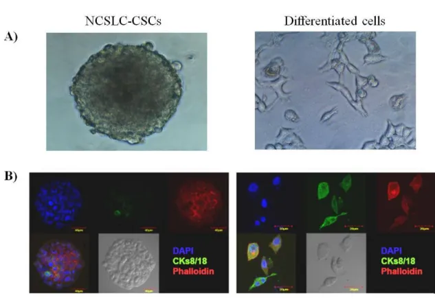
4.1. Isolation, culture and differentiation of NSCLC-CSCs 40
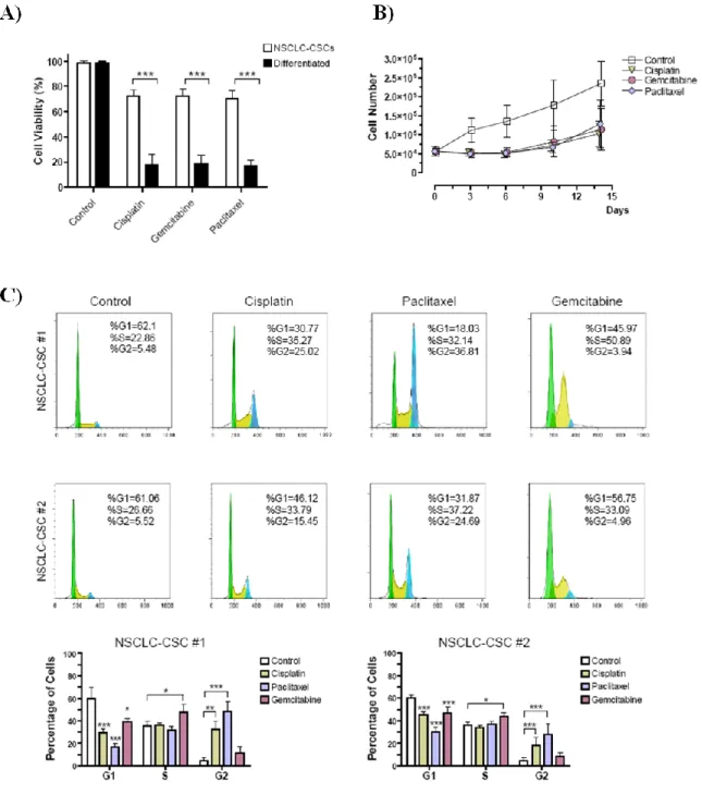
4.2. NSCLC-CSCs are resistant to chemotherapy and
efficiently repair chemotherapy-induced DNA damage 42
4.3. NSCLC-CSCs preferentially activate Chk1 46
4.4. Chk1 inhibitors prevent DNA repair and increase
the cytotoxicity of chemotherapy in NSCLC-CSCs 48
4.5. Chk1 inhibition induces premature activation of Cdc2/Cyclin B1 50
4.6. Mitotic catastrophe and cell death through apoptosis 52
4.7. Long term in vitro impact of Chk1 inhibition on NSCLC-CSCs 56
4.8. Combination of chemotherapy and Chk1 inhibitors
strongly affects tumor growth in vivo 58
5. Discussion
61
6. Publications and Posters
65
6.1. Publications 65
6.2. Presentations at scientific meetings 65
6.2.1. Poster presentations 65
6.2.2. Short talk 65
7. Acknowledgements
66
1. Introduction
1.1. Lung cancer
1.1.1. The human airway epithelium
The epithelium lining the human airways is important for gas transport and exchange, as well as regulation of host defence and reparation following tissue damage [1]. To accomplish these functions, the lung epithelium has evolved with different types of cells in different zones, such as basal mucous secretory cells of the trachea and bronchi, Clara cells of bronchioles, type 1 and type 2 pneumocytes of alveoli [1,2].
There are evidence supporting the presence of undifferentiated multipotent stem cells in each of the epithelial compartments of the lung and that these cells give rise to the mature differentiated cells of the lung [2-4].
1.1.2. General aspects of lung cancer
Lung cancer is one of the most common malignancies worldwide and a leading cause of cancer-related deaths [5-11]. Despite advances in lung cancer therapy, the average five-year survival rate is only 15% and early metastasis has become increasingly common [6,7]. The poor prognosis depends on late diagnosis and high rate of recurrence, which result in an advanced stage of cancer at the time of diagnosis, for which chemotherapy and radiotherapy have limited efficacy [11].
Lung cancer can be divided into two main subgroups that show major differences in histopathologic and genetic characteristics; small cell lung cancer (SCLC) and non-SCLC (Nnon-SCLC), where Nnon-SCLC accounts for more than 80% of all lung cancer cases [6,9-11]. NSCLC is further subdivided into squamous cell carcinoma (SCC), adenocarcinoma (AC) and large cell carcinoma (LCC) [6,7,9]. ACs are histologically heterogeneous peripheral masses that metastasize early, in many cases while the primary tumor is still a symptomless peripheral lesion [6,12]. SCCs are characterized by centrally located endobronchial masses, that unlike adenocarcinomas generally metastasize late in the disease course [6,12]. LCCs are poorly differentiated, usually large peripheral masses associated with early metastases [6,12]. SCLCs have a rapid
growth rate and are clinically aggressive. They are usually centrally located and are associated with early metastases. Despite their responsiveness to chemotherapy, small cell carcinomas often are advanced at the time of diagnosis, and patients have a poor prognosis [6,12].
1.1.3. Risk factors
Smoking is the predominant risk factor for all types of lung cancer. The effect of pipe and cigar use on the risk of lung cancer is similar to that of light cigarette smoking. Passive smoke exposure is also a risk factor and noteworthy is that passive smoking during childhood increases lung cancer risk in adulthood [6,9,10].
Environmental and occupational risk factors include exposure to asbestos, radon, arsenic, chromium, nickel, vinyl chloride, and ionizing radiation [6,10]. Lung cancer could also be a long-term effect of exposure to air pollution [10].
1.1.4. Symptoms of lung cancer
More than 90% of patients with lung cancer show symptoms at the time of diagnosis [13]. Most patients present with nonspecific symptoms, including fatigue, weakness and weight loss. A minority of patients present with direct signs and symptoms related to the primary tumor, to intrathoracic spread or to distant metastasis [6,13]. The most commonly presenting symptom caused by a primary lung tumor is cough. Other symptoms caused by the primary tumor include shortness of breath, hemoptysis (coughing up blood) and chest pain, which occurs in up to 50% of patients at diagnosis [6,13]. Intrathoracic spread of lung cancer, either by direct extension or lymphatic spread, produces a variety of symptoms and signs. These may be caused by involvement of the structures such as nerves, chest wall, vascular involvement and heart [13]. Approximately one third of patients present with symptoms as a result of distant metastases [13].
1.1.5. Diagnosis and staging of lung cancer
Diagnosis of lung cancer is based on methods such as chest radiograph, bronchoscopy, computer tomography scans and thoracotomy. Biopsy is often used to confirm the diagnosis of lung cancer and to identify the histological tumor type. The final
component of the diagnostic assessment is a functional evaluation of performance and pulmonary status of the patient [6].
Lung cancer staging, which can be determined based on the type of tumor identified and the presence or absence of metastatic disease, is an important factor affecting the possible treatment of lung cancer [6,9]. NSCLC is categorized using the TNM (tumor size – node involvement – metastasis status) staging system, whereas SCLC is categorized after a two-stage system defining tumors as being of limited stage or extensive stage [6].
1.1.6. Metastasis from lung cancer
The most common sites of distant metastasis from lung cancer are the bones, liver, brain, spinal cord, lymph nodes and skin.
The skeleton is one of the most common sites of metastasis in patients with lung cancer and the incidence of bone metastases is approximately 30-40% [14]. The primary symptoms resulting from metastatic bone disease include pain, pathologic fracture, vertebral malformation and spinal cord compression [13,14]. The prevention and treatment of bone metastases is mainly dependent on an effective treatment against the primary lung cancer tumor [14].
Liver metastasis occurs frequently with lung cancer and is associated with a very poor prognosis. Liver function is rarely abnormal until the metastases are numerous and large, which the give symptoms of weakness and weight loss [13].
Brain metastases occur in 10% of patients with lung cancer and are a significant cause of morbidity and mortality [13,15]. Spinal cord metastases are less common and tend to occur in patients with cerebral metastases [13].
NSCLC has a strong ability to metastasize to regional lymph nodes already at an early stages of tumor growth [16]. Detecting enlarged lymph nodes or subcutaneous nodules due to metastatic lung cancer is very helpful in facilitating both diagnosis and staging [13].
1.1.7. Genetic alterations associated with lung cancer
Lung cancers exhibit both common and type-specific genetic alterations. The profile of molecular and genetic alterations considerably differs between SCLC and NSCLC, as well as among the subtypes of NSCLC [9].
The most common genetic alteration occurs in p53, which is inactivated in nearly 50% of NSCLC and more than 70% of SCLC [9]. Activation of this tumor suppressor can be induced by carcinogenesis stresses or DNA damage, leading to the expression of downstream genes involved in cell cycle arrest, allowing DNA repair or initiation of apoptosis [17]. An essential effect of p53 inactivation that is significant in carcinogenesis is the avoidance of apoptosis and cell cycle arrest by neoplastic cells.
Rb gene alterations and protein loss are found in practically all SCLC, along with a normal p16/Ink4 gene and cyclin D1 protein expression. Rb mutations are infrequently detectable in NSCLC (15%). Instead, the Rb function is interrupted due to dysfunction of upstream components of the Rb pathway, either through p16/Ink4 gene silencing or by overexpression of cyclin D1 protein [18-20]. Rb protein is one of the critical regulators of the G1 to S phase cell cycle transition and exhibits a growth suppressive
function [20,21].
Activating mutations in epidermal growth factor receptor (EGFR) gene have been demonstrated in 40% to 80% of patients with NSCLC, and its overexpression correlates with a poor prognosis [10]. EGFR overexpression plays a significantly important role in SCC and AC carcinogenesis [22].
There are other less investigated genetic alterations that might contribute to lung cancer development or progression. K-ras gene mutations are rare in all lung cancers except AC, that show an alteration frequency of 30% and activation of PIK3 has been found in nearly half of SCC cases [9].
1.1.8. Treatment of lung cancer
Treatment of lung cancer differs according to the histologic type, the stage of the tumor and the patient’s functional evaluation [6]. Surgical resection remains the most reliable and successful option for cure of lung cancer patients [10]. Patients with resected lung
cancer have a high risk of relapse and adjuvant chemotherapy is standard after complete resection of lung cancer [6,10]. Treatment for unresectable lung cancer usually involve radiotherapy and chemotherapy [6]. Drugs commonly used for lung cancer treatment include cisplatin and carboplatin (alkylating agents that bind to and causes crosslinking of DNA), etoposide and irinotecan (topoisomerase inhibitors causing DNA strand breaks), docetaxel and paclitaxel (agents stabilizing microtubules and thereby inhibits mitosis), and gemcitabine (interferes with the nucleic acids during DNA replication and inhibits DNA synthesis) [12,23,24]. Although chemotherapy is appropriate for many patients with lung cancer, there are signs that the use of traditional chemotherapeutic agents has reached a therapeutic plateau and new therapeutic targets, such as signal transduction and angiogenesis pathways, are necessary [10].
1.2. Cancer stem cells
1.2.1. General aspects
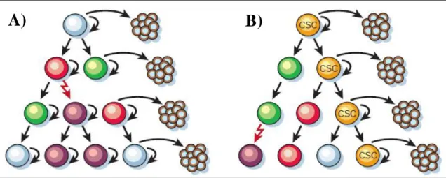
Solid tumors are composed of a heterogeneous population of cells with different proliferative, differentiative and tumor initiating potential [25]. To explain the heterogeneity of cells observed in cancer, two models of tumor cell proliferation and tumor expansion have been proposed; the stochastic model and the stem cell model (Figure 1) [26,27]. In the stochastic model, every cell has the same probability to proliferate extensively and to cause development and progression of malignancy. According to the stem cell model, only a small fraction of cells, the cancer stem cells (CSCs), have the potential to proliferate unlimitedly and to form tumors [26,27].
A)
B)
A)
B)
Figure 1. Two models of heterogeneity in solid tumors. Cancer cells are heterogenous in both models.
In the stochastich model (A), all phenotypes have the same potential to proliferate, while in the stem cell model (B), only the CSCs have the ability to proliferate extensively and form new tumors [26].
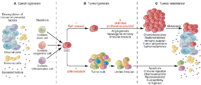
CSCs (or cancer initiating cells) are a rare population of undifferentiated tumorigenic cells responsible for tumor initiation, maintenance, spreading and therapy resistance [7,9,26,28]. These cells display unlimited proliferative potential, ability to self-renew and capacity to generate a progeny of terminally differentiated cells that constitute the major tumor population (Figure 2) [7,11,25,29-32]. Within an established tumor, only the CSCs have the capacity to reproduce the original tumor when transplanted into immunodeficient mice [7,9,11,25,26,30,31,33,34].
Figure 2. CSCs and their implication for carcinogenesis, tumorigenesis, and tumor resistance. A.
Tumors can arise from somatic cells through genetic mutations of cancer-critical genes and microenvironmental factors can contribute to the carcinogenic process. B. CSCs are capable of driving tumorigenesis through: (i) their ability for long-term self-renewal, (ii) their capacity to differentiate into tumor bulk populations, and (iii) their unlimited potential for proliferation and tumorigenic growth. C. CSCs exhibit increased resistance to chemotherapeutic agents and most likely drive neoplastic progression, tumor recurrence, and metastasis [35].
The origin of CSCs is still unknown, they could arise from somatic stem cells that acquire tumorigenic properties, or they could evolve from more differentiated progenitor cells that transform and acquire stem cell-like properties [27,36,37]. Stem cells have the self-renewing machinery already activated and the fact that CSCs and normal stem cells share many signalling pathways strengthens the hypothesis of a stem cell origin of cancer [26,27,29].
1.2.2. Discovery of CSCs
The existence of CSCs was first proven in the context of acute myeloid leukaemia (AML). It was shown that only a small subset of human AML cells, phenotypically similar to normal haematopoietic stem cells, were clonogenic in culture and capable of forming AML when transplanted into immunodeficient mice [27,38,39].
Since the initial discovery of CSCs in AML, subpopulations of tumor cells with stem cell-like characteristics have also been identified and expanded from several solid tumors, including breast [33], brain [40-43], melanoma [44,45], prostate [46,47], head and neck [48], pancreas [49,50], colon [51,52] and lung [7].
1.2.3. Identification of CSCs
The CSCs comprise a very small fraction of the entire tumor cell population and it is necessary to use specific markers to distinguish CSCs from the bulk of more differentiated tumor cells [11,25,30,37].
The use of cell surface markers for identification of CSCs has been widely investigated in different cancer types (Table 1). Several studies have demonstrated that CD133 (prominin-1) is an important marker of CSCs and it has been successfully used to identify CSCs in brain tumors [42], colon carcinomas [51,52], pancreatic cancer [49] and lung cancer [7]. Al-Haaj et al.[33] demonstrated that the CD44+CD24-/low cell population in breast cancer is significantly enriched in cancer-initiating cells. In AML, CD34+ and CD38- cells have highly enhanced tumor initiating potential [38,39].
Table 1. Cell surface markers for identification of CSCs [37].
Another functional approach used to distinguish CSCs within a population is by using Hoechst 33342. Uptake of the dye occurs universally in all cells, but efflux occurs only in specific cells. Cells with capacity to efflux the dye were first identified in the mouse bone marrow and they were termed side population (SP) cells as they fell to the side of the majority of positively stained cells in a FACS analysis [53]. SP cells have been identified in for example, skin, lung, liver, heart, brain and mammary gland and in
normal tissues the SP cells express high levels of stem cell genes and possess differentiation potential [25]. The Hoechst efflux requires the expression of the ABCG2 gene, a member ATP binding cassette (ABC) transporter superfamily [25,54].
Recently, Jiang et al. [55] reported that ALDH1-positive lung cancer cells exhibited properties consistent with those of tumor stem cells, including high ability of proliferation, self-renewal and differentiation, resistance to chemotherapy as well as expression of the CSC surface marker CD133. They also demonstrate that ALDH1-positive cells were able to induce tumor growth in vivo.
The selection of candidate markers for identifying CSCs can be very difficult, especially in the case of solid tumors where the normal tissue developmental hierarchy has not been characterized. A widely accepted assay to validate a candidate CSC population is by tumor initiation and serial transplantation in immunocompromised mice [37,56]. However, it is essential to confirm that these xenografts are phenotypically identical to patient tumors [56].
1.2.4. Signaling pathways in CSCs
Important signaling pathways that regulate self-renewal, proliferation and differentiation in stem cells include Bmi-1, Sonic Hedgehog (SHH), Notch and Wnt/β-catenin [28,57-59]. Involved in the process of self-renewal and proliferation is also the tumor suppressors, such as p53, PTEN, INK4A and ARF, that can inhibit proliferation, block self-renewal and regulate cell responses to DNA damage [28,58,60-62].
Signaling pathways regulating self-renewal of CSCs are poorly characterized, but based on the similarities between normal stem cells and CSCs and on the fact that signaling pathways often are deregulated in cells undergoing neoplastic transformation, the Bmi-1, SHH, Notch and Wnt/β-catenin pathways are thought to be implicated also in CSC regulation [28,59].
A few studies concerning signaling pathways activated in CSCs from solid tumors have been performed. Malanchi et al. [63] reported that Wnt/β-catenin signaling is essential to sustaining the CSC phenotype and to maintain skin tumorigenesis. It has been demonstrated that Wnt signaling activity is implicated in colon CSCs [64] and that
overexpression of Bmi-1 and inappropriate activation of SHH pathway might lead to medulloblastoma development [65].
1.2.5. Therapeutic implications of CSCs
CSCs are thought to be more resistant to chemo- and radiotherapy than the committed differentiated cells of the tumor population [26,28,31,66]. The difficulty in eradicating solid tumors may be due to the fact that existing therapies target the bulk of the tumor cells, while they fail to kill CSCs effectively [25,26,67]. The remaining CSCs could quickly allow regrowth of the tumor [26,31,37,66,67]. It has actually been shown that ionizing radiation treatment of human glioma cultures and glioma xenografts enriches the CD133+ CSC population [5,40].
Several properities of CSCs contribute to their resistance to chemotherapy. Firstly, CSCs are often found in a quiescent state or have a very low proliferation rate and are hence resistant to cell cycle specific chemotherapeutic agents, as well as chemotherapy that affect proliferating cells [5,68-70]. Secondly, stem cells are able to efficiently repair DNA damage, hence being more resistant to radiation and chemotherapy, including DNA damaging agents such as alkylating agents [40,68,71]. Thirdly, CSCs have impaired apoptosis pathway and express higher levels of antiapoptotic proteins, such as Bcl-2, compared to differentiated cells [28,32,68,72,73]. Finally, CSCs have high expression levels of drux efflux transporter proteins, such as p-glycoprotein, ABCG2 and other members of the ABC superfamily [5,66,74-77].
1.2.6. Lung cancer stem cells
Eramo et al. [7] identified lung cancer stem cells (LCSCs) as a rare population of CD133+ cells in both SCLC (SCLC-CSCs) and NSCLC (NSCLC-CSCs). LCSCs could be unlimitedly expanded and maintained in vitro as tumor spheres, while differentiated tumor cells lost the CD133 antigen and were only able to proliferate for 4 weeks before declining in number.
CD133+ lung cells showed higher tumorigenic potential than their CD133-counterparts and were able to generate tumor xenografts in immunocompromised mice, with morphological and immunohistological features closely resembling the original tumor [5,7]. LCSCs were shown to be resistant to chemotherapeutic drugs like cisplatin,
etoposide, paclitaxel and gemcitabine [5,7], which can explain the poor therapeutic effect of conventional chemotherapy on lung cancer patients. Even if tumor burden can be reduced by chemotherapy, the CD133+ tumor cells might be spared and enriched [5].
An increased expression level of genes involved in maintenance of stemness, like Oct3/4 and Nanog, was observed in CD133+ LCSCs compared with the CD133- counterpart [5,7,74].
1.3. DNA damage and the DNA damage response pathway
1.3.1. General aspects
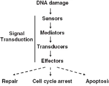
All cells are constantly under assault of different forms of DNA lesions. The damage can be classified as either endogenous (normal metabolic processes) or exogenous damage (environmental factors) like radiation, chemicals, UV, chemotherapy [78]. Unrepaired DNA damage can impede a cells ability to carry out its functions and may result in mutations, increased likelihood of tumor formation or even cell death [79]. Maintenance of genome stability depends on the appropriate response to DNA damage and cells have therefore evolved complex signaling networks, that coordinates cell cycle transitions, DNA replication, DNA repair and apoptosis [79-84]. In response to DNA damage, the DNA damage response (DDR) pathway is activated. The signaling pathways activated during DDR have three aims: (1) to block the cell cycle progression and cell division; (2) to increase accessibility to the damaged sites to the DNA repair machinery and (3) to induce apoptosis to cells with damaged DNA that cannot be repaired [85]. The DDR pathway consists of three main components; sensor proteins that recognize damaged DNA, signal transducers that relay and amplify the damage signal (ATR and ATM) and finally, the effectors that regulate the commitments of the cell (Chk1, Chk2 and p53) (Figure 3) [79,84,86].
Figure 3. Organization of the DNA damage response pathway. DNA damage is regocnized by sensor
proteins, thereafter the signal is transmitted to transducers (ATM and ATR) that finally regulate the effectors (Chk1 and Chk2) [87].
1.3.2. ATM and ATR
The major regulators of the DDR pathway are the protein kinases ATM (Ataxia telangiectasia mutated) and ATR (ATM and Rad3 related) that belong to the phosphoinisitide 3-kinase related protein kinases (PIKKs) [79,81,83,84,86]. Signals initiated by sensor proteins rapidly transduce to ATM and ATR kinases, which both phosphorylate a great number of substrates, initiating a cascade that results in cell cycle arrest, DNA repair or apoptosis [79,81].
DNA damage induces phosphorylation of ATM at Serine 1981, which in turn leads to activation of several key targets in the DDR pathway, including Chk2 and p53 [87-90]. ATM is mainly recruited and activated in response to DNA double-strand breaks (DSBs) [79,81-83,87,91] and the first response to DSBs is phosphorylation of histone H2A.X (γ-H2A.X) [88]. Cells lacking ATM could fail to perform many of the cellular responses necessary in response to DNA damage [84]. On the other hand, cells lacking ATM and ATM null mice are viable, suggesting that ATM is not essential for a normal cell cycle or differentiation [84,87].
ATR is activated in response to a broad range of damage, including UV, stalled DNA replication forks, DSBs and to a lesser extent, ionizing radiation (IR) [79,83,84,86,92,93]. Once ATR is activated, it phosphorylates Chk1, initiating a signal transduction cascade that leads to cell cycle arrest [87,92]. ATR deficiency in mice results in early embryonic death [87], it is essential for the viability of human and mouse cells [81,91] and ATR-/- blastocyst cells die in culture with a phenotype resembling mitotic catastrophe [84].
ATM and ATR have been shown to act either together or separately to organize the responses to specific types of DNA damage or stalled replication [83]. They share a number of phosphorylation substrates and their major functions in cell cycle control are overlapping and redundant [81,84]. ATM is activated rapidly irrespective of the cell cycle, whereas ATR is activated more slowly and predominantly in S and G2 phase cells
1.3.3. Chk1 and Chk2
Checkpoint kinases 1 and 2 (Chk1 and Chk2) are located downstream of ATM and ATR [79,84,86]. Chk1 and Chk2 are structurally unrelated but share a number of overlapping substrates, although it is clear that they have distinct roles in directing the response of the cell to DNA damage [79,84,93]. Once activated, the effects of Chk1 and Chk2 are mediated to proteins like Cdc25A, Cdc25B, Cdc25C, and p53 [79].
Chk1 is a Serine/Threonine kinase that is primarily responsible for initiating cell cycle arrest in response to DNA damage, allowing time for DNA repair and cell survival [79,84,86]. The Chk1 protein plays a fundamental role in cell cycle checkpoint control and is important in regulation of the S phase and G2-M phase checkpoints [79,84,87]. In
response to DNA damage, Chk1 is quickly phosphorylated at Serine 317/345 [81,87,91-95]. Serine 345 can be phosphorylated by ATR both in vivo and in vitro in response to UV [84]. Chk1-deficiency has been demonstrated to result in a premature onset of mitosis and Chk1-deficient mice die at an early embryonic stage of development [87,96,97].
Chk2 is also a Serine/Threonine kinase that is required for cell cycle arrest in response to DNA damage [84,86]. Chk2 is activated primarily by ATM in response to IR-induced DNA damage or to DSBs by phosphorylation of Threonine 68 [80,87,93]. Chk2 is not necessary for embryonic development [87] and Chk2-deficient mice are viable and seem normal [79]. However, tissues derived from Chk2-deficient mice show significant defects in G1-S checkpoint and IR-induced apoptosis [79].
In response to DSBs, ATM has been proposed to influence the activation and phosphorylation of Chk1 indirectly, through the regulation of DSB processing and subsequent activation of ATR, indicating that crosstalk takes place between the ATR and ATM signaling cascades [81,83,90,93,95,98,99].
1.4. Cell cycle checkpoints
1.4.1. General aspects
The cell cycle checkpoints mediate and regulate progression through the cell cycle and prevent the initiation of downstream events when defects are detected [100,101]. The checkpoints inspect the integrity and replication status of the genetic material before cells decide to replicate their DNA [91]. Defects in cell cycle checkpoints can lead to accumulation of mutations and chromosomal aberrations, which in turn can contribute to tumorigenesis [80,102].
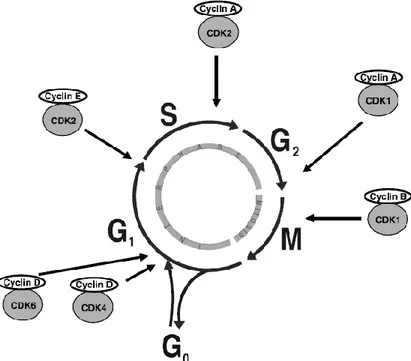
In response to DNA damage, interactions of the ATM/ATR pathways converge to regulate the cell cycle network and induce cell cycle arrest in order to provide time for DNA repair. Cell cycle arrest is induced by inactivating members of the Cdc25 family and thereby modulating the activity of Cyclins and Cyclin dependent kinases (CDKs) [103-105]. The CDKs are the key regulator proteins of the cell cycle and they control the transitions between different stages of the cell cycle [97,104,105]. The CDKs are positively regulated by Cyclins and at different phases of the cell cycle, diverse Cyclin-CDK complexes are responsible for blocking cell cycle progression (Figure 4), or alternatively inducing cell death if damage cannot be properly repaired [80,82,91].
Figure 4. The different phases of the cell cycle and the sites of activity for the diverse CDK/Cyclin
1.4.2. The G1 checkpoint
At the G1 checkpoint, cells make decisions whether to replicate DNA and complete the
cell division cycle or not [80]. DNA damage induces G1 arrest in a mechanism that
involves two different responses mediated by Chk1/Chk2; one initial rapid p53-independent response followed by a delayed, more sustained G1 arrest maintained by
p53–p21 signaling [79,80].
The immediate p53-independent response only lasts for a few hours and functions by decreasing abundance and activity of Cdc25A [80,86]. This results in inhibitory phosphorylation of CDK2, and thus inhibition of Cyclin E–CDK2 activity leading to the blockage of G1/S transition [80].
The delayed p53-dependent response occurs at transcriptional level [86]. p53 becomes post-translationally modified and stabilized, which results in an induced expression of genes required for cell cycle arrest [80,83]. Among the genes induced by p53 is the CDK inhibitor p21, capable of inhibiting the CDK2-cyclin E complex and thus preventing the progression from G1 phase to S phase of the cell cycle
[80,83,86,102,105].
1.4.3. The S phase checkpoint
During DNA replication, cells must ensure that there are no abnormalities that could cause a incorrect copy of the genome. When abnormalities or DNA damage are detected, the S phase checkpoint slows down the rate ofDNA synthesis, prevents new replication and stabilizes stalled replication forks [86,106].
The S phase checkpoint mechanisms respond in different manners to DNA damage, depending on the type of induced damage. In response to UVC, the S phase checkpoint response seems to be ATR-dependent [106]. However, in response to DNA DSBs, the S phase checkpoint requires ATM, but is p53-independent [79,80,106,107]. The ATM-Chk2 signaling cascade degrades Cdc25A, which leads to inhibition of the CDK2 kinase activity and thereby cell cycle arrest [80,106-108]. Chk1 has also been shown to retard progression through S phase via phosphorylation and degradation of Cdc25A [109].
1.4.4. The G2 checkpoint
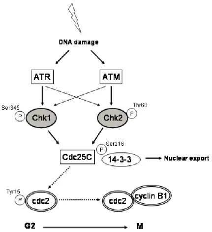
The G2 checkpoint represents the last barrier that can block the entry into mitosis of a
cell with damaged DNA. The G2/M transition of the cell cycle is controlled by the Cdc2
(CDK1)/Cyclin B complex, whose activity is essential for both the G2/M transition of
the cell cycle and completion of mitosis [79,84,100,102,110].
In response to DNA damage, a series of events will cause an arrest in G2 phase. DNA
damage-induced Chk1/Chk2 activation leads to Cdc25C phosphorylation on Serine 216, promoting its binding to 14-3-3 proteins, thereby inhibiting its activity by nuclear export and sequestration in the cytoplasm [83,87,97,103,105,110]. Cdc2 will be kept in its inactivated form through inhibitory phosphorylation on Tyrosine 15 and prevent activation of Cdc2-Cyclin B complex [97,100,105]. Cyclin B is kept inactivate until the beginning of the prophase, by active translocation from the nucleus to the cytoplasm [105]. When conditions are appropriate for mitotic entry, Cdc2 is dephosphorylated by Cdc25, leading to Cdc2 activation and initiation of mitosis (Figure 5) [87,97,100].
Figure 5. The G2 checkpoint. After detection of DNA damage, the G2 checkpoint primarily functions to
1.5. Cell Death
1.5.1. General aspects of cell death
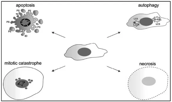
Programmed cell death is central to the development and homeostasis of multicellular organisms [111]. Three types of cell death have been distinguished in mammalian cells by morphological criteria. Type I cell death, better known as apoptosis, type II cell death, also known as autophagic cell death and finally, type III cell death, more known as necrosis [111,112]. Another form of cell death, termed mitotic catastrophe, has been discovered to be fundamentally different from type I – type III cell deaths [113].
1.5.2. Apoptosis
Apoptosis is a highly regulated form of cell death, which is morphologically characterized by cellular shrinkage, membrane blebbing, chromatin condensation, nuclear fragmentation, exposure of phosphatidylserine (PS) on the cell surface, and finally the formation of apoptotic bodies (Figure 6) [112-115]. In multicellular organisms, apoptosis is essential for development, tumor suppression, immune function and maintenance of homeostasis [36].
Two major pathways can induce apoptosis, the intrinsic pathway, which is controlled by mitochondrial membrane permeabilization, and the extrinsic pathway, in which death receptors trigger the apoptotic cascade [36,111]. The central players in both pathways are the caspases (cysteine-dependent aspartate-specific proteases) [36,116,117]. Caspase2, 8, 9 and 10 are regarded as initiator caspases, whereas caspase 3, 6 and -7, serve as effector caspases [114,11-7,118].
The intrinsic pathway is activated in the presence of cellular stresses such as growth factor withdrawal, cytotoxic drugs and DNA damage [36,111]. Initiation of the intrinsic pathway induces permeabilization of the outer mitochondrial membrane and the release of cytochrome C into the cytoplasm. Upon release, cytochrome C binds to Apaf-1 (apoptotic protease-activating factor-1), which in turn triggers the apoptotic cascade by activating procaspase-9 and forming the apoptosome complex. This complex activates the downstream effector caspases leading to DNA fragmentation and cell death [36,114,118].
The extrinsic pathway, or death receptor pathway, is activated through the TNF (tumor necrosis factor) family of cytokine receptors. This pathway is activated when a death ligand binds to the extracellular domains of the death receptor and leads directly to caspase activation [36].
1.5.3. Necrosis
In contrast to apoptosis, necrosis is a more uncontrolled form of cell death usually as a consequence of pathological traumas or harsh conditions. Morphologically, necrosis is characterized by vacuolization of the cytoplasm, loss of membrane integrity and cellular swelling (Figure 6) [111,114,115]. Necrosis is usually associated with inflammation, since the cellular contents leak into the extracellular environment [111]. There are growing evidence supporting the idea that necrosis can be a regulated form of cell death and that necrotic death can be induced by DNA damage via PARP-1, a protein involved in DNA damage repair [112,114].
1.5.4. Autophagy
The primary function of autophagy is to recycle proteins and organelles through degradation by lysosomal proteases and it is probably initiated as a survival response to cellular stress-associated damage or nutrient deprivation [111,114]. Various forms of environmental stress induce autophagy, which eventually results in either caspase-dependent or caspase-incaspase-dependent cell death [111].
Autophagy is recognized by the formation of autophagosomes, double membrane autophagic vacuoles that eventually fuse with lysosomes to form autolysosomes, where sequestered cellular components are digested (Figure 6) [111,114,115,119].
1.5.5. Mitotic catastrophe
Mitotic catastrophe is defined as a type of cell death that is caused by aberrant mitosis and is characterized by enlarged multinucleated cells, incomplete nuclear condensation, chromosome alignment defects, unequal DNA separation or mitosis in the presence of DNA damage (Figure 6) [114,115,120,121].
In mammalian cells and particularly in tumor cells, mitotic catastrophe is mainly associated with deficiencies in cell cycle checkpoints [113,114,121]. G2/M regulatory
proteins have been shown to be associated with mitotic catastrophe. High expression levels of proteins that promote entry of mitosis (such as Cdc2 and Cyclin B) as well as inhibition or knockout of proteins that prevent premature mitosis (such as ATR, ATM, Chk1, Chk2 and 14-3-3σ) can induce mitotic catastrophe [114]. Defective mitotic spindle checkpoints are also linked to mitotic catastrophe, since such defects usually lead to missegregation of chromosomes [113,114].
Figure 6. Morphological characteristics of apoptosis, autophagy, mitotic catastrophe and necrosis.
Apoptosis is characterized by membrane blebbing, cytoplasmic shrinkage, chromatin condensation, exposure of phosphatidylserine (PS) on the cell surface, and finally the formation of apoptotic bodies. Death by autophagy is characterized by the double-membrane vesicles containing cytosolic organelles. Cells dying from mitotic catastrophe are usually large with multiple micronuclei and contain uncondensed chromosomes. During necrosis, cells swell and loose their membrane integrity [114].
1.6. Checkpoint inhibitors in cancer therapy
1.6.1. General aspects
DNA damaging therapies are among the most common cancer treatments. Due to the efficacy of these anticancer treatments, DNA damaging agents are likely to remain a standard treatment of many cancers in the future [79]. Since the majority of these agents are used at the maximum tolerated dose, DNA damaging agents often cause significant side effects. Toxicities to different organs are commonly observed and many patients develop resistance and therefore become refractory to treatment [79,122].
Through increased knowledge about the DDR pathway and cell cycle regulation, the cell cycle checkpoints have emerged as attractive therapeutic targets for anticancer therapy. Tumors may be sensitized to DNA damaging agents by targeting the cell cycle checkpoints, since abrogation of cell cycle arrest prevents cancer cells from repairing DNA damage, thereby forcing them into mitotic catastrophe and apoptosis [123].
1.6.2. Cell cycle abrogation as an anticancer strategy
The rationale behind using cell cycle abrogation to target cancer cells depends on the difference in functional cell cycle checkpoints between normal cells and tumor cells. Many malignant cells suffer from defects in various tumor suppressor genes, including p53 and Rb pathway, and have therefore a defective G1 checkpoint mechanism
[123,124]. Cells deficient in the G1 checkpoint are highly dependent on the S and G2
checkpoints to maintain cell cycle arrest and for repair of DNA damage [123,124]. In normal cells, DNA damage would arrest cells mostly in G1 phase, whereas
p53-deficient tumors, accounting for over half of all tumor types, would have to rely on the S or G2/M checkpoints [125]. Therefore, G2 cell cycle arrest abrogation could be used to
specifically sensitize tumor cells to DNA damaging agents [79,124].
1.6.3. Candidate targets for cell cycle abrogation
The ideal S or G2 checkpoint abrogator needs to be selective and targeting a molecule
not involved in the G1 checkpoint [123]. Different candidate targets for cell cycle arrest
Cdc25C phosphatase, inhibition of the molecular chaperone heat shock protein-90 (HSP-90) and inhibition of Chk1 [123].
ATM /ATR inhibitiors are not specific G2 checkpoint abrogators since ATM and ATR
activate pathways involved in cell cycle checkpoints, apoptosis and DNA repair. However, ATM/ATR inhibition has been shown to disrupt the G2 checkpoint, inducing
damaged cells to undergo aberrant mitosis [123,126].
An alternative method of G2 abrogation is to activate Cdc25C, resulting in
de-phosphorylation and activation of Cyclin B/Cdc2 and subsequent cell cycle progression to mitosis. This can be achieved either through direct activation of Cdc25 or by inhibition of WEE1, a protein that opposes Cdc25 activity. The WEE1 inhibitor PD0166285 has demonstrated G2 checkpoint abrogation in preclinical models
[123,127].
An indirect and nonspecific method of checkpoint abrogation is provided by inhibition of HSP-90. In preclinical studies, the HSP-90 inhibitor 17-AAG has been shown to abrogate G2/M arrest when combined with SN38 in p53-deficient cells and when
combined with irradiation in human lung cancer cells [123].
The most relevant approach to checkpoint abrogation is by inhibition of Chk1 kinase. Chk1 plays a crucial role in the S phase checkpoint, the G2/M checkpoint as well as for
mitotic spindle checkpoint function. In this manner, Chk1 inhibitors are capable of not only enhancing the efficacy of DNA damaging agents that cause S or G2 arrest, but also
potentiating antimitotic agents [79,123]. Recent research have implicated checkpoint pathway activation as a major mechanism driving both chemoresistance and radioresistance, indicating that the use of Chk1 inhibitors will not only potentiate the efficacy of chemotherapy, but may also reduce drug resistance [40,79,128,129]. Based on the hypothesis that cells with defective G1 checkpoint can be sensitized to
chemotherapy by abrogating G2 arrest, it is natural to believe that Chk1 inhibitors would
preferentially enhance the cytotoxicity of DNA-damaging agents in cells with defective p53. Nonetheless, there are studies demonstrating that Chk1 inhibition also sensitizes p53-proficient cells, but the effects might be more pronounced in p53 mutant cell lines
[124,130-134]. The difference might in part relate to the different treatments and treatment schedules [135].
Based on the crosstalk between the Chk1 and Chk2 pathways, it has been claimed that it may be beneficial to target both Chk1 and Chk2 simultaneously. However, it has been shown that Chk1 is the only relevant checkpoint kinase as a cancer drug target and inhibition of other checkpoint kinases in addition to Chk1 has no benefit over inhibition of Chk1 alone [79,80,125]. Over the past decade many approaches to inhibit Chk1 have been made, for example by using small-molecule inhibitors and interference RNA, and several pharmaceutical compounds that target Chk1 are currently in clinical trials [79].
1.6.4. The Chk1 inhibitor UCN-01
The effects of inhibition of checkpoint pathways were first discovered in early work with caffeine, a nonspecific inhibitor of ATR and ATM, and with UCN-01 (7-Hydroxystaurosporine), an inhibitor of Chk1. It was shown that these agents could abrogate DNA damage-induced G2 arrest and potentiate the cytotoxicity of DNA
damaging agents [123,135-137]. UCN-01 has been shown to sensitize tumor cells to many agents, including, IR, cisplatin, camptothecin, gemcitabine and 5-fluoruoracil [24,138]. During phase I trials, the clinical utility of UCN-01 was found to be limited by its avid binding to human plasma proteins resulting in an unusually long half-life, however, the studies of UCN-01 collectively promoted Chk1 as a useful therapeutic target to induce enhanced cytotoxicity in tumor cells in response to DNA damaging agents [79,122,123,136,137].
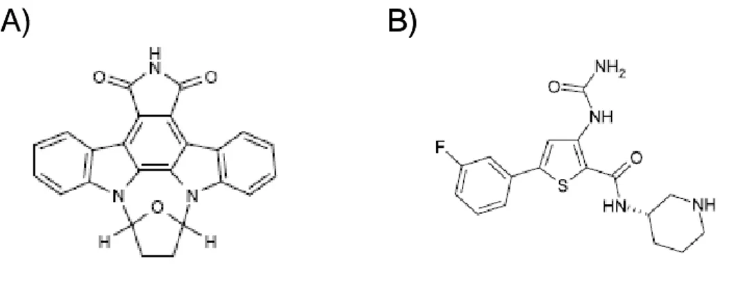
1.6.5. The Chk1 inhibitor SB218078
The indolocarbazole SB218078 (Figure 7A), a molecule related UCN-01, was demonstrated as an inhibitor of Chk1, which potently abrogated Chk1-induced phosphorylation of Cdc25C (IC50 = 15 nM). SB218078 also inhibited Cdc2 and PKC,
but at approximately 16-and 60-fold higher drug concentrations, respectively (IC50 =
250 nM and 1000 nM). This compound abrogates both camptothecin- and radiation-mediated cell cycle arrest and potentiates the cytotoxicity of camptothecin and topotecan in HeLa and HT-29 cells [122,137,139].
1.6.6. The Chk1 inhibitor AZD7762
AZD7762 (Figure 7B) is an ATP competitive Chk1/Chk2 inhibitor, currently in phase I clinical trials, that abrogates phosphorylation of a Cdc25C with an IC50 of 5 nmol/L
[122,123,134]. A 1000-fold selectivity for Chk1 over Cdc2 has been shown. In vitro, treatment with AZD7762 resulted in abrogation of G2 arrest induced by camptothecin or
gemcitabine and a reduction in the concentration of DNA damaging agents required to inhibit cell growth of several different cancer cell lines. In mouse and rat xenograft models, AZD7762 potentiated both the efficacy of gemcitabine and irinotecan, causing tumor growth delays [122,123,134]. It has also been shown that AZD7762 sensitizes human tumor cells to radiation both in vitro and in vivo [130,131].
A)
B)
A)
B)
Figure 7. Chk1 inhibitors. Structures of the Chk1 inhibitors SB218078 (A) and AZD7762 (B)
[134,139].
1.6.7. The Chk1 inhibitors XL-844 and PF-477736
Two other promising Chk1 inhibitors that have entered clinical trials are XL-844 and PF-477736. XL844 is a specific inhibitor of both Chk1 (IC50 = 2.2 nM) and Chk2 (IC50
= 0.2 nM) [122,123]. In vitro studies have shown potentiation of gemcitabine by abrogation of S phase arrest in multiple solid tumor cell lines and it has also been demonstrated to potentiate the effects of gemcitabine in vivo [109,122,123]. PF-477736 inhibits Chk1 with a 100-fold selectivity over Chk2 [122,123,140]. In vitro, PF-477736 induced checkpoint abrogation and potentiated the activity of a number of DNA-damaging agents, including gemcitabine, irinotecan and carboplatin, across several cell lines, with selectivity for p53-deficient cancer cell lines compared to p53-competent cells [122,123]. In vivo, PF-477736 enhanced the activity of gemcitabine and irinotecan in colon cancer xenograft models [123].
1.6.7. Validation of Chk1 inhibiton
It is necessary to be able to validate the inhibition of Chk1 in clinical studies and there are several possible markers for this purpose. Firstly, Cdc25C can be considered as marker for Chk1 inhibition, since Chk1 is known to phosphorylate Cdc25C following DNA damage and inhibition would cause a negative regulation. Secondly, an indirect but simpler way to evaluate Chk1 inhibition is to measure the extent of G2-M
checkpoint abrogation by using an antibody against phosphorylated histone H3, a marker of mitotic entry. Finally, activation of the DNA damage response as assayed by increased γ-H2AX levels compared to levels from chemotherapy alone can also be used as a downstream pharmacodynamic marker for Chk1 inhibition [123,135].
2. Aims
Despite the recent progress in molecularly targeted cancer therapies, advanced solid malignancies remain a therapeutic challenge, in part due to the development of resistance to radiation and chemotherapy. The purpose of this project was to study the cellular and molecular mechanisms of drug resistance in lung cancer tumors and convert it into the development of innovative therapies and clinical application for fighting lung cancer.
Identification of CSCs as an undifferentiated subpopulation of tumorigenic cells responsible for tumor maintenance, growth and spreading has become relevant for several human malignancies. To investigate the possibility that CSCs may represent the source of the drug-resistant subpopulation in lung tumors, our studies were specifically focused on the comparison of NSCLC-CSCs with their differentiated progenies to evaluate differences in response to chemotherapy and activation/regulation of the DNA damage response pathway.
The DNA damage response prevents cell cycle transition through surveillance mechanisms known as cell cycle checkpoints. Agents used for cancer treatment, such as cytotoxic chemotherapy and ionizing radiation (IR), also activate cell cycle checkpoints. Understanding how checkpoints are regulated is therefore important from the points of view of both tumorigenesis and cancer treatment.
We aimed to study the molecular hierarchy of the checkpoint signaling network in NSCLC-CSCs, especially the role of Chk1 in chemotherapy resistance and cancer therapy. Our approach was to treat NSCLC-CSCs with chemotherapy commonly used for lung cancer treatment in combination with chemical Chk1 inhibitors and thereafter evaluate the effects on DNA damage and reparation of damaged DNA, study changes in cell cycle profile and cell viability, characterize responsible molecular mechanisms and possible induction of cell death, and finally, to study the potential therapeutic effects of Chk1 inhibition on cancer progress in vivo.
3. Materials and Methods
3.1. Materials
3.1.1. Reagents
The following reagents were used: - cisplatin (Teva, Petach Tikva, Israel) - gemcitabine (Lilly, Fiesole, Italy)
- paclitaxel (Sigma-Aldrich, St. Louis, MO, USA) - SB218078 (Calbiochem, Nottingham, UK)
- AZD7762 (Axon Medchem, Groningen, The Netherlands)
3.1.2. Antibodies
The following antibodies were used:
- Chk1 (Cell Signaling Technology, Danvers, MA, USA) - phosphorylated Chk1 (Ser345) (Cell Signaling Technology) - Chk2 (Cell Signaling Technology)
- phosphorylated Chk2 (Thr68) (Cell Signaling Technology) - phosphorylated Cdc25C (Ser216) (Cell Signaling Technology) - phosphorylated Cdc2 (Tyr15) (Cell Signaling Technology) - ATM (5C2) (Santa Cruz Biotechnology, Santa Cruz, CA, USA) - phosphorylated ATM (0H11.E12) (Santa Cruz Biotechnology) - Cyclin B1 (clone D-11) (Santa Cruz Biotechnology)
- phosphorylated H2A.X (Ser139) (Upstate-Millipore, Billerica, MA, USA) - Caspase-2 (clone 35) (Upstate-Millipore)
- Caspase-3 (Upstate-Millipore) - β-actin (AC-15) (Sigma-Aldrich) - β-tubulin (TUB 2.1) (Sigma-Aldrich)
- anti-HLA class I (eBioscience, San Diego, CA, USA)
- PE-Cy5 anti-mouse CD45 (BD Pharmingen, San Diego, CA, USA)
3.1.3. NSCLC-CSCs
NSCLC-CSCs from human adenocarcinoma (NSCLC-CSC #1 and NSCLC-CSC #4), human squamous cell carcinoma (NSCLC-CSC #2 and NSCLC-CSC #3) and human large cell neuroendocrine carcinoma (NSCLC-CSC #5), were obtained from patients who underwent surgical resection of lung tumors [7].
3.2. Methods
3.2.1. Isolation and culture of NSCLC-CSCs
Surgically resected lung tumors were washed several times and left overnight in DMEM:F12 medium supplemented with 500 Units/mL penicillin, 500 μg/mL streptomycin and 5 μg/mL amphotericin B. Tissue dissociation was carried out by enzymatic digestion with 1.5 mg/mL collagenase II (Gibco-Invitrogen, Carlsbad, CA, USA) and 20 μg/ml DNase I (Roche, Basilea, SW, USA), agitating for 2h at 37˚C. In the presence of high quantity of blood cells, hypotonic lysis with ammonium chloride was performed. Recovered cells were cultured at clonal density in a growth medium specifically adapted for NSCLC-CSCs. The complete serum-free medium (SFM) for NSCLC-CSCs consists of:
- 50 μg/mL insulin (Sigma-Aldrich)
- 100 μg/mL apo-transferrin (Sigma-Aldrich) - 10 μg/mL putrescine (Sigma-Aldrich) - 0.03 mM sodium selenite (Sigma-Aldrich) - 2 μM progesterone (Sigma-Aldrich) - 0.6% glucose (Sigma-Aldrich) - 5 mM HEPES (Sigma-Aldrich)
- 0.1% sodium bicarbonate (Sigma-Aldrich)
- 0.4% BSA (ICN Biochemicals, Costa Mesa, CA, USA)
- 2 mM L-glutamine (PAA Laboratories GmbH, Pasching, Austria) - 100 Units/mL penicillin (PAA Laboratories GmbH)
- 100 μg/mL streptomycin (PAA Laboratories GmbH)
All these reagents were dissolved in DMEM:F12 medium (Gibco-Invitrogen) and supplemented with 20 μg/mL epidermal growth factor (EGF) and 10 μg/mL basic
fibroblast growth factor (bFGF) (PeproTech, Rocky Hill, NJ, USA). Non-treated, sterile polystyrene flasks for suspension cell cultures (Nunc, Thermo Fischer Scientific, Rochester, NY, USA) were used to reduce cell adherence and support growth as undifferentiated tumor spheres. The medium was replaced or supplemented with fresh growth factors (20 μg/mL EGF and 10 μg/mL bFGF) twice a week, until cells started to grow as floating aggregates. Cultures were expanded by mechanical dissociation of spheres, followed by re-plating of single cells and residual small aggregates in complete fresh SFM.
3.2.2. Differentiation of NSCLC-CSCs
To obtain differentiation of NSCLC-CSCs, spheres were dissociated to obtain single cells and plated in DMEM (Gibco-Invitrogen) supplemented with 10% FBS for 24h, to allow cell attachment. DMEM was then replaced with Bronchial Epithelial Cell Growth medium (Lonza, Basel, Switzerland) for 72h to facilitate the progress of differentiation. Differentiated cells could thereafter be maintained in DMEM supplemented with 10% FBS for several weeks.
3.2.3. Treatment
For in vitro experiments, the following concentrations of chemotherapeutic drugs and Chk1 inhibitors were used:
- 5 ug/mL cisplatin - 250 µM gemcitabine - 30 ng/mL paclitaxel - 20 nM SB218078 - 5 nM AZD7762
For in vivo studies we used: - 60 mg/kg gemcitabine - 3 mg/kg cisplatin - 10 mg/kg AZD7762
3.2.4. Cell viability assays
For chemoresistance comparison and cell viability studies, dissociated spheres and adherent differentiated cells were plated in 96 well plates at 5,000 cells/well in growth medium supplemented with cisplatin, gemcitabine or paclitaxel, for 96h. For cell viability studies, dissociated NSCLC-CSCs were seeded and treated as described above and in combination with Chk1 inhibitors. Cell viability was evaluated after 4 days of treatment by MTT (3-(4,5-dimethyl-2-thiazolyl)-2,5-diphenyl 2H-tetrazolium bromide) assay (Promega, Madison, WI, USA) or CellTiter-Glo Luminescent Cell Viability Assay (Promega) according to standard protocols and analyzed by a Victor 2 plate reader (Wallac, Turku, Finland).
3.2.5. Cell proliferation assays
A total of 75,000 dissociated NSCLC-CSCs were treated with cisplatin, gemcitabine or paclitaxel for 6 days. On day 3, cells were collected, counted and replated in the presence of chemotherapy. On day 6, cells were collected, counted and thereafter replated in fresh medium without chemotherapy. Cells were then counted and replated every 3 days until day 15, to study the effects of chemotherapy withdrawal. Viable cells were counted using trypan blue exclusion.
3.2.6. Cell cycle analysis
Cells were dissociated and treated with cisplatin, gemcitabine or paclitaxel for 48h. Thereafter, cells were dissociated with trypsin and stained with a PI staining solution (0.1% trisodium citrate, 9.65 mM NaCl, 0.3% NP40, 50 μg/mL PI, 200 μg/mL RNase A) for 30 minutes at room temperature. Cell cycle profile was acquired with a Facs Canto flow cytometer (Becton Dickinson, NJ, USA) and analyzed with FlowJo software (Tree Star Inc. www.FlowJo.com).
3.2.7. Immunofluorescence
NSCLC-CSCs were treated with cisplatin, paclitaxel, SB218078, AZD7762 or in combinations of drug and inhibitor for 48h or 96h. For CKs 8/18 staining, NSCLC-CSC spheres were fixed with 2% paraformaldehyde, attached to poly-L-lysine coated coverslips by sedimentation and permeabilized with 0.5% Triton X-100/PBS for 4h at 4°C. Differentiated progenies were cultured on coverslips to allow attachment, fixed with 2% paraformaldehyde and permeabilized with 0.5% Triton X-100/PBS for 2h at
4°C. Thereafter, slides were incubated with a FITC-conjugated CKs 8/18 antibody (Novocastra Laboratories Ltd) over night at 4°C. For anti-Cyclin B1 and anti-γ-H2A.X staining, treated NSCLC-CSCs were cyto-spun onto glass slides, fixed with 2% paraformaldehyde and then permeabilized with 0.1% Triton X-100/PBS for 1h at 37°C before incubation with Cyclin B1 (Santa Cruz Biotechnology) or γ-H2A.X (Ser139) (Upstate-Millipore) overnight at 4°C. Alexa Fluor 555 goat anti-mouse (1 μg/mL, Invitrogen) for 1h at RT was used as secondary antibody. TO-PRO-3 (4 μM, Invitrogen, Carlsbad, CA, USA) or DAPI (3 μM, Molecular Probes/Invitrogen) were used to visualize nuclei and Phalloidin-Alexa Fluor 488 (5 Units/mL, Molecular Probes/Invitrogen) was used to visualize cell membranes. Slides were analyzed using OLYMPUS FV-1000 confocal microscope with the Olympus objective Ultraplan Apochromatic 60X N.A.1.35 and the software Olympus Fluoview.
3.2.8. Western blot
NSCLC-CSCs were treated for 6h, 12h, 24h or 96h with cisplatin, gemcitabine, paclitaxel, SB218078, AZD7762 or in combinations of drug and Chk1 inhibitor. Whole cell lysates from treated cells were prepared in lysis buffer (NP40 1%, 20 mM TRIS (pH 7.2), 200 mM NaCl, Phosphatase Inhibitor Cocktail 1 (used 1:100, P2850, Sigma-Aldrich), Phosphatase Inhibitor Cocktail 2 (used 1:100, P5726, Sigma-Aldrich) and Protease Inhibitor Cocktail (used 1:100, P8340, Sigma-Aldrich)). 20 μg of whole cell extracts were subjected to 8-15% sodium dodecyl sulphate polyacrylamide gel electrophoresis, transferred onto Hybond-C membrane (Amersham Biosciences, Milan, Italy) and incubated with primary antibody over night at 4°C. Membranes were probed with secondary antibody for 1h at room temperature and detected using enhanced chemiluminescence detection kit (Pierce, Rockford, IL, USA).
3.2.9. Propidium iodide / Annexin V staining
NSCLC-CSCs were treated with cisplatin and SB218078 for 96h followed by 72h of cell culture in fresh medium. A total of 50,000 treated cells were washed with Annexin V-binding buffer (2.5 mM CaCl2, 140 mM NaCl, 10 mM Hepes) and thereafter
incubated with Annexin V Alexa Fluor 647 (1.25 nM, Invitrogen) for 15 minutes at room temperature. Cells were again washed with Annexin V-binding buffer, re-suspended in buffer containing 5 μg/mL PI and analysed by Facs Canto flow cytometer (Becton Dickinson).
3.2.10. May-Grünwald-Giemsa staining
NSCLC-CSCs were treated with cisplatin and SB218078 for 96h followed by 72h of cell culture in fresh medium. Treated cells were washed and cyto-spun onto glass slides. Slides were stained with concentrated giemsa staining (Sigma-Aldrich) for 30s and then with diluted giemsa staining (1:20) for 30 minutes.
3.2.11. Soft agar colony forming assays
Soft agar colony forming assays were carried out for dissociated NSCLC-CSCs treated with cisplatin or paclitaxel alone or in combinations with SB218078 or AZD7762 for 96h. Thereafter, cells were washed and 500 single cells were plated in the top agar layer in each well of a 24-well culture plate with 0.3% top agar layer and 0.4% bottom agar layer (SeaPlaque Agarose, Cambrex, New Jersey, USA). Cultures were incubated at 37°C for 20 days. Colonies from triplicate wells were stained with crystal violet (0.01% in 10% MetOH), visualized and counted under microscope and photographed with a Nikon D80 camera. For xenograft-derived cells, tumors were aseptically removed and dissociated. Recovered cells were extensively washed and plated in SFM for 3 days. Consequently, 500 cells for each treatment were plated in soft agar as described above.
3.2.12. In vivo studies
Female NOD-SCID mice were purchased from Charles River Laboratories Italia (Calco, LC, Italy). All procedures were conducted in accordance with the Institute for Laboratory Animal Research Guide for the Care and Use of Laboratory Animals and within the protocols approved by the Istituto Superiore di Sanità. NSCLC-CSCs were dissociated, counted and resuspended in a mix of PBS and Matrigel (1:1). 50,000 NSCLC-CSCs were implanted subcutaneously into the right flank of each mouse in a volume of 0.1 to 0.2mL using a 25-gauge needle. Tumors were allowed to grow to the size of 100 to 200 mm3 before the administration of compounds. Animals, 5 in each group, were dosed intraperitoneally with gemcitabine (60 mg/kg) or cisplatin (3 mg/kg) and intravenously with AZD7762 (10 mg/kg) every 3 days starting from day 0. Tumor growth was evaluated with an electronic caliper before every administration and volume was estimated by using the following formula: a * b2 / 2, where a and b represents the tumor length and width (in mm), respectively. After 30 days, levels of human carcinoembryonic antigen (CEA) were measured in serum obtained by retro-orbital withdrawal by immunoluminometric technique using Vitros ECI analyzer
(Ortho-Clinical Diagnostics Inc. Rochester, NY, USA). Tumors were subsequently removed and weighed using a PL202-L Precision Balance (Mettler-Toledo, Novate Milanese MI, Italy). Immunohistochemistry was performed on formalin fixed paraffin-embedded tissue or frozen tissue. Paraffin sections (5 μm) were dewaxed in xylene and re-hydrated with distilled water. The slides were subsequently incubated with anti- -H2A.X (Upstate-Millipore). The reaction was performed using Elite Vector Stain ABC systems (Vector Laboratories) and DAB substrate chromogen (DakoCytomation), followed by counterstaining with hematoxylin. Human origin of the tumor xenografts was confirmed by FACS analysis with a PE conjugated anti-HLA class I antibody (eBioscience) and a PE-Cy5 anti-mouse CD45 antibody (BD Pharmingen) was used to exclude unspecific staining of mouse cells .
3.2.13. Statistical analysis
All statistical analyses were performed using GraphPad Prism 4 (GraphPad Software Inc., www.graphpad.com). The statistical significance of the results shown in Fig. 9A, 9B and 9C was evaluated by two-way ANOVA and Bonferroni’s post-tests. The statistical significance of the results shown in Fig. 12C and 17H was evaluated by one-way ANOVA with Bonferroni’s multiple comparison test while results in Fig. 14B, 16B, 17B, 17D and 17E were evaluated by repeated measured one-way ANOVA with Bonferroni’s multiple comparison test. A P value <0.05 is represented by a single asterisk, a P value <0.01 is represented by a double asterisk, while three asterisks indicate P<0.001, all P values are two-sided.
![Table 1. Cell surface markers for identification of CSCs [37].](https://thumb-eu.123doks.com/thumbv2/123dokorg/4510788.34492/15.892.215.679.515.880/table-cell-surface-markers-identification-cscs.webp)