UNIVERSITÀ DEGLI STUDI DI CATANIA
DOTTORATO DI RICERCA IN BASIC AND APPLIED BIOMEDICAL
SCIENCES
XXXI CICLO
Dipartimento di Scienze Biomediche e Biotecnologiche (BIOMETEC)
Dott.ssa Giulia Russo
Novel computational strategies for the identification of new
therapeutic targets in melanoma and thyroid cancer
TESI DI DOTTORATO
Coordinatore: Relatore:
Ch.mo Prof. Massimo Libra Ch.mo Prof. Francesco Pappalardo
Novel computational strategies for the identification of new
therapeutic targets in melanoma and thyroid cancer
Giulia Russo
SOTTOMESSA IN PARZIALE ADEMPIMENTO AI REQUISITI PER
IL CONSEGUIMENTO DEL TITOLO DI
“DOTTORE DI RICERCA”
ALL' UNIVERSITÀ DEGLI STUDI DI CATANIA
20 NOVEMBRE 2018, ITALIA, CATANIA
UNIVERSITÀ DEGLI STUDI DI CATANIA
DOTTORATO DI RICERCA IN BASIC AND APPLIED BIOMEDICAL
SCIENCES
Dipartimento di Scienze Biomediche e Biotecnologiche (BIOMETEC)
I sottoscritti certificano che hanno letto e raccomandato alla Facoltà per gli Studi di Dottorato l'accettazione della tesi intitolata “Novel computational strategies for the identification of
new therapeutic targets in melanoma and thyroid cancer” di Giulia Russo, a parziale
adempimento ai requisiti per il conseguimento del titolo di “Dottore di Ricerca”.
20 Novembre, 2018
Tutor (Prof. Francesco Pappalardo): ____________________ Coordinatore (Prof. Massimo Libra): ____________________
UNIVERSITÀ DEGLI STUDI DI CATANIA
20 Novembre, 2018
Autore: Giulia Russo
Titolo: Novel computational strategies for the identification of new therapeutic targets in melanoma and thyroid cancer
Dipartimento: Scienze Biomediche e Biotecnologiche (BIOMETEC)
Grado: Dottore di Ricerca
L'autore concede il permesso all'Università degli studi di Catania di far circolare e di far ottenere copie della presente tesi di dottorato per soli usi didattici e non commerciali, sia a privati che istituzioni.
Firma dell'autore: _________________________
L'AUTORE SI RISERVA TUTTI GLI ALTRI DIRITTI DI PUBBLICAZIONE. NESSUNA PARTE DELLA TESI O RIASSUNTI ESTESI ESTRATTI DA ESSA POSSONO ESSERE RIPRODOTTI CON QUALUNQUE MEZZO, SENZA LA PREVENTIVA AUTORIZZAZIONE SCRITTA DA PARTE DELL'AUTORE.
INDEX
SUMMARY ... 6
SOMMARIO ... 7
ACKNOWLEDGEMENTS ... 8
1 INTRODUCTION ... 10
1.1 The importance of signaling pathways and cross-talks in cancer ... 10
1.2 MAPK and PI3K/AKT pathways: common target in cutaneous melanoma and thyroid cancer ... 13
1.3 Target therapy with BRAF inhibitors ... 19
1.4 Computational strategies for signaling networks ... 25
1.4.1 Database and tools for the visualization, simulation and analysis of signaling networks ... 25
1.4.2 Algorithms for Pathway visualization, investigation and prediction ... 27
1.4.3 Software for Pathway Analysis ... 29
1.4.4 Algorithms in finding alternative pathways and new drug targets ... 30
2 AIM OF THE THESIS ... 32
3 COMPUTATIONAL MODELING IN MELANOMA ... 33
3.1 Melanoma background ... 33
3.2 Methods ... 39
3.2.1 Pathway model ... 39
3.2.2 DNA methylation ... 44
3.3 Results and discussion ... 48
3.3.1 Pathway model results ... 48
3.3.2 DNA Methylation results ... 51
4 COMPUTATIONAL MODELING IN THYROID CANCER ... 55
4.1 Thyroid cancer background ... 55
4.2 Methods ... 58
4.3 Results and discussion ... 64
5 CONCLUSIONS ... 71
SUMMARY
Cancer signaling pathways have been extensively investigated. However, the way cross-talk processes and integrates pathway responses in cancer is still far from being completely elucidated. Genetic and epigenetic alterations lead cells to aberrant proliferation and escapement from physiological mechanism controlling cell growth, survival and migration. In this context, specific mutations transform cellular proto-oncogenes to oncogenes, triggering hyperactivation of signaling pathways, whereas inactivation of tumor suppressors removes critical negative regulators of signaling. MAPK and PI3K/AKT pathways often present mutated genes in different types of cancer, and are strongly involved in intensive cross-talk.
There is an ever-increasing awareness that computational modeling and simulation are more than helpful in improving the understanding at cellular and molecular levels, in speeding-up the drug discovery process through the identification of alternative strategies with the aim to overcome drug resistance in cancer.
The main objective of this thesis is to reveal biochemical and genetic mechanisms underlying drug resistance in melanoma and thyroid cancer through the application of ordinary differential equations based models coupled with algorithmic approaches. These tumors share both MAPK and PI3K/AKT signaling pathway, with the presence of BRAF V600E mutation. Computational approaches developed in this PhD project were demonstrated to be able to find novel therapeutic targets and prognostic biomarkers for a more effective treatment in melanoma and thyroid cancer.
SOMMARIO
Le vie di segnalazione del cancro sono state oggetto di ampio studio. Tuttavia, il modo in cui eventuali cross-talk processano e integrano le risposte delle diverse vie di segnalazione nel cancro è ancora ben lungi dall'essere completamente chiarito. Le alterazioni genetiche ed epigenetiche possono contribuire ad un’aberrante proliferazione cellulare e all’elusione del meccanismo fisiologico che controlla la crescita, la sopravvivenza e la migrazione delle cellule. In questo contesto, mutazioni specifiche trasformano i proto-oncogeni cellulari in oncogeni, innescando l'iperattivazione delle vie di segnalazione, mentre l'inattivazione dei soppressori tumorali rimuove i regolatori negativi critici della segnalazione. Le cascate di segnalazione MAPK e PI3K/AKT presentano spesso geni mutati in diversi tipi di tumore e sono fortemente coinvolti in complessi cross-talk.
E’ presente una sempre più crescente consapevolezza di come la modellizzazione e la simulazione computazionale risultino più che utili nel migliorare la comprensione a livello cellulare e molecolare dei fenomeni biologici, e contribuiscano all’accelerazione del processo di scoperta dei farmaci e l'identificazione di strategie alternative per superare la resistenza farmacologica presente in molte forme di cancro.
L'obbiettivo principale di questa tesi è quello di rivelare, attraverso l’utilizzo di modelli computazionali basati su equazioni differenziali ordinarie accoppiate ad approcci algoritmici, i potenziali meccanismi biochimici e genetici alla base della resistenza dei farmaci nel melanoma e nel cancro della tiroide. Questi tumori condividono entrambe le vie di segnalazione MAPK e PI3K/AKT e la presenza della mutazione BRAFV600E. Gli approcci computazionali sviluppati in questo progetto di dottorato sono stati efficaci nell’identificazione di nuovi bersagli terapeutici (nonché di biomarcatori prognostici) per un trattamento più efficace del melanoma e del cancro della tiroide.
ACKNOWLEDGEMENTS
Undertaking this PhD has been a truly life-changing experience for me and it would not have been possible to do without the support and guidance that I received from many people. Hence, I would like to extend thanks to who, so generously, contributed to the work presented in this thesis.
Special mention goes to my enthusiastic supervisor Prof. Francesco Pappalardo, for guiding and supporting me over the years. It has been a honor to be his first PhD student. He has taught me, both consciously and unconsciously, how a computational biologist should be. I appreciate all his contributions of time, ideas, feedbacks and opportunity to make my PhD experience productive and stimulating. I must confess that my PhD has been an amazing experience and I thank Francesco wholeheartedly, not only for his tremendous academic support, but also for giving me so many wonderful opportunities. Not many PhDs involve travelling a lot and through many countries, like to get to a conference in New Delhi! The joy and enthusiasm he owns for his research was contagious and intensively motivational for me. I am more than grateful for the excellent and invaluable example he has provided me as a researcher, mentor, instructor and go-to person. Without his guidance and constant feedback, this PhD would not have been achievable.
I am also hugely appreciative to Prof. Massimo Libra, especially for sharing his oncology expertise so willingly, and for being so dedicated to his role as my secondary supervisor.
The members of the COMBINE group have contributed immensely to my personal and professional time at University of Catania. The group has been a source of friendships as well as good advice and collaboration. I would like to acknowledge the senior group member Prof. Santo Motta for his insightful observations and precious suggestions he gave me since the beginning of my PhD I also would like to extend my special thanks to Marzio, for nurturing my enthusiasm for computational
modeling and to the junior part of the COMBINE group made of Giuseppe Sgroi and Giuseppe Parasiliti Palumbo. With all of them, I have had the pleasure to work with and alongside in a brilliantly and creatively way.
I would particularly like to acknowledge Guanglan Zhang, who has long been an inspiring research figure for me and a wonderful and generous friend. She constantly gave me enthusiasm, encouragement and suggestions during the thesis writing hell from the other part of the world. Nevertheless, unbelievably I feel her very close to me!
I would like to thank also the research group supervised by Prof. Massimo Libra and the one guided by Prof. Francesco Frasca that contributed to this research.
A special thank goes to my amazing family for the love, support, and constant encouragement I have gotten over the years. In particular, I would like to thank my parents, my brother (especially for his insights and suggestions about the state of the art of thyroid cancer), Andrea and my little niece Elena. You are the salt of the earth, and I undoubtedly could not have done this also without you.
And last but not the least, I would like to thank my passion for swimming and all the people around the swimming-pool center that allow me to feel constantly enthusiastic, encouraged, cheerful and full of life.
1 INTRODUCTION
1.1 The importance of signaling pathways and cross-talks in cancer
Biological cells communicate each other using physical signals but mostly through chemical signals. Within an organism, cells are immersed in an ocean of growth factors and hormones that represent the most important sources of chemical signals, secreted firstly from a cell and then released into the extracellular space1,2.
The chemical signal represents a real message with a specific biological meaning and is carried out by a ligand or a growth factor3. It is often communicated through a sequence of secondary messengers
inside the cell and then it is propagated towards the neighboring cells4. After that, cell changes its
status because of an alteration in the activity of a gene or when a whole process such as cell division has been initiated. Ultimately, the original intercellular signal is converted into a new signal that triggers a biological response5.
This complex and accurate function of communicating is guaranteed through a number of pathways that receive and process signals originating not only from the external environment and from other cells within the organism, but also from different regions within the cell, hence moving from a micro to a macroscale6. Furthermore, all of this cell machinery is strongly capable to adapt the function of
an organism to environmental changes in a signal-directed way and to control all the cellular functions as well. The capability to coordinate and regulate cellular function results from the complex network of communications among cells, where signals are transduced into intracellular biochemical reactions that follow, on a case-by-case basis, different kinetic laws such as mass action equilibrium7,
Michaelis-Menten kinetics8, constant flux equations9 and so forth10. The two general categories of
cell signaling include intercellular and intracellular signaling:
- intercellular signaling: it coordinates and regulates the physiological functions of a multicellular organism through the communication among cells11. In this case, a single cell
influences the behavior of other cells in a specific way. Signals propagated during intercellular signaling are delivered and processed in target cells with the aim to trigger biochemical reactions that absolve a specific cell function12. Intercellular communication uses messenger
substances, such as hormones13, secreted by signal-producing cells and gathered by target
cells. The extracellular signals are transduced into intracellular signaling sequences that control many of the biochemical activities of a cell and can trigger the formation of further extracellular signals14,15. Typical examples of signaling deal with physiological activities such
as response to external signals16, intermediary metabolism17, cell growth18, cell division19, cell
motility20, cell morphology21, cell differentiation22 and cell development23. Cells
communicate each other via messenger substances, gap junctions, surface proteins, electrical signals24–27. Communication steps between cells could be summarized as follows: i) formation
of a signal in the signal-producing cell as a result of an external trigger; ii) transport of the signal to the target cell; iii) recording of the signal in the target cell28,29.
- intracellular signaling: it coordinates and regulates signals within the cell, in response to extracellular and intracellular stimuli5,30. Sensory signals or external growth factors are
specifically recognized, processed and transduced by cell receptors that convert the external signal into an intracellular signaling chain. The intracellular signaling paths modulate intermediary metabolism17, cell division19, cell morphology4, and also the transcription
process31.
Both intercellular and intracellular signaling are regulated by specific control mechanisms and mediators in a certain tissue32. Modulation of intercellular signaling is mainly regulated via external
trigger signals, feedback loops, degradation and modification processes, and amount and activity of receptors33–35.
Typically, a large number of signaling components participate in the transduction of an extracellular signal into intracellular biochemical reactions that define the endpoint of signal transduction36. To
components involved, as well as their linkages, represent the essential features to consider. However, it is increasingly documented that the existence of subtypes of signaling proteins allow different signals to access and to be processed in the same type of signaling path, leading then to variable outcomes37. Besides, the features of branching and cross-talk of signaling in biology, in which one or
more components of one signal transduction pathway affect other pathways, usually include a large number of possible linkages within a signaling path and between different signaling paths38,39. In
these signal transduction pathways, there are often shared components that can interact with either pathway.
Commonly, signaling pathways are depicted through a sequential transmission of signals in a linear signaling chain. Linear pathway description is very useful to illustrate the main biochemical steps in a signaling cascade of events that help to outline the intrinsic biological and biochemical meaning of a signaling pathway. Each signal is listed by an upstream component of a signaling chain and is then transferred to the downstream constituent that will sequentially propagates the signal to the next protein40. This linear description of signaling comes out from in vitro experiments where signaling is
originated by strong signals produced by overexpression or mutation of signaling proteins41. A
component will activate the next component through intrinsic mechanisms of activation and deactivation by specific enzymes, commonly known as tyrosine and serine-threonine protein kinase42,43. They represent a complex system with elaborated internal and external interactions and
are known to play a fundamental role in protein phosphorylation44, the main enzymatic process for
the initiation of cellular processes such as cell division, metabolism, survival and apoptosis. The routing of signals depends on the amplitude, that is the signal intensity, and on the frequency that influences the duration of the signal. It is well known that many signaling proteins own multiple downstream reactions that can be activated for further signal transduction40. This feature leads to a
degeneracy of signals and to a distribution of alternative reaction partners (or branching reactions)45
Within the signaling pathways, different routes and alternative paths connect one pathway with another one, reinforcing each other and constantly receiving excessively signals simultaneously46.
Basically, the multiple outputs that originate from the same type of signaling protein lead to an activation of alternative routes and to the biological phenomenon of cross-talk.
A cross-talk is a biological process that involve the signaling cascades of transduction pathways and it refers to the interdependence among signaling pathways37–39,47.
In specific occurrences, one or more components of one signal transduction pathway affect the other one, sharing the same components that can interact and be linked with either pathway. These phenomena process a large number of signals at the same time and the information flow does not run through a single conduit.
Moreover, in cross-talk dynamics, when a signal propagates through different branches and meet a common target, the signaling responses along these branches will influence, with an incremental effect, the overall target response. Furthermore, each intersection that connects one pathway with another could potentially represent a regulatory checkpoint for the signal flow itself. Noteworthy is the multivalency of signaling proteins, that determines several effects on the components of a signaling, and the plausible role of cross-talk in achieving robust activation of key downstream targets by low physiological doses of external stimuli48. In the light of this extraordinary complexity of
signaling networks, it becomes more and more evident how sophisticated hypothesis and accurate predictions of cellular response and their intricate relationship represent a mandatory goal in signaling research.
1.2 MAPK and PI3K/AKT pathways: common target in cutaneous melanoma and
thyroid cancer
Many examples of cross-talk were found in cell signaling38. In particular, multiple levels of
modulation of cell fate47,49,50. In this view, specific feedforward and feedback loops, involving
interacting pathways, coordinate both input and output response of both pathway34,47.
The mitogen-activated protein kinase (MAPK) and the phosphoinositide 3-kinase (PI3K/AKT) pathway are the most common signaling pathways downstream of cellular growth factor receptor51– 54. These signaling pathways orchestrate the majority of cellular physiological processes, such as cell
growth, differentiation, metabolism, survival and mitogenesis43,55–57. A graphical summary of both
pathways is shown respectively in Figure 1 and Figure 2.
GFDL license via Wikimedia Commons
Figure 2. Schematic illustration of PI3K/AKT signalling cascade.
Robbins et al. Frontiers in Endocrinology 2016;6:188
MAPK cascade is a highly conserved pathway expressed in mammals58 in at least four distinct
regulated groups of MAPKs59, extracellular signal-related kinases (ERK)-1/260, Jun amino-terminal
kinases (JNK1/2/3)61,62, p38 proteins (p38alpha/beta/gamma/delta)63,64 and ERK565. All of these
kinases are activated by specific MAPKKs: MEK1/2 for ERK1/266, MKK3/667,68 for the p38,
MKK4/769 (JNKK1/2) for the JNKs, and MEK570 for ERK5. Each MAPKK, however, can be
activated by more than one MAPKKK71, increasing the complexity and diversity of MAPK
signalling. Apparently, each MAPKKK confers responsiveness to distinct stimuli. For example, activation of ERK1/2 by growth factors depends on the MAPKKK C-RAF72, but other MAPKKKs
may activate ERK1/2 in response to pro-inflammatory stimuli59. The fundamental protein network
involved in this signalling cascade is reported below in Table 1.
Signaling pathway KEGG
network Cell function Reference
EGF-EGFR-RAS-ERK Cell proliferation 73
PDGF-PDGFR-RAS-ERK Cell migration, proliferation and survival
74
FGF-FGFR-RAS-ERK Cellular proliferation, differentiation and migration
75
EGF-EGFR-PLCG-ERK Cell motility 76
IL1-IL1R-p38 Pro-inflammatory activities, innate immune reactions
77
IL1-IL1R-JNK Cellular apoptosis, response to stress stimuli
78
TGFA-EGFR-RAS-ERK Cell proliferation, differentiation and development
79
IGF2-IGF1R-RAS-ERK Pro-proliferative and anti-apoptotic effects
80
EREG-EGFR-RAS-ERK Tumorigenesis 81
AREG-EGFR-RAS-ERK Immunity, inflammation and tissue repair
82
Table 1. Signaling pathways involved in MAPK cascade
In physiological conditions, the activation of the MAPK signalling pathway initiates through ligand activation of receptor tyrosine kinases (RTKs)83 followed by guanosine triphosphate–bound RAS
binding84 to RAF kinase and its family members85, BRAF86 and/or CRAF87. This interaction
transposes the RAF kinase “activator” to the plasma membrane, where conformational changes and consequent phosphorylation lead to a heterodimerization or homodimerization of the activator RAF with a “receiver” RAF kinase88,89. In particular, BRAF, being the RAF activator, transactivates
CRAF, the bound receiver, that will be enable to phosphorylate MEK90. ARAF own a marginal role
even though it is able also to dimerize itself; ARAF kinase activity is weak in comparison to the other family members BRAF and CRAF and it seems that it works more than scaffold molecule for stabilizing the interactions between BRAF and CRAF91. Participation of BRAF in this signalling
reduce CRAF kinase activity induced by epidermal growth factor, by 90%, while CRAF depletion reduces BRAF activity by only 50%, and ARAF depletion present no significant effect88.
For what concerns PI3K-AKT signaling pathway, several types of cellular stimuli or toxic insults this cascade93. The binding of growth factors to their RTK or G protein-coupled receptors (GPCR)
stimulates respectively class Ia and Ib PI3K isoforms94. PI3K catalyzes the production of
phosphatidylinositol-3,4,5-triphosphate (PIP3) at the cell membrane95. PIP3 in turn works as a second
messenger that contributes to activate AKT53. Once active, AKT can regulate key cellular processes
by phosphorylating substrates involved in cell cycle, protein synthesis, metabolism and apoptosis96,97.
The fundamental protein network involved in this signalling cascade is reported below in Table 2.
Signaling pathway KEGG network
Cell function Reference
EGF-EGFR-RAS-PI3K Cell proliferation 73
EGF-EGFR-PI3K Survival, proliferation, migration, and differentiation
98
FGF-FGFR-PI3K Proliferation, migration,
angiogenesis and survival of cancer cells
99
PDGF-PDGFR-PI3K Survival, proliferation, growth, and metabolism
100
HGF-MET-PI3K Cell proliferation, migration, tumorigenesis, angiogenesis
101
KITLG-KIT-PI3K Cell survival, proliferation, hematopoiesis, stem cell
maintenance, gametogenesis, mast cell development, migration and function and in melanogenesis
102
CXCR-GNB/G-PI3K-AKT Migration, intracellular signalling and intercellular communication in the microenvironment,
transcription, translation,
proliferation, growth, and survival
103
IGF-IGFR-PI3K-NFKB Regulation of protein synthesis, glucose metabolism, cell development, inhibition of apoptosis and triggering of inflammatory responses
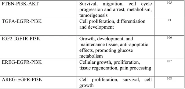
PTEN-PI3K-AKT Survival, migration, cell cycle progression and arrest, metabolism, tumorigenesis
105
TGFA-EGFR-PI3K Cell proliferation, differentiation and development
73
IGF2-IGF1R-PI3K Growth, development, and maintenance tissue, anti-apoptotic effects, promoting glucose metabolism
106
EREG-EGFR-PI3K Cellular growth, proliferation, tissue regeneration, pain processing
107
AREG-EGFR-PI3K Cell proliferation, survival, cell growth
108
Table 2. Signaling pathways involved in PI3K/AKT cascade
PI3K phosphorylates the phosphatidylinositol-3,4,5-trisphosphate (PI(3,4,5)P3), a fundamental second-messenger of survival signaling109. PI3K kinases are heterodimers made of a catalytic subunit
called P110 and a regulatory subunit named p85, which is activated by RTKs and by GPCR110–112.
The signaling downstream steps of RTK receptor include phosphorylation of the insulin receptor substrate (IRS)113 with the concomitant binding of the SH2-containing phosphatase (SHP-2)114 and
the phosphorylation of p85 subunit of PI3K115. This determines PI3K activation and, via PI(3,4,5)P3,
the stimulation of 3-phosphoinositide-dependent kinase-1 (PDK1) and the enhancement of the phosphorylation and, then, of the activity of AKT53,116–118.
Frequent genetic alterations, especially in cancer, were found in these signaling pathways: about 50% of melanoma patients present mutations in the serine/threonine kinase BRAF of melanocytes119,
which are regulated by the RAS/RAF/MEK/ERK MAPK pathway120, while PI3K pathway is often
dysregulated during melanomagenesis121. AKT itself is overexpressed during melanoma
progression122.
In more than 70% of papillary thyroid carcinoma, genetic alterations such as point mutations of BRAF and RAS gene, lead to an activation of MAPK123,124. Moreover, several aberrant RTKs and genetic
mutations result in a continuous activation of PI3K/AKT in its downstream effectors leading to a high cell proliferation in thyroid carcinomas125,126. Typical examples of genetic modifications are
represented by phosphatase and tensin homolog deleted on chromosome 10 (PTEN) encoding genes127, extra copies of phosphoinositide-3 kinase catalytic α (PIK3CA)128, phosphoinositide-3
kinase catalytic β (PIK3CB) and PDK1 encoding genes129.
1.3 Target therapy with BRAF inhibitors
BRAF gene, also known as BRAF proto-oncogene or serine/threonine kinase, is a human gene that allows the transmission of chemical signals from outside the cell to the nucleus130. The cytogenetic
location of BRAF gene is 7q34, which is the long (q) arm of chromosome 7 at position 34 and it encodes a protein belonging to the RAF family of serine/threonine protein kinases called BRAF. BRAF protein is a serine/threonine-specific protein kinase made of 766 amino acids and its mammalian RAF kinase family is composed by three RAF isoforms: A-RAF, BRAF and C-RAF131.
Several findings suggest that BRAF is the family member the most strongly involved in mediating MAPK activation132. In particular, it consists of three conserved domains characteristic of RAF kinase
family:
i) conserved region 1 (CR1), a Ras-GTP-binding self-regulatory domain;
ii) conserved region 2 (CR2), a serine-rich hinge region;
iii) conserved region 3 (CR3), a catalytic protein kinase domain that phosphorylates a sequence
of protein substrates.
Before becoming active, BRAF must be initially bound to RAS-GTP. Then, BRAF changes its conformation leading to a dimerization via hydrogen-bonding and electrostatic interactions of the kinase domains. BRAF catalyzes the phosphorylation of serine and threonine residues in a sequence of cascade proteins through the energetic contribution of ATP, yielding ADP and specific substrates of phosphorylated proteins as products. It is not surprisingly that, due to the high BRAF kinase activity compared to the other family members, a high frequency of BRAF point mutations and a constitutively activation of BRAF is observed in human cancers133,134. Mutations in this gene, mostly
V600E alteration, represent the most frequently detected cancer-causing mutations in melanoma (V600E has been examined in 66% of malignant melanomas)135–137 and in several other cancers
including non-Hodgkin lymphoma138, colorectal cancer139, thyroid carcinoma140, non-small cell lung
carcinoma141, hairy cell leukemia142 and adenocarcinoma of lung143. Approximately 80–90% of V600
BRAF mutations regard V600E144–146 and they deal with an amino acid substitution at position 600
in BRAF, from a valine (V) to a glutamic acid (E). This mutation is specifically localized to the serine/threonine kinase domain and occurs within the activation segment of the kinase domain, leading to a constitutive activation of the protein itself and insensitivity to negative feedback mechanisms147,148.
Among the BRAF mutations observed in melanoma, over 90 % are at codon 600 and, among these, over 90 % are single nucleotide mutations resulting in substitution of glutamic acid for valine149.
BRAFV600E mutation has been implicated in melanoma progression, through the activation of the downstream MEK/ERK within MAPK signalling pathway, evasion of senescence and apoptosis, angiogenesis, tissue invasion and metastasis process, and evasion of immune response137,150.
Activating mutation in the BRAF serine/threonine kinase represents also the most common genetic alteration in thyroid cancer, occurring in approximately 45% of papillary thyroid cancer, and in a lower proportion of poorly differentiated thyroid cancer (PDTC) and anaplastic thyroid cancer (ATC)140,151,152.
Over 95% of all BRAF mutations detected in thyroid cancer is a thymine to adenine transversion at exon 15 nucleotide 1799 (T1799A) of the BRAF gene leading to substitution of valine by glutamic acid at residue 600 of the protein chain (V600E)153. This alteration results in a constitutively active
BRAF molecule with sustained kinase activity that promotes chronic stimulation of MAPK pathway, thereby resulting in increased phosphorylation of downstream targets, including ERK kinase154. This
altered signalling results in an increased cell proliferation, survival and tumor progression, in a growth factor independent manner155.
In comparison to wild-type BRAF, activating mutations in BRAF are constitutively active and there is some evidence that these changes lead to bypass the dimerization process156. In melanoma, BRAF
V600E mutation permits BRAF to signal as a monomeric enzyme without the presence of activated RAS and upstream RTK inputs to amplify its dimerization157. In papillary thyroid cancer (PTC), the
BRAF V600E gene mutation is associated with more rapid cancer growth and a higher death rate158.
The discovery of genetic underpinnings in cancer has opened the way for targeted therapies consisting in drugs that directly target cells with specific gene changes, acting on specific cell processes and changing the way tumor cells signal to each other159–161. These drugs can stimulate the body to attack
or control the growth of cancer cells. Given the crucial role both in melanoma and in thyroid cancer, BRAF V600E represents the most promising therapeutic target for treatment of patients with metastatic melanoma and advanced thyroid cancer refractory to standard approaches162,163. Several
small molecule BRAF inhibitors have been developed during the last years, showing encouraging results in clinical trials both in melanoma and thyroid cancer164. These drugs work through a selective
competitive mechanism of action for the modified adenosine triphosphate binding site of the active forms of BRAF V600E kinase; in this way, they inhibit its ability to participate in MAPK pathway activation. Vemurafenib and dabrafenib have been shown to be highly selective for BRAF V600E mutant cells that are respectively 100 and 500 fold higher than those for cells with wild-type
BRAF165,166. Small molecules as vemurafenib, also known as PLX4032, and dabrafenib or
GSK2118436 belong to BRAF inhibitors and reduce or slow tumor growth in people whose metastatic melanoma has a BRAF gene change167. Such drugs can help some patients to live longer,
even though the melanoma typically starts increasing over again. Dabrafenib can be used after surgery in people with stage III melanoma (tumors that have spread to regional lymph nodes) and can contribute to decrease the risk of cancer recurrence.
MEK protein, which properties downstream from BRAF, represents an additional target in melanoma treatment because agents that block MEK proteins can indirectly help melanoma patients with BRAF mutation. MEK inhibitors as trametinib and cobimetinib have been shown to shrink BRAF-mutant
melanoma168. However, when used by themselves, these drugs do not seem to shrink the tumor as
BRAF inhibitors usually do. Hence, a combination therapy with BRAF and MEK inhibitors represents a more successful therapeutic approach in decreasing tumors for longer periods than administering either type of drug alone. The combination of dabrafenib with vemurafenib or trametinib has significantly extended the progression free survival compared to dabrafenib alone in advanced BRAF V600E mutated melanoma169–171 and in advanced BRAF V600E mutated anaplastic
thyroid cancer172,173. Unfortunately, response to BRAF inhibition differs among cell types: in most
patients who initially respond to these treatments, then resistance is inevitably acquired to BRAF inhibitors as cells develop alternative mechanisms to pathway activation. Up to now, several potential resistance mechanisms have been identified within the context of both cancers that lead to reactivation of the MAPK pathway171,174–176.
For what concerns melanoma, the resistance mechanisms include:
i) NRAS mutations177;
ii) activation of upstream RTKs (e.g.,insulin-like growth factor 1 receptor, platelet-derived growth factor receptor β [PDGFRβ], epidermal growth factor receptor [EGFR])178;
iii) BRAF V600E kinase splice variants unable to be inhibited by BRAF inhibitors179;
iv) transactivation of an uninhibited RAF dimer partner by the inhibited BRAF V600 mutant180;
v) acquisition of MEK-activating mutations181.
Thyroid cancer cells harboring BRAF V600E mutation show a not well-defined intrinsic resistance mechanism to BRAF kinase inhibitors (KIs). High levels of EGFR in thyroid cancer cells show a positive response to the combination of vemurafenib with an EGFR inhibitor, while poorly responses were observed when vemurafenib was administered alone172. In the light of this, EGFR expression
to be the deactivation of EGFR-negative feedback loops by vemurafenib and the following prompt activation of RTK183.
These mechanisms result in continuous signalling along the MAPK pathway or an alternative pro-survival pathway such as the PI3K pathway. Other alterations, both upstream and downstream of BRAF can alternatively activate other signalling pathways. Hence, growing concerns over drug resistance to molecular targeted therapies such as in melanoma and papillary thyroid cancer184,
including BRAF and MEK inhibitors, have stimulated researchers to discover alternative molecular targets for the treatment of disorders linked to BRAF mutations.
Pathways have not only facilitated researchers to understand the theoretical complexity of cell molecular mechanisms, at the same time, supported by the interdisciplinary framework of systems biology, but they have constituted a bridge for the development of innovative tools for complex biological events185.
From a systems biology perspective, there are many tools and resources for pathway analysis186–188,
an emergent discipline combining software tools, database models and computational algorithms with the aim to help biologists in converting protein interaction data into a set of computational models189.
High-throughput technologies also contribute to a significant amount of protein interaction data, generated through deep sequencing and microarrays190. All of these data help to acquire an overall
picture of cell regulatory processes.
An accurate classification of these methods remains discussable, according to the fact that pathway analysis methods evolve very fast. However, three main groups of methods in pathway analysis are available by now: i) Over-Representation Analysis or Enrichment Analysis (ORA); ii) Functional Class Scoring (FCS) and iii) Pathway Topology (PT).
ORA measures the percentage of genes in a specific pathway or any gene group (gene ontology (GO) groups, protein families and so on) that own differential expression. The main scope of ORA is to
provide a list of the most relevant pathways, sorted in accordance to a p-value. It is possible, hence, to identify relevant pathway, through the number of genes differently expressed in the experiment that pathways contain. The statistical significance of the correspondence among genes from a pathway and the list of differently expressed genes is determined by statistical tests as Fisher's test, hypergeometric distribution test and so forth191,192.
FCS analyzes the expression change of overall genes in the list (not ranking by statistical significance) of differently experimental expressed genes. FCS removes the ORA cut-off threshold limitation. The aim of FCS is to evaluate differently expressed genes enrichment scores using pathways as gene sets to execute their calculations193,194. One of the first and most popular methods using the FCS approach
is the Gene Set Enrichment Analysis (GSEA), also knowns as functional enrichment analysis195.
GSEA is useful to identify genes that display precise changes at the individual level and harmonious enrichment within a set196. It is worth mentioning that pathway enrichment methods can be
distinguished by the use or the absence of an explicit gene-wise statistic to measure the gene's association with a specific treatment and evaluate the relevance of a specific pathway in a specific treatment. Conversely, Gene Set Analysis (GSA)197 is able to rank genes applying the max-mean
statistic in order to summarize gene sets and re-standardize them for more accurate inferences.
PT is similar to FCS, but PT uses gene-level statistics through different databases integration. The critical difference is that PT is able to re-score the implication of a pathway as the connections change by taking into account the information about the role, the position and the direction of a biochemical interaction directly from the pathway database198. Oppositely, FCS will always provide the same
score. Signaling Pathway Impact Analysis (SPIA)199, EnrichNet200, Gene Graph Enrichment Analysis
(GGEA)201 and TopoGSA202 are some examples of PT approaches.
Thomas et al. suggested a method to analyse the topology of genes in a pathway employing a genetic algorithm for the estimation of the contribution of each gene, coupled with a system biology-based approach for the identification of significant perturbed genes in a specific pathway203.
Additionally, Bayesian Network (BN) models have gained popularity for learning biological pathways from microarray gene expression data204,205. Biological pathways as BN were modeled in
2014 by Korucuoglu et al., pioneers in developing the Bayesian Pathway Analysis (BPA) helpful for the identification of cancer-related pathways206.
At the same time, other methods such as cluster analysis with depth of inference approach207,
correlation statistics analysis208, weight matrices209, neural networks210, genetic algorithms211 and
supervised learning algorithms212 represent a helpful set of resources for pathway analysis and
discovery.
In this scenario, three major “actors” play a fundamental and necessary role in the game:
1. Databases: essential to storage molecular interaction network, collect pathways, molecular annotation and classifications (ontologies).
2. Algorithms: fundamental to allow the navigation of the network in the databases, the statistical analysis of the high - throughput data, the pathway inference and the network modeled. 3. Software client interface: useful for pathway and network layout and visualization.
1.4 Computational strategies for signaling networks
1.4.1 Database and tools for the visualization, simulation and analysis of signaling networks
A growing number of databases provides information and knowledge about function annotation, protein interactions and experimentally validated biological pathways. These databases represent excellent and essential resources to ease pathway predictions and models in general213,214. Most
researches and scientists have focused on integrating function annotation and protein–protein networks with expression data to ameliorate the accuracy and precision of a pathway model construction.
Several public universities have taken a pioneering lead in the attempt to become a fundamental authority for pathway and molecular interaction databases. In particular, Kyoto University has developed the Kyoto Encyclopedia of Genes and Genomes (KEGG) database promptly curated by its own staff and containing a comprehensive collection of pathways curated by scientists considered to be the top experts in the field215.
KEGG classifies pathways into seven specific classes: metabolism, genetic information processing, environmental information processing, cellular processes, organismal systems, human diseases and drug development. KEGG utilizes a sort of “maplink” to represent an interaction between a protein belonging to one pathway and a protein within the linked pathway. Moreover, KEGG associates more pathways from different species into one framework.
There are other databases that is worth mentioning for the investigation of biological pathway and the improvement of research in general:
A. WikiPathways: it is particularly useful to create manually electronic graphs of structured pathways both for cellular signaling and for metabolic processes216. Moreover, each pathway
sheet includes a brief description, a curated bibliography, an updated pathway version history and a list of all the component genes and proteins linked to public resources.
B. Gene Ontology (GO): it provides gene composition information of pathways pointing out the gene function and its relationships with three ontology categories, such as biological process, molecular function and cellular component217.
C. Protein ANalysis THrough Evolutionary Relationships (PANTHER): it represents a large curated biological database of gene/protein families based on a classification system that identify gene function and classify them with their functionally related subfamilies. PANTHER takes part of the Gene Ontology Reference Genome Project aimed to categorize proteins and genes for high-throughput analysis218.
D. Reactome: it is an open source curated bioinformatics database of human pathways and biochemical enzymatic reactions in which the concept of “reaction” is generalized through the typical transformations of specific entities such as nucleic acids, proteins (with or without post-translational modifications) and macromolecular complexes219. These transformations
include the transport of a specific entity from one compartment to another, the consequent interaction necessary to induce the protein complex formation and so on. This simple generalization allows to capture the range of biological processes that spans signaling, metabolism, transcriptional regulation, apoptosis and synaptic transmission in a single internally consistent, computationally navigable format.
1.4.2 Algorithms for Pathway visualization, investigation and prediction
Several applications can be found in the literature about two dimensional graph layout for pathway analysis220. The most popular are force-based and energy-based algorithms able to exploit the N-body
simulation method and helpful to reveal hubs and clusters in the signaling networks221.
Force-based algorithms are based on the ascription of definite physical properties to the nodes (treated as a set of interacting particles) and edges of the graph that will influence the equilibrium state of the entire system. This method uses both repulsive (i.e., electrostatic interaction between every pair of particles) and attractive forces (i.e., spring interaction along the graph edges) with the target to identify the stationary node position matching the equilibrium state of the entire system.
Energy-based algorithms are similar to force-based ones, as the equilibrium state of the system corresponds to a minimum of energy. These approaches offer a good basis for arbitrary graph placement but need huge computational resources222.
For every fixed node 𝑢̅ (𝑢̅ ∈ 𝑉) we have:
∑ 𝐹𝑟𝑒𝑝 𝑣∈𝑉 (𝑟𝑢̅− 𝑟𝑣) + ∑ 𝐹𝑎𝑡𝑡𝑟 (𝑢̅,𝑣)∈𝐸 (𝑟𝑢̅− 𝑟𝑣) = 0 (1)
Zero net force corresponds to the result of force-based algorithms, while the result of energy-based algorithms matches with the state corresponding to the minimum energy
⋃ = 𝑚𝑖𝑛 𝑚𝑖𝑛 [ ∑ ⋃ (𝑟𝑢− 𝑟𝑣) 𝑟𝑒𝑝 {𝑢,𝑣}∈𝑉 − ∑ ⋃ (𝑟𝑢− 𝑟𝑣) 𝑎𝑡𝑡𝑟 (𝑢,𝑣)∈𝐸 ] (2)
where 𝑈𝑟𝑒𝑝 and 𝑈𝑎𝑡𝑡𝑟 are absolute values of the repulsion and attraction potentials.
In both the approaches, the main goal is to achieve the stationary equilibrium for each node position. The force-based algorithm implementation consists of a solution of a nonlinear set of algebraic equations from a mathematical point of view, while the energy-based algorithm performs functional minimization. Both of these properties can be derived from one another.
For what concerns pathway investigation and prediction, genetic algorithms (GAs)223 and supervised
learning algorithms represent a class of methods to search knowledge and targets within gene interaction networks and also to automatically identify functionally cooperative genes.
GAs originate from the studies of cellular automata conducted by Holland and collaborators in the early 1970s224. From that moment forward, GAs have been increasingly applied to several
optimization problems in different fields, with a special attention in biomedicine. GAs consist on randomised, parallel search algorithms that model the principles of natural selection that leads to evolution. Like natural selection, GAs is a robust search method needing little information to explore effectively in large and poorly understood search spaces. In literature, several genetic algorithms were developed in order to take a data-driven approach to annotate and detect novel biological pathways, clusters in biological networks and to isolate functional and disease pathways.
Nguyen et al., described a method for orienting protein–protein interaction networks (PPIs) and discovering pathways, conducting multiple runs on the data of yeast PPI networks225. They tested the
best option for the problem through the design of a GA. Their GA is able to detect specific populations on a protein-protein interaction network and to evaluate their number and size against existing references.
Supervised learning algorithms are a class of machine learning algorithms that customs a known training dataset to make predictions. This dataset contains input data and response values from which the supervised learning algorithm tries to build a predictive model of the response values for a new dataset.
Dale and co-workers222 applied this machine learning method to obtain a metabolic pathway
prediction starting from a gold standard genome annotation dataset made of specific and well-known features, considered as input data. The final output was the estimation of the probability that a pathway is present or not in a specific organism.
1.4.3 Software for Pathway Analysis
Pathway analysis applications can be usually classified into: i) web-based applications; ii) desktop programs and, iii) programming packages.
Web-based applications for pathway analysis can offer both network visualization and simple analysis possibilities, such as STRING226, Cytoscape227, Ondex Web228, Visant229, CellMaps230.
Typical example of desktop programs are i) GSEA-P, a Java desktop program application for Gene Set Enrichment Analysis with a user-friendly graphic interface231; ii) Genome Informatics Data
Explorer (Guide), a desktop application designed to help biologists analyse RNA-seq and microarray gene expression data232.
Programming packages are principally written in R and Python languages, and are freely shared through the BioConductor233 project and GitHub234 service.
1.4.4 Algorithms in finding alternative pathways and new drug targets
One of the first undirected pathway prediction algorithms used was NetSearch235. Its function consists
on the enumeration of linear pathways and the generation of their ranking through a clustering of gene expression profile of each pathway entities. In parallel, NetSearch allows to generate a hypergeometric distribution-based score.
However, linear paths do not easily and fully capture the complexity of signaling networks. To this aim, researchers have been looking for other pathway prediction strategies. For example, Scott and colleagues236, employed a specific color-coding technique finalized to search paths and higher order
structures, namely trees and parallel paths, in a weighted protein interaction graph. Similarly to the work done by Scott and collaborators, Lu and co-workers237, showed a randomized
divide-and-conquer algorithm able to support complex non-linear pathway structures.
PathFinder, another tool commonly used for pathway analysis238, allows to: i) associate several data
sources and extract combining rules that describe protein function in a well-known signaling pathway;
ii) detect new pathways in the network of interest employing the extracted rules, along with
annotation and expression data.
These methods essentially are capable to search potential target pathways in an individual way, but it is worth to mention that other approaches, such as the one formulated by Zhao et al,239, own the
potentiality to recognize a single universal signaling subnetwork, using an unoriented edge selection algorithm240.
In this scenario, Yosef et al.241 tried to combine these two different approaches using an algorithm
able to recognize the trade-off between local and global search methods, and giving a preference to one or the other on a particular run of interest.
Even if these methods pointed out valuable outcomes, none of them is capable to generate directed pathways and specific targets. To overcome the orientation problem for length-bounded pathways in
weighted interaction networks, Gitter and collaborators242 implemented several algorithms based on
probabilistic selection and alternative methods to solve specific issues about weighted Boolean satisfiability (SAT)243. They applied these algorithms to PPI networks using simulated and
biologically derived sources and targets. These algorithms can recover many well-known pathways and improve upon previous approaches for pathway discovery, using real signaling networks. These algorithms are able to discover and analyzed pathways that do not appear in existing signaling databases and successfully they match notorious knowledge about the directionality of the interactions within pathways. Moreover, these algorithms rely on a number of reasonable biological assumptions including limiting the path length, using the confidence in the interaction edges and allowing for parallel pathways between sources and targets.
A very common need in pathway analysis is represented by the research of nodes that make significant contribution to a specific target. This is required, for example, in the identification of potential targets that may elicit an effect at cellular level through the controlling cascade of a specific biological function. These nodes, however, could not belong to the neighborhood of the biological target under investigation. It is also possible that effective targets could be hidden within pathways that are only connected to the analyzed one by only one or few nodes.
2
AIM OF THE THESIS
Computational modeling of signal transduction is currently attracting much attention as it promotes the understanding of complex signal transduction mechanisms. Although several computational models have been used to examine signaling pathways, little attention has been given to crosstalk mechanisms. In this PhD research project, we developed a computational model that automatically explores and detects the most relevant nodes within the MAPK and PI3K/AKT pathways, attributing a specific weight and simulating the dynamics of MAPK and PI3K/AKT signaling cascades involved in melanoma and thyroid cancer. Moreover, the dynamics of the protein activities were analyzed based on a set of kinetic equations fostered with data coming from both literature and experimental sources. In silico analysis revealed that the RAF and AKT pathways act independently in both diseases and that novel prognostic biomarkers and therapeutic targets could be identified for obtaining benefits and a more effective treatment in thyroid cancer and melanoma.
This PhD project has as the main goal the advancement of the state of the art in the development of computational strategies able to simulate both PI3K/AKT and MAPK pathways and their interactions, in order to deeper examine and investigate the cascade reactions involved in melanoma and thyroid cancer development and progression with a special attention to unresponsivity to conventional treatments.
3
COMPUTATIONAL MODELING IN MELANOMA
3.1 Melanoma background
Skin is the largest organ of the body and represents the first line of defense from external factors. To this aim, integumentary system protects body against pathogens and excessive water loss and is fundamental for vitamin D production, sensory stimuli and temperature regulation. Melanocytes are responsible for the production of melanin, a pigment that defends skin from damage effects of sunrays. In physiological conditions, melanocytes lead to dark agglomerations formation, visible on the skin, known as nevi244.
A tumor transformation of melanocytes could lead to cutaneous melanoma that represents a small percentage (about 5%) of all skin cancers245. It is estimated that one American dies of melanoma
every hour, and according to the American Cancer Society 3.5 million cases of basal and squamous cell skin cancer and about 73,000 cases of melanoma are diagnosed each year in the U.S., more than all other cancers combined, and 50 million people are treated for it annually246.
Epidemiological data coming from Italian Association of Tumor Registers (AIRTUM) report about 13 cases per 100000 people of melanoma in Italy, and a growing and doubled incidence in the last ten years. Cutaneous melanoma affects men and women around 40-50 years old and is quite rare in children. It originates from skin or pre-existing nevi that could be congenital, if present from birth, or acquired, if they appear during the course of life247. Cutaneous melanoma is classified into:
I. superficial spreading melanoma (the most common); II. lentigo malignant melanoma;
III. acral lentiginous melanoma; IV. nodular melanoma.
The main symptom of cutaneous melanoma is the change of a nevi shape or the formation of a new one. The characteristics of a nevi, that may indicate the onset of melanoma, are summarized in the acronym ABCDE248:
A= asymmetry form (a benign nevus has a circular form, while melanoma is more irregular);
B= border (irregular and indistinct);
C= color variable (with different shades within the same nevus);
D= dimensions (a clear increase in melanoma);
E= evolution of nevus (which also shows changes in a short time).
Other symptoms could be represented by a nevus that bleeds, surrounded by reddened area or itches.
Cutaneous melanomas are generally classified into four stages, I to IV, in which zero represents the melanoma in situ, restricted to the surface layer of the skin. These four stages are defined on the basis of the TNM system, which takes into account the characteristics of the tumor as the thickness, the rate of replication of cancer cells, the presence of ulcerations (T), the involvement of lymph nodes (N) and the possible presence of metastases (M)249. The prognosis is different according to the
thickness of the lesion.
The main risk factor for melanoma is an excessive exposure to ultraviolet light (UVA and UVB), mainly in the form of sun’s rays, but also of tanning beds that should be used with caution, without abuse. Prolonged exposure to UV radiation is potentially dangerous because it can damage the DNA of skin cells and trigger tumor transformation250. Other risk factors are the failure of the immune
system (due to previous chemotherapy treatments or transplant) and some genetic diseases such as xeroderma pigmentosum (in which patients DNA is not able to repair the damage caused by radiation). The risk increases in people with freckles, nevi, fair skin and eyes, in those who have a family history of melanoma and in those who was already affected from melanoma251.
To prevent the risk of development of skin cancer there are several good manners and recommendations that dermatologists suggest for a healthy skin. The European Society for Medical Oncology clinical practice guidelines for cutaneous melanoma highlight the importance of a detailed diagnosis for the establishment of the tumor stage and, in some tumors, a mutation test is also required252. A periodic skin self-examination usually allows to identify changes or suspicious nevi
and to consult a dermatologist in time. The dermatologist will assess a family history and the presence of typical signs and symptoms of cutaneous melanoma. The visual examination of the skin is more accurate when digital epiluminescence dermatoscope is used, in parallel to a magnifying glass and light illumination technique. However, the established clinical diagnosis of cutaneous melanoma requires a biopsy. Hence, early diagnosis is essential. Thanks to specific test analysis on tissue samples, it is possible to identify typical molecular mutations of different forms of cutaneous melanoma and establish relative prognosis and treatment. Diagnostic imaging tests such as x-rays of the chest, Computed Axial Tomography (CAT), Positron Emission Tomography (PET) and Magnetic Resonance Imaging (MRI) are useful to determine if and where the disease is spreading253.
Currently, the US Food and Drug Administration (FDA) have approved several options for treatment of cutaneous melanoma over the past years254. Current therapeutic approaches include surgical
resection, chemotherapy, photodynamic therapy, immunotherapy, biochemotherapy, and targeted therapy255. The therapeutic strategy can include single agents or combined therapies, depending on
the patient’s health, stage, and location of the tumor. The efficiency of these treatments can be decreased due to the development of diverse resistance mechanisms. New therapeutic targets have emerged from studies of the genetic profile of melanocytes and from the identification of molecular factors involved in the pathogenesis of the malignant transformation. The main approach is represented by surgery intervention that generally can permanently treat the disease at an early stage for patients with stage I-IIIB melanoma. Other following interventions depend on melanoma stage, as the removal of a portion of healthy tissue around the sick one. In some cases, the "sentinel" lymph nodes are also removed and radiotherapy is used as an adjuvant therapy after surgery or to treat cases
of recurrence. If melanoma has metastasized from the skin to other organs, the cancer is very unlikely to be treat by surgery. Even though resistance to apoptosis is probably the major cause of chemotherapy drug resistance in melanoma256, chemotherapy remains the most important palliative
treatment of refractory, progressive, and relapsed melanomas257. In Table 3, the most important and
conventional approaches in melanoma treatment258 are summarized and described in brief.
Unfortunately, for most of them, melanoma in its advanced stages, is generally considered to be resistant.
TREATMENT DESCRIPTION REFERENCE Dacarbazine (DTIC) For decades and until 2010, DTIC, a cell
cycle-nonspecific antineoplastic, was the current standard treatment for patients with inoperable metastatic melanoma. Nevertheless, it has never shown beneficial effects on survival of patients in phase III trials.
DTIC acts both as a cytotoxic agent and as an immunostimulatory drug because it is able to induce local activation of natural killer (NK) and T cells.
259,260
Temoxolomide (TMZ) TMZ is the first-choice treatment for patients with malignant melanoma who are not able to receive an intravenous chemotherapy
intervention. TMZ owns the same mechanism of DTIC, being the prodrug of DTIC and acting as an alkylating agent. TMZ demonstrates equal activity of DTIC with the advantage to be administered orally and to easily penetrate the blood-brain barrier.
261,262
Electrochemotherapy (ECT) ECT may be considered as an alternative strategy for local tumor control or as an additional treatment to the systemic ones. It uses a combination of physical properties of electroporation (through electric current) and chemical properties of chemotherapeutics with the aim to treat locally melanoma metastases.
263
Photodynamic therapy (PDT) PDT plays a possible role as an adjuvant therapy in the management of advanced melanoma (stage III and IV).
It involves a systemic or a local administration of a photosensitizer that, after its activation by irradiation, will take place within the tumor. The effects of PDT may be summarized as follows: induction of high levels of DNA damage, cytoskeleton alterations and enhanced pigmentogenesis.
264
Immunotherapy Since 2011, FDA has approved four new drugs for melanoma treatment. These drugs are ipilimumab, pembrolizumab, nivolumab, and talimogene laherparepvec (T-VEC). The last one is an oncolytic virus drug that stimulates stronger anti-tumor immune responses, while the other ones are checkpoint inhibitors that take the “brakes” off the immune system and enable it to fight cancer.
Oncolytic virus therapy This group includes viruses genetically modified as well as the ones found in nature to reproduce efficiently themselves in cancer cells without harming healthy cells. T-VEC (also known as OncoVEXGM-CSF) is approved from FDA for melanoma patients with injectable but non-resectable lesions in the skin and lymph nodes. It represents the first oncolytic virus approved for cancer therapy in the US.
268,269
IFN-α-2b FDA has approved high-dose of IFN-α-2b in patients after resection of high-risk melanoma (stage IIB and stage III).
IFN-α-2b may interfere with the growth of tumor cells and slow the growth of melanoma. Even though IFN is approved as adjuvant treatment in melanoma, in clinic its use is restricted due to the high toxicity and dubious effectiveness.
259,270
Peg-IFN On March 2011, pegylated-IFN-α-2b (Peg-IFN) has been approved for the treatment of melanoma patients with a high risk of recurrence after radical surgery and for adjuvant treatment of lymph node-positive melanoma. Peg-IFN owns a persistent absorption and a prolonged half-life; this means a better effectiveness compared with the non-pegylated form.
271
Biochemotherapy Biochemotherapy is defined as a therapeutic regimen that consists, at least, of a chemotherapy (in single or combination formulation) and IL-2. This strategy may provide better relapse-free survival (RFS) than high-dose interferon (HDI) in patients with high-risk melanoma.
272
IL-2 Immunotherapy based on IL-2 has been the mainstay of systemic therapy for advanced melanoma. When administered at high-doses, IL-2 produces a small number of durable remissions in patients with metastatic melanoma, and due to this fact, it was approved in the US in 1998 for metastatic melanoma. Unfortunately, IL-2 shows a significant toxicity and low-doses of IL-2 display low response rates and ineffectiveness. IL-2 effects consist on:
i)blockage of the reproduction and spread of
cancer cells; ii)stimulation of white blood cells helpful to attack cancer cells; iii)release of chemicals by cancer cells that attract cancer-killing immune system cells.
273,274
Treg inhibitors According to the major role of Tregs in promoting tumor progression, targeting Tregs seems to be promising approach in cancer immunotherapy. Several approaches have been developed for targeting Tregs, for example the depletion of Tregs, the suppression of Treg function, the disruption of Treg recruitment to the tumour microenvironment (TME) and inhibition of pTreg generation. Some examples of these therapeutic strategies are low‐dose chemotherapy drugs and immune checkpoint inhibitors (ICIs). The main idea is to target Tregs specifically in the TME rather than peripheral depletion to minimize the risk of autoimmune diseases.
275,276
CTLA-4 blockade Monoclonal antibodies directed against cytotoxic T lymphocyte–associated antigen-4 (CTLA-4), such as ipilimumab, harvests overall survival in patients with metastatic melanoma both in monotherapy and in combination with other checkpoint inhibitors.
277,278
PD-1/PD-1 ligand (PD-L1) blockade To maintain the discrepancy between immune surveillance and cancer cell proliferation, check-point antibody inhibitors, such as anti-PD-1/PD-L1, are a novel class of inhibitors that act as a tumor suppressing factor through the modulation of immune cell-tumor cell interaction. These checkpoint blockers are increasingly becoming a sophisticated and promising cancer therapeutic