Faculty of Medicine and Surgery
PhD in Neurobiology
Awarding University: University of Catania Associate University: University of Rome “La Sapienza”
Academic Year 2013-2014 PhD Thesis
Mechanism of action of Methylmercury on human astrocytes: neuroprotection of lipoic acid
Giuseppe A. Malfa
Tutor: Prof. Roberto Avola Co-tutor: Prof. Marcella Renis Coordinator: Prof. Roberto Avola
Ognuno sta solo sul cuor della terra trafitto da un raggio di sole: ed è subito sera. S. Q.
I am sincerely grateful to Prof. Roberto Avola for giving me the
opportunity to work on such an engaging and interesting project.
I am indebted to Prof. Marcella Renis for her endless encouragement, ever
useful advice and many discussions, which have guided me throughout
these years. She kindled my interest in biochemistry and molecular
biology and always gave me the freedom to find my own path during the
work. During my years at the university, I have learned so much from
them both and hope to continue doing so for many years to come.
I especially owe many thanks to my dearest colleague and friend Dr.
Barbara Tomasello who helped me in more ways than I could mention
during my PhD.
Special thanks also go to my dear collegues Prof. Sara Acquaviva, Prof.
Claudia Di Giacomo, Dr. Sonia Grasso, Dr. Vincenzo Bramanti and Dr.
Carla Motta for always being there for me.
I would like to thank my dear friend
David Shanahan for
proof-reading
the English manuscript and for his huge support.
My deepest gratitude goes to my loving parents who have always been
beside me and for their unconditional love and support.
ABSTRACT . . . .1
CHAPTER I - Introductio . . . 3
1.1 Toxic Metals . . . . . . 3
1.2 Mercury and Methylmercury . . . 7 1.3 Methylmercury Toxicity . . . 8
1.4 Physiology and Functions of Astrocytes . . . 11
1.5 Methylmercury Neurotoxicit y . . . 14 1.6 Methylmercury and Oxidative Stress . . . 17
1.7 Action of Methylmercury on Redox Systems . . . 18
1.8 Alpha-Lipoic Acid (ALA ) . . . 24
CHAPTER II - Aims . . . 28
CHAPTER III - Materials & Methods . . . 30
3.1 Chemicals and Reagents . . . 30
3.4 Lactate dehydrogenase (LDH) release . . . 32
3.5 Comet Assay . . . 32
3.6 Reactive oxygen species measurement . . . 33 3.7 GSH Thiol groups determination . . . 34 3.8 Western Blot Analysis . . . 34
3.9 Thiol labeling of proteins and electrophoresis 1D and 2D . . . 35
3.10 Carbonyl labelling of proteins and electrophoresis 1D . . . 36 3.11 Indirect Immunofluorescence (α-tubulin ) . . . 37
3.12 Direct Immunofluorescence (FITC- Phalloidin ) . . . 37
3.13 Scanning Electron Microscopy (SEM) . . . 38
3.14 Statistical analysis . . . 38
CHAPTER IV - Results . . . 39
4.1 MeHg Cytotoxicity and Genotoxicity on Human Astrocytes pretreated with ALA . . . 39
4.3 Intermediate Filaments (IF) expression increased with pre-treatment with ALA R+ . . . 45
4.4 Destabilisation of Human Astrocyte Cytoarchitecture as target of MeHg
neurotoxicity . . . 48 4.5 ALA enhances Cyclin D1 and MAPK expression and down-regulates NFkB signalling pathways in MeHg induced cytotoxicity . . . . . 53 4.6 ALA exerts a protective effect on some Redox parameters in HA exposed to MeHg . . . .56 4.7 Human Astrocyte stress response after MeHg treatments . . . 60 4.8 Measurement of Carbonyl and Thiols protein in HA treated with MeHg . . . . 65
CHAPTER V - Discussion . . . 71
CHAPTER VI - References . . . 77
Abstract
Astrocytes are actively involved in brain development, in mature CNS regulation and brain plasticity. They play a critical role in response to cerebral injuries and toxicants. A large amount of literature highlights the central role of astrocytes in mediating methylmercury (MeHg) neurotoxicity. In fact, mercury is the major neurotoxic pollutant that continues to arouse interest in research because of the severe risk it poses to human health. It has been widely demonstrated that this metal induces acute or chronic neurotoxicity exacerbating an oxidative stress condition and also age-related conditions, generating inflammatory reactions in the brain. These findings support the hypothesis that MeHg and many other environmental pollutants have the potential to cause neurodegeneration, through a variety of pathways similar to those described in human neurodegenerative diseases. However, despite numerous in vivo and in vitro studies, almost entirely on rat/mouse models, the issue remains controversial and the effects of MeHg in the human population are not completely known. Based on these findings, the aim of this study is to carry out for the first time, according to what has been reported in the literature, an in vitro experimental model that was as attributable as possible to humans, precisely human astrocytes (HA), so as to observe the effects of MeHg on some biochemical-molecular pathways. In parallel, I used HA cell cultures, which were treated with Alpha Dihydrolipoic Acid before being subjected to the action of MeHg in order to evaluate the possible protective effect exerted by this compound with antioxidant and chelating properties on cellular metabolism subjected to stress by MeHg.
Abstract
Gli astrociti sono attivamente coinvolti nello sviluppo del cervello, nella regolazione del sistema nervoso centrale maturo e nella plasticità cerebrale. Essi giocano un ruolo critico nella risposta a lesioni cerebrali e alle sostanze tossiche. Una grande quantità di letteratura evidenzia il ruolo centrale degli astrociti nel mediare la neurotossicità indotta dal metilmercurio ( MeHg ). Infatti, il mercurio è il principale inquinante neurotossico che continua a suscitare interesse nella ricerca a causa del grave rischio che comporta per la salute umana. È stato ampiamente dimostrato che questo metallo induce neurotossicità acuta o cronica esacerbando una condizione di stress ossidativo età correlato e generando reazioni infiammatorie nel cervello. Questi risultati supportano l'ipotesi che MeHg e molti altri inquinanti ambientali hanno il potenziale di causare neurodegenerazione, attraverso una varietà di percorsi simili a quelli descritti nelle malattie neurodegenerative umane. Tuttavia, nonostante numerosi studi in vivo e in vitro, quasi interamente su modelli di ratto/topo, la questione rimane controversa e gli effetti del MeHg nella popolazione umana non sono completamente noti. Sulla base di questi risultati, lo scopo di questo studio è quello di realizzare per la prima volta, secondo quanto riportato in letteratura, un modello sperimentale in
vitro che sia il più è possibile valido per l'uomo, attraverso l'impiego di astrociti
umani (HA ), in modo da osservare gli effetti del MeHg su alcune vie biochimico - molecolari. In parallelo, ho usato colture cellulari HA, che sono stati trattati con acido alfa diidrolipoico prima di essere sottoposto all'azione di MeHg, per valutare il possibile effetto protettivo esercitato da questo composto con proprietà antiossidanti e chelanti sul metabolismo cellulare sottoposte all'azione tossica del metallo .
CHAPTER I
Introduction
1.1 Toxic Metals
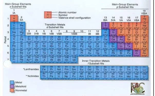
Metals are substances that account for three quarters of the elements in the periodic table (Fig. 1), but only some of them are essential for life. The majority of known metals are considerably toxic when present in excess, so their concentrations are strictly regulated at the cellular level. Man has been in contact with metals for thousands of years, but only in recent decades has the use of metals, and especially toxic metals, become very intense.
The progressive environmental and food pollution that characterises our industrialised society undermines, especially in some subjects, our detoxification system and this causes the spread of various chronic degenerative diseases including cancer, immunodeficiency, autistic spectrum diseases, Alzheimer's disease, fibromyalgia, multiple chemical sensitivity, chronic fatigue, etc. However, whether mercury, lead, aluminium, cadmium and arsenic play a principle role in these diseases is still not well-known.
Heavy metal pollution originates from several sources including coal, natural gas, chlorine-alkali industries and volcanic eruptions (Higueras P. et al. 2006), but the most common are represented by metal purification, copper smelting and from nuclear fuels (Zevenhoven R. et al. 2001). Marine water pollution is mainly due to the development of human activities that introduce, either directly or indirectly, substances capable of causing harmful effects in living organisms and, consequently, in human health into the aquatic environment (Fig 2). In particular, it depends on the contaminants carried into the sea by internal water basins, along which persist many industrial and agro-livestock enterprises, and urbanisation phenomena, while a significant proportion is due to direct input of municipal and industrial waste into coastal waters (Severino L. et al. 2007).
Although the adverse effects of heavy metals on human health have been known for a long time, exposure to such toxic agents continues, which is moreover on the rise in some geographical areas and is very worrying for some amongst them. An example is mercury, which is still used for gold mining in many parts of Latin America (Department of Environment, 2001).
Currently , the thirteen elements of greatest concern in the world are: As, Cd, Co, Cr, Cu, Hg, Mn, Ni, Pb, Sn and Ti (Zevenhoven R. et al. 2001), whose emissions are closely regulated in waste incinerators.
Figure 2. Sources of heavy metal pollution.
Heavy metals such as mercury, cadmium and lead are of particular concern because of their ability to bio-accumulate up the food chain until reaching highest contamination levels in predatory fish, such as tuna and swordfish (Storelli M.M. et al. 2005).
Some of these heavy metals are essential in small amounts (Co, Cu, Cr, Mn and Ni), but others are highly toxic (Hg, Pb and As). In particular, different heavy metals have different target organs. For example, Hg, Mn and Pb cause damage to the CNS, especially during its development stages. The kidneys and liver are susceptible to Cr and Cu, while the hard tissues such as bones and teeth are targets for Ni, Cd and Pb. Once released into the environment, heavy metal pollutants are able to localise and not decay; therefore, their toxicity may persist for generations. Man, therefore, may be exposed to chemical, physical and biological agents that are potentially harmful in
air, food, water or soil (Fig. 3).
Figure 3. Impact of environmental contaminants.
Exposure is a function of concentration and time. "It is an event that occurs for a certain period of time, when there is contact between the human being and the environment in which a contaminant is present at a specific concentration" (National Research Council, 1991). This stresses the importance of environmental factors and increases the possibility of an etiologic role in toxic exposures, which can be pre-natal or post-pre-natal, but also the combination of maternal and gestational effects, and direct exposure of the newborn (Adams J.B. et al. 2007). In the tissues of some people who have been exposed chronically, especially women, there may be (asymptomatically) high levels of one or more heavy metals that can, for the most part, be transmitted to the foetus, or poison infants through lactation (Younes B. et al. 1995). These metals interfere with reproduction and development, causing damage both during the growth of the foetus and at birth. They can also cause neurological damage, including developmental delays, learning disorders and behavioural
abnormalities (Fig. 4). Therefore, a potential diagnostic tool, which allows for the evaluation of previous exposure (Al-Ayadhi L. 2005), is the mineralogram analysis, generally carried out on a sample of hair taken, in appropriate conditions, from the nape. This investigation is backed up by the analysis of the levels of minerals in the urine, a useful method for detecting exposure levels among the various minerals. This is a very useful and often necessary practice to ensure an improvement in metabolism and also the elimination of toxic minerals.
Figure 4. Hg and MeHg toxicity from mother to foetus.
1.2 Mercury and Methylmercury
Mercury is a heavy metal whose presence in the environment is of both natural and anthropogenic origin. During the last century, industrialisation has significantly increased the release of this element into the environment (Mason R.P. et al. 1994). In recent decades, its industrial use has been greatly reduced, but its presence in the
environment appears to be constant, because of its elevated persistence in marine sediments, and because mercury discharged into the environment from natural sources, such as volcanic eruptions, arrives in the aquiferous system (Covelli S. et al. 2009). Once the mercury reaches the water, bacteria present in the oceans convert inorganic mercury into its organic form: methylmercury. This is the most prevalent form of mercury, the most toxic and the form that enters the food chain through plankton and then passes, through invertebrates and fish located at the lower levels of the food chain, to the large predators, where the highest concentrations are found (Fig. 5) (Mason R.P. et al. 1994; Covelli S. et al. 2009).
There is a greater accumulation in muscle tissue than in fat and about 90-99 % of the mercury we find is methylmercury (Ferrara F. et al. 2004).
The result is that the major source of human exposure is represented by consumption of certain types of fish, taking into consideration the type of product and place of capture (Clarkson T.W. 1997; Spry D.J. et al. 1991). The presence of mercury in food from the sea is not uniform. It depends on the concentration of Hg in the water and its pH, the redox potential, the species, age and size of specific fish. The conditions of the microenvironment, especially anoxia, favour the growth of microorganisms and increase the percentage of HG methylation (Boudou A. et al. 2005). But there are also other factors that can significantly increase the toxicity of the fish and / or harm to the individual, including the presence of other metals in the marine environment and, therefore, in fish. In this case, repeated exposure to the metal can lead to the manifestation of symptoms of intoxication. Even though exposure occurs at relatively low levels for each substance, we return to the concept of bioaccumulation (Wecker L. et al. 1985).
1.3 Methylmercury Toxicity
MeHg toxicity, exhibiting dyskinesia, mental retardation, primitive reflexes, growth disorders, salivation, intestinal dysbiosis and various other cognitive, learning and motility disorders (Tsubaki T. 1967; Harada M. 1995).
Figure 5. Routes of Transformation of Hg into MeHg.
From a clinical point of view, the symptoms of MeHg poisoning become significant when the levels of exposure to Hg exceed the values of 0.1 mg / Kg per day.
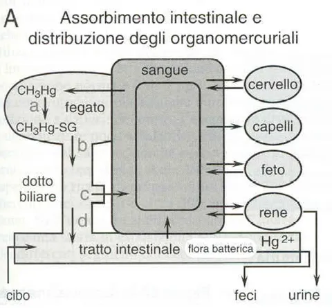
Inside the human body, methylmercury undergoes a redistribution from the gastro-intestinal tract to the majority of organs and systems. About 90 % is easily absorbed
and has a long retention time, with a half-life of about 70 days. Distribution in the blood, where it reaches about 7 % of the same ingested dose, occurs within 30 hours following ingestion ( Kershaw T.G. et al. 1980). The MeHg present in the systemic circulation accumulates mainly in red blood cells by binding to cysteine residues (-SH) also present in the beta chain of haemoglobin (Doi R. 1991), and is then slowly distributed in other tissues until reaching a balance over a number of days. (Fig. 6) ( Kershaw T.G. et al. 1980).
Figure 6. Absorption and elimination of MeHg.
In steady-state conditions, the ratio of MeHg concentrations in the blood and hair is approximately 1/250 (Skerfving S. et al. 1974), and in non-symptomatic conditions, the excretion rate of mercury is about 1 % of body weight (Swedish Expert Group 1971).
from the kidneys into the urine, but a substantial amount is reabsorbed in the intestine and enters enterohepatic circulation. The rate of excretion may also be affected by diet, as some dietary components may interfere with the absorption of MeHg in the lower intestine by interrupting enterohepatic circulation (Landry T.D. et al. 1979). A characteristic of this organic form of the metal is also its ability to cross the placenta and accumulate in the foetus in a concentration greater than that present in the mother. Therefore, its levels in umbilical cord blood are two times higher than those in the mother’s blood at delivery. About 5 % of the MeHg present in maternal blood is found in the breast milk (Bakir F. et al. 1973).
Today it is known that acute intoxication can evolve into pneumonia and pulmonary oedema, neuropathy, gastroenteritis, renal tubular necrosis and glomerular changes, while chronic poisoning leads to gingivostomatitis, pain in the limbs, cardiovascular disorders, tremor, neuropsychiatric disorders and in particular foetal and neonatal neurotoxicity and psychomotor retardation. MeHg, in the CNS, accumulates in particular in the glia, mostly in the astrocytes, which represent up to 50 % of the cell volume (Aschner M. et al. 2000).
1.4 Physiology and Functions of Astrocytes
Astrocytes, also known as astroglia, are a cell type of the CNS, present in the brain and spinal cord, characterised by a typical star shape due to the presence of numerous protrusions in their structure (Fig. 7). They play a fundamental role in maintaining the homeostasis of the CNS, participating in the formation of the blood-brain barrier, providing support and nutrients to neuronal cells and endothelial cells. They also play a major role in the processes of defence and regeneration of the brain and spinal cord following a trauma (Ramon Y. et al. 1909). They are also able to secrete growth factors, release cytokines and modulate synaptic transmission by removing neurotransmitters, such as glutamate, from the synaptic cleft (Schwabe T. et al. 2005;
Allen N.J. et al. 2005).
Astrocytes, considering morphological differences and different anatomical sites, can be classified into two main types (Ramon Y. et al. 1909):
1 - fibrous, with long, branched protrusions that wrap around the capillary wall;
2 - protoplasmic, present mainly in the grey matter, with short, widely branched protrusions. They are involved in maintaining extracellular ion balance, in the uptake of glutamate (Bechtholt-Gompf A.J. et al. 2010), in controlling GABA receptor function (Beenhakker M.P. et al. 2010), in controlling pH and in regulating the concentration of potassium (K +). The concentration of that ion in the extracellular fluid tends to increase due to the depolarisation of the neurons; this may cause interference in the normal propagation of an action potential from one neuron to another. Therefore, the presence of a high number of potassium channels in these cells facilitates the removal of the ion from the extracellular compartment, allowing the subsequent transfer to neighbouring cells via gap junctions (Walz W. 2000).
Figure 7. Human astrocytes.
Besides the presence of channels for potassium, astrocytes also present them for sodium; however, unlike neurons, they are not able to trigger or propagate any action potential (Nedergaard M. et al. 2003; Seifert G. et al. 2006). Nevertheless, it does not mean that astrocytes are physiologically silent. They show, in fact, a kind of cell excitability that, depending on the increase in the intracellular concentration of calcium (Ca++), represents the means of intercellular communication between astrocytes and between astrocytes and neurons (Halassa M.M. et al. 2007; Perea G. et al. 2009; Shigetomi E. et al. 2008; Volterra A. et al. 2005).
Recent studies have shown that these cells play a pivotal role in response to cerebral injury and their functional deficiency may contribute to the development of various neurodegenerative diseases, as well as being involved in 'brain ageing' (Araque A. 2006). A brain injury causes a reaction by one of the glial components, defined "reactive gliosis" (Pekny and Nilsson 2005; Norton W.T. et al. 1992). Astrocytes react to the injury by increasing in volume (hypertrophy), proliferating and starting inflammatory pathways, and so isolate the damaged area by forming a glial scar. This phenomenon is very useful in the short and medium term, because it contains the injured area and limits the inflammatory process (Kawano H. et al. 2012). In any event, in an excitotoxic state, the protective functions of reactive astrocytes, such as the uptake of glutamate, the maintenance of electrolyte homeostasis and the elimination of free radicals remain essential for the containment of neuronal damage (Iwata-Ichikawa E. et al. 1999; Chen Y. et al. 2001). It is clear that this kind of glial activation, typical of reactive gliosis, plays a key role in the mediation of the metabolic processes of neurons in physiological, toxic and neurodegenerative processes (Kawano H. et al. 2012).
1.5 Methylmercury Neurotoxicity
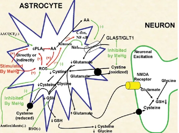
Astrocytes, even if not the only cell type to be negatively affected by exposure to MeHg, have been shown to play a key role in mediating neurotoxicity, which is induced by the organic form of this metal for several reasons (Fig. 8):
Astrocytes are the preferred site of accumulation of MeHg (Garman R.H. et al. 1975; Aschner M. et al. 2000);
MeHg inhibits the uptake of glutamate (and aspartate) and stimulates efflux. The resulting increased concentration of the two amino acids in the extracellular fluid sensitises neurons to excitotoxic injury (Allen J.W. et al. 2001; Brookes N. et al. 1989; Aschner M. et al. 1993; Dave V. et al. 1994); It has been shown that MeHg-induced neuronal dysfunctions are secondary to
disorders already present in astrocytes (Brookes N. 1992). In fact, the in vitro co-application of non-toxic concentrations of mercury with glutamate generate typical neuronal lesions, typical of excitotoxic stimulation (Matyja E. et al. 1993);
MeHg also promotes the cytotoxic cycle by activating cytosolic phospholipase A2 with the consequent release of arachidonic acid and inhibition of glutamate transport (Shanker G. et al. 2002; Shanker G. et al. 2003).
Figure 8. Schematic model proposed by M. Aschner et al. for MeHg neurotoxicity.
It should also be added, as previously mentioned, that as well as damaging the cells in different ways, mercury also binds to sulfhydryl groups, causes microtubules depolymerisation and neurotransmission alteration (Castoldi A.F. et al. 2001).
Since astrocytes are the cell type, which occupies up to 50 % of volume in the CNS, it is clear that one of the main causes of observed neurotoxicity is correlated to their dysfunction, resulting from harmful exposure to mercury (Chen S.H. et al. 2000). It is well known that upon contact with MeHg, about 10 % of this toxic form of the metal is retained in the encephalon, deposited preferentially in certain cell types of the CNS. This can be explained by considering the ease with which this element crosses the blood-brain barrier, accumulating in various brain areas, such as the cerebellum, cerebral cortex and retina (Mottet N.K. et al. 1984; Erie J.C. et al. 2005). The damage induced in the CNS, especially in a fully developed encephalon, is localised in certain areas of the visual cortex and includes the loss of the layer of granules of the
cerebellum. Differently, in a developing CNS, which is extremely sensitive to neurotoxicity induced by the metal, a diffuse cytoarchitecture disorganisation of the cerebral cortex is manifested, accompanied by the disappearance of the granule cells (Vettori M.V. et al. 2003). The onset of these kinds of alterations could also be caused by failure of the process of cell division, mediated by MeHg, in the course of the development phases of the CNS (Vettori M.V. et al. 2003).
Another mechanism of toxicity is the ability of the metal to inhibit the uptake of cysteine, an amino acid precursor in the synthesis of glutathione (GSH), whose levels cause a serious reduction with damage even at the neuronal level as these cells are replenished with GSH from astrocytes (Shanker G. et al. 2002; Allen J.W. et al. 2002). GSH, in fact, plays a key role as a scavenger of reactive oxygen species (ROS), whose concentration is known to increase in response to exposure to MeHg (Yin Z. et al. 2007), resulting in alteration of mitochondrial functions and an increase in oxidative stress, known to be involved in several neurodegenerative disorders, including Alzheimer's, Parkinson's and Amyotrophic Lateral Sclerosis (Kaltschmidt B. et al. 1993; Kitamura Y. et al. 1997). The increased production of MeHg-induced ROS in astrocytes and the consequent alteration of cellular homeostasis are phenomena also related to the increase in glutamate, which cause neurotoxicity. In fact, MeHg also induces inhibition of glutamate uptake in astrocytes with simultaneous stimulation of efflux of this amino acid, causing an excessive concentration at the synaptic level, and so generating neurotoxicity (Aschner M. et al. 1993; Yin Z. et al. 2007). The increase in the concentration of glutamate in the extracellular fluid, causes hyper-activation of the NMDA receptor for glutamate (N-methyl-D-aspartate) with consequent increase in the influence of ions Na+ and Ca++ (Choi D.W. 1992). In a sort of vicious circle, the increase in intracellular levels of Ca++ generates an increase in ROS (Lafon-Cazal M. et al. 1993), the production of which inhibits the transport of glutamate by astrocytes.
levels, increased levels of synaptic glutamate with increased expression of NMDA receptors on adjacent neurons, and therefore excitotoxicity.
Exposure to MeHg is also able to alter the cell cycle, interfering negatively with the proliferative process during the development phases of the CNS.
Glial cells provide neurotrophic signals to neurons, which are necessary for their survival, proliferation and differentiation. Various proteins are involved in these processes, such as:
proteins belonging to the cyclins family (cyclin D1);
the intermediate filament proteins: GFAP (glial fibrillary acidic protein), nestin and vimentin;
the MAP-kinase (mitogen-activated protein).
1.6 Methylmercury and Oxidative Stress
Although free radicals, reactive oxygen species (ROS) and reactive nitrogen species (RNS) are known to be key molecules in the cell where they act as "molecular beacons". An excess of these, caused by hyper-production or reduced scavenger activity of antioxidant systems, i.e. a condition known as oxidative stress is strongly implicated in several neurodegenerative diseases including Alzheimer's disease, Parkinson's disease and amyotrophic lateral sclerosis (Kaltschmidt B. et al. 1993; Hunot S. et al. 1997; Kitamura Y. et al. 1997), but also in other degenerative conditions such as autoimmune diseases and inflammatory diseases (e.g. ischemia and rheumatoid arthritis), cancer, diabetes mellitus and atherosclerosis (Kehrer J.P. et al. 1994; Halliwell B. et al. 1994; Betteridge D.J. 2000), as well as in the toxicity induced by the metal (LeBel C.P. et al. 1992).
ROS are known to mediate neurotoxicity induced by MeHg in different experimental models. For example, MeHg induces the formation of ROS in vivo (in the cerebellum of rodents), and in vitro (isolated from rat brain synaptosomes) (Ali S.F. et al. 1992),
as well as in neuronal cultures of the cerebellum, in a neuronal hypothalamic cell line and in mixed aggregates cell cultures (Sarafian T.A. et al. 1994; Park S.T. et al. 1996; Sorg O. et al. 1998). Furthermore, an increase in ROS was observed in: a) isolated mitochondria from a rat brain in which MeHg was injected (Yee S. and Choi B.H. 1996), b) isolated mitochondria in vitro from a rat brain and subsequently exposed to MeHg (Myhre O. and Fonnum F. 2001), and c) mitochondria of astrocytes and neurons exposed to Hg and glutamate (Dugan L.L. et al. 1995; Brawer J.R. et al. 1998). The production of ROS and glutamate dyshomeostasis induced by MeHg are related phenomena that influence each other.
1.7 Action of Methylmercury on Redox Systems
The interaction of MeHg with nucleophilic groups of biomolecules with both low and high molecular weight and its affinity for -SH groups, support and confirm the observations that oxidative stress is a central event in mediating MeHg-induced neurotoxicity (Aschner M. et al. 2007); in fact, levels of GSH (glutathione) are exhausted after MeHg intoxication.
MeHg targets specific proteins containing thiol groups and selenium that trigger molecular mechanisms involved in secondary MeHg neurotoxicity. In particular, MeHg can interrupt the activity of proteins containing a thiol group or selenium, such as glutathione peroxidase (GPX), thioredoxin (Trx) and thioredoxin reductase (TrxR) (Carvalho C.M. et al. 2008; Farina M. et al. 2009; Franco J.L. et al. 2009; Glaser V. et al. 2010; Branco V. et al. 2011), all major components of the cellular antioxidant system, thus contributing to the rupture of the normal redox balance of brain cells (Farina M. et al. 2011).
However, even if the fundamental role of oxidative stress in MeHg-induced neurotoxicity has been identified, the underlying molecular mechanisms are not yet
fully understood.
In fact, the initial oxidative damage caused by methylmercury in astrocytes, as described above, is also supported by its reaction with endogenous molecules containing thiol (-SH) or selenium (-SEH) proteins at the base of the cellular redox buffer, causing the formation of a very stable complex of the RSHgCH3 or
RSeHgCH3 type (Aschner M. et al. 2011; Farina M. et al. 2011, Branco V. et al.
2011) and in the case of proteins and enzymes containing these groups, the S-Hg Se-Hg bond formation can affect its function.
The thiol groups are much more abundant than the selenium groups (Nogueira C.W. and Rocha J.B.T. 2010), They are, in fact, found in proteins of low molecular weight (primarily cysteine and reduced glutathione) and high molecular weight, while the selenium groups are found in a small group of selenium-proteins (Araie H. and Shiraiwa Y. 2009; Lobanov A.V. et al. 2009; Lu J. and Holmgren A. 2009), but these groups may be critical for the catalytic activity of several enzymes (such as glutathione reductase etc.), being involved in intermediary metabolism and antioxidant processes. Since MeHg is able to influence enzymatic activity, it is reasonable to hypothesise that this interaction is responsible for imbalances in oxidative metabolism, as well as increasing levels of reactive species. In figure 9, some metabolic pathways in which some reactive species are the mediators of MeHg-induced neurotoxicity are shown:
Hydrogen peroxide (H2O2) is quantitatively the most important peroxide
generated by the cells (Dringen R. et al. 2005) and the main sources of H2O2
are the disproportionation of mitochondrial superoxide anion (Inoue M. et al. 2003) and reactions catalysed by oxidases, such as monoamine oxidase (Nicotra A. et al. 2004). Although it is known that hydrogen peroxide carries out important physiological functions in the modulation of cellular functions, high levels of H2O2 are dangerous for the cells, in large part due
and Day E.D. 1978).
MeHg is able to selectively inhibit transport of cysteine and cysteine in astrocytes, thereby negatively influencing their oxidation-reduction state and reducing glutathione content (GSH), an important antioxidant for humans, thus generating oxidative stress (Allen J.W. et al. 2001; Shanker G. et al. 2003);
Figure 9. Reactive radical species as mediators of MeHg neurotoxicity.
The superoxide anion (O2•-), a by-product of the normal operation of the mitochondrial respiratory chain is produced after the one-electron reduction of molecular oxygen. Even the superoxide anion plays an important role in MeHg-induced oxidative damage. Shanker and collaborators (2004), using a specific probe for the superoxide (hydroethidine), observed an increase in levels of ROS in cultured astrocytes treated with MeHg.
cytoplasmic glutathione peroxidase (GPx1) and peroxiredoxin (Prx) (Winterbourn C.C. and Hampton M.B. 2008). With particular attention to the pro-oxidant effects of MeHg, the evidence shows that it has the ability to increase levels of H2O2 in various experimental conditions (Manfroi C.B. et al.
2004; Shanker G. et al. 2004; Franco J.L. et al. 2007; Mori N. et al. 2007). The increased levels of H2O2 observed after exposure to MeHg represent the
consequences of various phenomena, such as the inhibitory effects of MeHg on glutathione peroxidase.
GPxs represent a family of selenoproteins whose catalytic activity depends on the reducing power of a selenium group, located in the active site (Brigelius-Flohé R. 2006). Due to the high affinity of MeHg for selenium groups, the decreased activity of GPX after exposure to MeHg has been attributed to direct inhibitory events (Farina m. et al. 2009). Furthermore, a recent study has proposed another molecular mechanism to explain the reduced activity of GPx after exposure to MeHg: MeHg induces a condition called “selenium deficiency,” which affects the synthesis of GPx through a post- transcriptional effect (Usuki F. et al. 2010), thus leading to decreased levels of GPX, reduced peroxidase activity and, consequently, higher levels of hydrogen peroxide.
Another mechanism linked to increased levels of H2O2 after exposure to MeHg
seems to be the direct effect of this toxic substance on the whole antioxidant system of glutathione.
In addition to direct depletion of reduced glutathione, MeHg also hinders the physiological maturation of many enzymes involved in the metabolism of GSH, thus leading to increased levels of H2O2 in the brain (Stringari J. et al.
2008).
While it is known that increased levels of H2O2 is a consequence of exposure to
MeHg, the precise role of this molecule in mediating MeHg-induced oxidative damage has not yet been fully determined.
Nitric oxide (NO) is a neurotransmitter widely used as a signalling molecule by all cells of the body. Although NO plays a number of important roles as a regulator of diverse biological processes (vascular tone, platelet adhesion, neurotransmission), it can, however, also cause deleterious effects, including inhibition of some enzymatic functions, the promotion of DNA damage and the activation of inflammatory processes (Hollenberg S.M. and Cinel I. 2009). Nitric oxide is synthesised from L-arginine and oxygen in a reaction catalysed by nitric oxide synthase (NOS), which has at least three distinct isoforms (inducible and constitutive), (NOS1, NOS2 and NOS3). Indeed, while the activity of NOS2 (inducible isoform) depends on transcription, NOS1 and NOS3 are constitutively expressed and are usually triggered by an increase in intracellular calcium (Alderton W.K. et al. 2001). In particular, an increase in the activity of the NOS enzyme and increased NO levels have been reported in MeHg-mediated neurotoxicity (Yamashita T. et al. 1997; Herculano A.M. et al. 2006), although the mechanisms involved in the interaction between MeHg and NO are not entirely clear.
Figure 10. A model that represents the role of oxidative stress in MeHg-induced neurotoxicity.
A number of articles indicate that activation of some signal molecules is induced by oxidative stress (for example, NFkB, protein-1 activator, c-fos, c-jun, etc.). This activation leads to the induction of various target genes (i.e. inducible NOS, COX and SOD, the inducible form (HSPs-72), cytokines, etc.), that contribute to cellular damage (Kumagai Y. et al. 1997; Hsieh H.J. et al. 1998; Goering P.L. et al. 2000). However, to date there is a lack of systematic studies that examine and correlate the formation of ROS in astrocytes with MeHg-induced toxicity, possibly examining the role of different signal pathways involved in the process. Figure 10 shows the objectives that may be affected by MeHg, as proposed by Aschner M. et al. (2007). Considering that hydrogen peroxide is involved in MeHg toxicity, both in vivo and in
vitro, numerous studies have examined the protective effects of treatment with
compounds) (Allen J.W. et al 2001 a, b; Farina M. et al. 2003; Ballatori N. et al. 1998). It has been shown that organoselenium compounds with thiol peroxidase activity exert protective effects, maintaining levels of H2O2 low in systems exposed to
MeHg (Farina M. et al. 2005; Moretto M.B. et al. 2005).
Although N-acetylcysteine, the precursor of GSH, may contribute to the maintenance of homeostasis of intracellular GSH, which is crucial in the detoxification of hydrogen peroxide by glutathione peroxidase, some of the beneficial effects elicited by N-acetylcysteine in in vivo conditions are also linked to its ability to accelerate the urinary excretion of MeHg in poisoned animals (Koh A.S. et al. 2002). To date, the use of molecules with antioxidant activity to prevent/reduce MeHg neurotoxicity continues to have great reticence. Currently, the only way to prevent or ameliorate the effects of MeHg toxicity caused by poisoning is to accelerate the elimination from the body through the following various therapeutic strategies that the medical expert must be able to adapt from individual to individual: hemodialysis, exchange transfusions and chelation therapy performed by intravenous perfusion based on disodium salt of Ethylene-Diamino-Tetraacetic acid (EDTA) (Clarkson T.W. et al. 1981; Lund M.E. et al. 1984).
In addition to traditional chelation therapy, in less serious conditions of intoxication, you can resort to oral "chelates" such as zeolite, chlorella and alpha-lipoic acid.
1.8 Alpha-Lipoic Acid (ALA)
ALA (Figure 11), an essential molecule for cell energy metabolism, a cofactor at entry to Krebs cycle, displays anti-oxidant effects by increasing glutathione peroxidase activity and reducing oxidative stress (Mantovani G. et al. 2003), regulates calcium homeostasis (Sen C.K. et al. 1996), and modulates the activity of the transcription factor NFkB (Packer L. 1998; Packer L. et al. 1997). Indeed, several studies have shown that ALA exerts multiple pharmacological actions that can
prevent nerve degeneration in experimental in vitro models of diabetes (Vincent A.M. et al. 2005), Parkinson’s disease (Bharat S. et al. 2002), and Alzheimer's diseases (Abdul H.M. and Butterfield D.A. 2007). It also inhibits oxidative stress in HIV infection in experimental studies (Packer L. et al. 1995) and reduces damage from ischemia-reperfusion in the central nervous system and cardio-vascular system in animal studies (Cao X. and Phillis J.W. 1995; Freisleben H.J. 2000).
Figure 11. Alpha-lipoic acid
Alpha-lipoic acid, also known as thioctic acid, was first isolated from bovine liver in 1950 (Reed L.J. 2001). Lipoic acid contains two thiol groups, which may be oxidised or reduced. Like the thiol antioxidant glutathione, ALA is part of a redox pair, being the oxidised partner of the reduced form dihydrolipoic acid (DHLA). Unlike glutathione, of which only the reduced form is an antioxidant, both the oxidised and reduced forms of lipoic acid are antioxidants. ALA is reduced in vivo to its dithiol form, DHLA, which also possesses biological activity. DHLA is a potent reducing agent with the capacity to reduce the oxidised forms of several important antioxidants, including vitamin C and glutathione (Jones W. et al. 2002). Although
reduced glutathione has twice the chemical reactivity in its thiol group, DHLA is superior to glutathione in regenerating vitamin C (Suh J.H. et al. 2004). ALA is a naturally occurring compound that is synthesised in small amounts by plants and animals, including humans (Smith A.R. et al. 2004). Endogenously synthesised ALA is covalently bound to specific proteins, which function as cofactors for mitochondrial dehydrogenase enzyme complexes. In addition to the physiological functions of protein-bound ALA, there is increasing scientific and medical interest in potential therapeutic uses of pharmacological doses of free ALA (Kramer K. et al. 2001). ALA exists as two enantiomers: the R-enantiomer and the S-enantiomer. Naturally occurring ALA is in the R-form, but synthetic ALA is a racemic mixture of R- and S- forms. Both forms seem to have different potencies; it was previously shown that the R-form is more potent than the S-form in its ability to stimulate glucose uptake in L6 myotubes, (Estrada D.E. et al 1996) as well as to increase insulin-stimulated glucose uptake in obese Zucker rats (Khanna S. et al. 1999). On the other hand, the S-form exerts a slightly increased affinity for glutathione reductase (Pick U. et al 1995), thus the two forms of ALA differ in the potency with which they exert various biological activities. As stated by Packer et al. (1997), an ideal therapeutic antioxidant should fulfil several criteria. ALA is unique among natural antioxidants in its ability to fulfil all of these requirements, potentially making it a highly effective therapeutic agent in a number of conditions in which oxidative damage has been implicated. The antioxidant properties of ALA comprise the following:
1. Its capacity to directly scavenge reactive oxygen species (ROS);
2. Its ability to regenerate endogenous antioxidants, such as glutathione, vitamins E and C;
3. Its metal chelating activity, resulting in reduced ROS production.
Due largely to its antioxidant properties, ALA has recently been reported to afford protection against oxidative injury in various disease processes, including
neurodegenerative disorders (Packer L. et al. 1997; Evans J.L. and Goldfine I.D. 2000). Although the ability of ALA to directly scavenge ROS appears to be responsible, at least partially, for its neuroprotective effects, it remains unknown whether the neuroprotective effects of ALA may also occur through other mechanisms, such as induction of the endogenous antioxidants and phase 2 enzymes in neuronal cells. ALA might also be able to induce endogenous antioxidants and phase 2 enzymes in neuronal cells, and the increasing endogenous defences may afford protection against oxidative/electrophilic neuronal cell injury.
ALA has neuroprotective effects in neuronal cells. One possible mechanism for the antioxidant effect of ALA is its metal chelating activity (Ou P. et al. 1995).
In a further study, Müller U. and Krieglstein J. (1995) tested whether pre-treatment with ALA can protect cultured neurons against injury caused by cyanide, glutamate, or iron ions. Neuroprotective effects were only significant when pre-treatment with ALA occurred for 24 h.
CHAPTER II
Aims
In light of what has been stated about MeHg toxicity and the lack of knowledge about the molecular mechanisms underlying its toxicity, most importantly in humans, I designed and developed the research subject of this thesis using human astrocyte cultures treated with MeHg, both at 24 h (acute treatment) and 72 h (chronic treatment), as my experimental model. The overall goal of my work was to carry out
in vitro research using, for the first time, according to what has been reported in the
literature, an experimental model that was as attributable as possible to humans, precisely human astrocytes, so as to observe the effects of MeHg on some biochemical-molecular pathways. In parallel, I used human astrocyte cell cultures, which were treated with Alpha Dihydrolipoic Acid before being subjected to the action of MeHg in order to evaluate the possible protective effect exerted by this compound with antioxidant and chelating properties on cellular metabolism subjected to stress by MeHg. Firstly, I examined the expression of some proteins capable of exhibiting possible astrocytic reactivity and the alteration of the cell cycle (GFAP, vimentin, nestin, etc) and, immediately afterwards, the protein expression of the cytoskeleton, in order to highlight a possible disintegration of the cytoskeleton (β-tubulin and actin). This research was supported by studying, in parallel, the cellular structure, which was carried out with electronic microscope analysis (SEM). The next step was to study some key markers of oxidative damage (ROS levels and GSH), and to analyse the expression of some proteins involved in the oxidative stress-induced alteration of cellular homeostasis, such as HSPs 27-60, e70KDa, iNOS, GGT1 and SOD, and the expression levels of metallothioneins. Before going on to study redox parameters in depth, I examined the expression levels of well-known markers of
apoptosis, caspase-3, AIF and Cyt C, in order to understand if programmed death was started in the cells (apoptosis), in both acute and chronic treatment. In all of the aforementioned phases, I always evaluated the effects of pre-treatment with lipoic acid by analysing the results, paying attention to possible differences between acute and chronic treatments. Therefore, encouraged and motivated by the results obtained in the first and second years, to study further the treatment-induced modulation of the cellular redox state and with the precise intention of individuating new specific targets, I went on to examine the modifications induced in both carbonyl and thiol groups, using redox proteomics. I then separated the proteins present in the cell lysate with both one-dimensional and two-dimensional electrophoresis, marking the free thiol groups with 5’-iodoacetamidofluorescein (IAF). The fluorescence images of all gels, 1D and 2D, were subsequently acquired and analysed using dedicated software.
CHAPTER III
Materials and Methods
3.1 Chemicals and Reagents
MeHg was purchased from Sigma Chemical Co. (St. Louis, MO, USA). Astrocyte Growth Supplement (AGS) was obtained from ScienCell Research Laboratories (USA). Dulbecco’s Modified Eagle’s Medium (DMEM), penicillin-streptomycin, trypsin-EDTA, foetal bovine serum (FBS) were obtained from GIBCO BRL (Invitrogen Corp., Carlsbad, CA, USA). ALA raceme or (R+) enantiomer were kindly provided by the School of Pharmacy, University of Camerino.
3.2 Primary Human Astrocyte Cell Line and Treatments
Primary Human Astrocytes (HA) were obtained from ScienCell. Cells were cultured and plated onto 75 cm2 cell culture flasks and grown at 37 °C (humidified 5 % CO
2
and 95 % air at 1 atmosphere) in DMEM supplemented with 10 % FBS, 1 % AGS, 100 Units/ml penicillin and 100 μg/ml streptomycin. When the culture reached the sub-confluence, the cells were treated with MeHg 1.125 µM for 24 h or 72 h. This concentration was chosen based on preliminary experiments to assess toxicity (data not shown). In order to simulate acute and chronic damage conditions, two different times of exposure (24 h and 72 h) were used. The cells were plated at a constant density to obtain identical experimental conditions in the different tests and cells not exposed to MeHg were used as a reference.
Astrocyte cultures were maintained under the following experimental conditions: • Untreated control HA non-pre-treated or pre-treated with 50 µM ALA raceme or (R+) enantiomer for 24 h
• Chronic treatment of HA with MeHg 1.125 µM for 24 h, non-treated or pre-treated with 50 µM ALA raceme or (R+) enantiomer for 24 h
with 0,5 mM glutamate for 24 h
• Acute treatment of HA with MeHg 1.125 µM for 72 h, non-treated or pre-treated with 50 µM ALA raceme or (R+) enantiomer for 24 h
ALA was dissolved in 0.5 M DMSO and then diluted in the culture medium at a concentration of 1 mM. Catalase (1000 U/ml) was added and the solution was incubated at 37 °C for 30 min. This solution was diluted in the culture medium in order to obtain 50 μM ALA final concentration and 1:10 in the culture medium in order to obtain 100 μM ALA final concentration.
3.3 Mitochondrial functionality assessed by MTT assay
HA were treated with MeHg (1.125 µM) for 24 h/72 h and non-treated or pre-treated with 50 µM ALA raceme or (R+) enantiomer for 24 h. The MeHg cytotoxic effect was evaluated as previously described (Malfa et al. 2010). This cell viability test is based on the conversion of 3-(4,5-dimethylthiazol-2-yl)-2,5-diphenyltetrazolium bromide (MTT) into insoluble blue formazan crystals by mitochondrial dehydrogenases of metabolically active cells. The optical density of each well sample was measured with a microplate spectrophotometer reader (Titertek Multiskan, Flow Laboratories) at λ 550 nm. The results are reported as a percentage of cell viability with respect to untreated control cells.
3.4 Lactate dehydrogenase (LDH) release
Lactate dehydrogenase (LDH) activity, as a membrane breakdown marker, was spectrophotometrically determined, both in the culture medium and in the cell lysate, at λ 340 nm, by analysing the decrease in NADH absorbance during the pyruvate– lactate transformation as described by Murphy and coll. (Murphy T.H. And Baraban S.M. 1990). The reaction was carried out in 100 mM potassium phosphate buffer (pH 7,5); 10 mM sodium pyruvate; 1 mM NADH. Briefly, the amount of LDH released was detected for each sample in 100 µl of supernatant medium obtained by centrifugation at 400 g for 10 min. The obtained pellet (dead cells floating in the medium) was mixed together with the corresponding cells in the dish. Cells were lysated with digitonin (2.5 mg/ml), centrifugated at 10,000 g for 15 min and endocellular LDH was measured on 30 µl cellular lysate. Values were expressed as a percentage of LDH released in the medium with respect to total LDH, considered the sum of the enzymatic activity present in the cellular lysate (intracellular) and that in the culture medium (extracellular). The decrease in NADH absorbance (Δmin) was measured by spectrophotometer (Hitachi U-2000) at λ=340 nm.
3.5 Comet Assay
Standard Comet assay was performed utilising Trevigen (Trevigen Inc., Gaithersburg, USA) pre-treated slides. For each treatment the aliquots of HA cells were embedded in 45 µl of low melting agarose (stabilised at 40 °C), dropped on the slides and maintained at 4 °C for 20 min. Then all the gel-supporting slides were immersed in lysis buffer (2.5 NaCl, 100 mM Na2EDTA, 1 % Triton X100, 1 % N-laurosyl sarcosine, 10 % DMSO, pH = 10) for 1 h at 4 °C, in order to leave gel-embedded DNA in the form of nucleoids. DNA unwinding was left for 20 min in fresh electrophoresis buffer A (300 mM NaOH, 1 mM Na2EDTA, pH = 13.1), afterwards, alkaline version of Comet assay was performed by submarine electrophoresis in the
same re-circulating pH 13.1 buffer A at 0.7 V/cm for 20 min and constant temp. (4 °C), as already reported (Renis M. et al. 2008). The samples, after the neutralisation treatment were 70 % ethanol dried and stained with SYBER GREEN (Trevigen kit) according to protocol suggestions. One hundred nucleoids per sample (50 for each of the two replicate slides) were randomly analysed using the Leica epifluorescence microscope All the operations were kept in the dark.
The DNA damage was quantified by CASP free software utilising as values the percentage of fragmented DNA in the tail of comet (% TDNA). The data is averaged from three different experiments in which each sample was processed in duplicate.
3.6 Reactive oxygen species measurement
The assay was performed, as previously described (Renis M. et al. 2000), by using the fluorescent probe dichlorodihydrofluorescein diacetate. 20,70-Dichlorodihydrofluorescein diacetate diffuses through the cell membrane; in the cytosol it is enzymatically hydrolysed by intracellular esterases and oxidised to the fluorescent 20,70-dichlorofluorescein in the presence of ROS.
The fluorescence (corresponding to the radical species oxidised 20,70-dichlorofluorescein) was monitored spectrofluorometrically using a Hitachi F-2000 spectrofluorimeter (Hitachi): lexcitation ¼ 488 nm, lemission ¼ 525 nm. A positive control was performed in each experiment by treating the cells with 200 mM-H2O2
for 20 min before adding 20,70-dichlorodihydrofluorescein diacetate to the cells. The total protein content was evaluated for each sample according to Bradford M.M. (1976). The results are reported as fluorescence intensity per mg protein and compared to the relative controls.
3.7 GSH Thiol groups determination
GSH thiol groups were measured, in 200 μl of plasma, using a spectrophotometric assay based on the reaction of thiols with 2,2-dithio-bis-nitrobenzoic acid (DTNB) at _=412 nm (Di Giacomo C. et al. 2003). The amount of thiol groups was calculated using an absorbance of 13,600 cm-1 M-1. Results are expressed as μmol/ml lysate.
3.8 Western Blot Analysis
HA were washed twice with ice-cold PBS, collected by lyses buffer (10 mM Tris– HCl plus 10 mM KCl, 2 mM MgCl2, 0.6 mM PMSF and 1 % SDS (pH 7.4),
incubated for 20 min at 0 °C and then sonicated. Equal amounts of total protein (30 μg/lane), measured according to Bradford M.M. (1976), were separated by different concentrations of TGX™ precast gel electrophoresis (Bio-Rad Laboratories, Inc.) and transferred into nitrocellulose membranes (Bio-Rad Laboratories, Inc.) with a semi-dry system. After verifying the protein transfer by Ponceau S staining, the membranes were blocked with Tris buffered saline containing 0.01 % Tween 20 (TBST) and 5 % non-fat milk, washed briefly, incubated with primary antibodies at 4 °C overnight and then incubated with corresponding conjugated secondary antibodies for 2 h at room temperature. All primary monoclonal and secondary antibodies were purchased from Santa Cruz, CA, USA (NFkB: sc8008; GAPDH: sc-365062; cyclin D1: sc-718; p38 MAPK: sc-4708; nestin: sc-23927; Cyt c: sc-13561; AIF: sc-9416; caspase 3: sc-7272; GGT1: sc-20639; SOD: sc-11407; iNOS: sc-7271; HSPs 70: sc-1060; HSPs
60: sc-13966; HSPs 27: sc-13132; MT: sc-11377), except for monoclonal antibodies
GFAP and vimentin (GFAP: MAB360 Clone: GA5 Chemicon society; vimentin: MAB3400 Clone: V9 Chemicon society); all the concentrations employed were set up experimentally according to the manufacturer’s instructions. The complex protein-primary-secondary antibody was detected by chemiluminescence methods using the
Gel Logic 2200 Imaging System. Chemiluminescent signals were densitometrically analysed using Kodak-MI software. Values were expressed as arbitrary densitometric units (A.D.U.). The values reported have been normalised with respect to
glyceraldehyde-3-phosphate dehydrogenase (GAPDH) levels.
3.9 Thiol labelling of proteins and 1D and 2D electrophoresis
Thiol groups in protein extracts of HA were labelled by adding 5′-iodoacetamide fluorescein (5′-IAF) and dimethyl sulfoxide (DMSO) to a final concentration of 800 μM and incubating at room temperature for 2 h in the dark. Proteins were then resolved using 1DE on 12 % polyacrylamide gels (Laemmli U.K. 1970). Gels were scanned in a Typhoon9400 scanner (GE Healthcare, UK; EXmax 490–495 nm; EMmax 515–520 nm) and were subsequently stained with Coomassie G250. Equal amounts of protein were loaded in 12 wells (3 replicates for each treatment) and repeated at least 3 times.
2D-SDS PAGE analysis was performed on protein extracts of control and treated samples incubated with 5′-IAF as described above (Tedesco S. et al. 2010). Samples (180 μg) were then precipitated with TCA/acetone and re-suspended in rehydration buffer containing 5 M urea, 2 M thiourea, 2 % CHAPS, 4 % ampholyte (Pharmalyte 3-10, Amersham-Pharmacia Biotech, Little Chalfont, Bucks., UK), 1 % Destreak reagent (Amersham-Pharmacia Biotech) and trace amounts of bromophenol blue. A final volume of 125 μl was loaded on 7 cm IPG strips pH 3–10NL on the bench overnight. Proteins were focused on a Protean IEF Cell (Bio-rad) with linear voltage increases: 250 V for 15 min: 4000 V for 2 h; then up to 20,000 V h. After focusing, strips were equilibrated for 15 min in equilibration buffer (6 M urea, 0.375 M Tris, pH 8.8, 2 % SDS, 20 % glycerol) containing 2 % DTT and then for 15 min in equilibration buffer containing 2.5 % iodoacetamide. Equilibrated strips were electrophoresed on 12 % SDS PAGE gels at a constant voltage (150 V) at 4 °C. 2D
Gels were scanned (Typhoon9400 scanner) as described above to reveal thiol-containing proteins labelled by 5′-IAF. Gels were then visualised by silver staining
(Rabilloud T. 1992) which revealed total protein. 2D-SDS PAGE gels were performed in digestive gland at least three times respectively for representative samples of control and MeHg, pre-treated or not with ALA.
For each 1DE gel, all bands detected by Typhoon9400 scanner were subsequently analysed by Quantity One image analysis software (Bio-Rad, Hercules, CA, USA) measuring the total intensity for each lane. They were quantified as arbitrary units (A.U.). All 1DE gels stained with Coomassie blue G250 were scanned in a GS-800 calibrated densitometer (Bio-Rad Laboratories) and the optical density from each lane was measured by Quantity One Image analysis software as described above. Total optical densities for each lane were normalised with those from Coomassie staining from the same gel. An average of at least three replicates from three different extracts for each treatment and tissue studied was determined.
3.10 Carbonyl labelling o
f proteins and
1D
electrophoresis
Protein extracts (70 μg) of each sample were brought to a final volume of 100 μl and treated with 1 ml of fluorescein-5-Thiosemicarbazide (FTSC) (100 mM) and incubated for 2 h in the dark at 4 °C. The proteins were precipitated with an equal volume of cold trichloroacetic acid (TCA) to 20 % (v / v) and then centrifuged at 11,000 g for 3 minutes at 25 °C. The pellets were re-suspended and washed with ethanol/ethyl acetate (1:1) x 5 times to remove FTSC excess. The final pellet was re-suspended in rehydration buffer containing 5M Urea, Thiourea 2M, CHAPS 2 %, 4 % ampholyte (Pharmalyte 3-10, Amersham-Pharmacia Biotech, Little Chalfont, Bucks. UK) and a minimal amount of blue bromophenol (Chaudhuri A.R. et al. 2006). The proteins were separated by one-dimensional electrophoresis (1DE) on 12 % polyacrylamide gels (Laemli U.K. 1970). The fluorescent images of the gels relative to the electrophoretic run was acquired by Typhoon 9400 scanner (GE Healthcare,
UK; ExMax 490-495 nm; Emmax 515-520 nm) and normalised by referring to the pictures of the same gels stained with Coomassie G250. Each experiment was set up in triplicate and repeated 3 times. The images of all 1DE gels, acquired by Typhoon9400 scanner, were subsequently analysed using the Quantity One software (Bio-Rad, Hercules, CA , USA), to quantify the total carbonyl-marked protein content of each well, obtained from all bands in the lane and expressed as arbitrary densitometric units (A.D.U.). The same gels stained with Coomassie blue G250 1DE were analysed with a densitometer, GS-800 (Bio-Rad Laboratories) and analysed with the same Quantity One software. The A.D.U. obtained from the analysis of fluorescence were normalised with those corresponding to the same gel stained with Coomassie blue G250 and expressed as mean ± SD.
3.11 Indirect Immunofluorescence (α-tubulin)
For the immunofluorescence staining, cells were fixed in 3.7 % paraformaldehyde in PBS for 30 min and permeabilised with 0.2 % Triton X-100 in PBS for 5 min at room temperature and blocked in 6 % BSA/PBS. After three washes in PBS, samples were incubated with 1:2000 mouse monoclonal anti α-tubulin (clone B 5-1-2; Sigma- Aldrich). After washes in 1 % BSA/PBS, samples were labelled with Alexa Fluor 594 Signal-Amplification Kit (Molecular Probes, Inc. OR, USA) for 1 h at room temperature in the dark. Finally, samples dried and mounted with mounting medium were examined with an Olympus BX50 and photographed with a Leica DC500 digital camera.
3.12 Direct Immunofluorescence (FITC- Phalloidin)
The analysis of cytoskeleton microfilaments was performed using FITC-Phalloidin (Sigma-Aldrich, Inc., St. Louis, MO, USA). Fluoriescein-labelled phalloidin was used as described by Dejana E. et al. (1988). Samples dried and mounted with mounting medium (Calbiochem) were visualised with an Olympus BX50 and
photographed with a Leica DC500 digital camera.
3.13 Scanning Electron Microscopy (SEM)
The morphological analysis was carried out on cell samples allowed to adhere to circular glass coverslips, 13 mm ø (EMS: Electron Microscopy Sciences, Fort Washington, PA, USA). HA were fixed in 2 % glutaraldehyde in 0.1 M sodium-cacodylate (EMS) buffer, pH 7.2, for 1 h at 4 °C and then postfixed in 1 % osmium tetroxide (EMS) for 1 h at 4 °C. After dehydration in graded ethanol and critical point drying using CO2 (Emscope-CPD 750), the coverslips were coated with vacuum
evaporated gold (Emscope-SM 300) and observed using a Hitachi S4000 Field Emission Scanning Electron Microscope.
3.14 Statistical analysis
Each experiment was repeated three times in triplicate and the means and standard deviations for each value were calculated. Statistical analysis of results was performed using paired Student’s t-test with the statistical software OriginPro 8.5. Differences were considered significant at p ≤ 0.05.
CHAPTER IV
Results
4.1 MeHg Cytotoxicity and Genotoxicity in Human Astrocytes
pre-treated with ALA
We preliminarily analysed whether MeHg treatment and ALA pre-treatment have an impact on HA survival, evaluating the cytotoxic and protective effects by MTT.
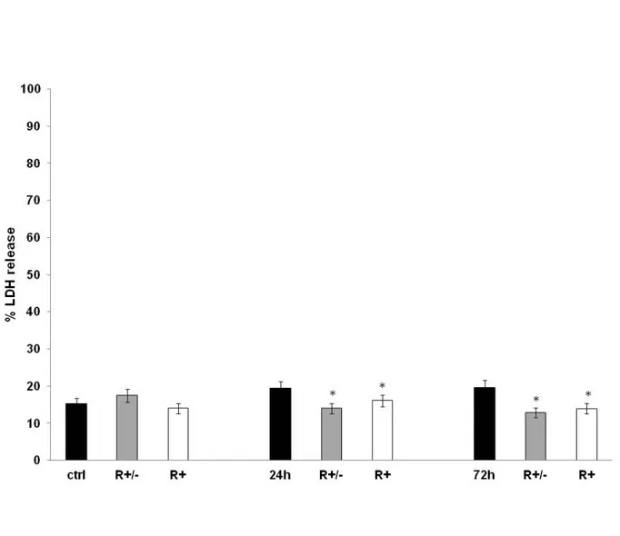
Figure 12 reports the results of cell viability showing that 24 h and 72 h after MeHg treatment cell survival was significantly reduced by approximately 40 % (p<0.05 vs control). Conversely, HA pre-treated with 50 µM ALA raceme or (R+) enantiomer for 24 h show significantly increased cell survival with respect to the non-pre-treated cells. It is clear that ALA, in particular or (R+) enantiomer, exerted a protective effect on HA treated with MeHg for 24 h and 72 h. Figure 13 shows the data of the release of lactate dehydrogenase in HA treated with MeHg compared with the untreated control and with astrocytes pre-treated with 50 µM ALA raceme or (R+) enantiomer. The results clearly indicate that methylmercury, at the concentration used, did not induce cytolysis in human astrocytes. The values, in fact, are completely superimposable in respect to the untreated control cells or those pre-treated with 50 µM ALA raceme or (R+) enantiomer.
DNA damage, assessed by alkaline comet assay and shown in Table 1, is expressed as % TDNA, or rather the percentage of DNA in the tail.
The results show that MeHg at 1.125 µM does not exhibit any genotoxic activity for 24 h and 72h, and also in the samples pre-treated with 50 µM ALA raceme or (R+) enantiomer.
Figure. 12. Cell viability analysis (MTT assay) performed on primary human astrocytes (HA) seeded in 96-well plates and treated with MeHg 1.125 μM in combination with Lipoic Acid (R± or R+) either for 24 h or for 72 h. The results are expressed as a percentage of the control value. #, * Mean values were significantly different from those of the control group or the corresponding 24 /72 h MeHg treatment (P<0.05) respectively.
Figure 13. LDH levels release in cultured primary human astrocytes (HA) treated with MeHg 1.125 μM for 24 h or for 72 h in combination with a pre-treatment with Lipoic Acid (R± or R+). The results are expressed as % release as Mean ± SD of three independent experiments each performed in duplicate. #, * Mean values were significantly different from those of the control group or the corresponding 24 /72 hr MeHg treatment (P<0.05) respectively.
Table 1. The data shown the results of the Comet assay at pH> 13 performed on primary human astrocytes (HA) treated with MeHg 1.125 μM for 24 h or for 72 in combination with a pre-treatment with Lipoic Acid (R± or R+). The results are expressed as% TDNA and represent the mean ± SD of three experiments performed in duplicate. Any significant differences are observed
4.2 Apoptosis Assessment after MeHg exposure of Human Astrocytes
pre-treated with ALA
The histograms related to the expression levels of the following proteins: Caspase 3, AIF and Cytochrome c are represented in figures 14, 15, 16 respectively. The results clearly indicate that methylmercury, at the concentration used, did not induce caspase 3 expression. In fact, there are no significant differences in expression levels in HA treated with MeHg for 24 h and 72 h in respect to the untreated control cells or those pre-treated with 50 µM ALA raceme or (R+) enantiomer (Fig. 14).
In figure 15, AIF expression is significantly down-regulated only at 72 h in respect to the untreated control cells (Fig 5, p<0.05 vs control). Moreover, pre-treatment with
ALA raceme or (R+) enantiomer did not change protein levels in respect to the untreated control cells or those treated with MeHg for 24 h and 72 h.
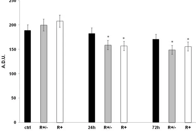
Figure 16 demonstrates that cytochrome c protein showed a peculiar expression trend: it reached the highest levels 24 h after MeHg treatment, but does not persist until 72 h when cytochrome c levels were slightly significant in respect to the control values (p< 0.05). ALA raceme or (R+) enantiomer pre-treatment attenuates the up-regulation of cytochrome c at 24 h of MeHg treatment and markedly decreases levels of the protein at 72 h of MeHg treatment to below the control levels.
Figure 14. Immunoblotting analysis for Caspase 3, performed on untreated HA and HA treated with MeHg 1.125 µM in combination with Lipoic Acid (R± or R+) either for 24 h or for 72 h. Values are expressed as arbitrary densitometric units (A.D.U.) corresponding to signal intensity and represent the mean of three independent experiments ±SD. #, * Mean values were significantly different from those of the control group or the corresponding 24 /72 hr MeHg treatment (P<0.05) respectively.
Figure 15. Immunoblotting analysis for AIF, performed on untreated HA and HA treated with MeHg 1.125 µM in combination with Lipoic Acid (R± or R+) either for 24 hr or for 72 hr. Values are expressed as arbitrary densitometric units (A.D.U.) corresponding to signal intensity and represent the mean of three independent experiments ±SD. #, * Mean values were significantly different from those of the control group or the corresponding 24 /72 hr MeHg treatment (P<0.05) respectively.