Periodontal evaluation of restorative
and prosthodontic margins
Edoardo Ferrari Cagidiaco
Digitally signed by
Edoardo Ferrari Cagidiaco 2021-03-08 14:02:15 +0100
UNIVERSITY OF SIENA
School of Dental Medicine
Department of Medical Biotechnologies PhD PROGRAM:
“BIOTECHNOLOGIES: SECTION OF DENTAL BIOMATERIALS”
PhD THESIS OF
Edoardo Ferrari Cagidiaco TITLE
Periodontal evaluation of restorative and prosthodontic margins Academic Year 2019/2020
February, 26th 2021
Siena, Italy
Committee
Promoter Prof. Simone Grandini Prof. Francesco Dotta
Prof. Francesco Iannelli Prof. Andrew Keeling Prof. Lorenzo Leoncini Prof. Teresa Marafioti Prof. Teresa Mezza Prof. Susanna Morano Prof. Maurilio Ponzoni Prof. Roberto Sorrentino Prof. Fernando Zarone
CANDIDATE
CONTENTS
Introduction
1.1 Prosthodontic and periodontal correlation on teeth...3 References...7
Chapter 2
2.1 Differences on different types of preparations...9
References...11 2.2 RCT on single zirconia crowns with knife edge vs chamfer finish lines: four-year
results...13
References...23
Chapter 3
3.1 FIT a new clinical score to evaluate single crowns...33 References...43 3.2 A pilot trial on lithium disilicate partial crowns using a novel prosthodontic
Functional Index for Teeth (FIT)...34 References...43 3.3 A randomized controlled clinical trial on two types of lithium disilicate partial
crowns...48 References...58 3.4 A randomized controlled clinical trial on press and block LiSi partial crowns: a
pilot study...61
References...69
2
Chapter 4 4.1 Evaluation of clinical failure, survival and success...78
References...79
4.2 Influence of cervical margin relocation (CMR) on periodontal health: 12- month results of a controlled trial...80
References...93
Chapter 5 5.1 Summary, conclusions and future directions...97
Bibliography...99
Curriculm Vitae...114
3
Chapter 1
Introduction
1.1 . Prosthodontic and periodontal correlation on teeth
In the daily dental practice 3 fundamental/empiric/clinical parameters have a role to establish the clinical success of prosthodontic treatment: function, aesthetic and longevity of the restorations. But, from a scientific point of view, how do we rate the success of the restorations?
When analyzing the existing literature, it can be noted that many authors focus their attention on the precision of the margin, to pursuit a small gap between the abutment and the crown, and to achieve the clinical success.
Christensen et al.1 and Mc Lean & Von Fraunhofer2 investigated the margins’ clinical acceptability
by dentists and asked to measure the gap between the abutment and the crown to a number of practitioners: it was shown that a clinician can clinically appreciate a gap not lower than 120 microns using a sharp explorer. This result may end in a not sure and sufficient seal between the crown and abutment, and consequently leakage at the margins.
This finding is not in agreement with the existing data coming from an in vitro study in which the acceptable marginal gap is lower than 50 microns3 Sorensen3 reported that small defects less or
equal then 0,050 mm were associated with significantly less fluid flow and bone loss than defects exceeding this value.
Martignoni4-5 reported that there are variable definitions regarding what constitutes a margin that
cab ne clinically acceptable, and there is no definite threshold for the maximum marginal discrep-ancy that is clinically acceptable. Many authors accept the criteria established by McLean and Von Fraunhofer2, they completed a 5-year examination of 1000 restorations and concluded that 120
microns should be considered the maximum marginal gap.
The adaptation, the precision and the quality of the restoration margin can be of greater significance in terms of gingival health, than the position of the margin6.
According to Lang et al. 7 following the placement of restorations with overhanging margins, a
subgingival flora was detected which closely resembled that of chronic periodontitis. Following the placement of the restorations with clinically perfect margins, a microflora characteristic for gingival health or initial gingivitis was observed.
intra-sul-4
cular margins were not predisposed to unfavorable gingival and microbial responses8.
Even among patients receiving regular preventive dental care, subgingival margins are associated with unfavorable periodontal reactions9.
Ercoli and Caton10, in a systematic review, describe how placement of restoration margins within
the junctional epithelium and supracrestal connective tissue attachment can be associated with gingival inflammation and, potentially, recession or periodontal pocket. The presence of fixed pros-theses finish line within the gingival sulcus or wearing of partial, removable dental prospros-theses does not cause gingivitis if the patients are complaint with self-performed plaque control and periodic maintenance. Procedures adopted for the fabrication of dental restorations and fixed prostheses have the potential to cause traumatic loss of the periodontal supporting tissue. They concluded that restoration margins located within the gingival sulcus do not cause gingivitis if the patients are complaint with self-performed plaque control and periodic maintenance.
Tooth-supported and/or tooth-retained restorations and their design, fabrication, delivery, and ma-terials, have often been associated with plaque retention and loss of attachment. Restoration mar-gins placed within the junctional epithelium and supracrestal connective tissue attachment can be associated with inflammation and, potentially, recession. Factors related to the presence, design, fabrication, delivery and materials of tooth-supported prostheses seem to influence the periodon-tium, generally related to localized increase in plaque accumulation and, less often, to traumatic and allergic reactions to dental materials10.
Jansson showd that the influence of a marginal overhang on pocket depth and radiographic attach-ment decrease with increasing loss of periodontal attachattach-ment in periodontitis-prone patients, and the effect on pocket depth of a marginal overhang may act synergistically, potentiating the effect of poor oral hygiene11.
Subgingival restorations with their apical borders still located subgingivally after periodontal treat-ment should be regarded as a risk factor in the progression of periodontitis12. Consequently,
place-ment of the restoration margin supragingivally is recommended, especially in periodontitis-prone patients with an insufficient plaque control12.
Dental restorations may be suggested as a risk indicator for periodontal disease and tooth loss. Routine SPT (Supportive Periodontal Therapy) was found to be associated with decrease in the prevalence of deep PPD over time, and it is of the utmost importance in maintaining periodontal health, especially adjacent to teeth with restorations. Finally, these findings may support the treat-ment of caries lesions and faulty restorations as part of a comprehensive cause-related therapy and
5 should be followed by a regular maintenance program13.
The relationship between dental restorations and periodontal status has been examined for some time. Research has shown that overhanging dental restorations and subgingival margin placement play an important role providing an ecologic niche for periodontal pathogens14.
An overhanging dental restoration is primarily found in the class II restoration, since access for interdental finishing and polishing of the restoration, and cleansing is often difficult in these areas, even for patients with good oral hygiene. Many studies have shown that there is more periodontal attachment loss and inflammation associated with teeth with overhangs than those without. Pres-ences of overhangs may cause an increase in plaque formation15-21 and a shift in the microbial
composition from healthy flora to one characteristic of periodontal disease14.
The location of the gingival margin of a restoration is directly related to the health status of the ad-jacent periodontium8. Numerous studies8-12-25
have shown that subgingival margins are associated with more plaque, more severe gingival inflam-mation and deeper periodontal pockets than supragingival ones. In a 26-year prospective cohort study, Schatzle et al. 25 followed middle class Scandinavian men for a period of 26 years. Gingival
index, and attachment level were compared between those who did and those who did not have restorative margins greater than 1mm from the gingival margin. After 10 years, the cumulative mean loss of attachment was 0.5 mm more for the group with subgingival margins. This was statistically significant. At each examination during 26 years of the study, the degree of inflammation in the gingival tissue adjacent to subgingival restorations was much greater than in the gingiva adjacent to supragingival margins. This is the first study to document a time sequence between the placement of subgingival margins and periodontal attachment loss, confirming that the subgingival placement of margins is detrimental to gingival and periodontal health.
Plaque at apical margin of a subgingival restoration will cause periodontal inflammation that may in turn destroy connective tissue and bone approximately, 1-2 mm away from inflamed area14.
Determination of the distance between the restorative margin and the alveolar crest is often done with bitewing radiographs; however, it is important to remember that a radiograph is a 2-dimension-al representation of 3-dimension2-dimension-al anatomy and structure. Thus, clinic2-dimension-al assessment and judgment are important adjuncts in determining if, and how much, bone should be removed to maintain ade-quate room for the dento-gingival supra crestal connective tissue height attachment14.
Although surface textures of restorative materials differ in their capacity to retain plaque26, all of
6
This includes underside of pontics. Composite resins are difficult to finish interproximally and may be more likely to show marginal defects than other materials28. As a result, they are more likely
to harbor bacterial plaque29. Intra-subject comparisons of unilateral direct compositive “veneers”
showed a statistically significant increase in plaque and gingival indices adjacent to the composites, 5-6 years after placement28. In addition, when a diastema is closed with composite, the restorations
are often overcontoured in the cervical-interproximal area, leading to increased plaque retention28.
As more plaque is retained, this could pose a significant problem for a patient with moderate to poor oral hygiene14.
For that, in absence of more specific prosthodontic parameters to evaluate the integration of crowns in to the periodontal environment, another way to determine the success and health of the resto-ration is to use the periodontal parameters such as: PPD (Periodontal Probing Depth) that is the measurement of the periodontal sulcus/pocket between the gingival margin and the bottom of the sulcus/pocket; REC (Recession) is the apical migration of the gingival margin measured with the distance between the gingival margin and the CEJ (Cement-Enamel Junction); PI (Plaque Index) the index records the presence of supragingival plaque; BOP (Bleeding On Probing) the presence or not of bleeding on surfaces of the teeth during the probing.
The aim of this study/thesis was to propose a clinical procedure to evaluate single unit restorations and their relations with periodontal tissues by a new clinical score: the FIT ( Functional Index for Teeth). FIT, that is a novel index for the assessment of the prosthetic results of lithium disilicate crowns, based on seven restorative-periodontal parameters, that evaluate crowns placed on natu-ral abutments, and want to be a reliable and objective instrument in assessing single partial crown success and periodontal outcome as perceived by patients and dentists.
7
References
1. Christensen GJ. Marginal fit of gold inlay casting. J Prosthet Dent. 1966;16:297-305.
2. McLean JW, von Fraunhofer JA. The estimation of cement film thickness by an in vivo tech-nique. Br Dent J 1971;131:107–11.
3. Sorensen J. A standardized method for determination of crown margin fidelity. J Prosthet Dent.1990;64:18-24.
4. Martignoni M, Schonenberg AJ. Limits of manual crown margin formation (1) Quintessenz Zahntech 1990 Jul;16:769-85.
5. Martignoni M, Schonenberg AJ. Limits of manual crown margin formation (2) Quintessenz Zahntech1990 Aug;16:883-94.
6. Richter-Snapp K, Aquilino S A, Svare C W, Turner K A. Change in marginal fit as related to margin design, alloy type, and porcelain proximity in porcelain-fused-to-metal restorations J Prosthet Dent:1988 60:435-9.
7. Lang N P, Kiel R A, Anderhalden K. Clinical and microbiological effects of sub-gingival resto-rations with overhanging or clinically perfect margins, J. Clin. Periodontol. 10 (1983) 563–578. 8. Kancyper SG, Koka S. The influence of intracrevicular crown margins on gingival health:
prelim-inary findings. J Prosthet Dent 2001:85:461-465.
9. Bader JD, Rozier RG, McFall WT Jr., Ramsey DL. Effect of crown margins on periodontal con-ditions in regularly attending patients. J Prosthet Dent 1991;65:75-79.
10. Ercoli C, Caton J. Dental prostheses and tooth-related factors. J Periodontol 2018;89(suppl 1):s223–s236.
11. Jansson L, Ehnevid H, Lindskog S, Blomlof L. Proximal restorations and periodontal status. J Clin Periodontol 1994:21:577-582
12. Jansson L, Blomster S, Forsgårdh A, et al. Interactory effect between marginal plaque and sub-gingival proximal restorations on periodontal pocket depth. Swed Dent J 1997;21:77–83.
13. Halperin-Sternfeld M. The association between dental proximal restorations and periodontal disease: A retrospective 10–18 years longitudinal study Quintessence international 2016;47:249-259 14. Matthews DC, Tabesh M. Detection of localized tooth-related factors that predispose to
periodontal infections. Periodontol 2000 2004;34:136–150.
15. Kells BE, Linden GJ. Overhanging amalgam restorations in young adults attending a periodon-tal department. J Dent 1992:20:85-89.
8
16. Keszthelyi G, Szabo I. Influence of Class II amalgam fillings on attachment loss. J Clin Peri-odontol 1984:11:81-86.
17. Lervik T, Riordan PJ, Haugejorden O. Periodontal disease and approximal overhangs on amal-gam restorations in Norwegian 21-year-olds. Community Dent Oral Epidemiol 1984:12: 264-268.
18. Parsell D, Streckfus C, Stewart B, Buchanan W. The effect of amalgam overhangs on alveolar bone height as a function of patient age and overhang width. Oper Dent 1998:23:94-99.
19. Raju PV, Varma BR, Bhat KM. Periodontal implications of Class II restorations. Clinical and SEM evaluation. Indian J Dent Res 1996:7:21-27;
20. Rodriguez-Ferrer HJ, Strahan JD, Newman HN. Effect of gingival health of removing overhang-ing margins of interproximal subgoverhang-ingival amalgam restorations. J Clin Period- ontol 1980:7:457-462.
21. Waerhaug J. Subgingival plaque and loss of attachment in periodontosis as evaluated on ex-tracted teeth. J Periodontol 1977:48:125-130.
22. Orkin DA, Reddy J, Bradshaw D. The relationship of the position of crown margins to gingival health. J Prosthet Dent 1987:57:421-424.
23. Reitemeier B, Hansel K, Walter M, Kastner C, Toutenburg 284. H. Effect of posterior crown margin placement on gingival health. J Prosthet Dent 2002:87:167-172.
24. Schatzle M, Lang NP, Anerud A, Boysen H, Burgin W, Loe H. The influence of margins of resto-rations on the periodontal tissues over 26 years. J Clin Periodontol 2001:28:57-64.
25. Waerhaug J. Presence or absence of plaque on subgingival restorations. Scand J Dent Res: 1975:83:193-201
26. Bollen CM, Lambrechts P, Quirynen M. Comparison of surface roughness of oral hard materials to the threshold surface roughness for bacterial plaque retention: a review of the literature. Dent Mater 1997:13: 258-269
27. Laurell L, Rylander H, Petterson B. The effect of different polishing of amalgam restoration on the plaque retention and gingival inflammation. Swed Dent J 1983:7:45-53
28. Peumans M, Van Meerbeek B, Lambrechts P, Vanherle G, Quirynen M. The influence of direct composite additions for the correction of tooth form and/or position on periodontal health. A ret-rospective study. J Periodontol 1998:69:422-427
29. van Dijken JWV, Sjostrom S, Wing K. The effect of different types of composite resin fillings on marginal gingiva. J Clin Periodontol 1987:14: 185-189
30. Peumans M, Van Meerbeek B, Lambrechts P, Vanherle G. The five-year clinical performance of direct composite additions to correct tooth form and position. Part II: marginal qualities. Clin Oral Investig 1997:1:19-26.
9
Chapter 2
2.1 Differences on different types of preparations
In prosthodontics there are different kind of margins preparation (chamfer, knife edge, feather edge, shoulder, ecc.) that can be differently located in relation with the periodontal tissues, in accordance with the clinical situation and needs of the patients. All of them have the same goal: good sealing, function and maintenance of the prosthetics margins.
In fact, the precision of the margins, and their location are important to achieve a good clinical result but also the possibility to clean them by home (care) oral hygiene is mandatory.
The effect of different finish line designs on the fatigue, fracture resistance and failure modality of veneered zirconia restorations was evaluated in a study: a complete narrow chamfer, a narrow chamfer with a lingual ledge, and a complete ledge are the three finishing margins that were pro-posed and analyzed for zirconia crowns1. The study showed that the finish line design did not have
any statistically significant influence on the fracture resistance or on the failure type of zirconia crowns. In another study it was showed that after experimentally induced veneering fracture the framework remained intact, independently of the finish line2 .
Some evidence is growing that crowns fabricated with digital procedures exhibit better marginal fit compared to conventional techniques3.
As to the overall fidelity, many authors reported that marginal discrepancy of single zirconia crowns is comprised in a clinical acceptable range of 0-78 microns4-10 whilst FDPs exhibit worse openings,
between 9-149 microns11-13.
However several aspects are not clarified yet.
The studies on marginal discrepancy usually report about mean value of gaps that may be in a clin-ical acceptable range14-18, but when all recorded values at the margins and the standard deviations
are evaluated it is clear that if only the mean value is considered the conclusions can be misinter-preted. Also, when external margins fidelity (marginal fit) is compared to internal fit, usually the latter shows higher discrepancies14.
Another open issue is how much marginal gap can be clinically accepted. Christensen18
10
Fraunhofer19 pointed out 120 microns as a limit for clinically acceptable marginal discrepancies,
Holmes et al. 20 placed the ideal marginal gap no more than 50 um, whilst Audenino et al., 21 found
gaps between 50-300 microns for metal-free restorations, Cagidiaco et al. 22 reported a marginal
gap of 20 microns with metal bevel margins and 120 microns with butt joint porcelain margins, but Jahangiri et al.23 specified that with clinical devices, such as the explorer, an assessment marginal
accuracy over 124 microns can be achieved.
Also, it must be pointed out that only few papers report about the value of clinically acceptable mar-gins: how much discrepancy at the margins can be clinically accepted is still unclear24.
Recently Navarra et al25 showed that after one year of simulated chewing activity, feather edge,
deep chanfer or slight chamfer zirconia crowns did not show any signs of t-m (temporo-mandibular) transformation, neither where the load was applied, neither at the margins.
In conclusions, although the chamfer is the most advocated finishing line of porcelain crowns, the choice of the marginal design of the preparation should be carefully evaluated, based on the condi-tion of the specific restored tooth.
11
References
1. Comlekoglu M, Dundar M, Ozcan M, Gungor M, Gokce B, 368 Artunc C. Influence of cer-vical finish line type on the 369 marginal adaptation of zirconia ceramic crowns. Oper Dent 2009;34:586–92.
2. Heintze SD, Rousson V. Survival of zirconia- and metal-supported fixed dental prostheses: a systematic review. Int J Prosthodont 2010;23:493-502.
3. Raigrodski, AJ. Contemporary materials and technologies for all-ceramic fixed partial dentures: a review of the literature. J Prosthet Dent. 2004; 92: 557–562.
4. Evans D. Hierarchy of evidence: A framework for the ranking of evidence evaluating nursing in-terventions, Journal of Clinical Nursing, 2003;12:77-84;
5. Ng J, Rose D, Wyatt C, A comparison of the marginal fit of crowns fabricated with digital and conventional methods. J Prosthet Dent 2014; 112(3):555-60;
6. Tinschert J, Schultze KA, Natt G, Latzke P, Heussen N, Spiekermann H. Clinical behavior of zirconia-based fixed partial dentures made of DC-Zirkon:3-year results. Int J Prosthodont 2008;21:217-222;
7. Luthardt R, Weber A, Rudolph H, Schone C, Quaas S, Walter M. Design and production of dental prosthetic restoreations: basic research on dental CAD/CAM technology. Int J Comput Dent, 2002; 5:165-176;
8. Guazzato M, Albakry M, Quach L, Swain MV. Strength, fracture toughness and microstructure of a selection of all-ceramic materials. Part I Zirconia based dental ceramics Dent Mater, 2004; 20:441-8;
9. Guazzato M, Albakry M, Ringer SP, Swain MV. Strength, fracture toughness and microstructu-re of a selection of all-ceramic materials. Part II. Zirconia-based dental ceramics Dent Mater 2004;20:449-456;
10. Guazzato M, Proos K, Sara G, Swain MV. Strenght, reliability, and mode of fracture of bilaye-red porcelain/core ceramics. Int J Prosthodont, 2004; 17:142-149.
11. Reich S, Kappe K, Teschner H, Schmitt J. Clinical fit of four-unit zirconia posterior fixed dental prostheses. Eur J Oral Sci, 2005; 113:174-179;
12. Conrad, HJ, Seong, WJ, and Pesun, IJ. Current ceramic materials and systems with clinical recommendations: a systematic review. J Prosthet Dent. 2007; 98: 389–404;
12
Rehabil, 2010; 14:Epub ahead of print.
14. Ferrari M, Mason PN, Poli L, Di Dente M, Marginal adaptation of crowns: a scanning electron microscopic investigation. Int J Perio Rest Dent 1994; 14:273-279;
15. Bind A, Mormann WH Marginal and internal fit of all-ceramic CAD/CAM crown-copings on chamfer reparations J Oral Rehabil 2005; 32: 441;
16. Coli P, Karlsson S. Fit of a new pressure-sintered zirconium dioxide coping. Int J Prosthodont 2004; 17: 59-64;
17. Addi S, Hedayati-Khams A, Poya A, Sjögren G. Interface gap size of manually and CAD/ CAM-manufactured ceramic inlays/onlays in vitro. J Dent 2002; 30: 53-8.
18. Christensen GJ. Marginal fit of gold inlay casting. J Prosthet Dent. 1966;16(2):297-305.
19. McLean JW, von Fraunhofer JA. The estimation of cement film thickness by an in vivo tech-nique. Br Dent J 1971;131:107–11.
20. Holmes JR, Bayne SC, Holland GA, Sulik WD. Considerations in measurement of marginal fit. J Prosthet Dent. 1989 Oct;62(4):405-8.
21. Audenino G, Bresciano ME, Bassi F, Carossa S. In vitro evaluation of fit of adhesively luted ceramic inlays. Int J Prosthodont. 1999 Jul-Aug;12(4):342-7.
22. Cagidiaco MC, Ferrari M, Bertelli E, Mason PN, Russo J. Cement thickness and microleakage under metal-ceramic restorations with a facial butted margin: an in vivo investigation. Int J Perio Rest Dent 1992; 12: 325-332.
23. Jahangiri L, Wahlers C, Hittelman E, Matheson P. Assessment of sensitivity and specificity of clinical evaluation of cast restoration marginal accuracy compared to stereomicroscopy. J Pro-sthet Dent. 2005 Feb;93(2):138-42.
24. Vichi A, Papini D, Bosco M, Goracci C, Ferrari M. IADR-PER Dubrovnik 2014 abstract #448. 25. Beuer F, Schweiger J, Eichberg M, Kappert HF, Gernet W, Edelhoff D. High-strength XAD/
CAM-fabricated veneering material sintered to zirconia copings-a new fabrication mode for all-ceramic restorations. Dent Mater 2009;25:1122-7.
13
2.2 RCT on single zirconia crowns with knife edge vs chamfer finish lines: four
years results
Abstract
Objectives: To evaluate the influence of two finish lines on the fracture resistance and periodontal response of porcelain zirconia crowns. Materials and Methods: Fifty zirconia single crowns were placed in posterior regions. Ethical committee approval was obtained. Abutments were randomly distributed into 2 groups. Group 1: feather-edge preparation (FP) and Group 2 chamfer preparation, (CP). Patients recalled after 1, 6 months, 1, 2, 3, and 4 years. The function, esthetics and marginal adaptation of the restorations were evaluated. Bleeding on probing (BoP) and distance of margins from bone crest were recorded. Statistical analyses were performed about survival and success rates. Results: Group 1: Success 21/25 (80%); survival 24/25 (96%); (one not reparable fractures of ceramic layer); Group 2: Success 20/25 (76%); survival 25/25 (100%). Chippings of four crowns were noticed in Group 1 (one crown replacement). In Group 2 five chippings without any replace-ment. No statistical significant differences between the two groups.
BoP was found in 18 of the 25 crowns of Group 1 (72%) and in 12 of the 25 crowns of Group 2 (48%). A statistically significant correlation between BoP and the distance of the margin to the bone crest was found. Conclusions: 1. No differences on survival and success rates clinically; 2. Sta-tistically significant correlation between BoP and the distance of the margin to the bone crest was found: margins should be placed at least at 3 mm from the bone crest; 3. In case of FP, higher probability of BoP can be faced.
Introduction
The clinical success in restorative/prosthodontic dentistry has been classically based on attainment of adequate function on occlusion precision and esthetics. These goals depend on many factors, such as the health of the periodontal tissues, the accuracy of the preparation margins, the precision and lack of fractures of the abutments, etc. 1,2.
In terms of periodontal health, it is clearly evidenced the preference for supra-gingivally placed res-torations3,4, however, this is not always possible in cases of high esthetic demands of in presence
of dental disease4. In these situations, subgingivally located margins are indicated, although even
slightly subgingival margins may affect periodontal health5. Due to this, the precision and type of
finish line6, its distance to the bone crest6 and quality of oral hygiene by the patient7-10 become key
14
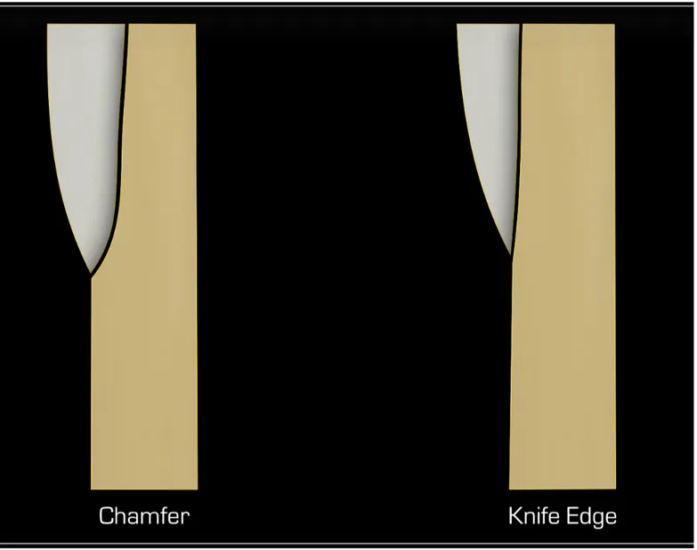
One of the most controversial of these factors has been the type of finish line when preparing the margins on natural abutments. This finish line has been defined as the final margin that separates the prepared axial tooth surface and the remaining unprepared tooth surface. When this finish line is prepared vertically without a defined margin (feather-edge), the resulting vertical area is the margin of the preparation. Conversely, when the finish line is designed by a horizontal sharp line (chamfer), there is a clear margin outlining this finish line. Consequently, the feather-edge leaves an undefined margin, whilst the chamfer results in a defined clear margin. It has been argued that the choice of the finish line depends on the: type of tooth4, crown length11, existing restorations11,
distance between adjacent abutments/teeth11 , location of the margin in relation with soft
periodon-tal tissue11, type of planned restorations11, type of selected material to fabricate the crown(s) or
bridge(s) 11, the periodontal health status12-13 and skill, experience and preference of the operators15.
However, there is no clear evidence from clinical trials on the superiority of a specific finish line10-15.
Carnevale et al. 16-17 introduced the feather-edge preparation during periodontal surgery and
report-ed retrospectively on the clinical success of this combinreport-ed periodontal-prosthetic procreport-edure. Paniz et al more recently designed a RCT to evaluate the 12-month periodontal response to two subgin-gival restorative margin designs reporting that the feather-edge in comparison with the chamfer was associated with a higher rate of bleeding on probing (BOP)18. Furthermore, other authors have
reported that the type of finish line could also affect the fracture resistance of esthetic crowns16,20.
It was, therefore, the objective of this randomized clinical trial to evaluate the influence of two mar-gin finish lines on the periodontal health and fracture resistance of zirconia single crowns layered with dedicated ceramics. Furthermore, this RCT was designed to evaluate whether the distance between the margin and the bone crest had a direct influence on the periodontal parameters, inde-pendently from the type of finish line.
The following null hypotheses were tested: 1) no difference between two different finish lines (feath-er-edge vs chamfer) on fracture resistance; 2) no difference between two different finish lines on periodontal tissue health and 3) there is a direct influence of the distance of the margin to the bone crest on the periodontal parameters, independently from the type of finish line,
Material and Methods
Study Design
15 effect of two different finish lines of posterior natural abutments on periodontal outcomes and the resistance to fracture of the restoration when loading. This RCT was approved by the Institutional Ethics Committee of the University of Siena (clinicaltrials.gov #NCT020906567) and was conducted according to the revised 2008 Declaration of Helsinki on experimentations involving human sub-jects20. The results of this RCT are presented in fulfillment of the CONSORT guidelines21.
Participants
Subjects were enrolled at the Department of Prosthodontics of the University of Siena (Italy) be-tween September 2013 and December 2013.
Patients were selected for the study on the basis of the following inclusion criteria: age above 18 years with one tooth in need of crowning, no active intraoral or systemic disease, no pregnancy or lactation, smoking less than 10 cigarettes/day, in good general health, good oral hygiene, low caries activity, vital or satisfactory endodontically treated tooth with no pathological signs on the X-ray and without clinical symptoms of inflammation, no history of previous periodontal flap surgery, periodon-tal pocket depth less than 3 mm, Bleeding On Probing and Plaque Index inferior to 20%, no tooth mobility, occlusal function with a natural tooth, possibility to placer the margins on sound dental structure, lack of excessive parafunctional activity leading to an extensive loss of tooth structure, abfraction lesions or cracks.
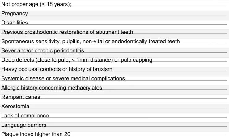
Conversely, the following exclusion criteria were adopted: addiction to alcohol and/or drugs, psy-chologically unstable patients, patients with acute symptoms of parafunctional disorders with the necessity of functional pretreatment before prosthodontic therapy, patients with systemic life-threat-ening diseases (physical status corresponding to group IV or higher of the American Society of Anesthesiologists classification), patients requiring hard/soft tissue augmentation, patients with un-treated periodontal disease/poor compliance, teeth with deep intrasulcular restoration.
Fifty consecutive patients (28 females and 22 males) with a mean age of 45.7 years (SD=10.2) and in need to receive 1 posterior layered zirconia single crown each in premolar and/or molar regions were selected for the study (Table 1). A total of 50 posterior teeth were selected: 30 molars (17 max-illa and 13 mandible) and 20 premolars (11 maxmax-illa and 9 mandible). All the treated posterior teeth had natural dentition in the opposite arch.
An experienced dental hygienist prepared the patients from a periodontal point of view and a first impression was taken with an irreversible hydrocolloid (GC Aroma Fine Plus, GC, Tokyo Japan) in order to pour the study casts and fabricate the composite resin temporary crowns. The casts of
16
both dental arches were mounted into a semi-adjustable articulator (Artex, Ammann Girrbach AG, Koblach, Austria) .
Randomization, Allocation Concealment, Masking of Examiners
Each patient was randomly assigned to 1 of the 2 experimental groups.
Allocation concealment was performed by opaque sealed, sequentially numbered envelopes. The statistician generated the allocation sequence by means of a computer-generated random list and instructed a different operator to assign a sealed envelope containing the type of finish line (i.e., feather-edge vs chamfer). The opaque envelope was opened before finish line election and treat-ment assigntreat-ment were communicated to the prosthodontist. Blinding of the examiners was main-tained throughout all experimental procedures.
The abutment teeth were randomly distributed into 2 groups of 25 samples each, according to 2 different finish line designs, as follows (Fig. 1):
- Vertical Preparation (Knife edge) - Horizontal Preparation (Chamfer)
Surgical and Prosthetic Procedures
A single, calibrated examiner, blinded to the experimental procedures, assessed all the clinical out-comes of the investigation both at the baseline and at the follow-up examinations.
A standardized tooth preparation was performed with occlusal and axial reduction of 1.5 mm and a chamfer or a knife edge finish line, that was placed juxta-gingivally. After one-two weeks, the tem-porary crowns were removed, a retraction suture silk cord (OOO) was placed into the sulcus and the tooth preparations refined using a stereomicroscope at 10x magnification placing the margins 0.5 mm into the sulcus (Zeiss OpMi1, Zeiss, Oberkochen, Germany). All internal line angles were rounded. All the preparations were made by the same experienced prosthodontist (MF). The inter-im restorations were relined intraorally on the prepared teeth; then, they were smoothed with soft rubbers and polishing cups to obtain an optimal marginal adaptation between the crowns and the soft tissues. Finally, the interim restorations were cemented in the same session with a eugenol-free temporary cement (Freegenol, GC). The interim restorations were worn by the patients for 3 weeks, so as to allow the soft tissues to recover from any possible preparation trauma and recover a com-plete health status.
17 (EXA’lence, GC) with custom auto polymerizing acrylic resin trays (SR-Ivolen, Ivoclar Vivadent AG, Schaan, Liechtenstein) made by the same dental technician at least 24 hours before the impres-sion. The impressions were poured using an extra-stone plaster type IV (Fuji Rock, GC) after 5 hours in order to allow the elastic return of the impression material. The composite resin temporary crowns were relined intraorally, polished and cemented as previously described.
In order to standardize as much as possible, the shape of the experimental copings, each frame-work was waxed-up by the same experienced dental technician with a minimum thickness of 0.5 mm; then, the copings were scanned by a Computer Aided Design-Computer Aided Manufacturing (CAD-CAM) software (Aadva, GC) and the zirconia cores were fabricated. The porcelain veneering was performed using a ceramic material dedicated to zirconia (Initial Zr-FS, GC), characterized by a special adaptation to the coefficient of thermal expansion (CTE) of the zirconia frameworks (9.4x10 -6K-1). Slow cooling was made, in order to dissipate the residual stresses within the bi-layered
res-torations. The pressure layering technique was adopted following the manufacturer’s instructions. At the intraoral try-in of the bisquebake crown and the slight occlusal adjustments were made by a diamond bur when needed, carefully checking the occlusal contacts. The final restorations were finally glazed and then cemented using a glass ionomer cement (Fuji-Cem, GC) following the manufacturer’s instructions. The luting agent was inserted into the crowns and the patients were requested to hold them under occlusal compression until cement set; then, excess cement was carefully removed.
Follow-up Examinations
The cementation time was considered the baseline to record data.
The patients were recalled for follow-up visits after 1 month, 6 months, 1, 2, 3 and 4 years of clinical service. The function and the esthetics were checked at the follow-up appointments by two inde-pendent examiners blinded to the group assignment and calibrated. In order to collect and classify the clinical outcomes, ‘success’ was defined by the percentage of restorations that remained in situ without any modification, ‘survival’ by the percentage of restorations that remained in situ with modifications but still under clinical acceptability, whilst ‘failure’ by the percentage of restorations that needed to be replaced23,24.
Data collection
for-18
mat 1:1). X-ray individual tray was made for each sample tooth of each patient, in order to be sure to have the radiogram in the same position at each recall.
The following clinical measurements at the baseline, immediately after luting the crown, were taken at the experimental sites by the blinded examiner: gingival bleeding on probing (BoP), at two differ-ent facial sites (mesial and distal), was reported as mean, according to Ainamo and Bay24.
Radiographic measurements at the time of placement of definitive restoration and final follow-up were made in order to calculate the distance between the bone crest and the margin of the crown. Bone level Mes: the distance from the tooth preparation to the bone at the mesial site.
Bone level Dis: the distance from the tooth preparation to the bone at the distal site.
The mean bone level was then calculated considering mesial and distal bone levels at the single dental abutment.
Statistical analysis
The patients’ characteristics and clinical variables were balanced between groups. The Fisher’s Exact Test was applied to assess the statistical significance of between-group differences in the 4-year success rate. The level of statistical significance was set at p<0.05.
A logistic regression analysis was applied to verify whether 4-year BoP at the interproximal level was significantly influenced by tooth type, preparation type, and distance of preparation margin from bone crest level. A separate logistic regression analysis was performed to assess whether 4-year BoP at the buccal site was significantly influenced by tooth type and preparation type.
The statistical analyses were performed using a statistical package software (IBM SPSS Statistics for Windows, Version 21.0, IBM Corp., Armonk, NY, USA).
Results
The following results were collected after the period of clinical service: Group 1: 80% success rates 21/25 (one not reparable fractures of ceramic layer) and 96% survival rate (24/25). Group 2: 76% success rate (20/25) and 100% survival rate (25/25). Four chippings were noticed in Group 1 but only 1 crown needed to be replaced after 4 years. In Group 2 five chippings were noted but no need for replacement was considered after 4 years of clinical service (Table 2). All chippings took place in coronal part of the sample crowns of patients with evident clinical signs of occlusal wear. One chipping (the catastrophic one) was recorded during the first year of clinical service, 2 were noted during the second year, 2 during the third and the other 4 during the fourth year of clinical service.
19 No chipping at the margins of both groups was noted.
Statistical analysis of survival and success rates did not show any statistically significant difference (Table 2).
Regarding the periodontal parameters, BoP at 4 years was present in 12 of 25 crowns of Group 1 (48%) and in 18 of the 25 crowns of Group 2 (55,5%) (Table 3). A statistically significant correlation was found between BoP and the distance of the margin to the bone crest. When the bone crest was less than 3 mm, a higher probability of BoP was detected. According to the regression analysis BOP at interproximal level was significantly dependent on the type of the preparation (p=0.004) and the distance between the bone crest and the crown margin (BC-CM) (p<0.001), while tooth type did not have a significant influence (p=0.821). Conversely, BOP at buccal sites, neither preparation type (p=0.721), nor tooth type (p=0.399) were statistically significant.
Discussion
The results of this study support rejection of the first null hypothesis: the survival rate of the zirconia crowns made with vertical or horizontal margins did not show statistically significant differences about survival and success rates after 4 years of clinical service. Within the limitations of this in
vivo study, due to the limited number of specimens tested, it was concluded that all zirconia crowns
created with feather-edge or chamfer demonstrated a similar and acceptable behavior with relation to the fracture resistance and periodontal response of zirconia single crowns layered with dedicated ceramics. Feather-edge would allow the use of precise zirconia restorations in abutments for fixed prostheses. Preserving a maximum amount of sound tooth structure during tooth preparation for fixed abutments, as it is commonly done in vertical preparations, might be a less invasive alternative to a chamfer margin. This would be true not only for periodontally treated teeth16-18, but also in other
clinical conditions such as endodontically treated teeth, vital teeth in young individuals, and teeth affected by caries at the cervical third of the clinical crown25.
There were certain limitations to this study. Only one specific zirconium-oxide-based ceramic CAD/ CAM system was evaluated with only one ceramic material dedicated to zirconia. Further clinical in-vestigation is necessary to evaluate the influence on clinical behavior of different tooth preparation designs with different total occlusal convergence (TOC) angles25-26. It would be also necessary to
undertake additional studies to determine the clinical risk of delamination of the veneering porcelain if different types of preparation are carried out.
20
speculated that other cements can reach similar clinical results of this study27. It should also be
noted that all the crowns included in this study had 360° zirconium-oxide margins: the zirconia mar-gins when a feather-edge was used are thinner than the zirconia marmar-gins obtained when a chamfer has been performed; therefore, it might be supposed that these thinner margins can be more easily altered during various clinical phases, for example during scaling procedures at oral hygiene re-calls28,29. Further studies should be accomplished to verify if these small irregularities may influence
the BoP of the two types of finish line. On the other hands a previous study showed that zirconia should not be altered during cementation and can be the material of choice to make esthetic crown with vertical finish line30.
The second null hypothesis tested in this study, that there was no difference between two different finish lines on periodontal response was rejected. The evaluation of the recorded BoP showed sta-tistically significant differences between the two groups and the highest score was recorded when feather-edge was used.
However, it should be noted that the deep position into the sulcus of this finish line and consequent-ly its close position to bone crest may be a real reason of increased BoP: because of the revealed statistically significant correlation between BoP and the distance of the margin to the bone crest, it should be clinically advocated that the margins have to be placed at least 3 or more mm far away from the bone crest independently from the type of finish line. For that reasons the third null hypoth-esis tested, that there was a direct influence of the distance of the margin to the bone crest on the periodontal parameters, independently from the type of finish line, was accepted. Another factor that can influence the BoP scores of the two groups might be different levels of home oral hygiene of the patients, aspects that can not be completely controlled in all patients.
Within the limitations of this study, it can be stated that clinicians might decide what type of finish line have to be chosen when using zirconia single crowns.
Accordingly with previous articles CAD/CAM systems were used to achieve good in vivo marginal fit for single-unit crowns made with chamfer and feather-edge with the advantages of homogeneous standardized materials31.
Clinicians should pay attention also on the periodontal parameters, as a matter of fact that the BoP can be correlated by the distance of the finish line from the bone crest as already demonstrated32-37.
Recently Ercoli and Caton10 evaluated if there was evidence in the literature that factors related to
teeth and dental prostheses can play a role in the initiation and progression of gingivitis and peri-odontitis. Along their narrative review Ercoli and Caton wanted to summarize the current evidence
21 about the role that the fabrication and presence of dental prostheses and tooth-related factors have on the initiation and progression of gingivitis and periodontitis10. They pointed out once more that
the placement of margins within the biological width causes gingival inflammation and possibly recession or pocket formation, and that the intraoral procedures to fabricate fixed prostheses can traumatize periodontal supporting tissue. However, it was evident that despite the existance of many clinical factors that can affect the periodontal tissue health, adequate periodontal assessment and treatment, appropriate instructions and motivation in self-performed plaque control and compli-ance to maintencompli-ance protocols appeared to be the most important factors to limit or avoid potential negative effects on the periodontium caused by fixed and removable prostheses10. Given the
lim-ited available evidence in humans, it was not possible to determine if the negative effects on the periodontium associated with a violation of the biologic width by restorative margins is caused by bacterial plaque, trauma or a combination of these factors10, both factors can play an important role.
For that the high rate of BoP recorded in the study can be related to the fact that when a margin is located slightly (0.5 mm) into the sulcus, in the interproximal area of posterior teeth, independently from the type of finish line was designed, the maintenance of healthy periodontal condition is mainly in the hands of the patient and his/her home plaque control38-40. The clinician must design the
pre-ferred margin in order to make easier to the patient all plaque control procedures and motivate the patient to properly and regularly perfom his/her oral hygien also interproximaly.
Another limitation of this study is that, after luting the sample crowns, was not possible to check the precision of the margin also based on the fact that a marginal gap of up 100-120 microns can be detected clinically41. For that reason, it can not be underestimated that the presence of a marginal
overhang can act as a plaque retentive factor and cause a qualitative shift toward a subgingival cultivable microflora more characteristic of periodontitis42.
Regarding the fact that 10% of both groups reported chipping of the crowns, a % of failures higher than that was expected; It might be observed that all crowns of this study were porcelain fused to zirconia crowns and all chipping were recorded in patients with signs of occlusal wear. Chipping of porcelain fused to zirconia crowns might be correlated to different factors such as the presence of natural opposing teeth and type of occlusal loading42-43, coping/framework design44-46, surface
finish-ing47-48, thermal misfit of veneering ceramic/zirconia composites49-55, slow heating and slow cooling
of porcelain on zirconia couping56, internal surface treatment of coping, type of luting material and
marginal gap56-61, hydrothermal degradation62, different clinical conditions and operators63-69.
differ-22
ent degrees of wear, more clinical information are needed to point out limitations of using porcelain to fused zirconia crowns on bruxist patients.
However, further research must be carried out; for example, concerning the clinical outcomes of horizontal and vertical finish lines in the case of multiple dental restorations: in multiple abutments different results can be achieved because of the insertion of the bridge and because higher the number of abutments less precision can be expected.
Conclusions
According to the results of the present in vivo study and under its limitations, the following conclu-sions were draw:
- the clinical performance of the zirconia crowns made with vertical or horizontal margins showed no differences of survival and success rates after 4 years of clinical service.
- statistically significant correlation between BoP and the distance of the margin to the bone crest was found: for that margins, both vertical or horizontal, should be placed at least at 3 mm or more far away from the bone crest.
- it must be also considered that in case of a vertical prep a higher probability of BoP can be faced.
Clinical Significance
The results of the present clinical study would allow clinicians to make a vertical or/and a horizontal finish line when using zirconia single crowns. However, clinicians must focus their attention also on the periodontal parameters, suggesting that the type of prep and its distance from the bone crest are key clinical factors.
Acknowledgments
Authors like to thank Prof. Mariano Sanz for his important contribution to edit this paper.
Also, Authors want to thank Mr. Gianni Bonadeo for his lab work on making all crowns that were tested in this study.
23
References
1. Hickel R, Peschke A, Tyas M, Mjor I, Bayne S, Peters M, Hiller KA, Randall R, Vanherle G, Hein-tze SD. FDI World Dental Deferation-Clinical Criteria for the Evaluation of Direct and Indirect Restorations. Update and Clinical Examples. J Adhes Dent 2010;12: 259-272.
2. Manhart J, Chen H, Hamm G, Hickel R. Buonocore Memorial Lecture. Review of the clinical survival of direct and indirect restorations in posterior teeth of the permanent dentition. Oper Dent 2004;29: 481-508.
3. Shenoy A, Shenoy N, Babannavar R. Periodontal considerations determining the design and location of margins in restorative dentistry. J Interdiscip Dentistry 2012;2:3-10.
4. Hunter AJ, Hunter AR. Gingival crown margin configurations a review and discussion. Part I: Terminology and widths. J Prosthet Dent.1990;64:548-52.
5. Broadbent JM, Williams KB, Thomson WM, Williams SM. Dental restorations: a risk factor for periodontal attachment loss? J Clin Periodontol 2006;33:803-10.
6. Ferrari M, Koken S, Grandini S, Ferrari Cagidaco E, Joda T, Discepoli N. Influence of cervical margin relocation (CMR) on periodontal health: 12-month results of a controlled trial. J Dent 2018;69:70-76.
7. Kosyfaki P, del Pilar Pinilla Martín M, Strub JR.Relationship between crowns and the periodon-tium: a literature update. Quintessence Int. 2010;41:109-26.
8. Kancyper SGJ, Koka S The influence of intracrevicular crown margins on gingival health: pre-liminary findings. J Prosthet Dent 2001;85:461-5.
9. Koha RJl, Pelz K, Strub JR. Effect of different crown contours on periodontal health in dogs. Microbiological results. J Dentistry 2004;322:153-159.
10. Ercoli C, Caton J. Dental prostheses and tooth-related factors. J Periodontol 2018;89:S223-S226. 11. Indian Dental Academy IDA. Finish lines in FPD. Retrieved on April 17, 2016 from www.
indiandentalacademy.blogspot.com/2013/07/finish-lines-in-fpd.html
12. Gargiulo AW, Wentz FM, Orban B. Dimensions and relations of the dentogingival junction in humans. J Periodontol 1961;32:261-7.
13. Gracis S, Fradeani M, Renato Celletti R, Bracchetti G, Biological integration of aesthetic res-torations: factors influencing appearance and long-term success. Periodontology 2000,2001; 27:29–44.
14. Croll BM. Emergence profiles in natural tooth contour. Part II: Clinical considerations. J Pros-thetic Dent 1990;63:374-379.
24
15. Beuer F, Aggstaller H, Edelhoff D, Gernet W. Effect of preparation design on the fracture resis-tance of zirconia crown. Dent Mater J 2008;27:362-7
16. Carnevale G, Di Febo G, Fuzzi M. An in vivo study of teeth reprepared during periodontal sur-gery. Int J Period Res Dent 1990;1:40-55.
17. Carnevale G, Di Febo G, Fuzzi M. A retrospective analysis of the perio-prosthetic aspect of teeth reprepared during periodontal surgery. J Clin Period 1990;17:313-6.
18. Paniz G, Nart J, Gobbato L, Chierico A, Lops D, Michalakis K. Periodontal response to two different subgingival restorative margin designs: a 12-month randomized clinical trial. Clin Oral Investig 2016;20:1243-52.
19. Carlini BJ, Cecchin D, Pereira GDS, Paulillo LAMS. Influence of remaining coronal struc-ture and finis line on the fracstruc-ture strength of roots restored with metallic posts. Braz Oral Res 2011;25:345-50
20. Needleman I, Worthigton H, Moher D, Schulz K, Altman DG. Improving the completeness and transparency of reports of randomized clinical trials in oral health: The CONSORT Statement. Am J Dent 2008;21:7–12.
21. Schmidt H, Mehring S, McMillan J. Interpreting the declaration of Helsinki (2008): “must”, “should” and different kind of obligation. Med Law 2010;29:565–591.
22. Ferrari M, Vichi A, Zarone F. Zirconia abutments and restorations: From laboratory to clinical in-vestigations. Dent Mater 2015;31:e-63-e76.
23. Anusavice KJ. Standardizing failure, success, and survival decisions in clinical studies of ceram-ic and metal-ceramceram-ic fixed dental prostheses. Dent Mater 2012;28:102-111.
24. Ainamo J, Bay I. Problems and proposals for recording gingivitis and plaque. Int Dent J 1975;25:229–235.
25. Schmitt J, Wichmann M, Holst S, Reich S. Restoring severely compromised anterior teeth with zirconia crowns and feather-edged. margin preparations: a 3-year follow-up of a prospective clinical trial. Int J Prosthodont 2010;23:107-09.
26. Gavelis JR, Morency JD, Riley ED, Sozio RB. The effect of various finish line preparations on the marginal seal and occlusal seat of full crown preparations. J Prosthet Dent 1981;45:138-45. 27. Vigolo P, Mutinelli S. Evaluation of Zirconium-Oxide-Based Ceramic Single-Unit Posterior
Fixed Dental Prostheses (FDPs) Generated with Two CAD/CAM Systems Compared to Porce-lain-Fused-to-Metal Single-Unit Posterior FDPs: A 5-Year Clinical Prospective Study. J Prostho-dont 2012;21:265-69.
25 28. Raigrodski AJ, Chiche GJ, Potiket N, Hochstedler JL, Mohamed SE, Billiot S et al. The efficacy
of posterior three-unit zirconium-oxide–based ceramic fixed partial dental prostheses: A pro-spective clinical pilot study. J Prosthet Dent 2006;96:237-44.
29. Vigolo P, Motterle M. An in vitro evaluation of zirconia surface roughness caused by different scaling methods. J Prosthet Dent 2010;103:283-87.
30. Fuzzi M, Tricarico MG, Ferrari Cagidiaco E, Bonadeo G, Ferrari M. Nanoleakage and internal adaptation of zirconia and lithium disilicate single crowns with feather edge preparation. J
Os-seointegr 2017;9:250-262.
31. Tinschert J, Natt G, Hassenpflug S, Spiekermann H. Status of current CAD/CAM technology in dental medicine. Int J Comput Dent 2004;7:25-45.
32. Gunay H, Seeger A, Tschernitschek H, Geurtsen W. Placement of the preparation line and peri-odontal health--a prospective 2-year clinical study. Int J Perio Restor Dent 2000;20:171-181 33. Boeckler AF, Lee H, Stadler A, Setz JM. Prospective observation of CAD/CAM titanium ceramic
single crowns: a three-year follow up. J Prosthet Dent 2009;102:290-297.
34. Boeckler AF, Lee H, Psoch A, Setz JM. Prospective observation of CAD/CAM titanium-ceram-ic-fixed partial dentures: 3-year follow-up. J Prosthodont 2010;19:592-597.
35. Hey J, Beuer F, Bensel T, Boeckler AF. Single crowns with CAD/CAM-fabricated copings from titanium: 6-year clinical results. J Prosthet Dent 2014;112:150-154.
36. Larato DC. Effects of artifical crown margin extension and tooth brushing frequency on gingival pocket depth. J Prosthet Dent 1975;34:640-643.
37. Heschl A, Haas M, Haas J, Payer M, Wegscheider W, Polansky R. Maxillary rehabilitation of periodontally compromised patients with extensive one-piece fixed prostheses supported by natural teeth: a retrospective longitudinal study. Clinical Oral Investig 2013;17:45-53.
38. Konradsson K, Claesson R, van Dijken JW. Dental biofilm, gingivitis and interleukin-1 adjacent to approximal sites of a bonded ceramic. J Clin Periodontol 2007;34:1062-1067.
39. Veterans Administration Cooperative Studies Project No 147. Part VIII: Plaque accumulation on metal ceramic restorations cast from noble and nickel-based alloys.A five-year report. J Prosthet Dent 1992;61:543–549.
40. Christensen GJ. Marginal fit of gold inlay castings. J Prosthet Dent 1966;16:297-3051
41. Lang N P, Kiel R A, Anderhalden K. Clinical and microbiological effects of subgingival resto-rations with overhanging or clinically perfect margins. J Clin Periodontol 1983;10:563-578. 42. Manfredini D, Poggio CE. Prosthodontic planning in patients with temporomandibular disorders
26
and/or bruxism: A systematic review. J Prosthet Dent 2017;5: 606-613.
43. Johansson A, Omar R, Carlsson GE. Bruxism and prosthetic treatment: A critical review. J Pro-sthodont Resear 2011;55:127–136
44. Ferrari M, Vichi A, Zarone F. Zirconia abutments and restorations: From laboratory to clinical investigations. Dental Materials 2015;31: e63–e76.
45. Bonfante EA, da Silva NR, Coelho PG, Bayardo-González DE, Thompson VP, Bonfante G. Ef-fect of framework design on crown failure. Eur J Oral Sci 2009;117:194–9.
46. Rafferty BT. Biomechanical evaluation of an anatomically correct all-ceramic tooth-crown sys-tem configuration: core layer multivariate analysis incorporating clinically relevant variables. J Biomech Eng 2001;132:15–9.
47. Deville S, Chevalier J, Gremillard L. Influence of surface finish and residual stresses on the age-ing sensitivity of biomedical grade zirconia. Biomaterials 2006;27:2186–92.
48. Mitov G, Gessner J, Lohauer U, Woll K, Muecklich F, Pospich P. Subcritical crack growth and be-havior and life data analysis of two types of dental Y-TZP ceramics. Dent Mater 2011;27:684–91. 49. Benetti P, Della Bona A, Kelly JR. Evaluation of thermal compatibility between core and veneer dental ceramics using shear bond strength test and contact angle measurement. Dent Mater 2010;26:743–50.
50. Guazzato M, Walton TR, Franklin W, Davis G, Bohl C, Klineberg I. Influence of thickness and cooling rate on development of spontaneous cracks in porcelain/zirconia structures. Aust Dent J 2010;55:306–10.
51. Belli R, Monteiro Jr S, Baratieri LN, Katte H, Petschelt A, Lohbauer U. A photoelastic assess-ment of residual stresses in zirconia-veneer crowns. J Dent Res 2012;91:316–20.
52. Belli R, Frankenberger R, Appelt A, Schmitt J, Baratieri LN, Greil P, et al. Thermal-induced resid-ual stresses affect the lifetime of zirconia-veneer crowns. Dent Mater 2013;29:181–90.
53. Fischer J, Stawarczyk B, Tomic M, Strub JR, Hammerle CH. Effect of thermal misfit between different veneering ceramics and zirconia frameworks on in vitro fracture load of single crowns. Dent Mater J 2007;26:766–72.
54. Fischer J, Stawarzcyk B, Trottmann A, Hammerle CH. Impact of thermal misfit on shear strength of veneering ceramic/zirconia composites. Dent Mater 2009;25:419–23.
27 cooling regimens to strengthen porcelain fused to zirconia. J Prosthet Dent 2012;107:163–9. 56. Son YH, Han CH, Kim S. Influence of internal-gap width and cement type on the retentive force
of zirconia copings in pullout testing. J Dent 2012;40:866–72.
57. Taira Y, Sakai M, Sawase T. Effects of primer containing silane and thiophosphate monomers on bonding resin to a leucite-reinforced ceramic. J Dent 2012;40:353–8.
58. Inokoshi M, Kameyama A, De Munck J, Minakuchi S, Van Meerbeek B. Durable bonding to me-chanically and/or chemically pre-treated dental zirconia. J Dent 2013;41:170–9.
59. Casucci A, Goracci C, Chieffi N, Monticelli F, Giovannetti A, Juloski J, et al. Microtensile bond strength evaluation of self-adhesive resin cement to zirconia ceramic after different pre-treat-ments. Am J Dent 2012;25:269–75.
60. Casucci A, Monticelli F, Goracci F, Mazzitelli C, Cantoro A, Papacchini F, et al. Effect of surface pre-treatments on the zirconia ceramic-resin cement microtensile bond strength. Dent Mater 2011;27:1024–30.
61. Kern M, Wegner SM. Bonding to zirconia ceramic: adhesion methods and their durability. Dent Mater 1998;14:64–71.
62. Kim JW, Covel NS, Guess PC, Rekow ED, Zhang Y. Concerns of hydrothermal degradation in CAD/CAM zirconia. J Dent Res 2010;89:91–5.
63. Ferrari M, Giovannetti A, Carrabba M, Bonadeo G, Rengo C, Monticelli F, et al. Fracture resis-tance of three porcelain-layered CAD-CAM zirconia frame designs. Dent Mater 2014;30:e163– 169.
64. Pjetursson BE, Tan K, Lang NP, Brägger U, Egger M, Zwahlen M. A systematic review of the survival and complication rates of fixed partial dentures (FPDs) after an observation period of at least 5 years. Clin Oral Implants Res 2004;15:625–32.
65. Pjetursson BE, Sailer I, Zwahlen M, Haeemrle CH. A systematic review of the survival and com-plication rates of all-ceramic and metal–ceramic reconstructions after an observation period of at least 3 years. Part I: Single crowns. Clin Oral Implants Res 2007;18(Suppl):73–85.
66. Sailer I, Pjetursson BE, Zwahlen M, Hammerle CH. A systematic review of the survival and com-plication rates of all-ceramic and metal–ceramic reconstructions after an observation period of at least 3 years. Part II: Fixed dental prostheses. Clin Oral Implants Res 2008;19:326–8.
28
rates of fixed partial dentures (FPDs) after an observation period of at least 5 years. Clin Oral Implants Res 2004;15:654–66.
68. Al-Amleh B, Lyons K, Swain M. Clinical trials in zirconia: a systematic review. J Oral Rehabil 2010;37:641–52.
69. Zarone F, Russo S, Sorrentino R. From porcelain-fused-to-metal to zirconia: clinical and exper-imental considerations. Dent Mater 2011;27:83–96.
Legends
29
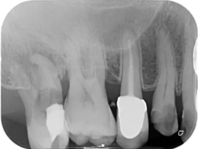
Fig. 2 (Group 1) x-Ray of a patient in need to restore the first premolar and to extract the second molar.
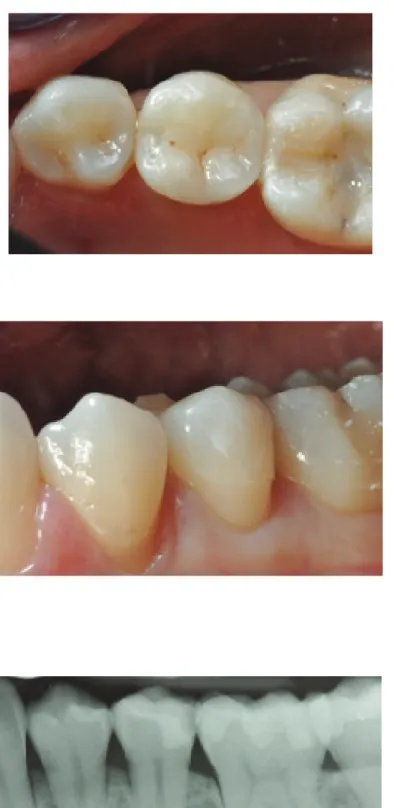
Fig. 3 and 4 After receiving a build up with resin composite, the premolar was prepared with feather edge.
30
Fig. 5, 6 and 7 The premolar after being restored with a porcelain fused to zirconia crown. Buccal and occlusal view and x-Ray.
31 Fig. 8 (Group 2) x-Ray of a patient in need to remove an old crown on the second
pre-molar made with a cantilever.
Fig. 9 The patient was treated with a fixture to replace the
32
Fig. 11 and 12 Buccal view and x-Ray at last recall. Fig. 10 Try-in of the two copings
33
Chapter 3
3.1 Fit a new clinical score to evaluate single crowns
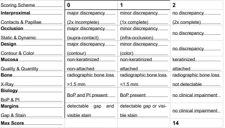
The aim of this study was to propose a clinical procedure to evaluate single restorations and their relations with periodontal tissues by a new clinical score: the FIT (Functional Index for Teeth). FIT, that is a novel index for the assessment of the prosthetic results of lithium disilicate crowns, based on seven restorative-periodontal parameters, that evaluate crowns placed on natural abutments, and want to be a reliable and objective instrument in assessing single partial crown success and periodontal outcome as perceived by patients and dentists.
The variables are the following: Interproximal Contacts and Papillae, Static and Dynamic Occlusion, Design Contour and Color, Quality and Quantity of Mucosa, Bone level in x-Ray, Biology related to Bleeding on Probing (BoP) and Plaque Index (PI) and Stain and Gap at Margins. Scoring for each variable from 0 to a maximum of 2, resulting a max score of 14.
34
3.2 A pilot trial on lithium disilicate partial crowns using a novel prosthodontic
Functional Index for Teeth (FIT)
Background
Due to the specific properties of lithium disilicate, particularly flexural strength, this restorative ma-terial is mainly indicated for single full and/or partial crowns1-3].Lithium disilicate provides high
aes-thetic results and, in comparison with porcelain and reinforced resin composites, its higher flexural strength makes it be preferable whenever the tooth defect exceeds a certain dimension4,5.
Lithium disilicate can be obtained using two different production processes: press technology and CAD/CAM technology. CAD/CAM technology is mainly used as chairside procedure, while the pressable technology is performed in the laboratory mainly using an analogic workflow. Pressed lithium disilicate results were very promising6,7 and recently the evaluation of a new lithium disilicate
material (Initial LiSi press, GC) has been reported8. Only few clinical trials are available on lithium
disilicate partial crowns, the majority of them being retrospective studies9-11 and only one being a
randomized controlled trial (RCT) 8.
Evaluation of clinical results of partial crowns on posterior teeth is usually performed following stan-dardized parameters, such as Ryge and Snyder clinical parameters12 or the modified FDI criteria13.
The evaluation is usually performed after luting at baseline, and then at recalls after 1,6,12, 24, or 36 months. The modified FDI criteria evaluate several categories with some sub-categories13. Also,
RCTs are done by blinded, calibrated and experienced dentists that can perform the follow-up eval-uation14,15.
It must be pointed out that Ryge and Snyder clinical parameters and modified FDI criteria were initially defined for direct restorations, therefore there is the need to determine clinical criteria ade-quate to evaluate indirect restorations. Clinical criteria should reflect the patients’ perception of the restorations, fulfilling teaching purposes and being easily applicable in daily practice. In order to ease the process of drafting a proper treatment plan16,17, some classifications and prognosis
evalu-ations have been proposed.
Recently, a novel Functional Implant Prosthodontic Score (FIPS) was proposed18-21; FIPS was
based on 5 clinical variables evaluated crowns placed on implants with an oral radiograph and a buccal and an occlusal picture. its potential to serve as an objective and reliable instrument in as-sessing implant success and restoration and periodontal outcome as perceived by patients, as well
35 as identifying the possible risk of failure, comparing follow-up observations, providing an effective teaching tool was demonstrated. Similarly, FIT, that is a novel index for the assessment of the pros-thetic results of lithium disilicate crowns, based on seven restorative-periodontal parameters, that evaluate crowns placed on natural abutments, and want to be a reliable and objective instrument in assessing single partial crown success and periodontal outcome as perceived by patients and dentists.
The aim of this study was to clinically evaluate two lithium disilicate systems using the novel prost-hodontic Functional Index for Teeth (FIT).
Materials and Methods
The aim of this RCT was to evaluate the clinical performance of two lithium disilicate pressed sys-tems using a novel Functional Index for Teeth (FIT), which is made up of seven clinical variables showing, among other things, the possible correlation with the level of appreciation perceived by the patients.
Functional Index for Teeth (FIT)
A novel Functional Index for Teeth (FIT) was used (Table 2). Seven clinical variables have been collected and main prosthodontic and periodontal parameters were evaluated simultaneously (Interproximal Contacts and Papillae, Static and Dynamic Occlusion, Design Contour and Color, Quality and Quantity of Mucosa, Bone level in x-Ray, Biology related to Bleeding on Probing (BoP) and Plaque Index (PI) and Stain and Gap at Margins).
The FIT evaluation was performed only at last recall (3-year follow-up) by an experienced operator (Figs. 1,2).
The null hypothesis tested in this clinical study was that there was no statistically significant dif-ference in the clinical performance of the two lithium disilicate systems. A sample of 60 patients in need of a single partial crown on posterior teeth (upper and lower premolars and molars), ac-cessing the Department of Prosthodontics and Dental Materials of the University of Siena, Italy, in the time period between September 2015 and January 2016 were included in the study. Selected patients, periodontally healthy or successfully treated in need for one posterior restoration, had a mean age of 37 (±7.5) years (between 18 and 70) (14F,16M). Exclusion criteria were: age <18 ye-ars, pregnancy, disabilities, prosthodontic restoration of the tooth, spontaneous sensitivity, pulpitic,