“AMEDEO AVOGADRO”
DIPARTIMENTO DI SCIENZE DEL FARMACO
Dottorato di Ricerca in “Scienza delle Sostanze Bioattive”
CHEMICAL AND METABOLIC STABILITY STUDIES OF
PROPARGYLAMINE-CONTAINING DRUGS
Coordinatore Prof. Luigi PANZA
Tutor Candidato
Prof. Giorgio GROSA Dott.ssa Rossana CANAVESI
TABLE OF CONTENTS
List of abbreviations... V
CHAPTER 1. Introduction ... 1
1.1 CHEMICAL STABILITY STUDIES ... 1
1.1.1 Experimental approach ... 2
1.1.2 Degradation conditions ... 2
1.1.3 Drug product ... 4
1.1.4 Stability-indicating method development ... 4
1.2 METABOLIC STABILITY STUDIES... 5
1.2.1 Xenobiotics biotransformation ... 6
1.2.2 Cytochrome P450 ... 7
1.2.3 In vitro models to study drug metabolism ... 10
1.3 PROPARGYLAMINE-CONTAINING DRUGS ... 14
1.3.1 Neurodegenerative disorders ... 14
1.3.2 Alzheimer’s Disease (AD) ... 15
1.3.3 Parkinson’s disease (PD) ... 16
1.3.4 Monoamine oxidase ... 18
1.3.5 Propargylamines ... 19
1.3.6 Multitarget-directed ligands (MTDLs) strategy ... 21
1.3.7 Conclusions ... 23
1.4 REFERENCES ... 24
CHAPTER 2. Aim of the work ... 25
CHAPTER 3. New insights in oxybutynin chemical stability: identification in transdermal patches of a new impurity arising from oxybutynin N-oxide rearrangement ... 27
3.1 INTRODUCTION ... 27
3.2 EXPERIMENTAL ... 28
3.2.1. Reagents and chemicals ... 28
3.2.2. Instrumentation and chromatographic conditions ... 29
3.2.3 Forced degradation study of Oxy ... 30
3.2.4. Isolation and purification of Oxy-EK ... 31
3.2.5. Synthesis of Oxy free base ... 32
3.2.6 Synthesis of degradation products ... 33
3.2.7. Evaluation of the mutagenic effects of Oxy-EK and Oxy ... 35
3.3 RESULTS AND DISCUSSION ... 38
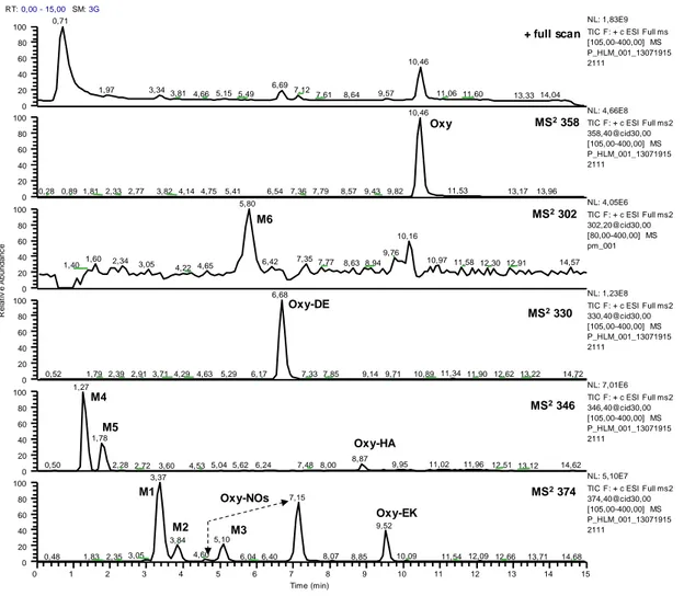
3.3.1 Forced degradation study of Oxy by LC-UV analysis ... 38
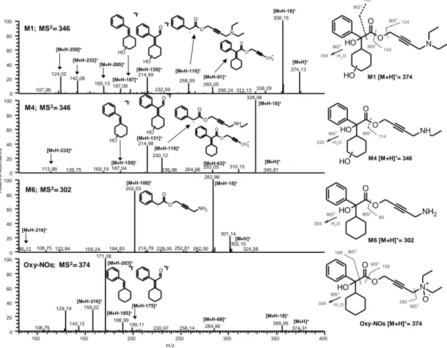
3.3.2. Structural characterization of degradation products by LC-ESI-MS/MS analysis ... 41
3.3.3 Synthesis of Oxy-EK... 46
3.3.4 Analysis of transdermal patches ... 47
3.4 CONCLUSIONS ... 50
3.5 REFERENCES ... 51
CHAPTER 4. New findings in the in vitro metabolism study of oxybutynin ... 53
4.1 INTRODUCTION ... 53
4.2 EXPERIMENTAL ... 54
4.2.1 Reagents and chemicals ... 54
4.2.2 Instrumentation and chromatographic conditions ... 55
4.2.3 Synthesis of the reference compounds ... 56
4.2.4 Human and rat Liver Preparations... 58
4.2.5 Phase I incubations with RLM, HLM ... 58
4.2.6 Phase II incubation with HLC fraction... 58
4.3 RESULTS ... 59
4.3.1 Phase II incubation with human liver subcellular fractions ... 63
4.4 DISCUSSION ... 65
4.5 REFERENCES ... 67
CHAPTER 5. Forced degradation study of selegiline... 68
5.1 INTRODUCTION ... 68
5.2 EXPERIMENTAL ... 69
5.2.1 Reagents and chemicals ... 69
5.2.2 Instrumentation and chromatographic conditions ... 70
5.2.3 Forced degradation conditions ... 71
5.2.4 Isolation and purification of SG-EA ... 72
5.2.5 Synthesis of degradation product ... 72
5.3 RESULTS AND DISCUSSION ... 73
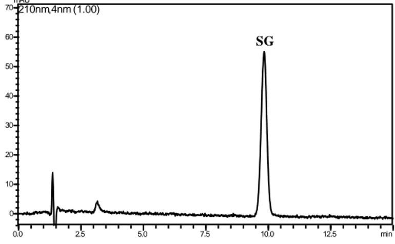
5.3.1 Forced degradation study and structural characterization of degradation products ... 73
5.3.2 Structural characterization of selegiline degradation products by LC-ESI-MS/MS analysis ... 78
5.4 CONCLUSIONS ... 82
5.5 REFERENCES ... 84
CHAPTER 6. New findings in the in vitro metabolism of selegiline ... 85
6.1 INTRODUCTION ... 85
6.2 EXPERIMENTAL ... 86
6.2.1 Reagents and chemicals ... 86
6.2.2 Instrumentation and chromatographic conditions ... 86
6.2.3 Rat and Human liver microsomes ... 87
6.2.4 In vitro RLM and HLM incubations of selegiline ... 87
6.2.5 In vitro RLM incubation of SG-EA ... 87
6.2.6 Incubation of selegiline in the presence of nucleophilic trapping agents ... 88
6.3 RESULTS AND DISCUSSION ... 88
6.4 CONCLUSION ... 92
CHAPTER 7. Forced degradation study of rasagiline: identification and characterization of degradation products
based on LC-UV and LC-ESI-MS/MS analysis ... 94
7.1 INTRODUCTION ... 94
7.2 EXPERIMENTAL ... 95
7.2.1 Reagents and chemicals ... 95
7.2.2 Instrumentation and chromatographic conditions ... 95
7.2.3 Forced degradation conditions ... 97
7.2.4 Sample preparation... 97
7.2.5 Synthesis of degradation product ... 98
7.3 RESULTS AND DISCUSSION ... 100
7.3.1 Forced degradation study and structural characterization of degradation products ... 100
7.3.2 Structural characterization of rasagiline degradation products by LC-ESI-MS/MS analysis ... 106
7.4 CONCLUSION ... 109
7.5 REFERENCES ... 110
CHAPTER 8. In vitro metabolism study of Rasagiline ... 111
8.1 INTRODUCTION ... 111
8.2 EXPERIMENTAL ... 113
8.2.1 Reagents and chemicals ... 113
8.2.2 Instrumentation and chromatographic conditions ... 113
8.2.3 Human intestinal and liver microsomes ... 115
8.2.4 Rat liver cytosol and microsomes ... 115
8.2.5 In vitro RLM, HLM and HIM incubations of rasagiline ... 115
8.2.6 Chemical stability of RG-NOH ... 115
8.2.7 Phase I metabolic stability of RG-NOH ... 116
8.2.8 Incubation of rasagiline in the presence of nucleophilic trapping agents ... 116
8.2.9 Phase II incubations ... 116
8.2.10 Synthesis of reference standards ... 117
8.3 RESULTS AND DISCUSSION ... 121
8.3.1 Metabolite 1-(R)-aminoindan (m/z 134) ... 123
8.3.2 Metabolites at m/z 188 ... 124
8.3.3 Metabolites RG-EA (m/z 226) and 3OH-RG-EA (m/z 242) ... 133
8.4 CONCLUSION ... 137
8.5 REFERENCES ... 138
CHAPTER 9. Conclusion ... 140
CHAPTER 10. In vitro phenotyping: new insights with the cocktail approach ... 142
10.1 INTRODUCTION... 142
10.2 EXPERIMENTAL ... 143
10.2.1 Materials and methods ... 143
10.2.2 UHPLC-QTOF instrumentation ... 144
10.2.4 Incubation method ... 145
10.3 RESULTS AND DISCUSSION ... 146
10.4 REFERENCES ... 149
Publications ... 152
List of abbreviations
AI: 1-(R)-aminoindanAI-EA: 3-(2,3-dihydro-1H-inden-1-ylamino)prop-2-enal Brij 58: polyoxyethylene 20 cetyl ether
CHMA: α-cyclohexylmandelic acid HLC: human liver cytosol
HLM: human liver microsomes
MS-PROP: (3-[(2-hydroxyethyl)thio]- 2-Propenal)
NADPNa2:Nicotinamide adenine dinucleotide phosphate disodium salt
Oxy: Oxybutynin hydrochloride; (4-(diethylamino)but-2-ynyl
(RS)-2-cyclohexyl-2-hydroxy-2-phenyl-acetate hydrochloride
Oxy-DE: N-desethyloxybutynin; Benzeneacetic acid,
α-cyclohexyl-α-hydroxy-4-(ethylamino)-2-butyn-1-yl ester
Oxy-EK: (3E)-4-(N,N-diethylamino)-2-oxo-3-buten-1-yl 1-cyclohexyl-1-phenylglycolate
Oxy-HA: Oxybutynin hydroxylamine; Benzeneacetic acid,
α-cyclohexyl-α-hydroxy-4-(ethylamino)-4-(hydroxy)-2-butyn-1-yl ester
Oxy-NOs: Oxybutynin N-oxides; (diastereomeric Oxy-NO1 and Oxy-NO2):
(4-(diethylamino)-but-2-ynyl-(RS)-2-cyclohexyl-2-hydroxy-2-phenylacetate) N-oxide
PAPS: 3'-phosphoadenosine-5'-phosphosulfate
RG: Rasagiline mesylate ((1R)-2,3-Dihydro-N-2-propynyl-1H-inden-1-amine methanesulfonate) RG-EA: 3-[2,3-dihydro-1H-inden-1-yl(prop-2-yn-1-yl)amino]prop-2-enal
RG-NOH: Rasagiline hydroxylamine; [2,3-Dihydro-N-hydroxy-N-2-propynyl-1H-inden-1-amine] RLM: rat liver microsomes
SG: Selegiline hydrochloride; [methyl(1-phenylpropan-2-yl)(prop-2-yn-1yl)amine] SG-EA: 3-[methyl(1-methyl-2-phenylethyl)amino]- 2-Propenal
SG-NOs: Selegiline N-oxides
TRIS HCl: Tris(hydroxymethyl)aminomethane hydrochloride UDPGA: uridine-5'-diphosphoglucuronic acid trisodium salt 3OH-AI: 3-hydroxy-1-aminoindan 3OH-RG: 3-hydroxy-N-propargyl-1-aminoindan 4OH-AI: 4-hydroxy-1-aminoindan; 4OH-RG: 4-hydroxy-N-propargyl-1-aminoindan 6OH-AI: 6-hydroxy-1-aminoindan 6OH-RG: 6-hydroxy-N-propargyl-1-aminoindan 7OH-AI: 7-hydroxy-1-aminoindan 7OH-RG: 7-hydroxy-N-propargyl-1-aminoindan
CHAPTER 1. Introduction
1.1 CHEMICAL STABILITY STUDIES
Chemical stability of drug molecule is a matter of great concern as it affects the safety and efficacy of the drug product.
The Food and Drug Administration (FDA) and the International Conference on Harmonization (ICH) guidance state the requirement of stability testing data to understand how the quality of a drug substance and drug product changes with time under the influence of various environmental factors.
In particular, the ICH guideline entitled “Stability Testing of New Drug Substance and Products” (ICH Q1A(R2), 2003) states that stress testing is intended to identify the likely degradation products which can in turn help establish the degradation pathways and the intrinsic stability of the molecule and validate the stability indicating power of the analytical procedures used.
Forced degradation or stress testing is undertaken to demonstrate specificity when developing stability-indicating methods, particularly when little information is available about potential degradation products. Besides these studies also provide information about the degradation pathways and degradation products that could form during storage. Forced degradation studies may help facilitate pharmaceutical development as well in areas such as formulation development, manufacturing, and packaging, in which knowledge of chemical behavior can be used to improve a drug product.
Even if the available regulatory guidance provides useful definitions and comments about degradation studies nonetheless it is very general concerning the scope, timing, and best practices for degradation studies. In particular, the issue of how much stress is adequate in stress testing is not addressed specifically. Overstressing a molecule can lead to degradation profiles that are not representative of real storage conditions and perhaps not relevant to method development, hence stress-testing conditions should be realistic and not excessive
(Reynolds et al. 2002). It is stated that the testing should include the effect of temperature, humidity (where appropriate), oxidation, photolysis and susceptibility to hydrolysis across a wide range of pH values. However no details are provided about the practical approach towards stress testing, for instance exact experimental conditions regarding temperatures,
duration, extent of degradation, etc. Nevertheless some general indications on how to best conduct forced degradation studies can be found in literature and are here mentioned.
1.1.1 Experimental approach
Forced degradation studies should be conducted whenever a stability-indicating method is required. Studies may need to be repeated as methods, processes, or formulations change. Forced degradation studies of active pharmaceutical ingredient (API) and drug product (DP) include appropriate solid state and solution state stress conditions (e.g. acid/base hydrolysis, heat, oxidation, and light exposure) in accordance with ICH guidelines.
Since it is advisable that the stress conditions should result in approximately 5–20% degradation of the API, the specific conditions (intensity and duration) used will depend on the chemical characteristics of the API. The stressed sample should be compared to the unstressed sample (control) and the appropriate blank. Finally, a compound may not necessarily degrade under a given stress conditions hence no further stressing is advised in these cases (Alsante et al. 2007).
1.1.2 Degradation conditions
1.1.2.1 Hydrolytic conditions
Hydrolytic reactions are among the most common processes for drug degradation. Hydrochloric acid or sulfuric acid (0.1–1 mol/L solution) for acid hydrolysis and sodium hydroxide or potassium hydroxide (0.1–1 mol/L solution) for base hydrolysis are suggested as suitable reagents for hydrolysis. Studies should be carried out in the solution state. For certain APIs that are partially soluble or insoluble in the described acidic or basic solution, addition of an appropriate co-solvent, may be required to achieve dissolution. Additionally, the sample may be heated for a defined time/temperature to accelerate degradation, depending on the API sensitivity to heat.
1.1.2.2 Oxidation
Oxidation can be carried out under an oxygen atmosphere or in the presence of peroxides. The use of oxygen is a more realistic model. Free radical initiators may be used to accelerate oxidation. Generally, a free radical initiator and peroxide will produce all primary oxidation degradation products observed on real-time stability. Therefore, free radical and/or hydrogen peroxide conditions are strongly recommended at all stages of development.
For solution state stress conditions, dissolve the API utilizing an appropriate solvent, add 5– 20 mol% of a free radical initiator at atmospheric pressure. To increase the solubility of oxygen in the solution, the reaction can be performed in a reaction vessel pressurized at 50– 300 psi with molecular oxygen. Additionally, the system is heated to accelerate degradation. The temperature depends on the free radical initiator selected.
For peroxide conditions, hydrogen peroxide reagent (up to 3%) can be used. As previously indicated, the addition of an appropriate co-solvent may be necessary, depending on API solubility. Hydrogen peroxide stress testing can be useful in DP studies where hydrogen peroxide is an impurity in an excipient. Solid-state stress conditions may be similarly investigated by placing the API (as is) in suitable closed containers filled with an oxygen headspace versus an argon or nitrogen control headspace. Additionally, the sample may be heated for a defined time/temperature to accelerate degradation, depending on the API sensitivity to heat.
For later stage development compounds when more time and effort can be focused on mechanistic understanding, the following oxidation conditions can be applied. The addition of metal ions to solutions of API can indicate whether there is a tendency for the API to be catalytically oxidized; indeed iron and copper ions are routinely found, as impurities, in APIs and formulation excipients. Transition metal ions can also reduce peroxide to generate hydroxyl radicals in a Fenton-type reaction. In addition, light can also effect oxidation reactions. Light absorbed by a photosensitizer can react with molecular oxygen to form the more reactive singlet oxygen species (Alsante et al. 2007).
1.1.2.3 Thermal/humidity
Solid state stability can be evaluated utilizing accelerated storage temperatures in general greater than 50 °C and > 75% relative humidity. The duration of exposure is dependent on the API sensitivity. If the forced degradation thermal/humidity conditions produce a phase change, it is recommended to also run thermal/humidity conditions below the critical thermal/ humidity that produces the phase change.
1.1.2.4 Photostability
The studies should be performed in accordance with ICH photostability guidelines (ICH Q1B, 1996) using Option 1 and/or Option 2. According to the ICH guideline, “the design of the forced degradation experiments is left to the applicant's discretion although the exposure
levels should be justified. The recommended exposures for confirmatory stability studies are an overall illumination of not less than 1.2 million lux hours and an integrated near ultraviolet energy of not less than 200 W-h/m2.
1.1.3 Drug product
Drug product (DP) degradation cannot be predicted solely from the stability studies of the API in the solid state or solution. The non-active pharmaceutical ingredients can also react with the API or catalyze degradation reactions. Impurities in the excipients can also lead to degradation in the DP not originally observed in the API. For DP formulations, heat, light, and humidity are often used. The DP stress conditions should result in approximately 5–20% degradation of the API or represent a reasonable maximum condition achievable for a given formulation. The specific conditions used will depend on the chemical characteristics of the DP. For a solid DP, key experiments are thermal, humidity, photostability and oxidation, if applicable. For solution formulations, key experiments are thermal, acid/ base hydrolysis, oxidation and photostability. It is recommended to compare stressed samples with unstressed samples and an appropriate blank. For DP studies, the blank sample is an appropriate placebo. The stressed placebo sample will provide information about excipient compatibility. It is advised to take kinetic time points along the reaction pathway for API and DP degradation studies to determine primary degradants and a better understanding of the degradation pathway.
1.1.4 Stability-indicating method development
A stability-indicating method is defined as an analytical method that accurately quantitates the active ingredients without interference from degradation products, process impurities, excipients, or other potential impurities. A method that accurately quantitates significant degradants may also be considered stability-indicating.
Forced degradation should be the first step in method development. If forced degradation studies are performed early, method development and identification of primary degradation products and unknown impurities can be run in parallel (Alsante et al. 2007).
1.2 METABOLIC STABILITY STUDIES
The drug development process involves several steps, from target identification and screening, lead generation and optimization, preclinical and clinical studies to final registration of a drug (Baranczewski et al., 2006). During the preclinical screening stage the main pharmacokinetic, pharmacodynamic, and toxicological properties of the candidate drug are investigated. Accordingly, efforts are being made to reduce attrition of drug candidates during the various stages of their development while bringing safer compounds to market. Among the major reasons for new chemical entities NCEs failure, other than poor clinical efficacy, are serious undesired side effects, adverse drug reactions, and unfavorable drug metabolism and pharmacokinetics (Prakash et al., 2007).
During the past years, pharmaceutical companies have invested and introduced a number of new approaches dedicated to improve the rate of success of development of new drugs. One of the new strategies is an in vitro approach for early determination and prediction of drug metabolism of NCEs. The use of in vitro methodology in drug metabolism studies has several advantages. First, it allows for determination of metabolic profiles of NCE early in the drug discovery process, and, therefore, this information can be used to guide further modifications of NCE in order to obtain favorable metabolic properties. Secondly, since it is possible to use human enzymes, cells and liver subcellular fraction the data are more relevant for the human
in vivo situation. Finally, the in vitro approach is cost and time effective (Baranczewski et al.,
2006).
Metabolism is a biochemical process by which endogenous compounds and xenobiotics (such as drugs) are converted to more hydrophilic (water soluble) entities, which enhance their elimination from the body. In general, metabolites are pharmacologically less active and less toxic than their corresponding parent compound. However, it is not uncommon that biotransformation reactions also lead to undesirable consequences, such as too rapid drug clearance, formation of pharmacologically active metabolites, drug-drug interactions via inhibition or induction of drug metabolizing enzymes, and/or formation of toxic metabolites. Therefore, determination of an NCE’s metabolic rate, biotransformation pathways in animals and humans, and pharmacological and toxicological consequences of its metabolites are very critical to pharmaceutical research and compound progression (Prakash et al., 2007).
Biotransformation occurs in many tissues, with the liver as the most important organ, but also the kidneys, skin, lungs, and intestine can be involved. The liver is the largest internal organ
of the human body and is strategically located between the digestive tract and the other parts of the body (Brandon et al., 2003).
1.2.1 Xenobiotics biotransformation
Drug biotransformation is divided into two types of reactions, namely phase I (functionalization) and phase II (conjugation). The biotransformation pathway is mediated by phase I, phase II, or a combination of both.
Phase I include: hydroxylation (aliphatic, aromatic, or nitrogen), epoxidation (aliphatic, aromatic), dealkylation (O-, N-, or S-), deamination, oxidation (N-, or S-), reduction (nitro, azo, disulfide, keto, aldehyde, olefin), and hydrolysis (amide, ester, carbamate, epoxide). These reactions introduce or unmask a functional group (e.g., –OH, –COOH, –NH2, or –SH)
within a molecule to enhance its hydrophilicity. These Phase I reactions are mediated primarily by liver enzymes, such as cytochrome P450 (CYPs), FAD-containing mono-oxygenases (FMOs), monoamine oxidases (MAOs), aldehyde oxidase/xanthine oxidase
(AO/XO), alcohol dehydrogenase (ADH), aldo-ketoreductase (AKR), esterases,
dehydropeptidase, and epoxide hydrolase (EH).
Phase II biotransformations include: glucuronidation, sulfation, methylation, acetylation, and amino acid (glycine, glutamic acid, and taurine) and glutathione (GSH) conjugation. Phase II reactions are catalyzed by conjugative enzymes, such as UDP-glucuronyltransferase (UGT), sulfotransferase (SULT), glutathione S-transferase (GST), N-acetyltransferase (NAT), and methyl transferase (N-methyl-, thiomethyl-, and thiopurinemethyl-). Glutathione conjugates are further metabolized to cysteine and N-acetyl cysteine adducts (i.e., mercapturic acid synthesis). Most phase II reactions result in a concomitant increase in hydrophilicity and decrease in volume of distribution of the metabolized compounds, facilitating their excretion from the body (Prakash et al., 2007). Some of the most important and common enzyme systems involved in drug biotransformation are presented in Figure 1.
Finally it is becoming increasingly apparent that drug transporters (phase III) influence not only the therapeutic efficacy but also the absorption, distribution, and elimination of a drug. The drug transporters are located in epithelial and endothelial cells of the liver, gastrointestinal tract, kidney, blood–brain barrier, and other organs. They are responsible for the transport of most of the commonly prescribed drugs across cellular barriers and thus for the concentration at the target or biotransformation site. Multidrug resistance proteins (MRP;
p-glycoprotein and others) have been shown to be important in explaining the
pharmacokinetics of a drug in man.
1.2.2 Cytochrome P450
Among the phase I enzymatic systems the CYP system is a superfamily of membrane-bound, heme-containing mixed-function oxygenases that are the principal enzyme system for the metabolism of drugs (Figure 1). These enzymes are expressed in many tissues, but are found at the highest levels in liver. CYPs principally function to introduce oxygen into a molecule to increase the hydrophilicity of the product and hence, the ease with which the product can be eliminated from the body. A nomenclature for these enzymes has been developed based on similarities in amino acid sequences. The catalytic activities of CYPs (Figure 2) require molecular oxygen and reducing equivalents from NADPH as summarized by the reaction below reported:
ROH + NADPH + O2 + H+ ROH + NADP+ + H2O
The flavoprotein NADPH-cytocrome P450 reductase is associated to the CYPs catalyzing the electron transfer from NADPH to cytocrome P450 through the coenzymes flavin-adenine dicucleotide (FAD) and riboflavin 5'-phosphate (FMN) (Figure 3). In addition, another membrane-bound, heme-containing protein, NADPH-cytocrome b5 reductase, stimulates the
Figure 2. Catalytic cycle of cytocrome P450.
Figure 3. Molecular components constituent of cytocrome P450 system.
There are 11 xenobiotic-metabolizing CYPs that are expressed in a typical human liver (CYP1A2, CYP2A6, CYP2B6, CYP2C8/9/18/19, CYP2D6, CYP2E1, and CYP3A4/5). A relatively limited subset of these enzymes (CYP1A2, CYP2C9, CYP2C19, CYP2D6, and CYP3A4) appears to be most commonly responsible for the metabolism of drugs and associated drug-drug interactions. The relative importance of this subset of enzymes is due to both the mass abundance of these enzymes (e.g., CYP3A4 is the most abundant P450 in human liver at ~30% of total P450) and the preference of these enzymes to bind and/or metabolize chemical structures commonly found in drugs.
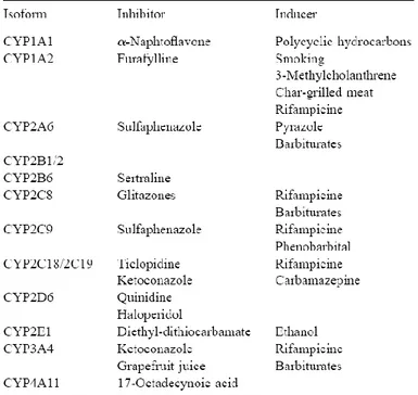
Human populations exhibit considerable variability in CYP activity levels. This is due to the fact that some human CYP enzymes are polymorphic, with a significant percentage of populations being deficient in a specific enzyme or having a functional enzyme with an altered amino acid sequence, which can change the kinetics of substrate metabolism. Expression levels of specific CYP enzymes vary substantially among individuals. The levels of other human CYP enzymes (e.g., CYP1A2 and CYP3A4) are induced by certain environmental exposures or drug treatments (Table 1). If an enzyme, which is polymorphic or subject to environmental regulation, is rate-limiting for the elimination of a drug, then substantial interindividual variation in pharmacokinetics is often observed (Crespi and Miller, 1999).
1.2.3 In vitro models to study drug metabolism
A key question in human drug biotransformation research is how to make reliable extrapolations from the in vitro or in vivo model to clinical practice.
Several in vitro models have been developed in the past. They are used to obtain early information about biotransformation pathways and to predict drug-drug interactions. The optimal model system depends on a number of factors, such as in vivo resemblance, expense, availability of the model, and ethical considerations. In vitro data from human and animal models can be used to choose the best in vivo model (e.g., mouse, rat, dog) for further testing. In conclusion, it can be stated that an in vitro model is always a compromise between convenience and relevance.
1.2.3.1 Liver microsomes
Microsomes can easily be prepared from different tissues, most commonly from the liver of animals and human donors. Therefore, human liver microsomes (HLM) have become a very commonly and widely used in vitro model. Liver microsomes consist of vesicles of the hepatocyte endoplasmic reticulum and are prepared by differential centrifugation and thus contain almost only CYP and UGT enzymes (Figure 4).
Figure 4. Liver fractions preparation
The enzymatic activities are stable during prolonged storage of the microsomes. In order to reflect the standard proportion of the enzymes in human or animal livers, liver microsomes
appropriate cofactors and other reaction components, it is possible to investigate and
distinguish between CYPs, flavin-containing monooxygenase (FMO) and
glucuronosyltransferase (UGT) activities.
The major advantages of microsomes are low costs, simplicity in use, and they are one of the best-characterized in vitro systems for drug biotransformation research. However, some major drawbacks exist. It should be noted that results obtained with microsomes cannot be used for quantitative estimations of in vivo human biotransformation, because CYPs and UGTs are enriched in the microsomal fraction and there is no competition with other enzymes (e.g., NAT, GST, and SULT) with the consequence that metabolites formed in intact liver could be unnoticed.
1.2.3.2 Liver cytosol fractions
The liver cytosolic fraction contains the soluble phase II enzymes, e.g., NAT, GST, and SULT. It is obtained by differential centrifugation of whole-liver homogenate, like microsomes (Figure 4). For the catalytic activity of the phase II enzymes, addition of exogenous cofactors, e.g., acetyl coenzyme A (acetyl CoA), dithiothreitol (DTT), and acetyl CoA regenerating system for NAT, 3'-phosphoadenosine-5'-phosphosulfate (PAPS) for SULT, and glutathione (GT) for GST, is necessary.
The main advantage of cytosol fraction is the presence of only three enzymes at higher concentrations compared to human liver S9 fraction. The biotransformation capacity of NAT, SULT, and GST can be studied separately or in combination depending on the cofactors added. A disadvantage is that only the soluble phase II enzymes are present in the liver cytosol fraction and that therefore the UGTs, which are located on the endoplasmic reticulum, metabolic pathways cannot be investigated with this model.
1.2.3.3 Liver S9 fractions
The liver S9 fraction contains both microsomal and cytosolic fractions. Like microsomes, a NADPH-regenerating system or NADPH solution is required to supply the energy demand of the CYP enzymes, as well as for the catalytic activity of phase II enzymes, addition of exogenous cofactors is necessary.
Compared with microsomes and cytosol, S9 fractions offer a more complete representation of the metabolic profile, by performing both phase I and phase II activities. In some cases with
S9 fractions, metabolites that are not produced by either the cytosolic fraction or the microsomal fraction alone are formed.
1.2.3.4 Human CYP and UGT supersomes (baculovirusinsect-cell-expressed)
Insect cells lack endogenous CYP and UGT activity and, therefore, microsomes, which consist of vesicles of the hepatocyte endoplasmic reticulum, of human CYP- or UGT transfected insect cells can be a useful tool in human biotransformation studies. Since the expression is baculovirus mediated, microsomes of these cells are sometimes referred to as baculosomes, but more often as supersomes™. The availability of specifically expressed human CYPs and UGTs in supersomes allows the investigation of the contribution of a single metabolic enzyme to the biotransformation pathway of the compound under investigation. At present all common human CYPs, coexpressed with NADPH-cytochrome P450 reductase and optionally cytochrome b5, and UGTs are offered in supersomes™. A control experiment, an
incubation with nontransfected supersomes, must always be conducted. NADPH-regenerating system or NADPH is required to supply the energy demand of the CYPs as well as UDPGA for the UGT activity has to be added as a cofactor. A major advantage of supersomes is that they can be used to study not only isozyme-specific drug biotransformation, but also drug– drug interactions. In the past few years the development of new CYP and UGT supersomes™ has increased considerably. The different genotypes of the CYP isozymes (e.g., CYP2C9*1,
CYP2C9*2, and CYP2C9*3) are now also commercially available
(https://www.corning.com). Hence, the influence of different polymorphisms on the drug biotransformation pattern also can be studied.
A disadvantage is that, in UGT supersomes, the UGT active site is shielded behind a hydrophobic barrier, resulting in latency of glucuronidations. However, this disadvantage can be overcome by using a pore-forming agent, alamethicin.
1.2.3.5 Hepatocytes Primary hepatocytes
Primary hepatocytes are a popular in vitro system for drug biotransformation research due to their strong resemblance of in vivo human liver. Human liver is mainly obtained from patients that undergo partial liver resection, e.g., because of liver metastasis.
Cultured hepatocytes
Once isolated, hepatocytes can be held in suspension, in which case they remain viable for only a few hours, or they can be maintained in monolayer culture for a maximum of 4 weeks. Both cultured hepatocytes and suspensions of primary hepatocytes have repeatedly proven to be powerful tools to analyze the specific metabolic profile of a variety of drugs with good in
vitro–in vivo correlations However, it has been widely recognized that cultured hepatocytes
are subject to a gradual loss of liver-specific functions, with special reference to a decreased CYP expression. This loss is different for the specific CYP isoforms; for some isoforms it becomes evident after a few days of culture (CYP 2E1 and CYP 3A4), while others remain nearly unaffected by the isolation and culturing processes (CYP 1A2 and CYP 2C9). An advantage of isolated hepatocytes compared to liver slices and perfused liver is the possibility of cryopreservation. Indeed cryopreserved hepatocytes have been shown to retain the activity of most phase I and phase II enzymes. A disadvantage is the lack of liver nonhepatocyte cells. Although hepatocytes account for the vast majority of the liver volume (about 80%), other cells such as Kupffer cells may be necessary for cofactor supply. Another problem encountered with human hepatocytes, as with human liver microsomes, is the considerable interindividual variation (Brandon et al., 2003).
1.3 PROPARGYLAMINE-CONTAINING DRUGS 1.3.1 Neurodegenerative disorders
Parkinson’s disease (PD) and Alzheimer’s disease (AD) are conditions constituting part of the broad cluster of diseases commonly referred to as neurodegenerative disorders (ND).
A classical feature of these diseases is the transient loss of neuronal cells in the brain due to apoptosis triggered by one of many factors including genetic, endogenous and environmental ones. Although each disease has its own molecular mechanisms and clinical manifestations, some general pathways might be recognized in different pathogenic cascades. They include protein misfolding and aggregation, oxidative stress and free radical formation, metal dyshomeostasis, mitochondrial dysfunction, and phosphorylation impairment, all occurring concurrently (Cavalli et al., 2008).
The multifactorial nature of NDs suggests that targeting any single mechanistic site will not result in successful retardation of the disease progression and many “single-site-targeting” drugs have failed to render sufficient neuroprotective and/or neurorestorative activity. The use of drugs aiming to a single receptor or enzymatic system is thus insufficient for treatment of these multifactorial diseases (Zindo et al., 2015).
It is now widely accepted that a more effective therapy would result from the use of multitarget-directed ligands (MTDLs) able to intervene in the different pathological events underlying the etiology of neuronal disorders. To obtain novel MTDLs, a design strategy is usually applied in which distinct pharmacophores of different drugs are combined in the same structure to afford hybrid molecules. In principle, each pharmacophore of these new drugs should retain the ability to interact with its specific site or sites on the target and, consequently, to produce specific pharmacological responses that, taken together, should slow or block the neurodegenerative process (Cavalli et al., 2008).
The last few decades have seen increasing research interest and exploration of the neuroprotective ability of compounds bearing propargylamine function seen in selegiline and rasagiline. The propargylamine moiety is now known to play an important role in providing the neuronal and mitochondrial protective properties inherent in these compounds and other reported activities ascribed to this moiety in literature include antiapoptotic and amyloid-β (Aβ) aggregation inhibition. These features of the propargylamine moiety have led to its incorporation into structures of many drug-like compounds designed for neuroprotection to afford resultant molecules with broader therapeutic profiles that may potentially meet the
1.3.2 Alzheimer’s Disease (AD)
Alzheimer’s disease (AD) stands out among neurodegenerative disorders as the fourth leading cause of death and the most common cause of acquired dementia in the elderly population, afflicting more than seven million people worldwide (Cavalli et al., 2008).
The predominant clinical manifestation is progressive memory deterioration and changes in brain function, including disordered behaviour and impairment in language and comprehension, which progressively worsen over 5–10 years. Most of these cognitive symptoms result from a depletion of basal forebrain cholinergic neurons leading to decreased cholinergic neurotransmission. Besides the cognitive deficits, patients frequently exhibit neuropsychiatric symptoms such as depression, psychosis and agitation.
From the histopathological viewpoint, two characteristic hallmarks accompany these features: the neurofibrillary tangles (NFT) which are intracellular fibrillar deposits mainly composed of the microtubule-associated protein tau, and the senile plaques (SP), formed by deposition of the aggregated amyloid-β peptide (Aβ). Although the pathogenesis of AD is not yet fully understood, it is a multifactorial disease triggered by several factors, including excessive protein misfolding and aggregation, oxidative stress and free radical formation, mitochondrial dysfunction, metal dyshomeostasis, excitotoxic and neuroinflammatory processes. In addition to the cholinergic dysfunction, disturbances in other neurotransmitter systems such as the monoaminergic have also been reported to account for AD symptoms (Bolea et al., 2012).
Of the above-mentioned hypotheses, the cholinergic one is the oldest and it has also had the strongest influence on the development of clinical treatment strategies, which led to the introduction of the acetylcholinesterase inhibitor (AChEI) tacrine (1), donepezil (2), galantamine (3) and rivastigmine (4) (Figures 5).
Figure 5.
These drugs have become the standard for AD therapy and, although beneficial in improving cognitive, behavioral, and functional impairments, they seem unable to address the molecular mechanisms that underlie the pathogenic processes.
1.3.3 Parkinson’s disease (PD)
Parkinson’s disease (PD) is the second most common neurodegenerative disorder. Like AD, it is currently an incurable disease. The available pharmacological therapies are unable to arrest or reverse the neurodegeneration associated with PD.
PD is characterized by the progressive loss of dopaminergic neurons in the substantia nigra pars compacta and other subcortical nuclei and by the presence of intraneuronal aggregates known as Lewy bodies (LBs), which are enriched in filamentous α-synuclein and other proteins that are often ubiquitinated. Depletion of dopamine (DA) causes dysregulation of the motor circuits that project throughout the basal ganglia, resulting in the clinical manifestations of PD, which include tremor, bradykinesia, rigidity, and postural instability. However, additional neurotransmitter systems are also involved in PD and, consequently, nonadrenergic, serotoninergic, and cholinergic neurons are also lost. This loss is responsible for nonmotor symptoms, such as cognitive decline, sleep abnormalities, and depression, which dominate the later stages of PD (Cavalli et al. 2008).
The crucial molecular step of PD pathogenesis is the formation of LBs, whose key constituent is represented by α-synuclein, a cytoplasmic soluble protein that contains a highly amyloidogenic domain within its mid-region. In pathogenic conditions, α-synuclein misfolds
and is converted into pathological oligomers and higher-ordered aggregates, which fibrillize and deposit into LBs as β-pleated sheets. Therefore, maintaining α-synuclein in the native and soluble random coil conformation, and consequently preventing α-synuclein-mediated neuron toxicity, emerges as a clear goal of innovative disease-modifying PD therapies. Moreover treatment with antioxidants might theoretically be of benefit in preventing neurodegeneration.
Despite an increased understanding of the molecular causes of PD, current PD therapies are based mainly on exogenous replacement of DA within the striatum. This improves the symptoms but without halting the progression of the neurodegenerative process or reversing the neuronal degeneration. Furthermore, although PD also involves degeneration of non-dopaminergic neurons, the treatment of the resulting predominantly non-motor features remains a challenge. Depletion of striatal DA from the loss of nigral projections is the main target for the currently available drugs. The classes of compound that still hold a prominent position in current anti-PD drug discovery are reported in Table 2.
Mechanism of action Drugs
DA precursor (prodrug) L-dopa
COMT inhibition Entacapone, Tolcapone
MAO-B inhibition Selegiline, Rasagiline
Dopaminergic receptor agonism (ergot derivative)
Bromocriptine, Pergolide, Cabergoline, Lisuride
Dopaminergic receptor agonism
(non-ergot derivative) Ropinirole, Pramipexole, Piribedil
Muscarinic receptor antagonism Biperiden, Triexyphenidyl, Metixene
Ion channel blockade Amantadine
Peripheral dopa decarboxylase inhibition Benserazide, Carbidopa
Table 2.
Levodopa (L-dopa) is the key compound in the treatment of PD, acting as a precursor of DA. However, besides offering only symptomatic relief for patients, this drug shows substantial side effects at the high doses required for therapeutic action.
Certain other available drugs, like MAO-B (selegiline and rasagiline) and COMT (entacapone and tolcapone) inhibitors, are used mainly with L-dopa, since they alter the in vivo metabolism of DA by increasing its plasma half-life. Anticholinergic compounds (such as
biperiden, triexyphenidyl) were among the first drugs used for PD therapy, since they were intended to correct the imbalance between DA and acetylcholine (ACh) brain levels. More recent therapeutic approaches to PD are represented by nicotine, anti-inflammatory agents, melatonin, selenium, iron chelators, and vitamins A, C, and E.
As previously mentioned, none of these therapies are able to arrest or reverse the progression of PD probably because these compounds treat only the symptoms of the disease rather than tackling the actual molecular PD causes. Moreover, the currently available drugs have been shown to interact with a single molecular target, instead of confronting the multifactorial nature of PD neurodegeneration.
1.3.4 Monoamine oxidase
Monoamine oxidase (MAO, E.C.1.4.3.4) is a flavin-containing enzyme that catalyses the oxidative deamination of a wide range of biogenic and xenobiotic amines, including dopamine (DA), noradrenaline, adrenaline, tyramine, serotonin (5-HT). This enzyme is known to exist in two isoforms namely MAO-A and MAO-B, which show different selectivities for substrates and inhibitors. The enzyme is inserted in the outer mitochondrial membrane, and is widely distributed in the body tissues, with a high degree of expression in the gastro-intestinal tract and liver, as well as neuronal tissue, and nearly all other organs. Monoamine oxidase A and B catalyze the oxidation of primary, secondary, and some tertiary amines to their corresponding aldehyde, hydrogen peroxide and ammonia (in case of primary amines) or a substituted amine (in case of secondary and tertiary amines) in a reaction shown below:
RCH2NH2 + H2O + O2 RCHO + NH3 + H2O2
Selective inhibitors for MAO-A have been shown to be effective antidepressants, whereas MAO-B inhibitors are useful for the treatment of Parkinson’s disease. The neuroprotective effect of monoamine oxidase inhibitors (MAOIs) in these disorders may result not only from the increased amine neurotransmission, but also from prevention of neurotoxic product formation, which promotes reactive oxygen species (ROS) generation and may ultimately contribute to increased neuronal damage. Despite some disadvantages found in using MAOIs in clinical practice, such as hepatotoxicity and the so-called ‘‘cheese-effect’’, which describes
MAO enzymes remain in the focus of drug design targeting neurodegenerative disorders. Nevertheless, it seems at present unlikely that the neuroprotective activities of MAOIs are exclusively related to the ability to decrease the production of free radicals and toxic aldehydes via the inhibition of enzymatic activity. With regard to this, several studies have suggested that they are rather related to the anti-apoptotic properties of the propargyl group present in these molecules (Bolea et al., 2014).
1.3.5 Propargylamines
Propargylamines are molecules containing a propargyl moiety that typically inhibits MAO-B including the well characterized compounds selegiline (l-deprenyl), rasagiline and PF9601N (Fig. 6).
Figure 6. Propargylamine-derived MAOIs (the propargylamine moiety is highlighted with dotted lines)
While selegiline was the first selective MAO-B inhibitor used clinically for the treatment of Parkinson’s disease, rasagiline and PF9601N belong to a second generation of MAO-B inhibitors that, unlike selegiline, do not generate amphetamine derivatives when metabolized. Since these compounds possess anti-apoptotic properties independent from their ability to inhibit MAO-B, diverse mechanisms have been suggested to be involved in the neuroprotective properties of propargylamine-containing compounds (Fig. 7).
Figure 7. Schematic representation of the sites of action of propargylamine-derived compounds as potential
targets for AD treatment figure taken from Bolea et al. (2014)
One of these mechanisms involves a significant antioxidant potency arising from the increase in the activities of superoxide dismutase (SOD) and catalase (CAT) enzymes, besides the prevention of the MAO reaction products formation, which are potentially neurotoxic since they contribute to oxidative stress and the formation of ROS. Moreover, the anti-apoptotic activity of these molecules has been attributed to their ability to prevent the fall in mitochondrial membrane potential (Ψm) and the blockade of the permeability transition pore (PTP) opening as a consequence of the up-regulation of Bcl-2 family protein and activation of protein kinase C (PKC) and mitogen-activated protein kinase (MAPK). These pathways may be additionally involved in the effect of propargylamines on the enhanced release of the non-amyloidogenic α-secretase form of soluble amyloid precursor protein (sAPPα), which precludes the formation of amyloid derivatives promoting the non-amyloidogenic pathway of APP processing. Further neuroprotective effects have been related to the induced increase in the expression of neurotrophic factors such as BDNF and GDNF (Bolea et al., 2014).
1.3.6 Multitarget-directed ligands (MTDLs) strategy
The advent of MTDL approach has now seen many researchers develop a number of compounds that can potentially confer neuroprotection by acting simultaneously on different receptors and target sites implicated in NDs. This approach has yielded a wide amount of interesting MTDLs to halt NDs: some examples of propargylamine-derived compounds as promising neuroprotective agents are here mentioned.
1.3.6.1 MAO/AChE inhibitor
A successful approach of combined MAO/AChE inhibition came from the combination of the carbamate moiety of rivastigmine (figure 5) with the indane scaffold present in the MAO-B inhibitor rasagiline (figure 6), leading to the compound ladostigil (Fig. 8) (Sterling et al., 2002).
Figure 8.
Ladostigil is able to inhibit both AChE and butyrylcholinesterase (BuChE) for a longer time than the parent compound rivastigmine. In addition, ladostigil selectively inhibits brain MAO-A and MMAO-AO-B resulting in an increase in noradrenaline, dopamine and serotonine levels, and thus exerting an antidepressant action. Ladostigil has also been shown to retain the neuroprotective and anti-apoptotic properties observed in the parent compound and propargylamine-derived rasagiline. Besides, ladostigil also possesses a cognition enhancing activity and is the most advanced MTDL on its category as demonstrated by the promising results obtained from a phase 2 clinical trial.
1.3.6.2.MTDLs targeting MAO and iron
It has been reported that iron contributes to Aβ aggregation, which in turn contributes to neuronal degeneration through the induction of oxidative stress. A link between high iron concentration and MAO activity and their involvement in ROS production has also been
reported. To address these problems, Youdim and collaborators have developed compounds with a bi-functional action on iron chelation and MAO inhibition obtained by combining the iron-chelating and antioxidant scaffold of VK28 with the N-propargylamine moiety of rasagiline. The most interesting compounds obtained were M30 and HLA20 (Fig. 9) possessing iron-chelating activity similar to that of VK28, but holding higher brain permeability.
Figure 9.
M30 and HLA20 possess neuroprotective properties comparable to rasagiline and potently
inhibit the iron induced membrane lipid peroxidation features. More importantly, M30 has been shown to selectively inhibit brain MAO (A and B) enzymes and to increase serotonin, dopamine and adrenaline neurotransmission, which confers to this multipotent compound an antidepressant action besides preventing the potentiation of tyramine-induced cardiovascular activity. Interestingly, M30 inhibits the Aβ aggregation induced by metals and reduces Aβ formation (Bolea et al., 2014).
1.3.7 Conclusions
The complex pathogenesis and etiology of neurodegenerative diseases make it clear that drugs targeting a single receptor or enzymatic system will not result in successful retardation and treatment of these multifactorial diseases. This is even more evident in that drugs used in the symptomatic treatment of these diseases fail to render sufficient neuroprotective and/or neurorestorative activity. The advent of MTDL approach has however seen the development of compounds that combine symptomatic treatment and neuroprotection in one molecule by acting simultaneously on different receptors and target sites implicated in NDs.
Compound bearing the propargylamine function have received significant attention based on the neuroprotective ability observed for the MAO-B inhibitors selegiline and rasagiline. This has led to its incorporation into many drug-like compounds designed for neuroprotection and to afford molecules with broader therapeutic profiles to potentially meet the curative needs of the multifactorial NDs.
1.4 REFERENCES
Alsante, K., Ando, A., Brown, R., Ensing, J., Hatajik, T., Kong, W., & Tsuda, Y. (2007). The role of degradant profiling in active pharmaceutical ingredients and drug products.
Advanced Drug Delivery Reviews, 59(1), 29–37.
Baranczewski, P., Stańczak, A., Sundberg, K., Svensson, R., Wallin, Å., Jansson, J., Garberg, P., Postlind, H. (2006). Introduction to in vitro estimation of metabolic stability and drug interactions of new chemical entities in drug discovery and development.
Pharmacological Reports, 58, 453–472.
Bolea, I., Gella, A., & Unzeta, M. (2013). Propargylamine-derived multitarget-directed ligands: fighting Alzheimer’s disease with monoamine oxidase inhibitors. Journal of
Neural Transmission, 120(6), 893–902.
Brandon, E. F. A., Raap, C. D., Meijerman, I., Beijnen, J. H., & Schellens, J. H. (2003). An update on in vitro test methods in human hepatic drug biotransformation research: pros and cons. Toxicology and Applied Pharmacology, 189(3), 233–46.
Cavalli, A., Bolognesi, M. L., Minarini, A., Rosini, M., Tumiatti, V., Recanatini, M., & Melchiorre, C. (2008). Multi-target-directed ligands to combat neurodegenerative diseases. Journal of Medicinal Chemistry, 51(3), 347–372.
Crespi, C. L., & Miller, V. P. (1999). The use of heterologously expressed drug metabolizing enzymes-state of the art and prospects for the future. Pharmacology & Therapeutics,
84(2), 121–31.
ICH Harmonized Tripartite Guidelines Q1B (1996). Stability Testing: Photostability Testing of New Drug Substances and Products
ICH Harmonized Tripartite Guidelines Q1A (R2) (2003). Stability Testing of New Drug Substances and Products.
Prakash, C., Shaffer, C. L., Nedderman, A. (2007). Analytical strategies for identifying drug metabolites. Mass Spectrometry Reviews, 26(2), 340–369.
Reynolds, D. W., Facchine, K. L., Mullaney, J. F., Alsante, K. M., Hatajik, T. D., & Motto, M. G. (2002). Available Guidance and best practices for conducting forced degradation studies. Pharmaceutical Technology.
Sterling, J., Herzig, Y., Goren, T., Finkelstein, N., Lerner, D., Goldenberg, W.,Weinstock, M. et al. (2002). Novel Dual Inhibitors of AChE and MAO Derived from Hydroxy
Aminoindan and Phenethylamine as Potential Treatment for Alzheimer ’s Disease.
Journal of Medicinal Chemistry, 45, 5260–5279.
Zindo F.T., Joubert J., M. S. F. (2015). Propargylamine as functional moiety in the design of multifunctional drugs for neurodegenerative disorders: MAO inhibition and beyond.
CHAPTER 2. Aim of the work
Chemical stability studies were performed on both drug substance and drug product to guarantee their efficacy, quality and safety and fulfill the requirements of regulatory authorities. In this area, forced degradation studies provide useful data to support: the intrinsic stability of the drug molecule, through the identification of possible degradants and the corresponding degradation pathways, and the development and validation of stability-indicating analytical procedures.
On the other hand, metabolic stability studies were performed on drugs to identify the metabolites arising from phase I and phase II reactions while delineating the corresponding metabolic pathways; these studies have become an integral part of the drug discovery/development process while playing a role also in post-marketing phase. Since drug metabolites may have intrinsic pharmacological activity or display specific toxicity, their knowledge is relevant for both pharmacokinetic, pharmacodynamic and for toxicokinetics. Generally, in academic research, chemical and metabolic stability studies were not conducted in an integrated manner being preferably done from researchers having different expertise. However, since the intrinsic stability of drugs can be strictly related to its metabolic stability, the integration of both aspects of stability could be conveniently performed defining a unique chemical and metabolic stability space.
In this work the chemical and metabolic stability of three propargylamine-containing drugs, whose structures are reported in the Figure 1, were studied.
Figure 1.
The interest regarding this class of drugs arises from the analyses in the course of stability studies performed on new formulations of oxybutynin hydrochloride (Oxy), a tertiary propargylamine used for the management of urinary frequency, urgency and incontinence in neurogenic bladder disorders. During the study, a new and unprecedented degradation product
was identified in topical formulations, whose structure and mechanism of formation has been determined through a forced degradation study. These results prompted us to extend the study of chemical and metabolic stability to other propargylamine-containing drugs. Selegiline (SG, tertiary and H-terminated propargylamine) and rasagiline (RG, secondary and H-terminated propargylamine), MAO-B inhibitors used in the treatment of Parkinson’s Disease, were included in the panel of compounds to increase the chemical diversity of propargylamine moiety.
To face the analytical tasks, required in performing the chemical and metabolic stability studies, LC-DAD-UV and LC-ESI-MS/MS techniques were employed: the analytical methods were developed to allow both qualitative and quantitative determination of analytes. Moreover, the new degradation products showing in their structure the presence of a structural alert were assessed for genotoxic potential evaluation; similarly, the new metabolites will be assessed for their potential pharmacodynamic activity.
CHAPTER 3. New insights in oxybutynin chemical stability: identification
in transdermal patches of a new impurity arising from oxybutynin N-oxide
rearrangement
3.1 INTRODUCTION
Oxybutynin hydrochloride (4-(diethylamino)but-2-ynyl (RS)-2-cyclohexyl-2-hydroxy-2-phenyl-acetate hydrochloride, Oxy) is an antimuscarinic drug which is used for the management of urinary frequency, urgency and incontinence in neurogenic bladder disorders and in idiopathic detrusor instability (Kennelly, 2010). With Oxy being the first choice in
treating urinary incontinence, different types of formulations are at present available on the market: immediate and extended release tablets, oral solutions, syrup and solution for intravesical administration. Alternatively, to mitigate the systemic adverse effects occurring during the therapy, Oxy free base was employed in topical formulations such as adhesive patch (Kentera™) and gel (Gelnique™) (Shaw et al., 2007). Indeed oxybutynin is associated with a high incidence of systemic side-effects, including dry mouth, constipation, blurred vision, dryness of the eyes, palpitations, drowsiness, dizziness and esophageal reflux (Lose et al., 2001).
From a structural point of view Oxy is characterized by the presence of a tertiary propargylamine moiety and a carboxyester function; as a consequence, the chemical stability of Oxy has been investigated(Miyamoto et al., 1994), demonstrating the formation of a single hydrolytic degradation product, namely α-cyclohexylmandelic acid (CHMA). The formation of CHMA was also observed by Wagieh et al. (2010) and El-Gindy (2005) during the stress studies performed in acidic and alkaline conditions. Moreover, CHMA was the only degradation product included in the list of specified impurities in both the Oxy monographs of the European and United States Pharmacopoeias (EP 8, 2013; USP 38, 2015). In accordance with this scenario, a literature survey showed that the stability indicating analytical methods (Wagieh et al., 2010; El-Gindy, 2005; De Schutter et al., 1988)for the determination of Oxy in drug substance and drug product take into account CHMA as the only degradation product. Interestingly, even if De Shutter et al. (1988) noted the formation of several minor peaks in the chromatogram of Oxy treated with hydrogen peroxide in severe conditions, no degradation product arising from oxidation has been identified so far. At first sight this is not
surprising, because oxybutynin is mainly used as hydrochloride salt and the corresponding protonated amino group is scarcely prone to oxidation. However, due to its better lipophylicity and permeability, Oxy free base is used in the new topical formulations: in that case the unionized tertiary amine can be more easily oxidized with the potential formation of degradation products.
These considerations, combined with the observation of an unknown impurity during oxybutynin adhesive patch analysis, prompted us to reinvestigate the Oxy chemical stability, focusing our attention on the oxidative degradation pathway.
In this report, we elucidated the chemical fate of Oxy in oxidative conditions showing the formation of the corresponding diastereomeric N-oxides (Oxy-NO1 and Oxy-NO2) and an unprecedented degradation product (Oxy-EK), whose structure and mechanism of formation were discussed. Finally, Oxy-EK synthesis was described, allowing us to propose it as the indicator of stability for the oxidative degradation in stability studies and, at the same time, to evaluate its potential mutagenic effects.
3.2 EXPERIMENTAL
3.2.1. Reagents and chemicals
Methanol (HPLC grade), tetrahydrofuran (THF), ammonium acetate, iron (III) trichloride hexahydrate, copper sulphate, pentahydrate, hydrogen peroxide solution (30% w/w), N,O-dimethylhydroxylamine hydrochloride acetoxyacetyl chloride, ethynylmagnesium bromide (0.5 M in THF), 4,4ˈ-azobis-(4-cyanovaleric acid) (ACVA), α-cyclohexylmandelic acid (97%), ninhydrin, histidine, tryptophan, biotin, 2-nitrofluorene (2NF), 2-aminoanthracene (2AA), sodium azide (SA), 9-aminoacridine (9AA) and methyl methanesulfonate (MMS), bacteriological agar, nutrient broth, nutrient agar were supplied by Sigma-Aldrich (St. Louis, MO, USA). Water (HPLC grade) was obtained from Milli-Q reverse osmosis system (Millipore Co., Billerica MA, USA). Column chromatography was performed on silica gel 60 (230-400 mesh ASTM Merck). TLC was carried out on plates with a layer thickness of 0.25 mm (silica gel 60 F254; Merck); when necessary, they were visualized after spraying with ninhydrin reagent. Oxy (potency: 99.3%), was obtained as gift sample from Pharmafar S.r.l. (Torino, Italy); Kentera™ transdermal patches (lot n° 356861C; exp. date: 11/2013 and lot n° 818002A exp. date: 09/2016; Nicobrand Ltd, Coleraine, Northern Ireland), were purchased
with the mixture phenobarbital/β-naphthoflavone to induce the hepatic microsomal enzymes) was supplied and certified by Moltox (Molecular Toxicology, Boone, NC, USA).
3.2.2. Instrumentation and chromatographic conditions
3.2.2.1 LC-UV analyses
A Shimadzu HPLC system, consisting in two LC-10AD Vp module pumps and a DGU-14-A on-line vacuum degasser, was used. The analyses were carried out on a Luna C18(2) (150 x 4.6 mm I.D., 5m dp; Phenomenex, Torrance, CA, USA) column. The isocratic mobile phase
(flow rate 1.2 ml/min) was composed of eluant A ammonium acetate buffer (20 mM pH=5.7) and eluant B methanol, being the A:B ratio 25:75 (v/v). The eluants A and B were filtered through a 0.45 m PVDF membrane filter prior the use. A SIL-10AD Vp autosampler was used for the injection of samples (20µl). The SPD-M10A Vp photodiode array detector was used to detect Oxy and the degradation products CHMA, Oxy-NOs at 210 nm and Oxy-EK at 311 nm. A LCsolution 1.24 software was used to process the chromatograms. All the analyses were carried out at room temperature.
3.2.2.2 LC-MS/MS analyses
A Thermo Finningan LCQ Deca XP Plus system equipped with a quaternary pump, a Surveyor AS autosampler, a Surveyor PDA detector and a vacuum degasser was used for LC-MS analysis (Thermo Electron Corporation, Waltham, MA, USA).
The analysis were performed on a Luna C18(2) (150 x 4.6 mm I.D., 5m dp; (Phenomenex,
Torrance, CA, USA) column maintained at 35°C. The mobile phase (flow rate 0.8 ml/min) was composed of eluant A: ammonium acetate buffer (pH 5.7; 10 mM) and eluant B: methanol, using an isocratic mode being 20:80 the ratio A:B; the sample injection volume was 20 l. The eluate was injected into the electrospray ion source (ESI) with a splitting of 40% and the MS and MS/MS spectra were acquired and processed using the Xcalibur® software (Thermo Electron Corporation, Waltham, MA, USA). The operating conditions of the ion trap mass spectrometer in positive ion mode were as follows: spray (source) voltage, 5.30 kV; source current, 80 A; capillary temperature, 350 °C; capillary voltage, 23.00 V; tube lens offset, 40.0 V; multipole 1 offset, -5.75 V; multipole 2 offset, -7.00 V; sheath gas flow (N2), 60 Auxiliary Units; sweep gas flow (N2): 6.0 A. U. Data were acquired both full
scan and MS/MS product ion scan modes using mass scan range m/z 105 to 500, optimizing the collision energy at 30%.
3.2.2.3 Spectroscopic analyses
1
H and 13C experiments were performed at 298K on a JEOL Eclipse ECP 300 FT MHz spectrometer (Jeol Ltd. Tokyo, Japan) operating at 7.05T. Chemical shifts are reported in part per million (ppm). FT-IR experiments were performed on AVATAR 370 FT-IR (Thermo Fisher Scientific, Madison WI, USA) spectrophotometers.
3.2.3 Forced degradation study of Oxy
The degradation study in solution was done at 1 mg/ml Oxy concentration. For the acidic
degradation study, Oxy was dissolved in 0.5 N HCl and the solution was left in the dark at 50°C for 24 h. The degradation in alkaline conditions was done in 0.01 N NaOH in presence of methanol/water 50/50 and the solution was left in the dark at 50°C for 30 minutes.
The oxidative degradation was studied, protecting the samples from light, using the following protocols:
a) Oxy was dissolved in 3% (v/v) H2O2 in acetate buffer (pH 4.5) and the solution was left
at room temperature (r.t.) for 24h.
b) Oxy was dissolved in 3% (v/v) H2O2 in 50/50 water/methanol mixture (8 mL) in the
presence of sodium hydrogen carbonate (100 mg): the solution was left at r.t. for 4h. c) Oxy was dissolved in acetate buffer (pH=4.5) in the presence of 1.5 mM Cu2+ or Fe3+
and left at r.t. for 4h.
d) To a solution of Oxy in 50/50 methanol/water, the radical initiator ACVA, previously dissolved in water (1 mg/ml) in the presence of sodium hydrogen carbonate, was added. The mixture of reaction (ACVA: 1 mM final concentration) was stored at 60 °C for 24h.
The thermal and photodegradation were studied by testing Oxy and Oxy free base. In particular, for thermal degradation, the samples were stored for 18 h at 60 °C in a hot air oven. Photodegradation was carried out, at r.t., by exposing a thin layer (10 mg) of Oxy or
3.2.3.1 Sample preparation
Before LC-UV and LC-MS/MS analyses, acidic and alkaline samples were first neutralized by addition of a suitable amount of sodium phosphate dibasic and diluted phosphoric acid (8.5% w/v) respectively, and then diluted with mobile phase. The other stressed samples were dissolved or tenfold diluted in mobile phase only; finally, solid samples were dissolved in mobile phase to attain ~0.1 mg/ml concentration. For comparison purposes a freshly prepared aqueous solution of Oxy (~1 mg/ml) was diluted and analyzed as above.
3.2.3.2 Procedure for the analysis of transdermal patch
After removal of the protective film (Kentera™ 36 mg/39 cm2), the patch was transferred into a Falcon™ conical tube and extracted by sonication (SweepZone Technology Quantrex S200) with methanol (3x20ml). The combined methanolic extracts were evaporated to small volume under reduced pressure and then transferred and diluted in a 20 ml volumetric flask with methanol/water 75/25. The solution was analyzed with LC-UV and LC-MS/MS. To evaluate the role of the extraction conditions on the chemical stability of Oxy, the procedure was repeated on a sample of Oxy free base.
3.2.3.3. Accelerated stability study of transdermal patch
The stability study was performed in a Binder-KBF-115 constant climate chamber (Binder GmbH, Tuttlingen Germany). Drug product (Kentera™ transdermal patch) packaged in the container closure system used for marketing was stored using the following condition: 40°C±2°C/75% RH±5%, t= 6 months.
3.2.4. Isolation and purification of Oxy-EK
To a solution of Oxy (500 mg, 1.27 mmol) in 5 ml of water, 240 mg of sodium hydrogen carbonate 5 ml of methanol and 5 ml of 30% w/w hydrogen peroxide were added. The mixture was reacted, under stirring at r.t., for 3h monitoring by TLC analysis (eluant: CH2Cl2/MeOH 9:1). At the end of the reaction, methanol was evaporated under reduced
pressure. The aqueous residue was extracted (3x15 ml) with CH2Cl2. The pooled organic
layers were washed with brine, dried over anhydrous sodium sulphate, filtered and evaporated in vacuo to dryness. The pale-yellow oily residue was left overnight at r.t. and then purified by column chromatography using dichloromethane/methanol 98/2 as eluant to give pure