Ch
p
_____FARM
hronic
pathog
mol
Dotto Dott.ssa Sa ___________ (firmaUn
DO
MACOLO
c lymp
genes
lecula
mic
rando accenti Elena ___________ a)nivers
F
OTTORA
OGIA E
COORDINAphocy
sis an
ar cyto
croRN
Settore Scie a ________ Asità
Ferra
ATO DI
ONCOL
CICLO XX ATORE Prof.ytic le
d pro
ogene
NAs ex
entifico Disci Anni 2011/2degl
ara
RICERC
LOGIA M
XVI Antonio Cuneukem
ognos
etic st
xpres
plinare MED _____ 2013li Stu
CA IN
MOLEC
neomia: as
is by
tudies
ssion
D/15 T Prof. Cu ___________ (fudi d
COLARE
ssess
mode
s and
Tutore uneo Antonio ___________ firma)di
E
sing
ern
o ________3
I
NDEX
ABSTRACT ... 5
ABSTRACT (ITALIAN) ... 9
CHAPTER 1 ... 13
CHRONIC LYMPHOCYTIC LEUKEMIA ... 13
1.1 Introduction... 13
1.2 Diagnosis and Staging ... 13
1.3 Biology and Pathogenesis of CLL ... 14
1.4 Prognostic factors in CLL ... 15 1.5 Treatment of CLL ... 18 1.5.1 Cytostatic Agents ... 19 1.5.2 Antibodies ... 19 1.5.3 Combination chemotherapy ... 20 1.5.4 Chemoimmunotherapy ... 20
1.5.5 New drugs targeting pathogenic pathways of CLL cells ... 21
CHAPTER 2 ... 23
PATHOGENESIS OF CLL: CD38 AND PROLIFERATIONS CENTERS ... 23
2.1 Introduction... 23
2.2 CD38 is associated with genetic heterogeneity and clonal evolution ... 24
2.2.1 Biology of human CD38 ... 24
2.2.2 CD38 as a marker in CLL ... 25
2.2.3 CD38 and genetic subclonal complexity ... 26
2.2.4 Methods and results ... 27
2.3 The pathogenetic role of “proliferation centers” in CLL: sites with CD38+ cells and a high frequency of genetic instability ... 32
2.3.1 The role of microenvironment in CLL ... 32
2.3.2 Proliferation centers ... 34
2.3.3 Tissue microarray ... 35
2.3.4 Methods and result ... 36
2.4 Discussion ... 41
Towards a better understanding of the pathogenesis of CLL: cell activation and genetic instability ... 41
CHAPTER 3 ... 47
TRANSLATING PATHOGENESIS INTO CLINICAL PRACTICE ... 47
4
3.2 Cytogenetic abnormalities in CLL ... 48
3.2.1 Clonal evolution in CLL ... 51
3.2.2 Methods and results ... 52
3.2.3 Conventional karyotyping using novel mitogens ... 57
3.2.4 Methods and results ... 58
3.3 Next-generation sequencing in CLL ... 61
3.3.1 Ion Torrent PGM (Personal Genome Machine) ... 64
3.3.2 Methods and results ... 66
3.4 Discussion ... 71
Assessing prognosis by modern molecular cytogenetic studies ... 71
CONCLUSIONS ... 77 APPENDIX I ... 79 APPENDIX II ... 89 APPENDIX III ... 99 APPENDIX IV ... 105 APPENDIX V ... 115 REFERENCES ... 129
5
A
BSTRACT
Chronic lymphocytic leukemia (CLL) is a B-cell clonal lymphoprolipherative disorder characterized by the accumulation of small lymphocytes in the peripheral blood, bone marrow and lymph nodes deriving from the transformation of CD5+ B-cell. Despite a homogeneous immunophenotype consisting of CD19+, CD20+, CD5+ and CD23+, CLL is clinically heterogeneous. Several adverse prognostic features have been identified including stage, CD38 positivity, the unmutated configuration of the variable region of the immunoglobulin heavy chain gene (IGHV), ZAP70 positivity, chromosome aberrations and molecular abnormalities.
Detailed immunophenotypic and genetic analysis allowed for the identification of a number of markers of activation and genetic instability, some of which are gaining relevance in clinical practice to predict outcome. Cell surface CD38 is one of these markers since it is an indicator of cell activation and proliferation that may prelude clonal evolution and worse clinical outcome.
We therefore studied the biological and clinical significance of the presence of genetic heterogeneity in the minor CD38+ leukemic population, in a cohort of untreated low-risk CD38-negative CLL patients, defined by the presence of <7% CD38+ cells, and by the absence of unfavourable genetic lesions. Our data showed that a significant proportion of CD38- CLL patients with low risk FISH findings presented genetic aberrations within CD38+ cells. Most of these abnormalities were high risk lesions (11q deletion and 17p deletion) and, in most of the cases, these lesions were found in different cells indicating that multiple cytogenetically unrelated minor clones may be present in the CD38+ cell fraction. Interestingly, the presence of these additional FISH lesions in the small CD38+ cell fraction was associated with shorter time to first treatment (TTT). To identify biomarkers associated with this phenomenon, we performed miRNA expression analysis. We were thus able to show a deregulated miRNA expression profile in CLL cases with additional FISH lesions in CD38+ cells. In particular, miR-125a-5p was found to be down-regulated both in CD38+ and CD38- cells in patients with FISH abnormal clones as compared to patients without FISH abnormal clones. The relevance of miR-125a-5p as a biomarker of inferior outcome and genetic complexity was then validated in a prospective cohort. In this validation cohort, we were able to confirm the predictive role of miR-125a-5p down-regulation in terms
6
of shorter TTT. In addition we found that CLL patients with lower levels of miR-125a-5p displayed an increased rate of mutations in CLL-related genes by next-generation sequencing.
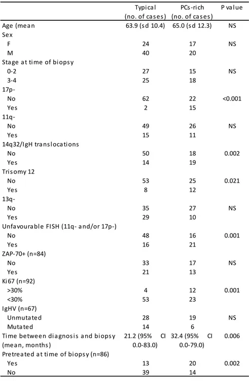
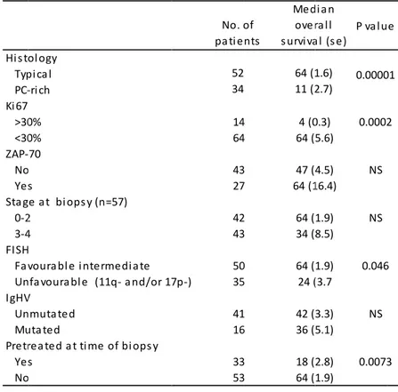
Several recent studies have shown that CD38 expression is higher in CLL cells in the bone marrow and lymphoid tissues, especially in the proliferation centres (PCs), which are regarded as the histologic hallmark of this disease. Indeed CLL is a disease in which the host’s microenvironment promotes leukemic cell growth, leading to sequential acquisition and accumulation of genetic alterations and proliferation centers may play an important role in the biology of CLL, as they represent its proliferative compartment. To better define the significance of proliferation centers, we studied lymph node biopsies taken from a cohort of patients by fluorescence in situ hybridization (FISH) studies using a 5-probe panel on tissue microarrays (TMA). The cases were classified into two categories: “PCs-rich” and “typical”. The PCs-rich group was associated with 17p-, 14q32/IGH translocations and +12. The median survival from the time of TMA for PCs-rich and typical groups was 11 and 64 months respectively. The PCs-rich pattern was the only predictive factor of an inferior survival. These findings establish an association between cytogenetic profile and the amount of PCs in CLL, and show that this histopathologic characteristic is of value for risk assessment in patients with clinically significant adenopathy.
CLL turned out to be a disease with multiple facets in its pathogenic mechanisms including genetic aberrations, antigen drive and microenvironmental interactions. In the first part of this work, we focused our attention on the correlation between CD38-positivity, proliferation centres and development of genetic aberrations. To translate this knowledge in clinical practice we planned further studies focusing i) on the correlation between chromosomal aberrations and clonal evolution and ii) on how to stratify patients into different risk-groups at diagnosis according to cytogenetic abnormalities and gene mutations.
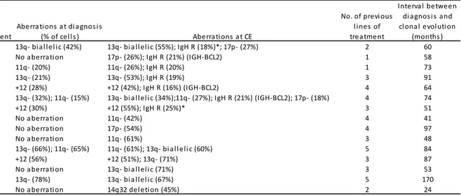
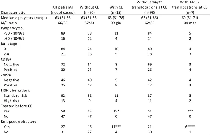
The presence of cytogenetic abnormalities is a hallmark of CLL. It was reported that a fraction of CLL patients developed new cytogenetic abnormalities at chromosome sites of known prognostic importance during the course of their disease (clonal evolution, CE). To better define the incidence and signature of CE, a cohort of patients were analysed sequentially by FISH. Recurring aberrations at
7
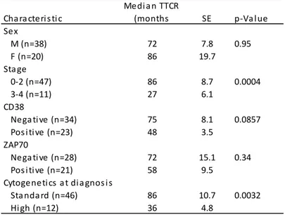
clonal evolution were 14q32/IGH translocation, 17p-, 11q-, 13q- and 14q32 deletion. The development of CE occurred only in previously treated patients. Our data show that the 14q32/IGH translocation may represent one of the most frequent aberrations acquired during the natural history of CLL. CE occurs in pre-treated patients with short TTT and survival, after the development of CE with and without 14q32 translocation, is relatively short.
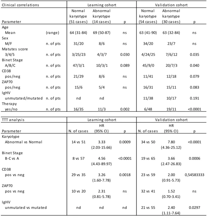
Having assessed the incidence of chromosome aberration in CLL with evolution and/or adenopathy we next moved to CLL with an apparently “favourable” profile of cytogenetic lesions, to establish if improved cytogenetic techniques could help refine prognostication. We therefore designed a study to assess whether karyotypic aberrations in patients without FISH anomalies correlate with established clinical and prognostic parameters. The clinical and prognostic significance of karyotypic aberrations in normal FISH CLL was first evaluated in a retrospective single centre series of patients and then validated prospectively in a multicentre series of cases diagnosed and analysed for karyotype with DPS30/IL2 stimulation. Conventional karyotyping using DSP30/IL2 stimulation is an effective method for the detection of chromosome aberrations in approximately one third of CLL with normal FISH. The abnormal karyotype correlated with shorter time to first treatment and shorter survival. This set of data also showed that, in CLL patients with normal FISH, conventional cytogenetic analysis identifies a subset of cases with adverse clinical and prognostic features to be considered for the design of risk-adapted treatment strategies.
In the last part of our experimental work we moved from the consideration that the cytogenetic lesions do not entirely explain the molecular pathogenesis and the clinical heterogeneity of CLL. Indeed, the advent of next-generation sequencing (NGS) technologies has enabled exploration of the CLL genome, uncovering genetic lesions that recurrently target the leukemic cells. NGS studies have further elucidated the genomic complexity of CLL. In order to improve understanding of genetic basis of CLL and to apply NGS to CLL, we sequenced DNA samples from untreated patients affected by chronic lymphocytic leukemia with a panel of 20 genes and we correlated mutational status with clinicobiological parameters.Mutations were identified in the following genes: TP53, SF3B1, POT1 , ATM , MYD88 , FBXW7 , MAPK1 , DDX3X , KLHL6 , KRAS. The presence of mutations correlated with high risk FISH (11q- and/or 17p-) and unfavourable
8
cytogenetic (11q-, 17p- or complex karyotype) findings. Patients carrying CLLs with gene mutations showed a significant shorter median time to first treatment in comparison to those without mutations (20 months vs not reached at 76 months). The frequency of mutations in the 20 investigated genes is in line with published data in the literature using whole exome sequencing. This study shows that the simultaneous sequencing of a panel of genes implicated in CLL is feasible.
In conclusion, in this work we tried to improve our knowledge on some fundamental pathogenetic aspects of CLL, including:
i) the development of genetic lesions in CD38+ activated cells, obtained from the PB in patients in an initial and indolent phase of the disease; ii) the pattern of cytogenetic lesions in lymph node microenvironment
(proliferation centres) in patients in a more advanced phase of the disease;
iii) to translate this knowledge in clinical practice, we assessed prognosis with modern techniques and we identified cytogenetic lesions associated with disease progression and shorter time to treatment; iv) we identified recurrent genetic lesions potentially useful for further
9
A
BSTRACT
(I
TALIAN
)
La leucemia linfocitica cronica (LLC) è un disordine linfoproliferativo a carico del linfocita B, caratterizzato dall’accumulo di piccoli linfociti nel sangue periferico, nel midollo osseo e nei linfonodi. Nonostante un immunofenotipo omogeneo costituito dalla presenza degli antigeni CD19, CD20, CD5 e CD23, la LLC è clinicamente eterogenea. Sono stati identificati diversi fattori di prognosi. Tra questi ritroviamo lo stadio di malattia alla diagnosi, la positività per il CD38, la configurazione non mutata della regione variabile della catena pesante dei geni immunoglobulinici (IGHV), la positività per ZAP70, le aberrazioni cromosomiche e le anomalie molecolari.
Analisi immunofenotipiche e genetiche hanno portato all'identificazione di diversi marcatori molecolari di attivazione, alcuni dei quali sono riconosciuti nella pratica clinica al fine di predire la prognosi. L’antigene di superficie CD38 è uno di questi. Esso, in particolare, è un indicatore di prognosi sfavorevole, di attivazione e proliferazione cellulare ed è in grado di predire un’evoluzione clonale ed una prognosi sfavorevole.
Abbiamo quindi studiato, in un gruppo di pazienti a basso rischio (cellule CD38+ <7% e anomalie FISH a basso rischio prognostico), il significato biologico e clinico della presenza di lesioni genetiche nella frazione cellulare CD38 positiva. I nostri dati hanno mostrato che una percentuale significativa di pazienti presentano aberrazioni genetiche all'interno delle cellule CD38+. La maggior parte di queste anomalie sono lesioni ad alto rischio (delezione 11q e 17p) e nella maggior parte dei casi queste lesioni sono stati trovate in cellule diverse indicando che più cloni minoritari possono essere presenti, in maniera indipendente, nella popolazione cellulare complessiva. La presenza di queste lesioni FISH nella piccola frazione CD38+ è associata ad un minore tempo al primo trattamento (TTT). Per identificare marcatori associati a questo fenomeno, abbiamo effettuato un’analisi di espressione dei microRNAs. Abbiamo quindi identificato un profilo di espressione dei miRNAs alterato nei casi di LLC con lesioni FISH supplementari a carico delle cellule CD38+. In particolare, il miR-125a-5p è risultato essere down-regolato sia nelle cellule CD38+ che nelle cellule CD38- nei pazienti con cloni aventi anomalie FISH, rispetto ai pazienti senza cloni anomali. L’importanza del miR-125a-5p come marcatore di prognosi inferiore e complessità genetica è stata
10
poi validata in una coorte di pazienti indipendente. In questa serie di validazione, abbiamo confermato il ruolo predittivo del miR-125a-5p in termini di TTT più breve. Inoltre abbiamo visto, attraverso tecniche di sequenziamento di ultima generazione, che i pazienti affetti da LLC con bassi livelli di miR-125a-5p presentano un aumento del tasso di mutazione nei geni implicati nella LLC.
Diversi studi recenti hanno dimostrato che l'espressione del CD38 è maggiore nelle cellule leucemiche all’interno del midollo osseo e del tessuto linfoide, in particolare all’interno dei centri proliferativi (PCs), che sono considerati la peculiarità istologica della LLC. Infatti la LLC è una malattia in cui microambiente promuove la crescita delle cellule leucemiche, conduce all'acquisizione ed al conseguente accumulo di alterazioni genetiche. In questo contesto, i centri proliferativi svolgono un ruolo fondamentale.
Per definire meglio il significato dei PCs, abbiamo studiato le biopsie linfonodali di una coorte di pazienti con metodica FISH su microarray tissutale (TMA). I casi sono stati classificati in due categorie: "PC-rich" e "tipici". Il gruppo “PC-rich” è stato associato alla presenza di delezioni 17p-, traslocazioni 14q32/IGH e trisomia del cromosoma 12. La sopravvivenza media, dal momento del TMA, per i gruppi “PC-rich” e “tipici” è risultato essere, rispettivamente, di 11 e 64 mesi. Questi risultati stabiliscono, quindi, un'associazione tra il profilo citogenetico e la quantità di PCs nella LLC, e dimostrano che questa caratteristica istopatologica è di grande valore per la valutazione del rischio nei pazienti con adenopatie clinicamente significative.
La LLC si è rivelata essere una patologia con molteplici sfaccettature nei suoi meccanismi patogenetici, tra cui la presenza di aberrazioni genetiche, la stimolazione da parte dell’antigene e le interazioni con il microambiente. Nella prima parte di questo lavoro, abbiamo focalizzato la nostra attenzione sulla correlazione tra positività per il CD38, centri proliferativi e sviluppo di aberrazioni genetiche. Per tradurre queste conoscenze nella pratica clinica abbiamo programmato ulteriori studi incentrati i) sulla correlazione tra aberrazioni cromosomiche e l'evoluzione clonale e ii) su come stratificare i pazienti in diversi gruppi di rischio al momento della diagnosi in base ad anomalie citogenetiche e mutazioni genetiche.
11
La presenza di anomalie citogenetiche è un segno distintivo della LLC. In letteratura è riportato che una frazione di pazienti affetti da LLC sviluppa nuove anomalie citogenetiche durante il corso della malattia (evoluzione clonale, CE). Per definire meglio l'incidenza e lo stato dell’evoluzione clonale, abbiamo analizzato una coorte di pazienti mediante FISH a diversi stadi di malattia. Aberrazioni ricorrenti nell’evoluzione clonale sono risultate essere: traslocazioni 14q32/IGH, delezioni 17p-, 11q-, 13q-e 14q32. L’evoluzione clonale si è verificata solo in pazienti precedentemente trattati. I nostri dati mostrano che la traslocazione 14q32/IGH rappresenta una delle aberrazioni acquisite più frequenti durante la storia naturale della LLC. Il fenomeno dell’evoluzione clonale si verifica in pazienti pretrattati con breve TTT, e la sopravvivenza dopo lo sviluppo di evoluzione clonale è relativamente breve.
Dopo aver valutato l'incidenza di aberrazioni cromosomiche in pazienti LLC con evoluzione clonale e/o adenopatia, ci siamo soffermati sul gruppo di pazienti LLC con lesioni citogenetiche "favorevoli". Abbiamo, quindi, effettuato uno studio per valutare se le aberrazioni cromosomiche in pazienti FISH negativi correlino con i parametri clinici e prognostici. Abbiamo dapprima studiato una coorte retrospettiva di pazienti aventi le suddette caratteristiche. In seguito, abbiamo validato prospetticamente in una serie multicentrica di casi analizzati mediante cariotipo convenzionale con stimolazione metafasica attraverso DPS30/IL2. Abbiamo visto che questa tecnica permette l'individuazione di aberrazioni cromosomiche in circa un terzo dei casi LLC con FISH normale. Il cariotipo anomalo correla con un minore tempo al primo trattamento ed una ridotta sopravvivenza. Questo insieme di dati ha quindi mostrato che, nei pazienti affetti da LLC con FISH normale, l'analisi citogenetica convenzionale identifica un sottoinsieme di casi con caratteristiche cliniche e prognostiche negative da prendere in considerazione al fine di adeguare le strategie di trattamento.
Nell'ultima parte del nostro lavoro sperimentale, abbiamo considerato che le lesioni citogenetiche non sono in grado di spiegare la patogenesi molecolare e l'eterogeneità clinica della LLC. L'avvento del sequenziamento di ultima generazione (NGS) ha permesso l’acquisizione di importanti conoscenze sulla caratterizzazione della LLC, chiarendone ulteriormente la complessità genomica. Al fine di migliorare la comprensione delle basi genetiche delle LLC e di applicare
12
le tecniche di NGS, abbiamo sequenziato una serie di pazienti non trattati con un pannello di 20 geni, ritrovati frequentemente mutati nella LLC, e abbiamo correlato lo stato mutazionale con i parametri clinicobiologici. Abbiamo ritrovato mutazioni somatiche nei seguenti geni: TP53, SF3B1, POT1, ATM, MYD88, FBXW7, MAPK1, DDX3X, KLHL6, KRAS. La presenza di mutazioni correla con la presenza di anomalie FISH ad alto rischio (11q- e/o 17p-), e cariotipo sfavorevole (11q-, 17p o cariotipo complesso). Pazienti recanti mutazioni genetiche hanno mostrato un tempo al trattamento significativamente più corto rispetto a quelli senza mutazioni. La frequenza di mutazioni nei 20 geni studiati è in linea con i dati pubblicati in letteratura.
In conclusione, in questo lavoro abbiamo cercato di migliorare le nostre conoscenze su alcuni aspetti patogenetici fondamentali della LLC, tra cui:
i) lo sviluppo di lesioni genetiche in cellule attivate CD38+, ottenute da sangue periferico in pazienti in una fase iniziale e indolente di malattia; ii) il tipo di lesioni citogenetiche nel microambiente linfonodale (centri
proliferativi) in pazienti in una fase più avanzata di malattia;
iii) traducendo queste conoscenze nella pratica clinica, abbiamo valutato la prognosi con tecniche moderne ed identificato lesioni citogenetiche associate a progressione di malattia e minor tempo libero da trattamento; iv) abbiamo identificato lesioni genetiche ricorrenti potenzialmente utili per un
ulteriore affinamento della nostra capacità di predizione della prognosi e della risposta al trattamento.
13
CHAPTER
1
C
HRONIC
L
YMPHOCYTIC
L
EUKEMIA
1.1
I
NTRODUCTIONChronic lymphocytic leukemia (CLL) is the most common adult leukemia in Western countries and is characterized by clonal proliferation and accumulation of mature B lymphocytes. CLL affects individuals with median age at diagnosis ranging between 67 and 72 years. Moreover, the proportion of younger patients with early stage CLL and minimal symptoms seems to increase due to more frequent blood testing. More male than female patients (1.7:1) are affected [1]. CLL is characterized by the clonal proliferation and accumulation of mature, typically CD5-positive B-cells within the blood, bone marrow, lymph nodes and spleen [2]. Typical immunophenotype of the CLL is the presence of the B-cell surface antigens CD19, CD22 and CD 23 with coexpression of T-cell lineage antigen CD5 and expression of CD20 and CD7b lower than that observed in normal B cells [3, 4].
1.2
D
IAGNOSIS ANDS
TAGINGThe diagnostic criteria as laid out by the International Workshop on Chronic Lymphocytic Leukemia (IWCLL-2008) [4] requires the presence of at least 5.000 B-lymphocytes/ul in the peripheral blood for the duration of minimum 3 months. In most cases the diagnosis of CLL is established by blood counts, differential counts, a blood smear and immunophenotyping. The World Health Organizaton (WHO) classification of hematopoietic neoplasias describes CLL as leukemic, lymphocytic lymphoma, being only distinguishable from SLL (small lymphocytic lymphoma) by its leukemic appearance [5].
CLL is always a disease of neoplastic B-cells and their clonality needs to be confirmed by flow cytometry [4]. The leukemia cells found in the blood smear are characteristically small, mature lymphocytes with a narrow border of cytoplasm and a dense nucleus lacking discernible nucleoli and having partially aggregated chromatin [6] (Fig. 1.1).
Two w system outcom FIGURE (4x); (B) The R lympho (Rai st splenom disease anemia The Bin the pre on whe having Patient that de less tha organo
1.3
B
Our un to be a a lack antiapo widely acce m. Both d mes. E 1.1. (A) Per Characterist Rai stagin ocytosis wi tage 0). P megaly an e (Rai stag a (stage III) net staging esence of e ether there Hb ≥10 g ts with Hb fined for st an 10 g/dL omegaly arB
IOLOGY derstandin a homogen of norma optotic prof epted stag escribe th ripheral blood tics appeara g system th leukem Patients w nd/or hep ge I or II). H ) or thromb g system is enlarged ly e is anem g/dL, platel b ≥10 g/dL, tage A are L and/or p e included Y ANDP
A ng of CLL h nous disea al cell de file, with s ging metho hree majo d smear in a nce of small m defines ia cells in with lymph atomegaly High risk d bocytopeni s based on ymph node mia or thro lets ≥100 x , platelets e defined a platelet cou in stage C ATHOGEN has change se, in whic eath. It is strong expr 14 ods co-ex or prognos a case of CLL CLL cells w low-risk blood and hocytosis, y are def disease inc ia (stage IV n the numb es of greate ombocytop x 109/L an ≥100 x 10 s having s unt of less C. NESIS OF ed over the ch mature currently ression of xist, the R stic group L with abnorm with frequent p disease d/or bone m enlarged ined as h cludes pati V). ber of invo er than 1 c enia. Bine nd up to tw 9/L and or stage B. All than 100 FCLL
e last deca B cells ac known th Bcl-2 prot Rai [7] and ps with dmally high lym prolymophoc as patien marrow (ly nodes in having inte ents with d lved areas cm or orga et stage A wo areas o ganomega l patients w x 109/L, ir ades. It wa ccumulated hat CLL c tein, which d the Bine iscrete cl mphocyte co cytes (40x). nts who ymphoid >3 any site termediate disease-re s, as define anomegaly A is define of involvem aly greater who have H rrespective as once tho d largely d cells show h suggests et [8] inical ounts have 30%) and risk lated ed by , and ed as ment. than Hb of ely of ought ue to w an s that
15
inhibition of apoptosis is responsible for CLL development. However, several reports have shown that the high lymphocyte count in CLL patients is caused not only by the prolonged survival, but also by proliferating cells. The disease probably develops is a result of accumulation of transformed B cells. In CLL cells, a great imbalance between cell birth and death rate is observed [3, 9].
The gene expression profile suggests that CLL cells originate from transformed, antigen-stimulated B cells [10]. In CLL cells, several mutated IGHV genes are expressed more frequently than in normal B lymphocytes. All those cells express restricted sets of B-cell receptors (BCR). Unique stereotypy of BCR suggests that antigens play a critical role in CLL pathogenesis.
Moreover, CLL has recently been established as a disease of remarkable diversity and differences in cell morphology, immunophenotype, cytogenetics and molecular characteristics. This heterogeneity translates into clinical course and the response to treatment [11]. Approximately one third of patients survive for 20 years or longer and never require treatment [12] and, alternatively, some patients may progress rapidly from the time of diagnosis.
1.4
P
ROGNOSTIC FACTORS INCLL
A number of clinical and biological markers of prognostic relevance have been identified. Due to great variability of CLL patients clinical course, there is a need to establish solid prognostic factors for this disease.
CLL patients are currently categorized into risk groups based on the clinical staging systems Rai [7] and Binet [8]. These classifications are helpful for dividing patients in regard to the expected overall survival (OS). However, both systems fail to indicate the higher risk of progression among patients in early stage of disease.
Among prognostic factors in CLL, there are lymphocyte doubling time (LDT), serum markers, IGHV mutational status, ZAP-70 and CD38 expression, cytogenetic abnormalities and somatic mutations [13-16].
LDT longer than 12 months correlates with increased progression-free survival (PFS) and OS. An increase in the lymphocyte count of more than 50% in two months or LDT during less than 6 months is a recommended criterion of active disease and indication for treatment [17].
Serum thymidi patients disease These status, express patients correla IGHV g mutate FIGURE genes. M months [ The ex cut-off correla cytome 70 estim prognosti ine kinase s. In fact e progress ones can somatic m sion of C s may hav tes with a gene have d IGHV ge E 1.2. Kaplan Median surv [14]. xpression o to class ting with u etry, is wide mation still ic factors, e were in they are sion, correl be immun mutations CD38 and ve either a favourabl significan ene (appro n-Meier survi vival for unm
of ZAP-70 ify patien unmutated ely propos l remains a such as ndicated a relevant ating with noglobulin and some intracellul somatically le or unfav tly shorter ximately 2 val curve co mutated CLL: remains c ts as ZA IGHV statu ed at 20% a challenge 16 β2-micro as an imp markers o other biolo heavy cha e cytogen ar expres y mutated vourable p r OS (appr 5 years) (F omparing CL : 117 month constant ov AP-70 pos us) or ZAP % threshold e. oglobulin, s portant pro of prolifera ogical prog ain variable etic abnor sion of Z d or unmu prognosis. roximately Fig. 1.2). L patients w hs; median s ver the co sitive (neg P-70 negat . However soluble CD ognostic f ative activ nostic fact e region (I rmalities, c AP-70 [4, tated IGHV Patients w 8 years) t ith mutated a survival for m urse of the gative pro tive, as me r, standard D23 or se factor for vity and a tors [18]. GHV) mut cell memb 18, 19]. V gene, w with unmu than those and unmutat mutated CLL e disease. ognostic f easured by dization of Z erum CLL a risk tation brane CLL which tated e with ted VH L: 293 . The factor y flow
ZAP-17
Similar correlation with outcome has been shown for CD38 expression. CD38-positive patients (the threshold proposed is >30% CD38+ CLL cells) were reported to have significantly worse prognosis regarding PFS and OS than those who were CD38 negative. It was observed that CD38 expression on CLL cells correlates with the absence of IGHV mutations [3, 4, 13, 18, 19].
A crucial prognostic importance is assigned to chromosomal aberrations. In approximately 80% of patients, there is a mutation detected by fluorescence in situ hybridization (FISH) [15]. The most common recurrent chromosomal abnormalities include deletion 13q, trisomy 12 and deletions 11q, 17p and 6q. A subdivision based on these aberrations is important, as they are predictor of disease outcome. Five prognostic categories have been identified in a hierarchical model, showing poor survival in patients with 17p deletion or 11q deletion (median survival 32 and 79 months, respectively) but better survival for patients with trisomy 12, normal karyotype, and deletion 13q as the sole abnormality (114, 111 and 133 months, respectively) [15]. Deletions of 11q22-q23 and 17p13, resulting in abnormalities of ATM and TP53 genes, respectively, are independent prognostic factors identifying patients with a rapid disease progression and a short OS in a multivariate analysis. Patients with del(11q) have more frequently B symptoms, advanced clinical stage and extensive peripheral, abdominal and mediastinal lymphoadenopathy. Deletions 17p and 11q are more often detected in advanced stages of the disease, among patients with unmutated IGHV gene. In contrast, deletion of chromosome band 13q14 is associated with a favourable CLL outcome. Moreover, patients with trisomy 12 have a shorter OS than those with 13q deletion [3, 4, 9, 13, 15, 18]. TP53 mutations have been described in 4-12% of patients with untreated CLL. Approximately 80-90% of cases with a deletion of one copy of TP53 locus will have a TP53 mutation on the remaining copy [16, 20-23]. TP53 mutations are more prevalent in progressive and refractory CLL [22] (Fig 1.3). A TP53 mutation is an independent predictor of poor prognosis and confers even shorter OS than del(17p) in the absence of TP53 mutation [20]. Mutations of the ATM gene may also have prognostic implications independent of those associated with the deletion of chromosome 11q. Patients harbouring ATM mutations have a more aggressive clinical course and are more resistant to traditional chemotherapeutic agents [24, 25].
In recent times, the improvements in next generation sequencing technologies have provided a novel opportunity to examine the CLL genome [26], and have
allowed mutatio (splicin protein progres across FIGURE parenthe progress transform pathway presenta
1.5
T
Despite disease or stab indicate or prog disease d previous ons of NOT g factor 3 3) [30]. M ssion and different c E 1.3. Gene esis) of gene sion: chemo mation to Ric ys in terms oation but are
REATME e the grea e still rema ble disease ed. Treatm gressive d e activity, a sly unkno TCH1 (neu 3B unit 1 Mutations o survival. E clinical pha etic lesions etic lesions is orefractorines chter Syndro of type and not enriched ENT OF
C
at improve ains difficu e patients. ment should disease. T age and co own genom urogenic loc ) [29] and of these ge Each of the ses and bi of CLL at d s shown for ss without e ome (RS). the frequency o d at CLL progCLL
ement in C lty curable In this gr d be introd he choice omorbiditie 18 mic altera cus notch d BIRC3 enes have ese lesions iological su different pha CLL at pres evidence of e two type of of genetic le gression are CLL treatm e. Chemoth roup, a “w duced in pa e of therap es. ations to homolog p (baculovira been asso s recogniz ubgroups o ases of the sentation and histologic t f CLL progre esions. Gene indicated in ment durin herapy is n watch and atients with py depend be identi protein 1) [ al IAP rep ociated wit es a differ of the disea disease. T d for two diff ransformatio ession follow etic lesions t gray. ng the last ot recomm wait” appr h advanced ds on clin ified, such [27, 28], SF peat-conta th short tim rent distrib ase. The frequen ferent type o on, and hist w distinct mol that occur a t decades mended in roach is w d, symptom ical stage h as F3B1 aining me to bution cy (in of CLL tologic ecular at CLL , the early widely matic , the19
1.5.1 Cytostatic Agents
For many years, chlorambucil (with or without steroids) was the drug of choice in previously untreated patients with progressive or advanced CLL [31]. Even today, this drug remains an appropriate option for elderly and unfit patients. The advantages of chlorambucil are its moderate toxicity and low cost; the major disadvantages are its low complete remission (CR) rate and some side effects that occur after extended use.
Three purine analogs are currently used in CLL: fludarabine, pentostatin and cladribine. These drugs showed high antileukemic activity. Significantly higher overall response (OR), complete remission and progression free survival (PFS) in patients treated with fludarabine or cladribine were documented in several randomized clinical trials [32, 33]. Next, combination of fludarabine or cladribine with cyclophosphamide appeared to be more effective than purine analogs in monotherapy in regard to OR, CR and PFS [34-36].
1.5.2 Antibodies
Rituximab. CD20 is an activated, glycosylated phosphoprotein expressed on
the surface of mature B-cells. The protein has unknown natural ligand and its function is not yet discovered [37]. As CD20 is expressed on most B-cell malignancies, the introduction of the anti-CD20 antibody Rituximab in 1998 improved the treatment of most CD20-positive on-Hodgkin lymphomas including CLL [38]. In CLL, Rituximab is less active as single agent than in follicular lymphoma, unless very high doses are used [39, 40]. In contrast, combinations of Rituximab and chemotherapy have proven to be very efficacious therapies of CLL.
Ofatumumab. It is a fully humanized antibody targeting a unique epitope on the
CD20 molecule expressed on human B-cells. Ofatumumab was found to be effective in a phase III randomized study, as monotherapy for heavily pretreated patients with CLL resistant to fludarabine or fludarabine-alemtuzumab regimen [41].
Alemtuzumab. It is another MoAb, recombinant, humanized anti-CD52, highly
active in CLL. In previously untreated patients, an OR rate of more than 80% was achieved, being effective in patients with high risk genetic markers such as deletions of chromosome 11 or 17 and TP53 mutations [42, 43], and in those patients with Alemtuzumab can be used as a first-line treatment [44].
20
Obinutuzumab (GA101). The humanized and glycol-engineered monoclonal
antibody Obinutuzumab showed impressive results in vitro with higher rates of apopotosis in B-cells in comparison to Rituximab [45]. Encouraging results were reported in the interim analysis of CLL11 trial on CLL patients with comorbidity. The primary endpoint was PFS [46].
1.5.3 Combination chemotherapy
A major advance in CLL treatment was achieved by the combined use of different treatment modalities. Because purine analogs and alkylating agents have different mechanisms of action and partially nonoverlapping toxicity profiles, they were combined to achieve synergistic effects. The most thoroughly studied combination chemotherapy for CLL is fludarabine plus cyclophosphamide (FC) [47]. In noncompertive trials, the overall response rates did not appear to be better than with fludarabine alone, but the addition of cyclophosphamide appeared to improve the CR rate up to 50% [47]. A phase II study of cladribine in combination with cyclophosphamide has also demonstrated activity in advanced CLL, but results seemed inferior to FC [48].
1.5.4 Chemoimmunotherapy
The results of recent studies showed that Rituximab in combination with purine analogs or purine analogs and cyclophosphamide can increase the OR and CR rates and PFS time [49]. The combination of Rituximab with FC (FCR regimen) demonstrated particularly high rates of OR, CR and duration of PFS in both previously untreated and relapsed/refractory CLL [50]. However FCR is acceptable for younger, physically fit patients. This regimen has limitations in the unfit group, mainly due to the risk of myelosuppression and other side effects. Because CLL often occurs in elderly patients with relevant comorbidities, a dose modified FCR-Lite regimen was designed to maintain the efficacy but decrease the toxicity of the FCR regimen [51]. This regimen reduced the dose of fludarabine and cyclophosphamide and increased the dose of Rituximab. Recent trials proved that combination of Rituximab with pentostatin or cladribine and cyclophosphamide is a highly active treatment modality in CLL. More recently, bendamustine, a bifunctional agent composed of an alkylating nitrogen mustard group and a
purine-21
like benzimidazole ring, has been included in CLL treatment regimens. In a randomized trial compared activity of bendamustine or chlorambucil in untreated CLL patients [52], OR and CR rates were significantly higher in patients treated with bendamustine. Several other combinations have been investigated, such as, cladribine with rituximab, methylprednisolone plus rituximab followed by alemtuzumab, or rituximab plus alemtuzumab.
1.5.5 New drugs targeting pathogenic pathways of CLL cells
There are an increasing number of interesting new compunds in clinical development. The common denominator of these compounds is that thei mechanism of action targets a relatively specific signalling abnormality or redirects the immune system against CLL cells.
Agents targeting BCR signalling. Idelalisib (CAL-101) is an inhibitor of
phosphatidylinositol 3-kinase (PI3K) signalling. CAL-101 reduces survival signals derived from the BCR and inhibits BCR and chemokine-receptor-induced AKT and MAP kinase (ERK) activation [53]. In preclinical studies, it was found to induce apoptosis of CLL cells. Protein kinase C and PI3K pathways have an influence on the survival of B cells in CLL. In a phase I clinical trial, Idelalisib showed acceptable toxicity, positive pharmacodynamics effects and favourable clinical activity [54]. Results on Idelalisib in combination with Rituximab, Ofatumumab or bendamustine/rituximab were presented in preliminary form and showed encouraging results .
Ibrutinib is an orally active small molecule inhibiting BTK that plays a role in the signal transduction of the BCR. Inhibition of BTK might induce apoptosis in B-cell lymphomas and CLL cells [55]. Ibrutinib showed significant activity in patients with relapsed or refractory B-cell malignancies, including CLL [56].
Bcl-2 inhibitors. Proteins in the B-cell CLL/Lymphoma 2 (Bcl-2) family are
key regulators of the apoptotic process [57]. The Bcl-2 family comprises proapoptotic and prosurvival proteins. Shifting the balance toward the latter is an established mechanism whereby cancer cells evade apoptosis. ABT-263 (Novitoclax) is a small molecule Bcl-2 family inhibitor that binds with high affinity to multiple antiapoptotic Bcl-2 family proteins. Initial studies showed very promising results for this drug as single agent [58]. However its therapeutic use seemed somewhat limited by severe thrombocytopenia being a prominent side effect.
22
Therefore the compound was reengineered to create a highly potent and cl-2 selective inhibitor, ABT-199 [59]. This compound inhibits the growth of Bcl-2-dependent tumors in vivo and spares human platelets. Recent data from clinical trials indicate that selective pharmacological inhibition of Bcl-2 holds great promise for the treatment of CLL.
Given the impressive choice of options, the right choice of treatment becomes a task that requires experience, a good clinical judgement and an appropriate use of diagnostic tools. The following parameters should be considered before recommending a treatment for CLL: the clinical stage of disease; the fitness of the patient; the genetic risk of leukemia; the treatment situation (first versus second line, response versus non-response of the last treatment).
23
CHAPTER
2
P
ATHOGENESIS OF
CLL:
CD38
AND PROLIFERATIONS
CENTERS
2.1
I
NTRODUCTIONIn 1995, chronic lymphocytic leukemia (CLL) was defined as a homogeneous disease of immature, immune-incompetent, minimally self-renewing B cells [2], which accumulate relentlessly because of a faulty apoptotic mechanism. Since ten years, these views have been transformed by a wealth of new information about the leukemic cells. CLL is currently considered a clinically heterogeneous disease originating form B lymphocytes that may differ in activation, maturation state or cellular subgroup. Leukemic cell accumulation occurs because of survival signals delivered to a subgroup of leukemic cells from the external environment through a variety of receptors and their cell-bound and soluble ligands [60].
CLL is seen as a disease characterized by a dynamic balance between cells circulating in the blood and cells located in permissive niches in lymphoid organs [61]. The former are primarily mature-looking small lymphocytes resistant to apoptosis, whereas the latter are composed of lymphocytes that undergo either proliferation or apoptosis according to environment [60].
B lymphocytes mature in the bone marrow and in the process rearrange immunoglobulin variable (V) gene segments to create the code for an immunoglobulin molecule that serves as the B-cell receptor for antigen. When an antigen of adequate affinity engages the receptor, the cell enters a germinal center in lymphoid follicles, where, as a centroblast, it rapidly divides and its V genes undergo somatic hypermutation. This process introduces mutations in the rearranged VHDJH and VLJL gene segments that code for the binding site of the
receptor. Through these mutations, the receptors of the descendent B cells, called centrocytes, acquire new properties. Centrocytes with receptors that no longer bind the antigen or do bind autoantigens are normally eliminated [62].
24
This stimulation and selection pathway usually requires the help of T lymphocytes and occurs in germinal centers [62], the structure of which ensures the selection of B cells capable to recognize antigens. However, the process can proceed without T cells [63] and outside germinal centers, in the marginal zones around lymphoid follicles [64]. Both process lead to the development of plasma cells or memory (antigen-experienced) B cells. Concomitant with B-cell activation, the proteins on the surface of the B cell change. These modifications help activated B cells to interact with other cells and soluble mediators. One surface molecule that supports B cell interactions and differentiation is CD38 [65].
The heterogeneous clinical outcome of CLL patients is dictate, at least in part, by the nature of these microenvironmental signals and interactions that can promote or impair accumulation of genomic alterations [66]. Detailed immunophenotypic and genetic analysis of different neoplastic clones have led to the identification of a number of molecular markers of activation, some of which are gaining relevance in clinical practice to predict outcome [14, 15, 17, 67]. Cell surface CD38 is one of these markers.
2.2
CD38
IS ASSOCIATED WITH GENETIC HETEROGENEITY AND CLONAL EVOLUTION2.2.1 Biology of human CD38
CD38 is expressed at high levels by B lineage progenitors in bone marrow and by B lymphocytes in germinal center, in activated tonsils, and by terminally differentiated plasma cells [68]. On the other side, mature virgin and memory B cells express low levels of this molecule. CD38 is located in the cytosol and/or in the nucleus and not on the cell membrane [68].
CD38 is a 45 kDa type II transmembrane glycoprotein. The functional CD38 molecule is a dimer, with the central part hosting the catalytic site [69]. The transition from monomer to dimer modulates the function of the molecule. CD38 has the tendency to associate with other proteins, forming large supramolecular complexes. Molecules that associate with CD38 include the CD19/CD81 complex, the chemokine receptor CXCR4, ad adhesion molecules, such as CD49d [70, 71]. CD38 is also found in exosomes [72], membrane vescicles secreted by B cells, and is probably part of an intracellular communication network.
25
The functional properties of CD38 on human B cells appear to be strictly linked to the stage of maturation. The presence of blocking mAbs in cultures of CD19+ B-cell precursors on stromal layers markedly suppress B-B-cell lymphopoiesis by inducing apoptosis [73]. In contrast, in mature circulating B lymphocytes and tonsillar germinal center B cells, CD38 ligation is followed by activation, apoptosis inhibition, proliferation and cytokine secretion [74, 75]. In both cases, the mechanisms are attributed to the activation of an intracellular signalling pathway ruled by CD38 and requiring an association with CD19.
2.2.2 CD38 as a marker in CLL
CD38 identifies two subgroups of CLL patients with different clinical outcomes [67]; this distinction is based on the percentage of CD38+ leukemic cells within a CLL clone. The two patient subgroups differ clinically in several ways, including overall survival [67, 76], time to first treatment [77, 78], number of leukemic cells with atypical morphology [79], extent and level of adenopathy [76] and absolute lymphocyte counts [80]. These subgroups also differ in responsiveness to various therapies [80].
The past decade has highlighted several molecular differences between CD38+ and CD38- members of the same clone, including expression of specific activation markers [81], which reflect quantitative and temporal differences in response to stimulation. CD38+ CLL cells express high levels of CD69 and HLA-DR [82], both indicative of recent activation. CD38 also marks a cellular subset enriched in Ki-67+ and ZAP-70+ cells. In addition, CD38+ CLL clones display enhanced ability to migrate in response to chemokine and to transduce BCR-mediated signals.
These findings suggest that cellular proliferation might be at the root of the association between higher levels of CD38, aggressively growing CLL clones, and poor patient outcome [60]. This suggestion is consistent with in vivo labelling experiments using 2H2O (“heavy water”) that demonstrated larger than anticipated
rates of CLL birth, especially in patients with poor clinical course and outcome [83] and a several-fold higher percentage of newly born cells in CD38+ fractions of CLL clones than in CD38- counterparts of the same clones [84]. Furthermore, preliminary results from a large clinical study suggest that the percentage of CD38+ CLL cells strongly correlates with the level of leukemic cell turnover observed in vivo [85].
26
2.2.3 CD38 and genetic subclonal complexity
Current models of cancer progression are based on the concept that tumors are subject to the Darwinian process of evolution and selection. Recent studies in acute lymphpoblastic leukemia have provided pivotal insights into the complex sequence of events during leukemogenesis, showing that the initiating mutation is followed by copy number alterations (CNAs) that drive the emergence of the disease [86, 87]. These data imply that at least some CNAs/copy neutral loss of heterozigosity regions (cnLOHs) are likely to be involved in driving leukemia progression and therefore might contribute to relapse. Knight and colleagues [88] hypothesized that subclones containing driver CNAs/cnLOHs would newly occur or expand in relapse samples compared with samples taken before treatment. They tested their hypothesis by systemically tracking the presence and subclonal distribution of CNAs/cnLOHs in CLL patients before treatment and at subsequent relapse. The results reveal complex changes in the subclonal architecture of paired samples at relapse compared with pre-treatment, suggested that genomic complexity correlate with a poor clinical outcome. Clinically, the existence of multiple, genetically distinct, subpopulations that escape therapeutic intervention might present formidable challenges for the development of effective treatment for patients with relapsed refractory CLL.
In order to better understand the ongoing evolution of genetic lesions in patients with CLL, Grubor and colleagues [89] compared the leukemic genome with the patient’s normal DNA by using a high-resolution CGH technique called representational oligonucleotide microarray analysis (ROMA) [90]. This technique have an incredible resolution and it is so sensitive to permits the examination of the clonal heterogeneity within the same CLL patient form mixed population. To find clearer evidence of intraclonal heterogeneity within patients, they searched for genomic differences between CD38+ and CD38- populations in the same patient. So they profiled the genomes of CLL cells separated by the surface marker CD38 and found evidence of distinct subclones of CLL within the same patient. In fact, they observed copy number differences between CD38+ and CD38- fractions in 3 of 4 cases. In one case, this involved a loss of p53 locus in the CD38+ fraction, a marker that was not observed in the parallel CD38- fraction.
Another molecular basis for the association between CD38 expression and inferior clinical outcome emerged in the study of Pepper and colleagues [91]. They analysed sorted CD38+ and CD38- CLL cells derived from the same patient by
27
gene expression profiling. Their conclusion is that CD8+ fraction possess a distinct gene expression profile when compared with CD38- subclones. Subclonal populations, having identical IgHV rearrangements, derive from a single malignant transforming event and differences in gene expression between them cannot be explained by heterogeneous genetic background. Importantly, CD38+ CLL cells relatively over expressed vascular endothelial growth factor (VEGF). Elevated VEGF expression was associated with increased expression of the anti-apoptotic protein Mcl-1. Moreover, the expression of the CD38 antigen defines a sub-population of CLL cells with a distinct transcriptional profile that may be the cause or effect of an increase in cellular activation and reduced apoptosis.
More generally, within an overall apparently constant leukemic burden, the outgrowth of a subclone with additional genomic lesions and distinct gene expression profile might signal the start of a new phase of the disease.
In order to better understand the biological and molecular features predicting disease progression in CLL patient, we choose a subset of patients with favourable prognostic features and designed a two-phase study having the following aims:
- Phase I, a) to assess whether genetic lesions may be present in the minority of CD38+ cells in a series of untreated low risk patients as defined by CD38 negativity (CD38+ cells <7%) and favourable genetic findings b) to identify biologic factors associated to genetic lesions in the small CD38+ fraction of CD38- CLL patients and predicting for disease progression;
- Phase II, to validate our findings in an independent cohort of consecutive untreated CD38- CLL patients with favourable FISH findings.
Details are shown in Appendix I (paper Rigolin GM et al., Oncotarget 2013).
2.2.4 Methods and results 2.2.4.1 Patients
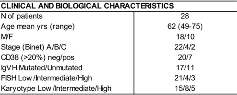
In this study 2 cohorts of patients have been considered. Cohort one (C1) included 28 untreated CLL patients seen between 2005 and 2006. Cohort two (C2) consisted of 71 consecutive untreated CLL patients diagnosed between 2007 and 2011. The principal characteristics of C1 and C2 are reported in Table 2.1.
28
TABLE 2.1. Principal clinical and biologic characteristics of the patients of the cohort 1 and cohort
2
2.2.4.2 FISH analysis on immunomagnetically sorted cells in patient C1
In the 28 patients in C1, CD38+ and CD38- CLL cells were isolated by immunomagnetic sorting. The purity of sorted fractions was >98% as determined by flow cytometric analysis. For CLL risk assessment, FISH was performed in all patient on peripheral blood samples obtained at diagnosis using probes for 13q14, 12q13, 11q22/ATM, 17p13/TP53. In C1 patients, FISH analysis was performed on both CD38+ and CD38- sorted cells, and the following region was investigated: 13q14, 12q13, 11q22/ATM, 17p13/TP53 and 14q32. Co-hybridization experiments were performed in order to evaluate the coexistence on the same cells of more genetic lesions.
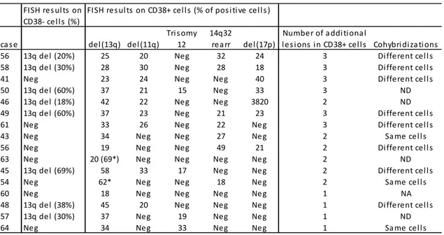
TABLE 2.2. FISH results in CLL patients with detectable genetic lesions in CD38+ cells
*biallelic 13q deletion.
Result of FISH analysis in CD38- cells and CD38+ cells in C1 are reported in Table 2.2. In 16/28 patients, genetic aberrations were detected in CD38+ cells and
Cohort 1 Cohort 2 N of pati ents 28 71 M/F 16/12 49/22 Age mean yrs (range) 65 (50‐91) 64 (38‐86) Stage (Bi net) A/B/C 28/0/0 63/8/0 FISH neg/13q del eti on 14/4 40/31 ZAP70 (>30%) neg/pos 22/5 61/10 IGHV mut/unmut 20/2 60/11 TP53 mut/unmut 0/18 0/69 FISH res ul ts on CD38‐ cel ls (%) ca s e del (13q) del (11q) Tri s omy 12 14q32 rea rr del (17p)
Number of a ddi tional
les i ons in CD38+ cel ls Cohybri diza ti ons 56 13q del (20%) 25 20 Neg 32 24 3 Different cel l s 58 13q del (30%) 28 30 Neg 28 18 3 Different cel l s 41 Neg 23 24 Neg Neg 40 3 Different cel l s 50 13q del (60%) 37 21 15 Neg 33 3 ND 46 13q del (18%) 42 22 Neg Neg 3820 2 ND 49 13q del (60%) 37 23 Neg 21 23 3 Different cel l s 61 Neg 33 26 Neg 22 Neg 3 Different cel l s 43 Neg 34 Neg Neg 27 Neg 2 Sa me cel ls 56 Neg 19 Neg Neg 49 21 2 Different cel l s 63 Neg 20 (69*) Neg Neg Neg Neg 2 ND 45 13q del (69%) 58 33 17 Neg Neg 2 Different cel l s 54 Neg 62* Neg Neg 18 Neg 2 Sa me cel ls 60 Neg 18 Neg Neg Neg Neg 1 NA 48 13q del (38%) 45 20 Neg Neg Neg 1 Different cel l s 57 13q del (30%) 37 Neg 19 Neg Neg 1 ND 64 Neg 34 Neg 33 Neg Neg 1 Sa me cel ls
not i foun FIGU miRN (Blue and w of the miRN regul n CD38- c nd in the CD URE 2.1. Clu NA profiling o e) FISH lesio withput FISH e genes rep NA mean ex ated. cells. In the D38+ popu ster analysis of CD38+ (A) ons in the CD lesions both presented on xpression acr e remainin ulation as c s of patients ) and CD38-D38+ fraction h in the CD3 n the heatma
ross all sam
29 g 12 patie compared with (W) and - (B) cells fro n. A distincti 8+ (23 micro ap correspo mples: green nts no add to the CD3 d without (W om CD38- CL ive miRNA p oRNAs) and nd to the ex indicates d ditional gen 38- cells. WO) lesions in LL patients w profile charac CD38- (9 mi xpression va own-regulate netic lesion n the CD38+ with (red) an cterized pati icroRNAs). T alues norma
ed; red indic
ns were + fraction. d without ents with The coors alized on cates
up-30
To assess whether the genetic lesions were on different clones, co-hybridization experiments using appropriate probes were performed in 11 cases with >1 aberration in CD38+ cells. In these experiments it was shown that genetic lesions involved different CD38+ cells in 8 cases and involved the same cells in remaining 3 cases.
2.2.4.3 miRNA profiling on immunomagnetically sorted cells in C1
MiRNA expression was investigated using Agilent Technologies platform. Samples were grouped according to the presence or not of genetic lesions by FISH analysis in the CD38 positive cells. So we evaluated the global miRNA expression profile of 19 patients by considering CD38+ and CD38- cell populations separately.
We found that at diagnosis most of the patients with genetic lesions in CD38+ (W) had a distinctive miRNA profile when compared to those without genetic lesions (WO), both in CD38+ (Fig. 2.1 A) and CD38- fraction (Fig. 2.1 B).
Twenty-three miRNAs were found to be differentially expressed in CD38+ population (corrected p<0.05) and nine miRNAs were found to be differentially expressed in CD38- population (corrected p<0.05). Four miRNAs were found to be down-regulated in patients with in contrast to those without genetic aberrations in CD38+ cells, both in the CD38+ and CD38- subpopulations: let-7e-5p, miR-125a-5p, miR-181b-5p and miR-338-3p. Interestingly, miR-125a-5p showed the higher degree of significance both in CD38+ and CD38- cell fractions and was therefore chosen for further clinical correlations. The down-regulation of miR-125a-5p was confirmed by RT-qPCR analysis, using Taqman MicroRNA assay specific for miR-125a-5p and normalized on 18S ribosomal RNA.
2.2.4.4 Clinical outcome
In C1, the presence of additional FISH abnormalities in the CD38+ cells correlated with a more aggressive course of the disease that was characterized by a shorter time to treatment (TTT) (HR 8.052, range 1.332-16.760, p=0.0162) (Fig. 2.2 A). Having shown that miR-125a-5p down regulation was strongly associated with additional FISH lesions on CD38+ cells and with shorter TTT, we investigated in an independent cohort of 71 consecutive untreated low-risk CLL the clinical relevance of miR-125a-5p expression.
For patie grou of m 0.00 perc with expr more disea univa anal 2.2.4 Beca gene scre (NG NGS regio KIT, TP53 kit s were man expr with p=0. FIGU coho acco (WO to th (low the purpo ents were ups based miR-125a-5 0276-6.575 centile 0.85 a lo ression we e aggress ase and a ariate an ysis (Fig. 2 4.5 Mutatio ause down etic lesion ened 20 S). S analysis ons of 20 KLHL6, K 3, XPO1, Z starting fro e loaded in nufacturer ression dis no patien .015). URE 2.2. T
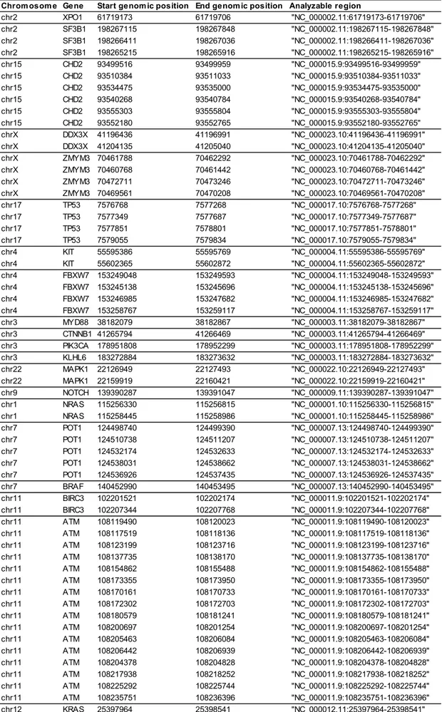
ort 1 (A) and ording to th O) of FISH le he level of ex w or high), res ose of th e subdivid on the 50t 5p distribu 599 ΔΔC 5082 ΔΔCq ower m ere charact sive cour a shorter T nd in m 2.2 B). onal analy n-regulatio s in a mi consecutiv was perfo genes (AT KRAS, MA ZMYM3) w m genomi n one Ion c instruction splayed m nts out of 8 Time to first cohort 2 (B) he presence esions in CD xpression of spectively. is analysi ded into 2 h percentile tion (rang Cq, 50t q). Patient iR-125a-5 terized by se of th TTT both i multivariat ysis by Ne on of miR-inor CD38 ve patient ormed usi TM, BIRC3 APK1, MYD were constr c DNA. E chip and s ns. Seven utations in 8 cases w treatment i ) respectively e (W) or no 38+ cells an miR-125a-5 31 s 2 e e th ts p a e n e ext Genera -125a-5p w 8+ fraction ts in C2 ing Ion To 3, BRAF, C D88, NOTC ructed usin nriched lib sequenced n out of n the CLL with high m in y, ot nd 5p ation Sequ was assoc n of total using Ne orrent PGM CDKN2A, CH1, NRA ng Agilent braries link using Ion 12 patien population miR-125a-5 uencing (N ciated with neoplastic xt Genera M. Librarie CTNNB1, AS, PIK3CA Haloplex T ked to Ion Torrent P nts with l n (Table 2 5p express NGS) h the prese c cells in ation Sequ es of spot DDX3X, F A, POT1, Target Enri Sphere P PGM, acco low miR-1 2.3) as co sion (7/12 ence of C1, we uencing exonic FBXW7, SF3B1, ichment Particles rding to 125a-5p mpared vs 0/8,
32
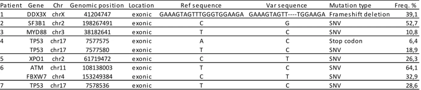
TABLE 2.3. NGS mutations as assessed by Ion Torrent technology in 7 patients with low
miR-125a-5p expression
SNV, non synonymous variation.
2.3
T
HE PATHOGENETIC ROLE OF“
PROLIFERATION CENTERS”
INCLL:
SITES WITHCD38+
CELLS AND A HIGH FREQUENCY OF GENETIC INSTABILITY2.3.1 The role of microenvironment in CLL
Available evidence exists suggesting that the interaction of leukemic cells with stromal and T cells in the microenvironment has a key role in CLL pathogenesis and evolution. As there is increasing evidence that individual cancer samples are heterogeneous and include subclonal populations and that tumors likely evolve through competition and interaction between different sublclones, this tumoral microenvironment appears to be the site where acquisition of additional genetic lesions in the clone occur, which, should greatly influence clinical outcome [92]. In this hypothesis CLL could emerged as a chronic disease in which the host physiologically provides essential elements and conditions leading to the acquisition and accumulation of genetic alterations by leukemic cells. This scheme accommodates the existence of structures that provide replicating and surviving signals to B cells on their way to neoplastic transformation [5, 10]. A key element in this view is that leukemic cells can and do proliferate, with division taking place not in the blood, but primarily in specialized morphologically discrete structures of lymph nodes and bone marrow known as proliferation centers (PCs) [66, 84, 93, 94].
So important advances in the molecular pathogenesis were obtained through the study of the role of the microenvironment.
CLL can be defined as a low-grade CD5+ B-cell tumor, whose tumoral cells have previously encountered the antigen, escaped programmed cell death and undergone cell cycle arrest in the G0/G1 phase. In CLL cells, elevated levels of
Pa ti ent Gene Chr Genomi c pos i ti on Loca ti on Ref s equence Va r s equence Muta ti on type Freq. % 1 DDX3X chrX 41204747 exoni c GAAAGTAGTTTGGGTGGAAGA GAAAGTAGTT‐‐‐‐TGGAAGA Fra mes hi ft del eti on 39,1
2 SF3B1 chr2 198267491 exoni c C G SNV 52,7 3 MYD88 chr3 38182641 exoni c T C SNV 10,8 4 TP53 chr17 7577575 exoni c A C Stop codon 6,4 TP53 chr17 7577580 exoni c T C SNV 18,9 5 XPO1 chr2 61719472 exoni c C T SNV 26,3 6 ATM chr11 108138003 exoni c T C SNV 64,1 FBXW7 chr4 153249384 exoni c C T SNV 32,9 7 TP53 chr17 7578536 exoni c T C SNV 28,6
33
the cyclin-negative regulator p27-Kip1 protein are found in a minority of patients [95]. Given the key role of this protein in cell cycle progression, its overexpression in CLL cells may account for the accumulation of B cells in early phases of the cell cycle [96]. In addition, overexpression of the anti-apoptotic BCL-2, BCL-XL, BAG-1 and MCL-1 molecules and the absence of microRNAs miR-15 and miR-16 [97], whereas proapoptotic proteins like BAX and BCL-XS are under expressed [98] could explain the resistance of tumoral cells to apoptosis. Despite the fact that most leukemic cells are arrested in cell cycle G0/G1 stages, Messmer et al. [83] demonstrated that CLL is not only a static disease but also a disease in which a proliferative pool coexist. In consequence, evolution of this leukemia depend on the relative balance between these subpopulations.
In contrast with in vivo results, apoptosis occurs after in vitro culture, suggesting a role of the microenvironment in CLL cell survival [99]. Within leukemic microenvironment, two cellular components appear to be potential players: stromal cells and T-lymphocytes. Direct contact between CLL and mesenchymal stromal cells (MSCs) is required for this inhibition of apoptosis, and tumor cell-stromal cell interactions are also important in controlling migration into and retention of CLL cells within tissue compartments (bone marrow or lymphoid tissue). MSCs are an important component of the bone marrow and support the maintenance of normal hematopoietic stem cells [100]. When co-coltured with CLL cells, MSCs have also been shown to protect the neoplastic B cells from apoptosis induced by fludarabine, bendamustine and steroids [101, 102]. Closely related to MSCs is a population of monocyte-derived “nurse-like” cells (NLC) that are also able to protect CLL cells from apoptosis [103, 104]. Both MSCs and NLCs therefore have anti-apoptotic activity and the combination of these two cell types provides a supportive microenvironment for tumor cells in CLL. In the other side, also T lymphocytes are attractive candidates to the role of elements that amplify microenvironment able to inhibit the malignant B-cell apoptosis and to favour disease progression. The weight of evidence points to a dialogue between CLL cells and CD4+ T cells, based upon bidirectional interactions that are regulated by adhesion molecules and chemokine and translate into the production of several cytokines by both cell types [99]. Many reports suggest that CD40/CD40L interactions are central to the dialogue between CLL cells and T cells. CD40 is a member of the tumor necrosis factor (TNF) receptor superfamily that is expressed by B cells, dendritic cells and monocytes [105]. CD40L is a member of the TNF
34
family expressed by activated T cells. The stimulation of CD40 rescues CLL cells from apoptosis and induces their proliferation [98, 106].
2.3.2 Proliferation centers
At this time, it is clear that crosstalk with accessory cells in specialized tissue microenvironments favours disease progression by promoting malignant B-cell growth and the emergence of new genetic alterations, which will lead to drug resistance. Therefore, understanding the crosstalk between malignant B-cells and their milieu could give us new keys on the cellular and molecular biology of CLL that can finally lead to novel strategies for disease treatment. In this regard, the isolation and analyses of the tumoral subset that is being triggered in the proliferative compartments of progressive CLL cases is an important aim to understand CLL pathogenesis.
The CLL proliferating compartment is represented by focal aggregates of proliferating prolymphocytes and para-immunoblasts that give rise to the so-called pseudofollicles or proliferation centers (PCs) [107]. Proliferation centres are present in approximately 90% of lymphocytic lymphomas. They are the histological CLL hallmark in lymph nodes, white pulp of the spleen and bone marrow where they appear as vaguely nodular areas never surrounded by a mantle zone. They consist of loosely arranged larger cells that often contain prominent nucleoli. In contrast to true B-cell follicle, which may be found entrapped within the small lymphocytic infiltrate in sections of B-CLL, proliferations centers are said not to contain follicular dendritic cells, although their presence has been occasionally recorded. Proliferation centers contain numerous T cells, most of which are CD4+. Notably, as compared to the non-proliferation center component of CLL, cells clustered in the proliferation centers have increased expression of the proliferation-associated markers Ki-67 and CD71, co-expression of survivin and BCL-2 and also higher expression of CD20, CD23 and MUM1/IRF-4 [81, 107]. Cells are surrounded by and interspersed with new vessels. It is still unclear whether these newly formed vessels are fully functional and bring nutrients to the proliferating cells or whether they represent an epiphenomenon of the angiogenetic factors that are produced by actively proliferating malignant B cells [108].