Università degli Studi di Ferrara
DOTTORATO DI RICERCA IN CO-TUTELA con
« Université Pierre et Marie Curie » di Parigi
(Sorbonne Université)
in
"Scienze Farmaceutiche" e “Chimie Moléculaire”
CICLO XXIII
COORDINATORI Prof. Stefano Mandredini e Prof. Emmanuel Lacôte
Enantioselective Approaches and Domino Sequences
for the Synthesis of Compounds of Biological Interest
Settore Scientifico Disciplinare CHIM/06
Sostenuto l’ 8 Aprile 2011
Davanti alla commissione costituita da:
Prof. Pollini Gian Piero e Prof. Poli Giovanni: Direttori della Tesi
Prof.ssa Ongeri Sandrine e Prof.ssa Costi Maria Paola: Controrelatori
M. Karoyan Philippe et M. Trapella Claudio: Esaminatori
Dottorando Tutori
Dott.ssa Pelà Michela Prof. Pollini Gian Piero
Prof. Poli Giovanni
THESE DE DOCTORAT DE
L’UNIVERSITE PIERRE ET MARIE CURIE
Spécialité
Chimie Moléculaire, ED : 406
(Ecole doctorale)
Présentée par
Mme Pelà Michela
Pour obtenir le grade de
DOCTEUR de l’UNIVERSITÉ PIERRE ET MARIE CURIE
Sujet de la thèse :
Enantioselective Approaches and Domino Sequences
for the Synthesis of Compounds of Biological Interest
soutenue le 8 Avril 2011
devant le jury composé de :
M. Poli Giovanni et M. Pollini Gian Piero: Directeurs de thèse
Mme Ongeri Sandrine et Mme Costi Maria Paola : Rapporteurs
M. Karoyan Philippe et M. Trapella Claudio : Examinateurs
Université Pierre & Marie Curie - Paris 6
Bureau d’accueil, inscription des doctorants et base de données
Esc G, 2ème étage
15 rue de l’école de médecine 75270-PARIS CEDEX 06
Tél. Secrétariat : 01 42 34 68 35 Fax : 01 42 34 68 40 Tél. pour les étudiants de A à EL : 01 42 34 69 54 Tél. pour les étudiants de EM à MON : 01 42 34 68 41 Tél. pour les étudiants de MOO à Z : 01 42 34 68 51
E-mail : [email protected]
Contents
List of abbreviations ………... 7
Genaral Introduction ……….... 8
Chapter 1: “Ferrara’s Project”……… 12
1. Introduction ………... 13
1.1 G-protein coupled receptor (GPCR): structure, biological activation
and signal transduction ... 13
1.2 NPS/NPSR system ... 16
1.3 Structure-activity relationship study of NPS leading to
identified peptide and non peptide ligands ... 19
1.3.1 Peptide ligands of NPSR ... 19
1.3.2 Non-peptide ligands of NPSR ... 21
2. Aim of the project ………... 24
3. Synthesis ... 25
3.1 Synthesis of Takeda’s compound ... 25
3.2 Synthesis of SHA-68 compound in racemic mixture ... 27
3.3 Asymmetric Synthesis of (R)-SHA 68 and (S)-SHA 68 ... 30
4. Chiral chromatography profiles ... 35
5. NMR spectrometry profile ... 37
6. X ray spectroscopy analysis ... 38
7. Biological activity
...
... 40
Chapter 2: “Paris' Project”………... 43
I.
Introduction ... 44
II.
Aim of the project ... 49
III.
Synthesis ... 50
III.
I
Retrosynthetic analysis ... 50
III.
II
Synthesis of allenol intermediate ... 51
III.
III
Synthesis of cyclization precursor ... 52
III.
IV
Phosphine-free Pd-catalyzed Domino Sequence and
benzylation reaction ... 54
III.
V
Removal of the methoxycarbonyl group ... 56
III.
VI
Further steps ... 57
IV.
Conclusion ... 59
References ... 60
Chapter 3: Experimental Section ... 65
General methods... 66
Synthesis of Takeda’s compound ... 68
Synthesis of SHA-68 compound in racemic mixture ... 76
Asymmetric Synthesis of (R)-SHA 68 and (S)-SHA 68 ... 80
Synthesis of (-)-Steganacin aza analogue ... 90
Supporting Information ... 104
Résumé de Thése ... 119
Riassunto della Tesi ... 121
List of abbreviations
Ac
acetyl
Ar
aryl
Bn
benzyl
Boc
tert-butoxycarbonyl
n-Bu
butyl
d. r.
diastereomeric ratio
DBU
1,8-Diazabicyclo[5.4.0]undec-7-ene
DHP
3,4-dihydro-2H-pyran
DMF
N,N-dimethylformamide
DMSO
dimethyl sulfoxide
ee
enantiomeric excess
equiv.
equivalent
Et
ethyl
h
hour
LiHMDS
Lithium bis(trimethylsilyl)amide
L-Sélectride Lithium tri-sec-butylborohydride
Me
methyl
min
minute
Ph
phenyl
iPr
isopropyl
pTsOH
p-toluenesulphonic acid
Y
yield
NMR
nuclear magnetic resonance
TBAB
tetra-n-butylammonium bromide
TBAF
tetra-n-butylammonium fluoride
TBS
tert-butyldimethylsilyl
tBu
tert-butyl
THF
tetrahydrofuran
TMEDA
tetramethylethylene diamine
General Introduction
Ryōji Noyori in “Asymmetric Catalysis in Organic Synthesis”, John Wiley&Sons, New
York, 1994 wrote: “Life depends on chiral recognition, because living systems interact
with enantiomers in decisively different manner”.
We know that the majority of biological processes take place as a consequence of the
different ways in which enantiomers with different configuration react with receptors
and life itself is in many ways chiral. Nature provides a vast diversity of chiral species
in several classes of compounds, including “inter alia” aminoacids, carbohydrates,
terpenes, carboxylic acids and alkaloids, several in great abundance, and the chiral
information is derived from enantiomerically pure building blocks enzymatically
synthesized by various organisms.
Interestingly, chiral biomolecules usually exist in Nature prevalently as one of the two
possible enantiomeric forms, e.g., amino acids in the L-form and sugars in the D-form.
Chirality is an important factor in determining bioactivity. The recognition that
enantiomers often have different bioactivity and metabolic fates
1in a number of chiral
chemicals with important pharmacological activity led to a significant increase in the
development of chiral pharmaceuticals, chirality becoming a major concern in the
modern pharmaceutical industry.
2This interest can be attributed largely to a deeper awareness that enantiomers of a
racemic drug may have different pharmacological activities, as well as different
pharmacokinetic and pharmacodynamic effects.
Each racemic drug could interact differently and metabolize each enantiomer by a
separate pathway to produce different pharmacological activity.
Thus, one isomer may produce the desired therapeutic effect, the other one being
1 D.E. Drayer, Clin. Pharmacol. Theor., 1986, 40 125.
2 S. Lam and G. Malikin, Chirality, 1992, 4, 395; I.W. Wainer (Editor), Drug Stereochemistry Analytical Methods and Pharmacology, Marcel Dekker, Inc., New York, New York, 1993.
inactive or, in
worst cases, noxious. For example, the (L)-Thyroxine, an amino acid
produced by thyroid gland, is known to speed up metabolic processes, causing
nervousness and loss of weight, while its enantiomer, (D)-Thyroxine served to lower the
cholesterol levels.
HO
I
I
O
I
I
COOH
NH
2HO
I
I
O
I
I
COOH
NH
2(L)-Thyroxine (D)-Thyroxine
Figure 1
The use of thalidomide (n-phthalyl-glutamic acid imide), which was marketed as the
racemate, led to a tragedy in the 1960s in Europe. The sedative-hypnotic drug
thalidomide prescribed to pregnant women to counter morning sickness exhibited
irreversible neurotoxicity and mutagenic effects. It was discovered only after hundred of
babies were born deformed that the S-enantiomer was teratogenic. Studies later
suggested that these effects were caused by the S-enantiomer and that the R-enantiomer
contained the desired therapeutic activity.
3N
NH
O
O
O
O
N
NH
O
O
O
O
R- Thalidomide S- Thalidomide
Figure 2
In 1992 the U.S. Food and Drug Administration issued a guideline for chiral drugs: only
the therapeutically active isomer could be marketed and each enantiomer should be
studied separately for its pharmacological and metabolic pathways.
4In addition, a
3Stephens TD, Fillmore BJ., Hypothesis: Thalidomide Embryopathy – Proposed Mechanism of Action. Teratology 2000; 61: 189-195.
rigorous justification is required for market approval of a racemate of chiral drugs.
Currently, a large number of chiral drugs are marketed as racemic mixtures.
5Nevertheless, to avoid the possible undesirable effects of a chiral drug, it is imperative
that only the pure, therapeutically active form should be prepared and marketed.
When a chiral molecule is synthesized in an achiral environment using achiral starting
materials, an equal mixture of the two possible enantiomers (i.e. a racemic mixture) is
produced. The special problem of the separation of the enantiomers could be solved
using a chiral resolving agent. This technique relies on the fact that while enantiomers
have identical physical properties, diastereomers generally have different properties.
Another technique is to use chiral chromatography. In this process, the racemate is run
through a column filled with a chiral substance. The enantiomers will interact
differently with the substance and will then elute at different rates.
As more and more enantiomerically enriched active agents are required today,
the
selective synthesis of enantiomers is the subject of many research projects aimed at the
discovery of
reactions or reaction sequences in which one configuration of one or more
new stereogenic elements is selectively formed. In an asymmetric synthesis, an achiral
molecule is enantioselectively converted into a chiral molecule or a chiral molecule is
diastereoselectively converted into a new chiral molecule that contains at least one more
chirality element.
In an asymmetric synthesis the enantiomers (or diastereomers) of a
chiral product are formed in different yields.
Thus, the study of stereoselectivity has evolved from issues of diastereoselectivity,
through auxiliary-based methods for the synthesis of enantiomerically pure compounds
to asymmetric catalysis. In the latter instance, enantiomers (not diastereomers) are the
products, and highly selective reactions and modern purification techniques allow
preparation - in a single step - of chiral substances in 99% ee for many reaction types.
Physical resolution of racemates for example using HPLC (High Performance
Liquid Chromatography) with specific chiral columns;
5 B. Lin, X. Zhu, B. Koppenhoeffer and U. Epperlein, LC·GC, 1997,15 40. P. van Eikeren, in S. Ahuja (Editor) Chiral Separations
Indirect enantiomeric resolution, via chiral auxiliary-based approaches,
involves the coupling of the enantiomers with an auxiliary chiral reagent to
convert them into diastereomers. The diastereomers can then be separated by
cromatography or other achiral separation techniques;
Enzymatic catalysis in which the action of an enzymatic transformation is
exploited to obtain enantioenriched compounds;
Catalytic enantioselective trasformations using chiral pool as catalytic
compounds (for example the best know catalyst is the (L)-Proline).
6Chapter 1
1. Introduction
1.1 G-protein coupled receptor (GPCR): structure, biological activation and signal
transduction
Many drugs produce therapeutic activities through interaction with G-protein-coupled
receptors (GPCRs), which actually represent the most important biological target for
drug discovery.
7, 8GPCR (G-protein coupled receptor)s constitue a superfamily of integral membrane
proteins consisting of seven transmembrane helices connected by loops with a
N-terminal extremity always located on the extracellular side, the C-terminus being
extended into the cytoplasm. GPCRs can be divided into different families on the basis
of their structural and genetic properties.
Receptors of Family 1 are characterized by several highly conserved amino
acids and a disulphide bridge that connects the first and second extracellular
loops (ECLs). Most of these receptors at the carboxy-terminal tail present
palmitoylated cysteine residue that bind to the membrane. They are also
characterized by the presence of amino acids such as proline that produces a
distortion of the helical transmembrane domain.
Family 2 GPCRs are characterized by a relatively long amino terminus, which
contains several cysteines forming a network of disulphide bridges. Their
morphology is similar to some family 1 receptors but lacking the palmitoylation
site. Moreover, the conserved residues and motifs are different from the
conserved residues in the family 1 receptors.
7 Gilchrist, A., Expert Opin. Ther. Targets, 2004, 8, 495–498 8 Saito, Y., and Civelli, O., Int. Rev. Neurobiol. 2005, 65, 179–209
Receptor of the Family 3 are characterized by a long amino terminus and
carboxyl tail. The ligand-binding domain is located in the amino terminus. None
of the features that characterize family 1 and 2 receptors are present in Family 3
receptors.
These receptors are activated by an external signal in form of a ligand or other signal
mediator, including peptide (e.g. Neuropeptide S) and non-peptide neurotransmitters,
hormones, growth factors, odorant molecules and light. The conformational change
created in the receptor caused activation of a G-protein. Further effect depends on the
type of G protein.
Figure 3
The transduction of the signal through the membrane by the receptor is not completely
understood. It is known that the inactive G protein is bound to the receptor in its
inactive state. (Figure 4)
GPCR bound to Gαβγ complex; “G-proteins” are a trimer of α, β, and γ subunits
(known as Gα, Gβ, and Gγ, respectively);
Gα complex bound to Guanosine diphosphate (GDP) when the protein is
inactive;
Upon receptor activation, there is a consequent allosterically activation of the
protein by facilitating the exchange of a molecule of GDP for GTP at the
G-protein's α-subunit (2);
Once the G protein is activated, the subunits of the G-protein dissociate from the
receptor, Gα looses affinity for βγ dimer to yield a Gα-GTP monomer and a Gβγ
dimer, which are now free to modulate the activity of other intracellular proteins
(3);
Cascade events:
o
βγ dimer stays bound to the membrane and interacts with Effectors based
on the specific βγ subunits like Adenylyl Cyclase (4);
o
Gα GTP complex enters the cytosol and interacts with Effectors, such as
PLC-β (4);
Hydrolysis of GTP, linked to Gα subunit, provides Gα-GDP (5) product that is
inactive but more related to the βγ dimer;
The trimer protein are reformed (Gαβγ) (6);
Gαβγ complex binds to GPCR.
There is another subclassification of “G-protein” in particular at the α subunit: Gαs, Gαi
and Gαq. Activation each of them produces different cascade events.
In Figure 5a the mechanism of signal transduction is described: Gαs protein is activated
by a ligand; a subsequent activation of adenylate cyclase takes place increasing the
AMPc concentration.
The section 5b outlines the mechanism after activation of Gαq: the released
phospholipase C (PLC) provokes the cleavage of the membrane lipid
phosphatidylinosityl-bisphosphate (PIP2) into diacylglycerol and IP3. The latter serves
mainly to open calcium channels of the endoplasmic reticulum (ER) leading to the
release of Ca
2+into the cell with consequent increase of its intracellular concentration.
These profiles representing the two activated pathways by NPS (neuropeptide S) after
interaction with its receptor are shown below.
a.
b.
Figure 5
1.
2
NPS/NPSR system
At least 800 different genes codifying for putative GPCRs have been identified in the
human genome, ~360 of which encode transmitter GPCRs
9. Moreover, the endogenous
9Vassilatis, D. K., Hohmann, J. G., Zeng, H., Li, F., Ranchalis, J. E., Mortrud, M. T., Brown, A., Rodriguez, S. S., Weller, J. R., Wright, A. C., Bergmann, J. E., and Gaitanaris, G. A. Proc. Natl. Acad. Sci. U. S. A., 2003, 100, 4903–4908
ligand is known for only ~240 receptors, whereas the others are still orphans. The first
step in understanding the function and the potential of an orphan receptor as drug target
is the identification of its endogenous ligand. In the last decade, the reverse
pharmacology technique
10, i.e. the use of a recombinant orphan GPCR as a target for
identifying its endogenous ligand, has been validated as a successful approach for the
identification of novel transmitter systems. In particular, several novel peptide receptor
systems have been identified through this approach, including nociceptin/orphanin FQ,
prolactin-releasing peptide, urotensin II and many others. The latest neuropeptides
identified by the reverse pharmacology approach was Neuropeptide S (NPS): a crucial
discovery firstly reported in the patent literature.
11A subsequent, elegant study by Xu et
al.
12demonstrated that hNPS (as well as the rat and mouse isoforms of this peptide)
selectively binds and activates a previous orphan GPCR, known as GPR154, that was
renamed NPS receptor, then abbreviated as NPSR.
Several splice variants and multiple single nucleotide polymorphisms have been
reported for the human NPSR. The most intensely investigated NPSR isoforms are
hNPSR Asn107 and hNPSR Ile107. This receptor polymorphism seems to have
functional implications since the hNPSR Ile107 receptor displayed similar binding
affinity but higher NPS potency (by approx. 10-fold) than hNPSR Asn107.
13It is
worthy of mention that the rat and mouse NPSR contain Ile at position 107.
14The primary sequence of human (h) NPS, a 20-residue peptide, which is highly
conserved across species, is reported (Figure 6).
SFRNGVGTGMKKTSFQRAKS
H-Ser-Phe-Arg-Asn-Gly-Val-Gly-Thr-Gly-Met-Lys-Lys-Thr-Ser-Phe-Gln-Arg-Ala-Lys-Ser-OH
Figure 6
10 Civelli, O. , Trends Pharmacol. Sci., 2005, 26, 15–19
11
Sato, S., Shintani, Y., Miyajima, N., and Yoshimura, K. (April 18, 2002) Japan Patent WO 0231145
12 Xu, Y. L., Reinscheid, R. K., Huitron-Resendiz, S., Clark, S. D., Wang, Z., Lin, S. H., Brucher, F. A., Zeng, J., Ly, N. K., Henriksen, S. J., de Lecea, L., and Civelli, O. Neuron, 2004, 43, 487–497.
13 Reinscheid, R. K., Xu, Y. L., Okamura, N., Zeng, J., Chung, S., Pai, R., Wang, Z., and Civelli, O. J. Pharmacol. Exp. Ther. 2005, 315, 1338–1345
14
The N-terminal serine residue, which is present in all the species analyzed so far, gave
the name to this novel neuropeptide (Neuropeptide S). hNPS is cleaved from a larger
precursor protein (ppNPS) which is expressed in few discrete brain areas. On the
contrary, the receptor NPSR is widely distributed in the brain. This profile of receptor
expression suggests the involvement of the NPS–NPSR system in the regulation of
multiple central functions.
In cells expressing the recombinant NPSR receptor, NPS selectively binds and activates
its receptor, producing intracellular calcium mobilization and an increase of cAMP
levels. This indicates that the increase cellular excitability, after NPSR stimulation, it is
due to the activation of both Gq and Gs protein. In vivo studies in rodents showed that
the supraspinal administration of NPS produced a rather unique pattern of actions:
anxiolytic-like effects associated with the stimulation of locomotor activity and clear
arousal promoting effects.
5Thus, NPS could be defined as an activating anxiolytic.
7In addition, NPS has been reported to inhibit food intake, facilitate memory and elicit
antinociceptive effects, while recent evidence suggests an involvement of the
NPS/NPSR system in drug addiction.
1515
1.
3
Structure-activity relationship study of NPS leading to identified
peptide and non peptide ligands
1.
3
.1. Peptide ligands of NPSR
The development of novel ligands for NPSR is required in order to determine the role
played by the NPS-NPS receptor system in the regulation of different biological
functions and ultimately to predict the therapeutic potential of novel drugs interacting
with this receptor.
Thanks to structure-activity relationship studies reported for the first time by our
research group, we were able to identify the main requirements for biological activity in
the primary structure of NPS.
16Thus, single residue replacement, either by an alanine residue (Ala-scan) or by the
corresponding enantiomer (D-scan) with N- and C-terminal truncation, demonstrated
that the N-terminus of the peptide was of crucial importance for biological activity. In
particular, the sequence Phe
2-Arg
3-Asn
4is likely to act as message domain crucial for
receptor binding and its activation, while the sequence Gly
5-Val
6-Gly
7is important for
inducing the bioactive conformation of the peptide.
In parallel, we also performed a conformation/activity relationship study
17that
demonstrated that helicity can be tolerated in the C-terminal part of NPS and this
conformational structure is not required for bioactivity and it is not tolerated around
position Gly
7.
16Roth, A. L.; Marzola, E.; Rizzi, A.; Arduin, M.; Trapella, C.; Corti, C.; Vergura, R.; Martinelli, P.; Salvadori, S.; Regoli, D.; Corsi, M.; Cavanni, P.; Calo`, G.; Guerrini, R.. J. Biol. Chem. 2006, 281, 20809-20816.
17 Tancredi, T.; Guerrini, R.; Marzola, E.; Trapella, C.; Calo, G.; Regoli, D.; Reinscheid, R. K.; Camarda, V.; Salvadori, S.; Temussi, P. A., J. Med. Chem., 2007, 50, 4501–4508.
Figure 7
Conformational changes induced by substituting Gly
5with the achiral R helix
promoting amino acid Aib or with D-Ala seem to provoke a decreased agonist efficacy,
as indicated by structure–activity studies performed at position 5. Therefore, we
planned a SAR study mainly focusing at Gly
5position by replacing with a series of L-
and D- amino acids characterized by hydrophobic aromatic and aliphatic side chains.
18As a result, we demonstrated that substitution of Gly
5with D-amino acids bearing a
short lipophilic-branched side chain could generate a fairly potent, pure and selective
NPSR antagonists.
a. H-Ser-Phe-Arg-Asn-Gly-Val-Gly-Thr-Gly-Met-Lys-Lys-Thr-Ser-Phe-Gln-Arg-Ala-Lys-Ser-OH b. H-Ser-Phe-Arg-Asn-(D)Val-Val-Gly-Thr-Gly-Met-Lys-Lys-Thr-Ser-Phe-Gln-Arg-Ala-Lys-Ser-OH c. H-Ser-Phe-Arg-Asn-(D)Cys (tBu)-Val-Gly-Thr-Gly-Met-Lys-Lys-Thr-Ser-Phe-Gln-Arg-Ala-Lys-Ser-OH
Figure 8
18R.Guerrini, V.Camarda, C.Trapella, G.Calò, A.Rizzi, C.Ruzza, S.Fiorini, E.Marzola, R. K.Reinscheid, D.Regoli, S.Salvadori, J. Med. Chem. 2009, 52, 524–529.
Message Conformation C-Terminal Domain Inducing Domain Domain
Above, the structures of first two NPSR-peptide antagonists actually known in literature
are reported, namely: [D-Val
5]NPS (Figure 8b) and [D-Cys(tBu)
5]NPS (Figure 8c)
whose pharmacology activity has been evaluated in a calcium mobilization assay using
HEK293 cells stably expressing mouse NPSR (HEK293mNPSR) and the fluorometric
imaging plate reader FlexStation II.
1.
3
.2 Non-peptide ligands of NPSR
Non-peptide derivatives have been widely used to examine in details and improve the
knowledge of the NPS system
19. However, at the beginning of this work only ligands
featuring an oxazol-piperazine scaffold were known able to bind the NPSR receptor.
A series of compounds with a claimed activity at NPSR has been recently reported in a
patent application by Takeda Pharmaceuticals Inc
20without the support of
pharmacological or biological data. In the patent, structures containing a
3-oxo-tetrahydro-oxazolo[3,4-a]pyrazine scaffold mainly substituted in position 1 and 7, have
been described to possess NPSR antagonistic activity.
N
N
O
O
R
R'
R''
1 2 3 4 5 6 7Figure 9
Two of these compounds, namely (SHA 66 and SHA 68), differing exclusively for the
presence of a fluoro substituent at the para position of the benzyl moiety (SHA 68),
19Reinscheid R.K.. Peptides 2007, 28, 830–837; Reinscheid R.K. and Xu, Y.L.. Neuroscientist, 2005, 11, 532–538; Guerrini R., Salvadori S., Rizzi A., et al.. Medicinal Research Reviews, 2010, 30 (5), 751-77.
20Fukatzu K, Nakayama Y, Tarui N, Mori M, Matsumoto H, Kurasawa O, Banno H.; Takeda Pharmaceuticals; 2004, PCT/JP04/12683.
were prepared by Okamura et al.
21who studied their pharmacological proprieties as
potential NPSR antagonists in vitro and in vivo. The two closely related bicyclic
piperazines have been described as potent and selective antagonists at NPSR in vitro,
able to antagonize NPS-induced effects in vivo.
Thus, SHA 68 shoved selectivity for this receptor and was able to block NPS-induced
Ca
2+mobilization in central and peripheral site after binding.
However, its
pharmacokinetic profile indicated a limited BBB penetration.
SHA 68 (see Fig. 10b) i.e. the racemic mixture
(9R/S)-3-oxo-1,1-diphenyl-tetrahydro-oxazolo[3,4-a]pyrazine-7-carboxylic acid 4-fluoro-benzylamide represents the first
generation of non-peptide NPSR antagonists.
H
N
N
N
O
O
O
H
N
N
N
O
O
O
F
a. (±) SHA-66 b. (±) SHA-68
Figure 10
Structure–activity studies performed at position 7 of SHA 68
22have indicated that a
potent NPSR antagonist activity required a free urea moiety since alkylation of the urea
nitrogen or its replacement with carbon or oxygen atoms generated derivatives with a
decreased activity. In addition, compounds with α-methyl substitution or elongated
alkyl chains showed reduced potency, indicating a limited tolerance for position 7
substituents. Interestingly, removal of the fluorine atom at the para position of the
phenyl ring generated a compound (SHA 66) displaying similar potency in comparison
21 Okamura N, Habay SA, Zeng J, Chamberlin AR, Reinscheid RK.; J. Pharmacol. Exp. Ther., 2008, 325, 893–901. 22 Y. Zhang, B. P. Gilmour, H. A. Navarro, S. P. Runyon, Bioorg. Med. Chem. Lett., 2008, 18, 4064–4067.
to the parent compound, indicating that the fluorine atom does not affect receptor
binding
.
6,7
Very recently, new series of NPSR antagonists has been developed by the same group
of researchers (Melamed et al..
23and Trotter et al..
24) in order to identified new
antagonists of the Neuropeptide S receptor (NPSR) with high potency, good
permeability into the brain trough the brain-blood-barrier (BBB), namely: (Figure 11):
a quinolinone class of potent NPSR antagonists that readily cross the blood–
brain barrier (NPSR-QA1).
a tricyclic imidazole antagonist of NPSR, represented by NPSR-PI1, that
demonstrates potent in vitro NPSR antagonism and central exposure in vivo.
N
N
H
3CO
Cl
N
Et
Et
O
N
O
N
O
R R'
R = Me
R' = Et
N
O
NPSR-PI1 NPSR-QA1
Figure 11
In both cases, the racemic mixture of NPSR-PI1 and NPSR-QA1 was separated using
chiral column chromatography affording the two potent enantiomeric compounds
showed in Figure 11, but the absolute configuration of these antagonists remained
unknown.
23
J. Y. Melamed, A.E. Zartman , N.R. Kett , A.L. Gotter, V.N. Uebele, D.R. Reiss, C.L. Condra, C. Fandozzi, L.S. Lubbers, B.A. Rowe, G.B. McGaughey, M.Henault, R. Stocco, J.J. Renger, G.D. Hartman, M.T. Bilodeau, B.W. Trotter, Bioorg. Med. Chem. Lett., 2010, 20, 4700–4703.
24
B. W. Trotter, K. K. Nanda, P.J. Manley, V. N. Uebele, C. L. Condra, A. L. Gotter, K. Menzel, M. Henault, R. Stocco, J.J. Renger, G. D. Hartman, M. T. Bilodeau, Bioorg. Med. Chem. Lett., 2010, 20, 15, 4704-4708 .
2. Aim of the project
The NPS-NPSR system is a quite recent discovery and many studies are still required to
deeply understand its biological functions and better define the therapeutic potential of
selective NPSR ligands.
The availability of selective NPSR antagonists is an important target in this field and
my work has been mostly addressed to their discovery.
In details, this work has been maily focused in the asymmetric synthesis of chiral
2,4-disubstituted- and 2,4,6-trisubstitued-piperazines starting from cheap, commercially
available materials. The piperazine moiety is a well recognized “privileged scaffold” in
medicinal chemistry,
25being the common structural feature of a wide range of
biologically active natural and synthetic products.
The variety of synthetic methods that allow for the fast and efficient assembly of
piperazine skeletons reported in the literature has been comprehensively surveyed in
four excellent reviews.
26In this project, the asymmetric construction of the substituted piperazine ring has been
accomplished through two efficient methodologies featuring a different way for the
introduction of the required chirality, namely:
using readily available optical active natural materials such as aminoacids
(alanine) as starting materials;
using a chiral auxiliary reagent to obtain separable diastereomers.
The first class of compounds showing antagonism activity for the NPS receptor has
been reported in the literature by the Takeda’s group. The synthetic pathway has been
previously described by Schanen and coworkers in 1996.
2725a) Horton, D. A.; Bourne, G. T.; Smythe, M. L., Chem. Rev., 2003, 103, 893–930; b) Tullberg, M.; Grøtli, M.; Luthman, K. J. Org. Chem., 2007, 72, 195–199.
26 a)Dinsmore, C. J.; Beshore, D. C. Tetrahedron, 2002, 58, 3297–3312; Dinsmore, C. J.; b) Beshore, D. C. Org. Prepar. Proced. Int., 2002, 34, 367–404; c) Quirion, J.-C. Asymmetric Synthesis of Substituted Piperazines. In Targets in Heterocyclic Systems; Attanasi, O. A., Spinelli, D., Eds.; Italian Society of Chemistry: Roma, 2008; Vol. 12, pp 438–459; d) De Risi C., Pelà M., Pollini G.P., Trapella C., Zanirato V., Tetrahedron: Asymmetry, 2010, 21, 255-274.
3. Synthesis
3.
1
Synthesis of Takeda’s compound
In order to obtain the first non-peptide antagonist of NPS for pharmacological
evaluation we planned to prepare the “Takeda compound” following the Schanen’s
synthetic approach
27even if some modifications.
Thus, the starting move was the reaction between R-(-)phenylglycinol 1 and
N-Boc-(D)-Alanine 2 to produce amide 3 under standard condensation conditions for the peptide
bond formation (activation via dicyclohexylcarbodiimide) introducing a defined
stereogenic centre at the beginning of the piperazine synthon construction, differently
from the Schanen’s approach.
The two carbonyl functions of the intermediate 3 could be selectively reduced with
LiAlH
4in diethylether leading to the formation of 4 and its hydroxyl function was
subsequently protected with tert-butyldimethylsilyl chloride in THF producing the
amine 5. Its condensation reaction with with bromoacetic acid was conveniently
accomplished via dicyclohexylcarbodiimide activation (Scheme 1) leading to the
bromoacetylated derivative 6 which was smoothly cyclized to the piperazinone 7 by
treatment with NaH in a mixture of DMF/THF.
HO NH2 COOH NH Boc HO NH NH Boc O HO NH NH Boc O NH NH Boc N NH Boc O Br N N Boc O 1 2 3 4 5 6 7 Si Si O O Si a b c d e
Reagents and conditions : (a) N-Boc-(D)-Ala-OH, CH
2Cl
2, WSC, room temp., Y=64%; (b) LiAlH
4, Et
2O,
0°C, Y=100%; (c) TBDMS-Cl, imidazole, DMF, 0°C, Y=76%; (d) Bromoacetic acid, CH
2Cl
2, WSC,
room temp., Y=35%; (e) NaH 60%, THF/DMF, room temp., Y=74%.
Scheme 1
Removal of the silyl protective group of 7 was easily achieved by action of fluorine ions
giving rise to 8 which was treated with borane dimethyl sulfide complex in order to
reduce selectively the lactam moiety producing the piperazine derivative 9. It was well
known that the alkylation at position 2 of the piperazine ring system does not occur in
the presence of the free hydroxyl function.
27Therefore, the alcoholic functionality has
been protected as the corresponding methyl ether 10 using NaH and MeI. A solution of
this compound in freshly distilled THF was treated with sec-BuLi/TMEDA at -78 °C
and reacted with diisopropyl ketone furnishing exclusively the
trans-1,1-diisopropyl-7-(2-methoxy-1-phenyl-ethyl)-5-methyl-hexahydro-oxazolo[3,4-a]pyrazin-3-one 11 .
Informations about the conformation of the piperazine ring was obtained by NMR
analysis that showed coupling constants between the proton on C
2and the adjacent
methylene protons (on C
3) of 12 and 5 Hz. These data confirm that proton in C
2was in
axial position and consequently the alkylated substituent in equatorial conformation.
Interestingly, the exclusive formation of trans-2,6-disubstitued product has been already
observed by Beak
28in the piperidine series. Thus, the synthesis of the first example of a
non-peptide compound able to interact with the NPSR, also named “Takeda
Compound”, was completed in nine steps with very low overall yield (~4%).
TBDMSO N N Boc O HO N N Boc O HO N N Boc O N N Boc O N N O O 7 8 9 10 a b c d 11
Reagents and conditions : (a) TBAF, THF, room temp.
Y=72%; (b) (CH
3)
2S*BH
3, THF, 0°C,Y=88%; (c)
NaH 60%, MeI, THF/DMF, 0°C,Y=62%;
(d) sec-BuLi, TMEDA, isopropyl ketone, THF,-78°C, Y=48%.
Scheme 2
3.
2
Synthesis of SHA-68 compound in racemic mixture
While this work was in progress, two small molecules described in the Takeda patent
were also synthesized and tested by Okamura et al.
21namely, the closely related
bicyclic piperazines SHA 66 and SHA 68 that are potent and selective antagonists at
NPSR in vitro and are able to antagonize NPS-induced effects in vivo.
H N N N O O O F H N N N O O O
SHA 66 SHA 68
Figure 12
The low overall yield of the synthetic process to prepare 11 and the interesting data
related to the two new compounds described by Okamura et al.,
21prompted us to
explore an asymmetric synthesis of the two enantiomers of the Neuropeptide S receptor
(NPSR) antagonist
(9R/S)-3-oxo-1,1-diphenyl-tetrahydro-oxazolo[3,4-a]pyrazine-7-carboxylic acid 4-fluoro-benzylamide ((R/S)-SHA 68) to complete their structural
characterization and to confirm the pharmacological evaluation. Initially, in order to
confirm the published data, we decided to synthesize (R/S)-SHA 68 in racemic form
envisaging a very easy pathway starting from a cheap, commercially available
N-benzylpiperazine, already possessing a synthetically suitable substituent at one of the
two nitrogen of the heterocyclic skeleton.
H N N N O O O F H N N O O N N O O O N C O F Commercially available Commercially available Commercially available N N H
Scheme 3
The first step was the orthogonal protection of second nitrogen of the piperazine
scaffold. As the corresponding tert-butoxycarbonyl group (Boc) and this operation was
easily achieved under standard condition to produce quantitatively the intermadiate 13.
The construction of the second heterocycle condensed to the piperazine ring 14 was
smoothly accomplished in good yield (76%) by treatment 13 with
sec-butyllithium/N,N,N’,N’-tetramethylethylene diamine (TMEDA), at -78°C, followed by
trapping of the intermediate anion with benzophenone. As already observed by
Okamura et al., removal of the benzyl-protecting group of 14 was unsuccessful via
palladium catalysed hydrogenolysis, but occurred smoothly through a simultaneous
substitution in situ of the N-benzyl function with the fluorenyl methylenoxycarbonyl
protective group (Fmoc-) by adding FmocCl to an acetonitrile solution of 14 at room
temperature. After approximately 10 min, the precipitated 15 could be isolated by
filtration with 70% yield.
The last step of this pathway to obtain (R/S)-SHA 68
compound required removal of the Fmoc-protecting group by treatment of 15 with
1,8-diazabicyclo[5.4.0]undec-7-ene (DBU) followed by reaction with the commercially
available p-fluorobenzyl isocyanate.
N N N N O O N N O O H N N N O O O F Fmoc N N H N N O O O O 12 13 14 15 a b c d O O (R/S)-SHA 68 15
Reagents and conditions: (a) Boc2
O, H
2O/tert-butanol, NaOH (2N), room temp.; (b) sec-BuLi, TMEDA,
benzophenone, THF, -78°C, Y=49%; (c) Fmoc-Cl, CH
3CN, 100°C, Y=65%; (d) DBU,
4-Fluorobenzyl-isocyanate, THF, room temp., Y=59%.
Scheme 4
Quenching of the reaction mixture after 15 min allowed to obtain (R/S)-SHA 68 in
almost quantitative yield after purification via flash cromatography (Scheme 4).
The structure of (R/S)-SHA 68 was fully characterized by
1H and
13C NMR and mass
spectrometry.
3.
3
Asymmetric Synthesis of (R)-SHA 68 and (S)-SHA 68
Enantioselective synthesis of chiral compound is an important goal for organic and
medicinal chemists and different strategies are available for the synthesis of optically
active compounds.
In this project aimed at the construction of optical active piperazine compounds as a
new class of non-peptide NPSR antagonists, we envisaged the chiral auxiliary approach
as a convenient pathway for their asymmetric synthesis as outlined retrosynthetically in
scheme 5.
H N N N O O O F H N N O O N N O O O N C O F Cl Cl O NH NH O O NH2 COOH NH Boc Commercially available Commerciallyavailable Commercially available
Scheme 5
The piperazine scaffold was synthesized in seven steps utilizing as chiral auxiliaries the
readily available S-phenyl-ethylamine and R-phenyl-ethylamine. In the following
schemes are reported the synthetic pathway for only the S-enantiomer, the same results
being obtained with the other one. WSC-promoted condensation of N-Boc-glycine with
the chiral amine give the amide 16, which underwent LiAlH
4reduction of the carbonyl
moieties to give 17, which was acylated by action of chloro acetyl chloride in the
presence of NaHCO
3to furnish 18. Its subsequent cyclization was performed by
treatment with NaH at room temperature to produce the piperazinone derivative 19,
which was subsequently reduced in a good yield at the lactame function employing
borane dimethyl sulfide complex.
NH2 COOH NH Boc NH Boc NH O NH Boc NH b a c NH Boc N Cl O d N Boc N O e N Boc N 16 17 18 19 20
Reagents and conditions : (a) Boc-Gly-OH, CH2
Cl
2, WSC, room temp., Y=60%; (b) LiAlH
4, Et
2O, 0°C,
Y=90%; (c) Chloroacetyl-chloride, NaHCO
3, AcOEt, 0°C, Y=100%; (d) NaH 60%, THF/DMF, room
temp., Y=45%; (CH
3)
2S*BH
3, THF, 0°C, Y=87%.
Scheme 6
First attempts to build up the second heterocycle through the key alkylation step was
rather unexpectedly unsuccessful although we payed careful attention to the choice of
reagents (benzophenone, TMEDA and THF were freshly distilled and a new bottle of
sec-BuLi was opened and used immediately) and reaction conditions (anhydrous and
under nitrogen).
N
N
Boc
O
TMEDA
N
N
O
O
sec -BuLi
THF
-78°C; -30°C;-78°C; R.T.
20
21
X
20a
Scheme 7
Beside starting material, the only detectable reaction product characterized by NMR and
mass spettroscopy was the secondary alcohol derived by reduction of benzophenone,
which is likely to be produced by action of a residual piperazine-borane complex
formed during the previous carbonyl reductive step.
N
N
Boc
O
TMEDA
sec- BuLi
THF
-78°C; -30°C;-78°C; R.T.
OH
BH
220a
20b
Scheme 8
This hypothesis has been confirmed since the dihydropyrazine 19a was obtained using
LiAlH
4instead of borane dimethyl sulfide complex.
N
Boc
N
O
19
19a
N
Boc
N
LiAlH
4THF
0°C
Y=90%
Scheme 9
The formation of this unexpected product could be accounted for considering the basic
character of H
-.
N Boc N O N Boc N O AlH 3 N Boc N 19a N Boc N 19 20 nucleophylic character basic character Al H H H H Li N Boc N O Li AlH3 N Boc N H H H H H LiScheme 10
Moreover, since the Boc-protective group too seemed to be the responsible for the
unexpected formation of 20, we decided to remove it using a solution of trifluoroacetic
acid (TFA) in CH
2Cl
2. The reduction of amide group carried out on the derived free
amine 19b using LiAlH
4gave the desired piperazine 19c, subsequently protected at the
second nitrogen atom as the Boc-derivative 20 in quantitative yield.
N
Boc
N
O
N
H
N
O
N
H
N
N
N
Boc
19
a
b
c
19b
19c
20
Reagents and conditions: (a) TFA, CH
2Cl
2, 0°C, Y=97%; (b) LiAlH
4, Et
2O, 0°C, Y=87%; (c) Boc
2O,
H2O/tert-butanol, NaOH (2N), room temp., Y=100%.
While this work was in progress, an interesting paper by Reginato et al.
29described the
selective reduction of 2-oxo-piperazines as strategy to perform the easy alkylation at the
carbon C2. Thus, treatment of 20 with sec-butyllithium/TMEDA at -78 °C, followed by
trapping of the intermediate anion with benzophenone, the smooth formation of two
diastereoisomers, 21a and 21b, occurred in good yield (76%) and the mixture was easily
separated by flash cromatography.
Further steps were similar to the ones previously used for the preparation of (R/S)-SHA
68. The amine debenzylation was accomplished by addition of FmocCl to an
acetonitrile solution of 21a and 21b at room temperature. After 10 min, crude 22a and
22b were isolated as a white solids by filtration and used without further purification.
Compounds 22a and 22b were treated with DBU and the urea moiety was formed using
p-fluoro-benzylisocyanate to give the final products (S)-SHA 68 and (R)-SHA 68 in
almost quantitative yield (Scheme 12).
29 G.Reginato, B.Credico, D.Andreotti, A.Mingardi, A.Paio, D.Donati, B.Pezzati, Al. Mordini, Tetrahedron: Asymmetry, 2010, 21, 191–194;
N N Boc O N N O O a N N O O H N N N O O O F 20 22a (S)-SHA 68 O O N N O O N N O O 22b O O H N N N O O O F (R)-SHA 68 + 21a 21b b b c c
Reagents and conditions: (a) Benzophenone, sec-BuLi, TMEDA, THF, -78°C, Y=45%; (b) Fmoc-Cl,
CH
3CN, 100°C, Y=67%; (c) DBU, p-fluorobenzyl-isocyanate, THF, room temp, 76% and 83% yields.
Scheme 12
4. Chiral chromatography profiles
The structures of the two diastereoisomers 21a and 21b and the two optical active
compounds were fully characterized by
1H,
13C , DEPT, COSY, HMQC, HMBC, NOE,
NMR and mass spectrometry. The enantiomeric excess of (R)-SHA 68 and (S)-SHA 68
were determined by chiral HPLC analysis. The Figure 6 shows the chromatograms for
the single enantiomers (S)-SHA 68 (first eluted component, red line) and (R)-SHA 68
(second eluted species, blue line), no trace of (R)-SHA 68 in the chromatogram
corresponding to the elution of (S)-SHA 68, nor of (S)-SHA 68 in that of (R)-SHA 68
could be detected. The optical purity of the two enantiomers was further supported by
values of the reported peaks area .
Figure 13
In Figure 14 we compare the chromatographic analytical profiles of the two enantiomers
with the racemate.
(R)-SHA 68
(S)-SHA 68
Figure 14
5. NMR spectrometry profile
In Figure 8 the enlarged [
1H]NMR spectra of the C9 proton region of the (R)-SHA 68 is
reported.
Evaluation of the coupling constants between the proton Hx on C9 and the two
adjacent protons Ha/Hb on C8 was useful to define the conformation of the enantiomer.
The coupling constant values of Hx with Ha/Hb protons were 11.2 and 3.7 Hz for
(R)-SHA 68. These data confirms that the proton on C9
was in axial position and
consequently the alkylated substituent in equatorial position.
(R/S)-SHA 68
(R)-SHA 68
(S)-SHA 68
J ax J ax
JJ bxbx
Figure 15
6. X ray spectroscopy analysis
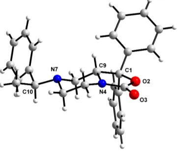
In order to define the absolute configuration of the C9 chiral centre crystals of the
diastereoisomer 21b were produced using ethanol/ethyl acetate as crystallization
solvents. The absolute configuration of the enantiomer (R)-SHA 68 has been assigned
by reference to the configurations of the two stereogenic centers present in the
diastereoisomer 21b.
As shows in figure 16, knowing the unchanged chiral centre C10 (in configuration S) of
the compound 21b we can determine the configuration of the chiral centre C9, that
shows a configuration R.
N
N
O
O
O
H
N
F
H
xH
aH
bFigure 16
Figure 17 represents another angulation of the X ray structure of the diestereoisomer (S,
R) 21b; the piperazine ring assumes a perfect chair conformation and we can confirm
that the proton bind at C9 is placed in axial position as demonstrate by NMR analysis.
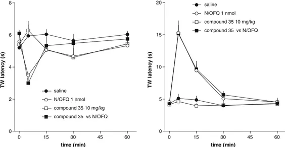
7. Biological activity
Next, in collaboration with the pharmacology researchers of the "Università degli Studi
di Ferrara” we planned a study able to furnish information about the biological activity
of this new class of compounds.
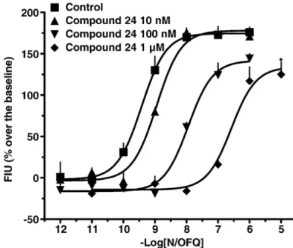
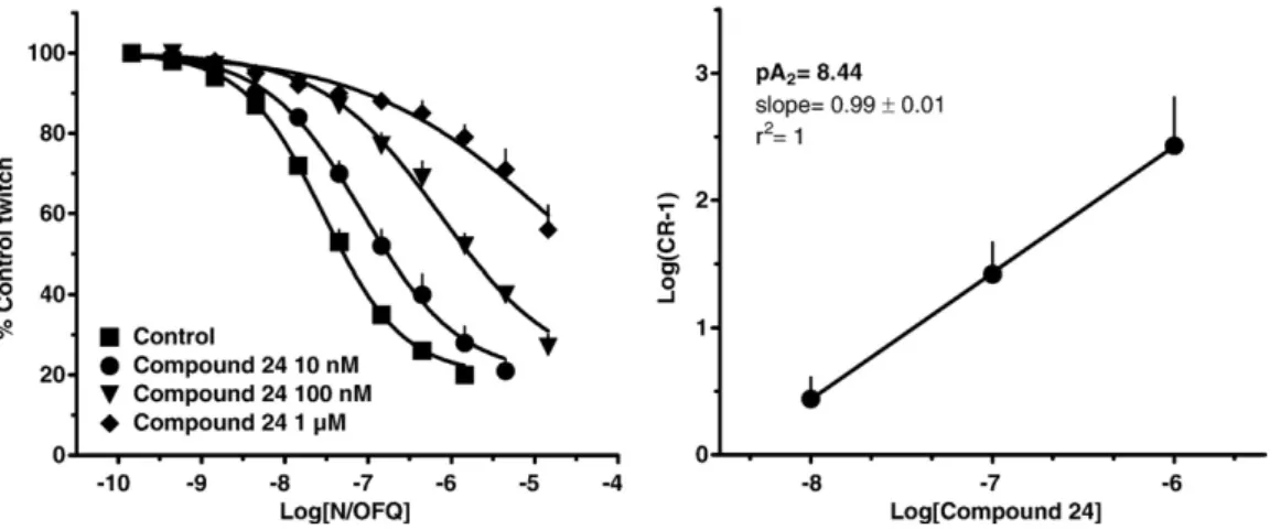
We evaluated and compared the in vitro NPSR antagonist properties of (R/S)-SHA 68,
(R)-SHA 68 and (S)-SHA 68. The three compounds were tested in calcium
mobilization studies performed on HEK293 cells expressing the murine NPSR or the
two isoforms of the human receptor (hNPSRAsn107 and hNPSRIle107).
13The natural peptide NPS was able to induce calcium mobilization in a concentration
dependent manner in HEK 293 mNPSR (pEC
508.97 ± 0.11; E
max250 ± 11%),
hNPSRAsn107 (pEC
50: 9.07 ± 0.11; E
max316 ± 13%) and hNPSRIle107 (pEC
50: 9.17 ±
0.15; E
max333 ± 17%).
(R/S)-SHA 68 inhibited in a concentration dependent manner the stimulatory effect of
NPS showing similar high values of potency (pK
B≅ 8). These values of potency are
superimposable to those previously published.
21,30(R)-SHA 68 was also able to antagonize in a concentration dependent manner the
stimulatory effect of NPS displaying values of potency similar or slightly higher than
the racemic mixture.
By contrast, (S)-SHA 68 showed a slight inhibitory effect only at micromolar
concentrations.
The values of potency of the three compounds, in the three cells lines, are summarized
in Table 1.
30C. Ruzza, A. Rizzi, C. Trapella, M. Pelà, V. Camarda, V. Ruggieri, M. Filaferro, C. Cifani, R. K. Reinscheid, G. Vitale, R.
mNPSR
hNPSR Ile107
hNPSR Asn107
pK
BpK
BpK
B(R/S)-SHA 68
8.16
8.03
7.99
(R)-SHA 68
8.29
8.18
8.28
(S)-SHA 68
<6
<6
<6
Table 1
Collectively, these results demonstrated that (R)-SHA 68 is the active enantiomer while
the contribution of (S)-SHA 68 to the biological activity of the racemic mixture is
negligible. As already mentioned in the introduction, the relevance of ligand chirality
for NPSR binding is also corroborated by the fact that the biological activity of
chemically different molecules, such as the quinoline
23and the tricyclic imidazole
24compounds, could be attributed to a single bioactive enantiomer.
8.
Conclusions
The present study described the enantioselective synthesis of 2,4-disubstituted and
2,4,6-trisubstitued chiral piperazines.
Initially, envisaged the synthesis of (R/S)-SHA 68 in racemic form in order to confirm
the chemical and biological published data through an efficient synthetic scheme which
can be easily scaled up to multi-grams.
Later, we focused our attention to the asymmetric synthesis of the two enantiomers
(R)-SHA 68 and (S)-(R)-SHA 68 starting from cheap commercially available reagents.
In order to define the conformation of the piperazine ring we performed a series of
NMR experiments leading to define a chair conformation where the substituent in C9
was placed in equatorial position.
To know the absolute configuration of this new chiral centre X ray analysis was
performed on suitable crystals obtained by the diastereoisomer 21b. The new
stereogenic centre showed (R) configuration
Furthermore, we studied these molecules from a pharmacological point of view;
evaluating and comparing the in vitro NPSR antagonist properties of (R/S)-SHA 68,
(R)-SHA 68 and (S)-SHA 68.
The results demonstrated that (R)-SHA 68 is the active enantiomer while the
contribution of (S)-SHA 68 to the biological activity of the racemic mixture is
negligible.
(R)-SHA 68 was demonstrated to be the antagonist of the receptor of the neuropeptide
S.
Nowadays, this molecule represents the standard non peptide NPSR antagonist and
surely will be used to investigate the biological functions controlled by the NPS / NPSR
system and to evaluate the therapeutic potential of innovative drugs acting as NPSR
selective ligands.
Chapter 2
I. Introduction
In the frame of the synthesis of biological active chiral compounds I have spend nine
months at the Pierre et Marie Curie University in Paris under the supervision of
Professor Giovanni Poli, focusing the attention on the synthesis of natural product
(-)-Steganacin. This stage allowed me to view a different approach for the selective
generation of new structures using a palladium catalysed domino reactions instead of
the use of chiral auxiliaries used for the synthesis of (R) and (S)-SHA 68.
The isolation and structure determination of a novel class of dibenzocyclooctadiene
lignan lactones, represented by the antileukemic esters steganacin 23 and steganangin
23a, was reported in 1973 by Kupchan and his collaborators.
31O
O
O
O
OAc
H
3CO
OCH
3H
3CO
23
O
O
O
O
H
3CO
OCH
3H
3CO
23a
COOH
Steganacin
Steganagin
Figure 18
Steganacin and steganangin were isolated from a plant of South Africa, Steganotaenia
araliacea Hochst (Figure 18), which was found to show significant antitumor activity in
vivo against P388 leukemia in mice.
31 M. Kupchan, R.W. Britton, M.F. Ziegler, C.J. Gilmore, R.J. Restivo, R.F. Bryan,Journal of the American Society, 1973, 95:4, 21, 1335-1336.
In vitro both compounds inhibit the assembly of tubulin into microtubules by interacting
with the colchicine binding site and have been shown to possess cytotoxic activity
against several cancer cell lines.
32Figura 19
Steganacin also causes a slow depolymerization of preformed microtubules.
This
molecule inhibits the binding of colchicine to tubulin and thus resembles
Podophyllotoxin, which also competitively inhibits colchicine binding (Figure 20).
33Steganacin presents a trimethoxybenzene ring and probably interacts with that portion
of the colchicine binding site that recognizes the trimethoxybenzene ring of Colchicine.
O O O O OAc O O O 23 Steganacin HN O O O O O H3CO Colchicine O O O O H H O O O OH Podophyllotoxin 23b 23c
Figure 20
32a) L. Wilson, Biochemistry, 1970, 9, 4999; b) R. W. J. Wang, L. I. Rebbun, and M. S. Kupchan, Cancer Res., 1977, 37, 3071. 33For the synthesis of an aza-analogue of podophyllotoxin from our group see: Poli, G.; Giambastiani, G. J. Org. Chem. 2002, 67, 9456-9459.