Review Article
Role of Acupuncture in the Management of Severe
Acquired Brain Injuries (sABIs)
Loredana Cavalli
,
1Lucia Briscese,
1Tiziana Cavalli,
2Paolo Andre,
3and Maria Chiara Carboncini
11Severe Acquired Brain Injuries Unit, Cisanello University Hospital, Pisa, Italy 2Dipartimento Chirurgico Ortopedico, Ospedale Carlo Poma di Mantova, Italy
3Dipartimento di Scienze Mediche, Chirurgiche e Neuroscienze, University of Siena, Italy
Correspondence should be addressed to Loredana Cavalli; [email protected]
Received 15 February 2018; Revised 25 July 2018; Accepted 15 August 2018; Published 12 September 2018 Academic Editor: Morry Silberstein
Copyright © 2018 Loredana Cavalli et al. This is an open access article distributed under the Creative Commons Attribution License, which permits unrestricted use, distribution, and reproduction in any medium, provided the original work is properly cited. Acupuncture therapy has been used to treat several disorders in Asian countries and its use is increasing in Western countries as well. Current literature assessed the safety and efficacy of acupuncture in the acute management and rehabilitation of patients with neurologic disorders. In this paper, the role of acupuncture in the treatment of acute severe acquired brain injuries is described, acting on neuroinflammation, intracranial oedema, oxidative stress, and neuronal regeneration. Moreover, beneficial effects of acupuncture on subacute phase and chronic outcomes have been reported in controlling the imbalance of IGF-1 hormone and in decreasing spasticity, pain, and the incidence of neurovegetative crisis. Moreover, acupuncture may have a positive action on the arousal recovery. Further work is needed to understand the effects of specific acupoints on the brain. Allegedly concurrent neurophysiological measurements (e.g., EEG) may help in studying acupuncture-related changes in central nervous system activity and determining its potential as an add-on rehabilitative treatment for patients with consciousness disorders.
1. Introduction
Severe acquired brain injuries (sABIs) include a variety of acute brain lesions characterized by the occurrence of variably prolonged coma (24 hours), and simultaneous motor, sensory, cognitive, and/or behavioural impairment that causes a certain degree of disability. Congenital, perinatal onset, or degenerative-progressive brain injuries are excluded from this definition.
A common consequence of sABIs is disorders of consciousness (DOCs), a prolonged cognitive impairment including the loss of awareness of oneself and environment. DOCs represent one of the greatest challenges that modern medicine faces today, with a huge burden of care for families and facilities. On the basis of the current taxonomy of DOCs, a state of altered consciousness can be categorized into coma, vegetative state (VS), also referred to as unresponsive wakefulness syndrome (UWS), and minimally conscious state (MCS) [1].
The most common cause of sABI is traumatic brain injury (TBI), a major source of death and disability worldwide. Two further causes of sABI are anoxic encephalopathy (AE), usually due to cardiocirculatory arrest (secondary to exten-sive myocardial injury and/or malignant arrhythmias), and ischemic or haemorrhagic stroke. These conditions mostly affect subjects from the fifth decade onwards and represent about 40% of the sABIs. The main involvement of neurons leads to a worse prognosis than TBI. In AE, the neurons disruptions, with a low regenerative potential, cause a high risk of irreversibility of the consciousness disorder. Moreover, the time interval within which a recover is reasonable, up to 12 months from the event for TBI, is unlikely more than 3-6 months from the event for anoxic or vascular brain injuries. Late recoveries are possible, but rare. Further nontraumatic sABI arises from brain tumors, infections, and toxic-metabolic encephalopathy [2].
Despite the fact that steady progress has been made toward prolonging patients survival and several Volume 2018, Article ID 8107508, 10 pages
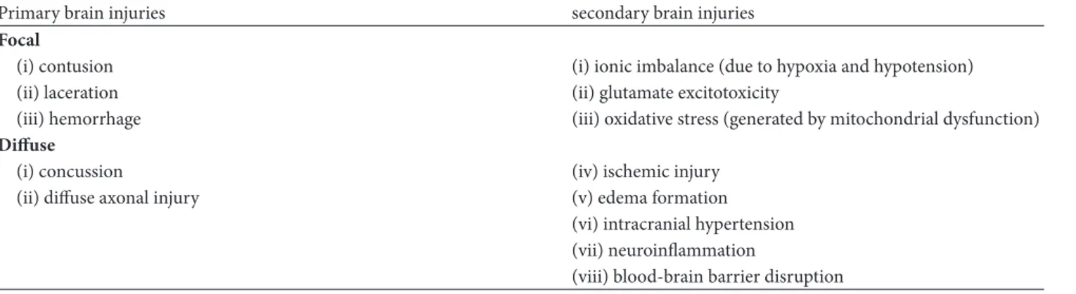
Table 1: Physiopathology of DOCs.
Primary brain injuries secondary brain injuries
Focal
(i) contusion (i) ionic imbalance (due to hypoxia and hypotension)
(ii) laceration (ii) glutamate excitotoxicity
(iii) hemorrhage (iii) oxidative stress (generated by mitochondrial dysfunction)
Diffuse
(i) concussion (iv) ischemic injury
(ii) diffuse axonal injury (v) edema formation
(vi) intracranial hypertension (vii) neuroinflammation
(viii) blood-brain barrier disruption
pharmacologic and neuromodulating strategies have been proposed, results on functional recovery of DOCs are still scarce.
2. Physiopathology of DOCs
The immediate effect of the impact forces on intracranial tissues, i.e., the primary brain injury, can be focal or diffuse. Focal injuries, such as contusion, laceration, and haemorrhage, are early detectable upon imaging and their consequences depend on their location and severity. Diffuse brain injuries, like concussion or diffuse axonal injury, require magnetic resonance to be detected [3]. However, what is susceptible of treatment is secondary brain injury (SBI), the cascade of biochemical and cellular events devel-oping minutes to months after the insult: ionic imbalance (due to hypoxia and hypotension), glutamate excitotoxicity, oxidative stress generated by mitochondrial dysfunction, ischemic injury, edema formation, intracranial hypertension, neuroinflammation (by both systemic and central neuron system immunoactivation), and blood-brain barrier (BEE) disruption [3, 4], as schematized in Table 1.
Several secondary processes are based on intracellular calciumoverload, due to excitatory amino acids and inflam-matory cytokines released. In absence of oxygen, glucose enters the glycolytic pathway, where it is converted to NADH and pyruvate; the latter becomes lactic acid just producing 2 ATP molecules, while Krebs cycle is precluded. Thus, a down-regulation of ATP-dependent Na+/K+-ATPase pump leads to sodium overload, which acting on Na+/Ca2+exchange pump provides calcium retention.
Analogously, the huge release of glutamate following a head trauma, in hypoxic conditions, is not counterbal-anced by astrocyte reuptake of glutamate, which acts on N-methyl-d-aspartate (NMDA) receptor, thus increasing calcium influx. Calcium is therefore sequestered by mito-chondria causing their dysfunction, lysis, and release of byproducts [3]. Moreover, calcium stimulates neuronal and endothelial cells overproduction of nitric oxide (NO), which can be associated with oxidative stress. A further effect of calcium overload is mitochondrial-mediated apoptosis following the release of cytochrome c.
In response to primary brain injury, an inflammatory response is provided by microglia and astrocytes, which
attract leukocytes via cytokines, chemokines, and NO. The disruption of the BEE often leads to cerebral oedema and intracranial hypertension [3].
3. Reactions to Brain Injury
A series of mechanisms are activated at neuronal (neuroge-nesis, synaptoge(neuroge-nesis, and dendritic remodelling) glial, and vascular level, referred to asneuroplasticity [5]. These events are promoted by increased expression of growth factors involved in brain development, such as nerve growth factor (NGF), neurotrophin 4/5, basic fibroblast growth factor, and brain derived neurotrophic factor (BDNF); an increasing importance has also emerged for insulin-like growth factor 1 (IGF-1), regulating metabolic function, oligodendrocyte proliferation, and survival, angiogenesis, myelinisation, and neurite outgrowth [5] and receptors for IGF-1, virtually ubiquitary, are mainly expressed by mesenchymal origin-derived cells in the hippocampus, parahippocampus, amyg-dala, cerebellum, and cortex [6].
4. Available Strategies to Manage sABIs
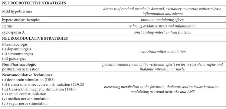
New insights into the pathophysiology of sABIs initiated new therapeutical approaches (neuroprotective strategies) aimed at interrupting secondary brain injury development and promoting mechanisms of arousal (see Table 2). Among the neuroprotective strategies, mild hypothermia (which decreases cerebral metabolic demand, excitatory neurotrans-mitter release, inflammation, and oedema), hyperosmolar therapies (also known for their immune-modulating prop-erty), statin (reducing oxidative stress and inflammation), and cyclosporin A (ameliorating mitochondrial function) have been proposed [3]. For arousal recovery, dopaminergic, serotoninergic and GABAergic drugs have been explored, with occasional results. Several nonpharmacological strate-gies have been utilized. Early verticalization has been shown to increase the spectral power of the EEG higher frequen-cies and the subject’s arousal [7, 8], probably enhancing vestibular afference on locus coeruleus, raphe, and thalamic intralaminar nuclei. Enriched environment, equipped with emotional stimuli and biographically meaningful objects, showed greater range of behavioural responses [9]. Finally neuromodulatory techniques, including deep brain stimula-tion (DBS), transcranial direct current stimulastimula-tion (TDCS),
Table 2: Available strategies to manage sABIs and their mechanisms of action. NEUROPROTECTIVE STRATEGIES
Mild hypothermia decrease of cerebral metabolic demand, excitatory neurotransmitter release,inflammation and edema
hyperosmolar therapies immune-modulating effects
statins reducing oxidative stress and inflammation
cyclosporin A ameliorating mitochondrial function
NEUROMODULATIVE STRATEGIES Pharmacologic (i) dopaminergics (ii) serotoninergics (iii) gabaergics neurotransmitter modulation Non Pharmacologic postural verticalization
potential enhancement of the vestibular effects on locus coeruleus, raphe and thalamic intralaminar nuclei
Neuromodulative Techniques: (i) deep brain stimulation (DBS)
(ii) transcranial direct current stimulation (TDCS) (iii) transcranial magnetic stimulation (TMS) (iv) spinal cord stimulation
(v) median nerve stimulation (vi) vagus nerve stimulation
increasing metabolism in the forebrain, thalamus and reticular formation. modulating neuronal networks and ANS
transcranial magnetic stimulation (TMS), spinal cord stimu-lation, median nerve stimustimu-lation, or vagus nerve stimustimu-lation, have been proposed.
5. A Complementary Approach for
the sABIs’ Management Derivating from
Ancient Chinese Medicine:
Acupuncture
The aim of this review is to explore the potential role of acupuncture (1) in the acute treatment of sABIs, in containing secondary brain injury by acting on different pathophysio-logic mechanisms, and (2) in the subacute/chronic manage-ment of sABI outcomes, through a limitation of spasticity, pain, dysautonomia, and a possible action on the arousal recovery.
Acupuncture is a traditional medicine traced back to over 3000 years ago in China [10], consisting in inserting needles into specific points of the patient’s body (“acupoints”) chosen on the basis of the Meridian theory of Traditional Chinese Medicine (TCM) [11]. It has been used in the treatment of stroke and its consequences for over 2,000 years in China.
Under the influence of neuroanatomy, neurophysiology, and bioholographic principle of modern medicine, in the early 1970s, scalp acupuncture, one of the several specialized acupuncture techniques in which a filiform needle is used to penetrate specific stimulation areas on the scalp mainly for the treatment of brain diseases, was set up and separated from traditional acupuncture system [12].
Scalp acupuncture has been reported to (1) improve cerebral blood circulation, promoting regional energy metabolism; (2) upregulate expression of glial cell-line derived neurotrophic factor (GDNF), possibly promoting proliferation and differentiation of neural stem cells in the focal cerebral cortex and hippocampus; (3) reduce contents
of excitatory amino acid and increase level of GABA, thus lowering neurogenic toxicity; (4) ease cerebral vascular immunoinflammatory reactions; (5) regulate blood lipid metabolism to resist cerebral free radical damage; and (6) inhibit cerebral cortical apoptosis [13].
5.1. Acupuncture and Inflammation in DOCs. Few studies
considered the diachronic assessment of inflammatory mark-ers after a sABI in order to monitor the local and systemic stress response in DOCs. Amico and colleagues evaluated the subacute phase of DOCs in five patients in vegetative state (VS) and one in minimal consciousness state (MCS), by clinical assessment and biochemical analyses [14]. A positive correlation was found between the serum levels of osteopon-tin(OPN), a cytokine involved both in neuroinflammation and neurorepair, and prognosis, with the lowest level detected in the patient who then emerged from MCS, the highest in the one who then died. Moreover, the lymphocyte subset presented a general increase of CD4+/CD3+ ratio, with a suspect unbalance of CD4+ toward Th2; prolactin resulted to be the best endocrinological marker of sABI [14].
Applied at Baihui (GV20) and left Zusanli (ST36), acupuncture significantly reduced the infiltration of inflam-matory cells and the expression of the proinflaminflam-matory enzyme MMP2 in cerebral ischemia/reperfusion injury (CIRI) model rats. In particular they attenuated the expres-sion of the water channel proteins P4 and AQP9 in the ischemic brain, leading to the mitigation of inflammation-related brain edema. Consistent with the smaller observed infarct size, acupuncture and EA both promoted significant improvements in the Modified Neurological Severity Score (mNSS) in CIRI model rats, indicative of enhanced neuro-logical function [15].
Moreover, acupuncture successfully downregulates tumor necrosis factor alpha (TNF-𝛼), which results in
anti-inflammatory responses. The neural pathways by which acupuncture signalling stimulates anti-inflammatory effects have been mapped. By testing the effects that a splenic neurectomy and vagotomy have on TNF-𝛼 levels in the spleen and the brain, Lim et al. found that the anti-inflammatory effects of manual acupuncture at ST36 rely on the vagus nerve pathway. Moreover, both manual acupuncture stimulation (MAC) and electroacupuncture (EA) induce c-Fos protein generation [16].
5.2. Acupuncture and Redox Equilibrium. Oxidative stress,
the imbalance between the production of reactive oxygen and nitrogen species (ROS/RNS) and the endogenous antioxidant system, causing a cascade of chain reactions resulting in cellular damage, is a critical feature in the pathological process of various diseases [17].
Recently, a large body of evidence demonstrated that acupuncture has antioxidative effect in various conditions [18–20], although the exact mechanism, especially the influence of acupuncture on signalling pathways, remains unclear. Through redox system, antioxidant system, anti-inflammatory system, and nervous system, acupuncture could make the oxidative damage and the antioxidant defence remain in a relatively constant redox state. However, the recent acupuncture researches about oxidative stress are sporadic and preliminary [21].
5.3. Acupuncture and Intracellular Calcium. The insertion
of the needle represents an effective mechanical stimulus, leading to tissue displacement and to intracellular calcium increase and signalling. The modulation of calcium channels seems to be the primary mechanism for endorphin secretion and release from immunocytes and for the inhibitory effects of opioids on peripheral neurons [22]. Thus, calcium ion, whose increase is so crucial in the pathogenesis of SBI, may be taken as the carrier of the biological modulation system provided by acupuncture, where the mechanical wave onsets an acoustic shear wave, and this drives to calcium signalling [22].
5.4. Acupuncture and Neuron Regeneration. In the brain of
human adults, neural stem cells (NSCs) have been demon-strated in the pallium, subependymal region, hippocam-pus, and corpus striatum, which have the ability of self-duplication, self-regeneration, and differentiation into neu-rons and glial cells. During cerebral ischemia reperfusion, astrocytes play a crucial role in limiting neuronal lesion, as they release epoxyeicosanoic acids in order to enlarge brain vessels, release Nerve Growth Factors to make neurons sur-vive and axons grow, produce neurotransmitters, metabolize toxic molecules, and have also the potentiality to become NSCs [23, 24].
The astrocyte activation and proliferation marker is Glial Fibrillary Acidic Protein (GFAP), while Neuron Specific Enolase (NSE) is one of the neurons’ markers. Acupuncture on the conception (CV) and governor vessels (GV) has been shown to inhibit excessive proliferation of astrocytes and promote NSCs differentiation in the ischemic brain [25, 26]. In particular, needling acupoints GV20 and GV26 could
downregulate the number of GFAP+ cells, while increasing the GFAP/NSE double–labelled cells in the hippocampal dentate gyrus [25, 26]. Another study, which employed a rat TBI model, proved that during the early post-TBI stage, acupuncture (GV20, GV26, GV15, GV16, and LI4) can promote the proliferation and differentiation of NSCs and glial cells, which is crucial to control neuronal necrosis; in the late phase, it can inhibit glial proliferation and differentiation, driving to neuron and oligodendrocytes regeneration and tissue repair [27].
Moreover, needling CV24, CV4, and CV3 has been shown to upregulate the expression of basic FGF, EGF, and NGF after cerebral ischemia reperfusion, activating nerve repair and proliferation of neuronal precursors [28, 29].
As regards human studies, recent evaluations used single-photon emission computed tomography (SPECT) and T2-weighted imaging (T2WI) [30], or functional magnetic res-onance imaging (fMRI) [31]. Shen and colleagues com-pared serial diffusion tensor imaging (DTI), fluid-attenuated inversion recovery (FLAIR), and T2WI performed in 20 patients with recent cerebral infarction in the basal ganglia, randomly divided into an acupuncture group and a control group [32]. The apparent diffusion coefficient (ADC) of infarction lesions, decreased at stroke onset, was showed significantly elevated after the acute stage, while the ADC of the bilateral cerebral peduncle was reduced on the infarction side. Fractional anisotropy (FA) values of abnormal signals on DTI in the infarction areas and cerebral peduncles underwent a significant reduction from stroke onset to the chronic stage. Interestingly, a significant difference in ADC and FA values between the two groups was observed, with a higher FA value in the acupuncture group than the control group, thus suggesting the effectiveness of acupuncture for protecting neurons by postponing Wallerian degeneration of brain infarction, and facilitating recovery [32].
5.5. Acupuncture and Arousal. The pathology of disorders of
consciousnesscan be represented by (A) damage of Reticular Ascending System (B) large-scale damage to cerebral cortex, (C) injury to links (e.g., thalamus) between cerebral cortex and brain stem, and (D) injury to connections (e.g., corpus callosum) within the cerebral cortex, i.e., severe diffuse axonal injury (DAI).
The production of inhibitors (including GABA) induced by brain injury generates a response resembling automatic shutdown, probably aimed at conserving energy and promot-ing cell survival, but causpromot-ing a comatose state [33]. Therefore, any treatment affecting the reticular activating system may be worth trying, and, among the possible treatments, acupunc-ture has the highest potential [34].
Recent studies on resting state (RS) in DOC, by using functional magnetic resonance imaging (fMRI), showed that functional connectivity is severely impaired above all in the Default Mode Network (DMN). In the vegetative and minimally conscious state, DMN integrity seems to correlate with the level of remaining consciousness.
The DMN is among the most robust networks found with resting state fMRI and encompasses the posterior gulate cortex (PCC)/precuneus, mediofrontal/anterior cin-gulate cortex, and temporoparietal cortex [35]. Activity in the
DMN diminishes when the brain is involved in attention-demanding cognitive tasks [36] and returns to its prominent presence when no such task is being performed.
The DMN seems to be of particular interest, as its connec-tivity has been shown to decrease during loss of conscious-ness, and PET studies have shown an increase in neuronal activity in DMN regions (especially in the PCC/precuneus) upon recovery from VS [37]. Indeed, Vanhaudenhuyse [38] and Fern´andez-Espejo [39] et al. observed a correlation between the DMN integrity and the level of consciousness in noncommunicative brain-damaged patients.
Imaging evidence has been provided to support that electroacupuncture at GV20, employed to treat major depres-sive disorders, may modulate the Default Mode Network (DMN), the cerebral functional network encompassing the posterior and anterior cortical midline structures, which is considered to be involved in stimulus-independent thought, mind-wandering, and self-consciousness. EA at GV20 would increase functional connectivity (FC) between the pre-cuneus/posterior cingulate cortex (PC/PCC) and bilateral anterior cingulate cortex (ACC) and reduce FC between the PC/PCC and left middle prefrontal cortex, left angular gyrus, and bilateral hippocampus/parahippocampus [40]. These findings are of particular importance when considering DOCs, where resting state network activity reveals reduced interhemispheric connectivity and correlates with levels of consciousness.
The acupoint Shuigou (GV26), placed at the junction of the upper one-third and lower two-thirds of the philtrum midline, also has been described as promoting the function of GV meridian, closely related to brain function, decreasing cognitive impairment, and promoting neurogenesis in the APP/PS1 transgenic mice [41].
Interestingly, enhanced bodily attention can be triggered by genuine acupuncture at PC6 and HT7 acupoints, which were exhibited to activate the salience network (insula, ACC, secondary somatosensory cortex, and superior parietal cortex) and deactivate the DMN (medial prefrontal cortex, PCC, inferior parietal cortex, and parahippocampus) [42].
Combined with western medicine, electroacupuncture therapy at Baihui (GV20), Shuigou (GV26), and Yongquan (KI1), resulted effective in improving consciousness recovery of patients in coma due to TBI, both reducing awake time and increasing awake rate, compared to a control group receiving only western treatments [43].
5.5.1. Autonomic Dysfunction in sABI. Autonomic nervous
system (ANS) deregulation and/or dysautonomia is another severe consequence of brain injury, not well cleared. Dysau-tonomia affects in particular ninety percent of TBI patients during the first week, leading to sleep and heart rhythm disorders, and increasing specific biomarkers of neural dam-age [6]. Clinically, patients suffering from DOC can show the so called “paroxysmal sympathetic hyperactivity” (PSH), episodic sudden increase in vital signs, particularly heart rate, blood pressure, respiratory rate, and temperature, with possible diaphoresis (i.e., excessive sweating) and abnormal, unintentional movements, spontaneously or in response to external painful stimuli [44, 45]. A growing body of evidence
suggests that ANS may mediate large-scale brain activation, in an extreme attempt to preserve body system homeostasis and regain consciousness [46]. The primary and secondary brain lesions have the potential to compromise both cortical and subcortical control mechanisms of the ANS.
Most often, TBI leads to sustained sympathetic activation, contributing to the high morbidity, with oxidative stress in the ANS and activated hypothalamic-pituitary-adrenal axis and hypothalamic-sympathoadrenal medullary axis [4].
5.6. Acupuncture and ANS Modulation. An increasing
clin-ical evidence demonstrates that acupuncture is helpful in treating ANS dysfunctions, such as nausea and vomiting [47]. For example, acupoints stimulation has been shown to change the sympathovagal balance toward vagal predominance [48, 49].
Abnormalities in the ANS, such as sympathetic overacti-vation and/or parasympathetic hypoactioveracti-vation, may generate and sustain chronic pain [50, 51].
Acupuncture at certain points could reduce sympathetic nervous system activity associated with pain [52] or during mental stress in patients with heart failure [53]. However, the neurobiological basis of these effects is not yet clear [54]. In order to explore the regulative effect on ANS by acupuncture, Sakatani and colleagues monitored heart rateby placing photoelectrical sensor on the first finger of eighteen healthy male adults, and thus low frequency (LF) amplitude (0.04–0.15 Hz) and high frequency (HF) amplitude (0.15–0.4 Hz) were calculated by power spectral analysis [54]. Real acupuncture performed at point Large Intestine 4 (LI4) of the right hand (r-LI4) was shown to determine significant decreases of HR and LF/HF and a significant increase of HF, indicating a parasympathetic activation as well as sympathetic depression [55].
Moreover, vagus nerve stimulation (VNS) increases metabolism in the forebrain, thalamus, and reticular for-mation [56]. It also enhances neuronal firing in the locus coeruleus, which leads to massive release of norepinephrine in the thalamus and hippocampus, a noradrenergic path-way essential for arousal, alertness, and the fight-or-flight response [57]. Recently, based on this rationale, Transcuta-neous Auricular VNS (taVNS), a noninvasive stimulation developed for treating epilepsy and depression without the surgery-related risks [58, 59], was firstly employed by Yu-tian Yu and colleagues on a patient in vegetative state [60]. A further case with implanted VNS recovered behavioural responsiveness and enhanced brain connectivity patterns [61].
5.7. Acupuncture against Neuroendocrine Dysfunction. An
imbalance of the pituitary and hypothalamus hormones and their axes is often associated with sABI, due to compression, edema, skull base fracture, haemorrhage, intracranial hyper-tension, or hypoxia. In the acute phase of sABI, low IGF-1 with elevated GH levels have been detected, with increasing IGF-1 and normalizing GH concentrations in the following weeks. The IGF-1 upregulation and the disruption of BEE that persists until 7 days after injury, allowing a wide level of hormone to reach the surviving neurons, probably play a
role in promoting neurite overgrowth, inducing progenitor cell differentiation and inhibiting neuron apoptosis [6].
In rat model of renal failure- (RF-) induced hypertension, stimulation with acupuncture, and most significantly with EA, at ST36 and KI3, not only attenuated glomerulosclerosis and tubulointerstitial fibrosis, but corrected the decreases in RF-induced IGF-1 mRNA and protein levels, thus counteract-ing oxidative stress [62]. These findcounteract-ing may suggest the ability of acupuncture in restoring IGF-1 function in any situations where its levels are reduced, including sABI, although no studies have been conducted on this purpose.
5.7.1. Pain and DOC. The experience of pain in disorders of
consciousness is still debated. Neuroimaging studies, using functional magnetic resonance imaging (fMRI) [63, 64], Positron Emission Tomography (PET) [65], multichannel electroencephalography (EEG), and laser-evoked potentials [66], suggest that the perception of pain increases with the level of consciousness.
VS and MCS are by definition incompatible with a reliable and consistent ability to communicate about pain experi-ences, while the nature of these conditions is characterized by various factors that can give rise to pain (e.g., spasticity, contractures, etc.) [67].
5.8. Acupuncture and Pain Relief. Many different
mech-anisms may explain the analgesic effect of acupuncture. Among these is thegate control theory of pain proposed by Melzack and Wall in 1965 [68]. Specific nerve fibers would transmit pain to the spinal cord, while other nerve fibers inhibit pain transmission. Both groups of fibers met at the substantia gelatinosa in the spinal cord, where pain and pain inhibitory stimuli were integrated. Pain would be perceived only if the noxious input exceeded the inhibition of pain. However, gate control theory cannot explain the full spectrum of acupuncture effects, and in particular the prolonged pain relief.
Since the 1970s, the secretion of a range of biochem-icals or neurotransmitters has been considered among the mechanism of acupuncture analgesia, such as adeno-sine, opioid peptides, cholecystokinin octapeptide, 5-hydroxytryptamine, noradrenalin, glutamate, GABA, sub-stance P, calcium ions, angiotensin II, somatostatin, argi-nine vasopressin, and dopamine[69–81].
Different subtypes of opioid receptor were also believed to mediate the frequency-dependent electroacupuncture anal-gesia [82, 83]. For example, EA at a low frequency of 2 Hz would facilitate the release of enkephalin, but not dynor-phin, while a high frequency of 100 Hz would stimulate the dynorphin but not enkephalin release in rats [82], as well as in humans [83]. The primary foundations for acupuncture effects seem to be bioelectromagnetic, while biochemical factors would be secondary. By the way, the opioid peptide secretion was recently proposed as being due to mechan-ical acoustic shear wave activation and calcium signalling induced by needle rotation [22].
Recently, the inflammatory reflex (via the ANS) has been observed as potentially crucial for the antihyperalgesic effect of acupuncture: by regulating the immune system
it can elucidate not only the analgesic, but also the anti-inflammatory mechanism of acupuncture [76].
5.9. Acupuncture and Control of Spasticity. Spasticity is a
frequent consequence of sABIs, arising from an anarchic reorganization of the pyramidal and parapyramidal fibers, and leading to hypertonia and hyperreflexia of the affected muscular groups and, if untreated, to possible irreversible joint lesions. Current treatment options include intrathecal baclofen and soft splints, botulinum toxin, or cortical acti-vation by thalamic stimulation [84]. There is a low quality evidence for rehabilitation programs, extracorporeal shock-wave therapy, transcranial direct current stimulation, tran-scranial magnetic stimulation, and transcutaneous electrical nerve stimulation targeting spasticity, while a moderate evi-dence has been shown for electroneuromuscular stimulation and acupuncture as an adjunct therapy to conventional routine care (pharmacological and rehabilitation) [85]. In patients with DOCs, acupuncture at GV26, Ex-HN3, LI4, and ST36was proven to reduce spastic muscle hypertonia by decreasing the excitability of the spinal motor neurons, both ten minutes after needles insertion and ten minutes after their removal [86].
6. Final Considerations and Conclusions
The World Health Organization has recommended acupunc-ture in 1980 as an effective complementary therapy for several diseases. Among the indications, neurologic disorders have been shown to benefit from acupuncture.
In this review, we analysed scientific studies and clinical reports that explored the acupuncture’s effects in several acquired brain injuries, aiming to
(1) limit brain secondary injury, by acting on systemic and local inflammation, oxidative stress, intracellular calcium overload, neuron regeneration, and growth factors release;
(2) manage sABI consequences, such as neuroendocrine and autonomic dysfunction, muscle spasticity, and pain.
Research in this field has obtained significant improve-ment with the technical support of the life sciences, and the studies of acupuncture have in turn accelerated the devel-opment of biomedical science. However, intrinsic aspects of this medical approach make it difficult to run a clinical study, and several data derive from animal studies or from small-size and heterogeneous samples of patients. Moreover, the acupoints selected for treating sABI can differ between research groups.
In addition, patients with disorders of consciousness are
per se difficult to study. Given the impossibility of
communi-cating with the patient, the content of consciousness can be only inferred by response behaviour. Diagnostic errors may depend not only on the operator but also on wakefulness fluctuations of the patient, who may be drowsy or agitated, or have epileptic seizures or aphasia. The neurophysiologic evaluations are made difficult by the presence of sweat of the head (which worsens the EEG impedance), muscle hypertonus (that obstructs mobilization), and noise from electromedical equipment, while functional imaging tech-niques are expansive and not easily available.
Table 3: Acupuncture and neuroplasticity.
MAIN MECHANISMS BY WHICH ACUPUNCTURE MAY INFLUENCE THE PHYSIOLOGIC PLASTIC REACTIONS TO BRAIN INJURY
(i) Inhibition of brain neuronal apoptosis (vii) Reduction of blood-brain barrier permeability in intracerebral haemorrhage (caveolin-1/matrix metalloproteinase)
(ii) Inhibition of aberrant astrocyte activation (viii) Regulation of blood lipid metabolism to counteract cerebral free radical damage
(iii) Upregulation of neurotrophins expression (ix) Promoting cerebral vascular immunoinflammatory reactions (iv) Upregulate expression of GDNF (x) Increase of GABA level
(v) Increased functional connectivity (xi) Reduce contents of excitatory amino acids (vi) Enhanced neuroblast proliferation and differentiation
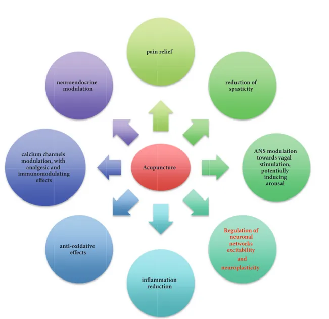
Acupuncture pain relief reduction of spasticity ANS modulation towards vagal stimulation, potentially inducing arousal Regulation of neuronal networks excitability and neuroplasticity inflammation reduction anti-oxidative effects calcium channels modulation, with analgesic and immunomodulating effects neuroendocrine modulation
Figure 1: Role of acupuncture in the management of sABIs.
Further studies are needed to identify the most efficient and customized therapeutical protocol, aiming in particular at eliciting arousal.
The available data suggest that, in patients with sABIs/DOCs, acupuncture may represent an interesting
frontier in the years ahead, as it seems to limit the secondary brain injury development, modulate ANS, and ameliorate their quality of life (Table 3, Figure 1). The absence of side effects or drug interactions make it particularly indicated for such fragile subjects.
Conflicts of Interest
The authors declare that they have no conflicts of interest.
References
[1] J. T. Giacino, S. Ashwal, N. Childs et al., “The minimally conscious state: definition and diagnostic criteria,” Neurology, vol. 58, no. 3, pp. 349–353, 2002.
[2] A. De Tanti, M. Zampolini, S. Pregno, and CC3 Group, “Recommendations for clinical practice and research in severe brain injury in intensive rehabilitation: the Italian Consensus Conference,” European Journal of Physical and Rehabilitation
Medicine, vol. 51, no. 1, pp. 89–103, 2015.
[3] L. V. Tran, “Understanding the pathophysiology of traumatic brain injury and the mechanisms of action of neuroprotective interventions,” Journal of Trauma Nursing, vol. 21, no. 1, pp. 30– 35, 2014.
[4] H. Toklu and N. T¨umer, “Oxidative stress, brain edema, blood–brain barrier permeability, and autonomic dysfunction from traumatic brain injury,” in Brain Neurotrauma, Frontiers in Neuroengineering Series, pp. 43–48, CRC Press, Boca Raton, FL, USA, 2015.
[5] S. Madathil and K. Saatman, “IGF-1/IGF-R signaling in trau-matic brain injury: impact on cell survival, neurogenesis, and behavioral outcome,” in Brain Neurotrauma, Frontiers in Neuroengineering Series, pp. 61–78, CRC Press, Boca Raton, FL, USA, 2015.
[6] A. Mangiola, V. Vigo, C. Anile, P. De Bonis, G. Marziali, and G. Lofrese, “Role and importance of IGF-1 in traumatic brain injuries,” BioMed Research International, vol. 2015, Article ID 736104, 12 pages, 2015.
[7] L. Elliott, M. Coleman, A. Shiel et al., “Effect of posture on levels of arousal and awareness in vegetative and minimally conscious state patients: a preliminary investigation,” Journal of Neurology,
Neurosurgery & Psychiatry, vol. 76, no. 2, pp. 298-299, 2005.
[8] A. Greco, M. C. Carboncini, A. Virgillito, A. Lanata, G. Valenza, and E. P. Scilingo, “Quantitative EEG analysis in minimally conscious state patients during postural changes,” in Proceedings
of the 35th Annual International Conference of the IEEE Engi-neering in Medicine and Biology Society (EMBC ’13), pp. 6313–
6316, July 2013.
[9] C. Di Stefano, A. Cortesi, S. Masotti, L. Simoncini, and R. Piperno, “Increased behavioural responsiveness with complex stimulation in VS and MCS: Preliminary results,” Brain Injury, vol. 26, no. 10, pp. 1250–1256, 2012.
[10] J.-S. Han and Y.-S. Ho, “Global trends and performances of acupuncture research,” Neuroscience & Biobehavioral Reviews, vol. 35, no. 3, pp. 680–687, 2011.
[11] B. R. Xu, Clinical Acupuncture, Liaoning Science Publishing House Chine, 1986.
[12] Z. Liu, L. Guan, Y. Wang, C.-L. Xie, X.-M. Lin, and G.-Q. Zheng, “History and mechanism for treatment of intracerebral hemor-rhage with scalp acupuncture,” Evidence-Based Complementary
and Alternative Medicine, vol. 2012, Article ID 895032, 9 pages,
2012.
[13] L. Tian, X. Du, J. Wang et al., “Scalp acupuncture twisting manipulation for treatment of hemiplegia after acute ischemic stroke in patients: study protocol for a randomized, parallel, controlled, single-blind trial,” Asia Pacific Clinical and
Transla-tional Nervous System Diseases, vol. 41, no. 1, pp. 87–93, 2016.
[14] A. P. Amico, T. Annamaria, M. Marisa, M. Gianfranco, and D. Sabino, “Immune endocrinological evaluation in patients with severe vascular acquired brain injuries: Therapeutical approaches,” Endocrine, Metabolic & Immune Disorders—Drug
Targets, vol. 13, no. 2, pp. 204–208, 2013.
[15] H. Xu, Y. Zhang, H. Sun, S. Chen, and F. Wang, “Effects of acupuncture at gv20 and st36 on the expression of matrix metalloproteinase 2, aquaporin 4, and aquaporin 9 in rats subjected to cerebral ischemia/reperfusion injury,” PLoS ONE, vol. 9, no. 5, 2014.
[16] H.-D. Lim, M.-H. Kim, C.-Y. Lee, and U. Namgung, “Anti-inflammatory effects of acupuncture stimulation via the vagus nerve,” PLoS ONE, vol. 11, no. 3, 2016.
[17] S. R. Thomas, P. K. Witting, and G. R. Drummond, “Redox control of endothelial function and dysfunction: molecular mechanisms and therapeutic opportunities,” Antioxidants &
Redox Signaling, vol. 10, no. 10, pp. 1713–1765, 2008.
[18] T. Wang, C. Z. Liu, J. C. Yu, W. Jiang, and J. X. Han, “Acupunc-ture protected cerebral multi-infarction rats from memory impairment by regulating the expression of apoptosis related genes Bcl-2 and Bax in hippocampus,” Physiology & Behavior, vol. 96, no. 1, pp. 155–161, 2009.
[19] G. Shi, C. Liu, Q. Li, H. Zhu, and L. Wang, “Influence of acupuncture on cognitive function and markers of oxidative DNA damage in patients with vascular dementia,” Journal of
Traditional Chinese Medicine, vol. 32, no. 2, pp. 199–202, 2012.
[20] C. Liu, J. Yu, X. Zhang, W. Fu, T. Wang, and J. Han, “Acupunc-ture prevents cognitive deficits and oxidative stress in cerebral multi-infarction rats,” Neuroscience Letters, vol. 393, no. 1, pp. 45–50, 2006.
[21] X.-H. Zeng, Q.-Q. Li, Q. Xu, F. Li, and C.-Z. Liu, “Acupuncture mechanism and redox equilibrium,” Evidence-Based
Comple-mentary and Alternative Medicine, vol. 2014, Article ID 483294,
7 pages, 2014.
[22] E. S. Yang, P.-W. Li, B. Nilius, and G. Li, “Ancient Chinese medicine and mechanistic evidence of acupuncture physiology,”
European Journal of Physiology, vol. 462, no. 5, pp. 645–653, 2011.
[23] B. Seri, J. M. Garc´ıa-Verdugo, B. S. McEwen, and A. Alvarez-Buylla, “Astrocytes give rise to new neurons in the adult mammalian hippocampus,” The Journal of Neuroscience, vol. 21, no. 18, pp. 7153–7160, 2001.
[24] Z.-X. Yang, P.-D. Chen, H.-B. Yu et al., “Research advances in treatment of cerebral ischemic injury by acupuncture of con-ception and governor vessels to promote nerve regeneration,”
Journal of Chinese Integrative Medicine, vol. 10, no. 1, pp. 19–24,
2012.
[25] Z. X. Yang, H. B. Yu, W. S. Luo et al., “The effect of elec-troacupuncture at Ren and Du Vessels on hippocamp horizon-tal cells of focal cerebral ischemia,” China Medical Herald, vol. 5, no. 31, pp. 7–9, 2008.
[26] Z. Yang, H. Yu, X. Rao, Y. Liu, and M. Pi, “Effects of elec-troacupuncture at the conception vessel on proliferation and differentiation of nerve stem cells in the inferior zone of the lateral ventricle in cerebral ischemia rats,” Journal of Traditional
Chinese Medicine, vol. 28, no. 1, pp. 58–63, 2008.
[27] S. Jiang, W. Chen, Y. Zhang et al., “Acupuncture induces the proliferation and differentiation of endogenous neural stem cells in rats with traumatic brain injury,” Evidence-Based
Complementary and Alternative Medicine, vol. 2016, Article ID
2047412, 8 pages, 2016.
[28] Z. X. Yang, X. M. Ma, and H. B. Yu, “Expression of growth factor after local cerebral ischemia-reperfusion in rats and effects of
electroacupuncturing Ren Vessel on it,” Chinese Archives of
Traditional Chinese Medicine, vol. 27, no. 6, pp. 1152–1155, 2009.
[29] X. M. Ma, Z. X. Yang, and H. B. Yu, “Effects of electroacupunc-turing Ren Vessels on expression of IGF after focal cerebral ischemia-reperfusion in rats,” Chinese Archives of Traditional
Chinese Medicine, vol. 29, no. 7, pp. 1602–1605, 2011.
[30] J. D. Lee, J. S. Chon, H. K. Jeong et al., “The cerebrovascular response to traditional acupuncture after stroke,”
Neuroradiol-ogy, vol. 45, no. 11, pp. 780–784, 2003.
[31] T. Schockert, R. Schnitker, B. Boroojerdi et al., “Cortical acti-vation by Yamamoto new scalp acupuncture in the treatment of patients with a stroke: A sham-controlled study using functional MRI,” Acupuncture in Medicine, vol. 28, no. 4, pp. 212-213, 2010.
[32] Y. Shen, M. Li, R. Wei, and M. Lou, “Effect of acupuncture therapy for postponing wallerian degeneration of cerebral infarction as shown by diffusion tensor imaging,” The Journal
of Alternative and Complementary Medicine, vol. 18, no. 12, pp.
1154–1160, 2012.
[33] R. Clauss and W. Nel, “Drug induced arousal from the perma-nent vegetative state,” NeuroRehabilitation, vol. 21, no. 1, pp. 23– 28, 2006.
[34] W. L. Hu, Y. C. Hung, and C. H. Chang, “Acupuncture for disorders of consciousness—a case series and review, acupunc-ture,” Clinical Practice, Particular Techniques and Special Issues,
Marcelo Saad (Ed.), 2011.
[35] M. F. Mason, M. I. Norton, J. D. van Horn, D. M. Wegner, S. T. Grafton, and C. N. Macrae, “Wandering minds: the default network and stimulus-independent thought,” Science, vol. 315, no. 5810, pp. 393–395, 2007.
[36] M. E. Raichle, A. M. MacLeod, A. Z. Snyder, W. J. Powers, D. A. Gusnard, and G. L. Shulman, “A default mode of brain function,”
Proceedings of the National Acadamy of Sciences of the United States of America, vol. 98, no. 2, pp. 676–682, 2001.
[37] S. Laureys, M. Boly, and P. Maquet, “Tracking the recovery of consciousness from coma,” The Journal of Clinical Investigation, vol. 116, no. 7, pp. 1823–1825, 2006.
[38] A. Vanhaudenhuyse, Q. Noirhomme, L. J.-F. Tshibanda et al., “Default network connectivity reflects the level of consciousness in non-communicative brain-damaged patients,” Brain, vol. 133, no. 1, pp. 161–171, 2009.
[39] D. Fern´andez-Espejo, C. Junque, D. Cruse et al., “Combination of diffusion tensor and functional magnetic resonance imaging during recovery from the vegetative state,” BMC Neurology, vol. 10, 2010.
[40] D. Deng, H. Liao, G. Duan et al., “Modulation of the default mode network in first-episode, drug-na¨ıve major depressive disorder via acupuncture at Baihui (GV20) Acupoint,” Frontiers
in Human Neuroscience, vol. 10, article 230, 2016.
[41] J. Cao, Y. Tang, Y. Li, K. Gao, X. Shi, and Z. Li, “Behavioral changes and hippocampus glucose metabolism in APP/PS1 transgenic mice via electro-acupuncture at governor vessel acupoints,” Frontiers in Aging Neuroscience, vol. 9, article 5, 2017. [42] W.-M. Jung, I.-S. Lee, C. Wallraven, Y.-H. Ryu, H.-J. Park, and Y. Chae, “Cortical activation patterns of bodily attention triggered by acupuncture stimulation,” Scientific Reports, vol. 5, 2015. [43] J.-P. Liu, Z.-L. Yang, M.-S. Wang, R. Shi, and B.-P. Zhu,
“Obser-vation on therapeutic effect of electroacupuncture therapy for promoting consciousness of patients with coma,” Chinese
Acupuncture & Moxibustion, vol. 30, no. 3, pp. 206–208, 2010.
[44] C. Chatelle, A. Vanhaudenhuyse, A. N. Mergam et al., “Pain assessment in non-communicative patients,” Revue M´edicale de
Li`ege, vol. 63, no. 5-6, pp. 429–437, 2008.
[45] I. Perkes, I. J. Baguley, M. T. Nott, and D. K. Menon, “A review of paroxysmal sympathetic hyperactivity after acquired brain injury,” Annals of Neurology, vol. 68, no. 2, pp. 126–135, 2010. [46] C. Takahashi, H. E. Hinson, and I. J. Baguley, “Autonomic
dysfunction syndromes after acute brain injury,” Handbook of
Clinical Neurology, vol. 128, pp. 539–551, 2015.
[47] K. Streitberger, J. Ezzo, and A. Schneider, “Acupuncture for nausea and vomiting: An update of clinical and experimental studies,” Autonomic Neuroscience: Basic and Clinical, vol. 129, no. 1-2, pp. 107–117, 2006.
[48] S.-T. Huang, G.-Y. Chen, H.-M. Lo, J.-G. Lin, Y.-S. Lee, and C.-D. Kuo, “Increase in the vagal modulation by acupuncture at Neiguan point in the healthy subjects,” American Journal of
Chinese Medicine, vol. 33, no. 1, pp. 157–164, 2005.
[49] K. Nishijo, H. Mori, K. Yosikawa, and K. Yazawa, “Decreased heart rate by acupuncture stimulation in humans via facilitation of cardiac vagal activity and suppression of cardiac sympathetic nerve,” Neuroscience Letters, vol. 227, no. 3, pp. 165–168, 1997. [50] M. Passatore and S. Roatta, “Influence of sympathetic
ner-vous system on sensorimotor function: whiplash associated disorders (WAD) as a model,” European Journal of Applied
Physiology, vol. 98, no. 5, pp. 423–449, 2006.
[51] G. D. Schott, “Pain and the sympathyetic nervous system,” in
Autonomic Failure Oxford, C. J. Mathias and S. R. Bannister,
Eds., pp. 520–526, Oxford University Press, 1999.
[52] Y.-C. P. Arai, T. Ushida, T. Osuga et al., “The effect of acupressure at the extra 1 point on subjective and autonomic responses to needle insertion,” Anesthesia & Analgesia, vol. 107, no. 2, pp. 661– 664, 2008.
[53] H. R. Middlekauff, K. Hui, J. L. Yu et al., “Acupuncture inhibits sympathetic activation during mental stress in advanced heart failure patients,” Journal of Cardiac Failure, vol. 8, no. 6, pp. 399– 406, 2002.
[54] K. Sakatani, T. Kitagawa, N. Aoyama, and M. Sasaki, “Effects of acupuncture on autonomic nervous function and prefrontal cortex activity,” Advances in Experimental Medicine and Biology, vol. 662, pp. 455–460, 2010.
[55] K. Streitberger, J. Steppan, C. Maier, H. Hill, J. Backs, and K. Plaschke, “Effects of verum acupuncture compared to placebo acupuncture on quantitative EEG and heart rate variability in healthy volunteers,” The Journal of Alternative and
Complemen-tary Medicine, vol. 14, no. 5, pp. 505–513, 2008.
[56] T. R. Henry, J. R. Votaw, P. B. Pennell et al., “Acute blood flow changes and efficacy of vagus nerve stimulation in partial epilepsy,” Neurology, vol. 52, no. 6, pp. 1166–1173, 1999. [57] A. E. Dorr, “Effect of vagus nerve stimulation on serotonergic
and noradrenergic transmission,” The Journal of Pharmacology
and Experimental Therapeutics, vol. 318, no. 2, pp. 890–898,
2006.
[58] J. L. Fang, P. J. Rong, Y. Hong et al., “Transcutaneous vagus nerve stimulation modulates default mode network in major depressive disorder,” Biological Psychiatry, vol. 79, no. 4, pp. 266–273, 2015.
[59] P. Rong, A. Liu, J. Zhang et al., “Transcutaneous nerve stim-ulation for refractory epilepsy: a randomized controlled trial,”
[60] Y.-T. Yu, Y. Yang, L.-B. Wang et al., “Transcutaneous auricular vagus nerve stimulation in disorders of consciousness moni-tored by fMRI: The first case report,” Brain Stimulation, vol. 10, no. 2, pp. 328–330, 2017.
[61] M. Corazzol, G. Lio, A. Lefevre et al., “Restoring consciousness with vagus nerve stimulation,” Current Biology, vol. 27, no. 18, pp. R994–R996, 2017.
[62] Y. Oh, E. J. Yang, S. Choi, and C. Kang, “The effect of elec-troacupuncture on insulin-like growth factor-I and oxidative stress in an animal model of renal failure-induced hyperten-sion,” Kidney and Blood Pressure Research, vol. 35, no. 6, pp. 634– 643, 2013.
[63] P. Zanatta, S. Messerotti Benvenuti, F. Baldanzi et al., “Pain-related somatosensory evoked potentials and functional brain magnetic resonance in the evaluation of neurologic recovery after cardiac arrest: A case study of three patients,” Scandinavian
Journal of Trauma, Resuscitation and Emergency Medicine, vol.
20, article 22, 2012.
[64] A. Markl, T. Yu, D. Vogel, F. M¨uller, B. Kotchoubey, and S. Lang, “Brain processing of pain in patients with unresponsive wakefulness syndrome,” Brain and Behavior, vol. 3, no. 2, pp. 95–103, 2013.
[65] S. Laureys, M. E. Faymonville, P. Peigneux et al., “Cortical processing of noxious somatosensory stimuli in the persistent vegetative state,” NeuroImage, vol. 17, no. 2, pp. 732–741, 2002. [66] M. De Tommaso, J. Navarro, K. Ricci et al., “Pain in prolonged
disorders of consciousness: Laser evoked potentials findings in patients with vegetative and minimally conscious states,” Brain
Injury, vol. 27, no. 7-8, pp. 962–972, 2013.
[67] A. Thibaut, C. Chatelle, S. Wannez et al., “Spasticity in disorders of consciousness: A behavioral study,” European Journal of
Physical and Rehabilitation Medicine, vol. 51, no. 4, pp. 389–397,
2015.
[68] R. Melzack, “Acupuncture and pain mechanisms,” Anaesthesist, vol. 25, 1976 (German).
[69] J. Sims, “The mechanism of acupuncture analgesia: a review,”
Complementary Therapies in Medicine, vol. 5, no. 2, pp. 102–111,
1997.
[70] R. Staud and D. D. Price, “Mechanisms of acupuncture analgesia for clinical and experimental pain,” Expert Review of
Neurother-apeutics, vol. 6, no. 5, pp. 661–667, 2006.
[71] G. A. Ulett, S. Han, and J.-S. Han, “Electroacupuncture: mech-anisms and clinical application,” Biological Psychiatry, vol. 44, no. 2, pp. 129–138, 1998.
[72] Z.-Q. Zhao, “Neural mechanism underlying acupuncture anal-gesia,” Progress in Neurobiology, vol. 85, no. 4, pp. 355–375, 2008. [73] D. Irnich and A. Beyer, “Neurobiological mechanisms of acupuncture analgesia,” Der Schmerz, vol. 16, no. 2, pp. 93–102, 2002.
[74] J. Kong J, R. Gollub R, T. T. Huang et al., “Acupuncture de qi, from qualitative history to quantitative measurement,” The
Journal of Alternative and Complementary Medicine, vol. 13, pp.
1059–1070, 2007.
[75] J. Sun, Y. Zhu, Y. Yang et al., “What is the de-qi-related pattern of BOLD responses? A review of acupuncture studies in fMRI,”
Evidence-Based Complementary and Alternative Medicine, vol.
2013, Article ID 297839, 11 pages, 2013.
[76] J.-G. Lin and W.-L. Chen, “Acupuncture analgesia: a review of its mechanisms of actions,” American Journal of Chinese Medicine, vol. 36, no. 4, pp. 635–645, 2008.
[77] N. Goldman, M. Chen, T. Fujita et al., “Adenosine A1 receptors mediate local anti-nociceptive effects of acupuncture,” Nature
Neuroscience, vol. 13, no. 7, pp. 883–888, 2010.
[78] J. S. Han and L. Terenius, “Neurochemical basis of acupuncture analgesia.,” Annual Review of Pharmacology and Toxicology, vol. 22, pp. 193–220, 1982.
[79] J. S. Han, “On the mechanism of acupuncture analgesia,” Journal
of Acupuncture Research, vol. 3, pp. 236–245, 1984.
[80] J.-S. Han, “Acupuncture and endorphins,” Neuroscience Letters, vol. 361, no. 1–3, pp. 258–261, 2004.
[81] S. S. Cheng and B. Pomeranz, “Monoaminergic mechanism of electroacupuncture analgesia,” Brain Research, vol. 215, no. 1-2, pp. 77–92, 1981.
[82] H. Fei, G. X. Xie, and J. S. Han, “Low and high frequency elec-troacupuncture stimulation release metenkephalin and dynor-phin A in rat spinal cord,” Chinese Science Bulletin, vol. 32, pp. 1496–1501, 1987.
[83] J. S. Han, X. H. Chen, S. L. Sun et al., “Effect of low- and high-frequency TENS on Met-enkephalin-Arg-Phe and dynorphin A immunoreactivity in human lumbar CSF,” PAIN, vol. 47, no. 3, pp. 295–298, 1991.
[84] G. Martens, S. Laureys, and A. Thibaut, “Spasticity management in disorders of consciousness,” Brain Sciences, vol. 7, no. 12, 2017. [85] F. Khan, B. Amatya, D. Bensmail, and A. Yelnik, “Non-pharmacological interventions for spasticity in adults: An overview of systematic reviews,” Annals of Physical and
Reha-bilitation Medicine, 2017.
[86] J. Matsumoto-Miyazaki, Y. Asano, Y. Ikegame, T. Kawasaki, Y. Nomura, and J. Shinoda, “Acupuncture Reduces Excitability of Spinal Motor Neurons in Patients with Spastic Muscle Overactivity and Chronic Disorder of Consciousness Following Traumatic Brain Injury,” The Journal of Alternative and
Stem Cells
International
Hindawi www.hindawi.com Volume 2018 Hindawi www.hindawi.com Volume 2018 INFLAMMATIONEndocrinology
International Journal ofHindawi www.hindawi.com Volume 2018 Hindawi www.hindawi.com Volume 2018
Disease Markers
Hindawi www.hindawi.com Volume 2018 BioMed Research InternationalOncology
Journal of Hindawi www.hindawi.com Volume 2013 Hindawi www.hindawi.com Volume 2018 Oxidative Medicine and Cellular Longevity Hindawiwww.hindawi.com Volume 2018
PPAR Research
Hindawi Publishing Corporation
http://www.hindawi.com Volume 2013 Hindawi www.hindawi.com
The Scientific
World Journal
Volume 2018 Immunology Research Hindawi www.hindawi.com Volume 2018 Journal ofObesity
Journal of Hindawi www.hindawi.com Volume 2018 Hindawi www.hindawi.com Volume 2018 Computational and Mathematical Methods in Medicine Hindawi www.hindawi.com Volume 2018Behavioural
Neurology
Ophthalmology
Journal of Hindawi www.hindawi.com Volume 2018Diabetes Research
Journal ofHindawi
www.hindawi.com Volume 2018
Hindawi
www.hindawi.com Volume 2018 Research and Treatment
AIDS
Hindawi
www.hindawi.com Volume 2018
Gastroenterology Research and Practice
Hindawi www.hindawi.com Volume 2018