CONTENTS Sommario... 3 Summary ... 7 Chapter I ... 10 Tissue engineering ... 10 Chapter II ... 22
Polymeric membranes as biomaterials ... 22
Chapter III... 38
Membrane Approaches for Liver and Neuronal Tissue Engineering ... 38
Chapter IV ... 84
Biodegradable and synthetic membranes for the expansion and functional differentiation of rat embryonic liver cells .. 84
Chapter V ... 114
Chitosan biodegradable films for neuronal tissue regeneration ... 114
Chapter VI ... 137
Sommario
L’enorme progresso delle conoscenze nel campo della biologia cellulare e delle biotecnologie ha consentito, negli ultimi anni, lo sviluppo di tecnologie mirate alla coltivazione ed alla ricostruzione in vitro di tessuti od organi, definendo una nuova branca di scienze biomediche conosciuta con il termine di “ingegneria dei tessuti”. Questa tecnologia permette di poter far crescere cellule autologhe ex vivo e riutilizzarle nella riparazione di lesioni e rigenerazione di tessuti mediante coltura in matrici polimeriche biocompatibili tridimensionali. Modulando opportunamente le caratteristiche chimiche, meccaniche e fisiche di tali matrici è possibile teoricamente rigenerare in vitro tipi diversi di tessuti. Queste strutture bioartificiali rappresentano la seconda generazione di sistemi di sostituzione di organi e tessuti. La prima generazione era essenzialmente costituita da organi artificiali tradizionali (reni,
macchina cuore-polmoni, protesi valvolari cardiache, pacemakers cardiaci protesi di articolazione ileo-femorale e ginocchio), la cui alternativa clinica è il trapianto di
organi umani ottenuti da donatori. L’ingegneria tessutale rappresenta un’evoluzione
di tali interventi terapeutici consentendo la possibilità di associare la potenzialità del
trapianto di cellule viventi con la tecnologia degli organi artificiali per la realizzazione di strutture funzionali. Tale strategia implica lo studio sia delle strutture dei costrutti e delle forze fisiche che su questi agiscono, sia dei fattori biochimici e molecolari della crescita e del differenziamento delle cellule e dei tessuti. I prodotti dell’ingegneria dei tessuti derivano direttamente da queste ricerche ed hanno lo scopo di condurre a dispositivi che associano le strutture artificiali con quelle viventi, rendendone disponibili le importanti funzioni.
Lo studio dei materiali utilizzati nell’ingegneria tessutale rappresenta un importante settore di ricerca. I materiali naturali hanno spesso il vantaggio di contenere nella
loro struttura, in particolari sequenze segnale, informazioni atte a promuovere l’adesione delle cellule e a mantenerne le funzioni. D’altra parte i materiali di sintesi, nonostante permettano la riproducibilità delle procedure di produzione ed un preciso controllo di alcuni parametri (ad esempio, peso molecolare, tempi di degradazione, idrofilicità o idrofobicità della superficie di contatto), possono talvolta interagire in maniera indesiderata con le cellule. Quando i materiali di sintesi si fondono con i materiali naturali, si realizzano materiali dotati di elevate caratteristiche e prestazioni denominati biomateriali innovativi.
Tra le diverse aree di ricerca che concorrono a formare le competenze in questo campo un ruolo fondamentale, è quello svolto dalla biologia cellulare. E‘ infatti, sempre più necessario comprendere nei dettagli i meccanismi che regolano la crescita e la differenziazione cellulare e le modalità attraverso le quali i componenti della matrice extracellulare interagiscono con le funzioni cellulari. La cellula è, infatti, l’unità strutturale e funzionale comune a tutti gli organismi viventi e ne possiede tutte le caratteristiche. L’organismo umano non fa eccezioni ed è costituito da miliardi di cellule organizzate tra loro per formare tessuti, organi ed apparati. Grande importanza in questa organizzazione la rivestono le membrane cellulari che, oltre a compartimentare l’interno della cellula e a rivestire i diversi organuli, ne regolano le complesse interazioni con l’ambiente esterno. Le membrane sono strutture dinamiche e complesse che regolano, in modo estremamente selettivo, il traffico di molecole dall’esterno verso, l’interno della cellula e viceversa. La permeabilità altamente selettiva nei confronti di soluti polari consente di regolare e mantenere una determinata concentrazione ionica all’interno della cellula. Le membrane non sono strutture statiche ed inerti; al contrario, esse rappresentano sistemi complessi nei quali molecole di tipo diverso si associano tra loro costituendo una struttura ordinata. Il ruolo complesso delle membrane cellulari diventa particolarmente evidente negli organismi pluricellulari, per i quali i rapporti ed i contatti non si limitano esclusivamente all’ambiente esterno, come avviene
nell’organismo unicellulare, ma interessano anche le altre cellule che compongono i tessuti ed i diversi organi. Grazie alle membrane, le cellule possono comunicare tra loro e scambiarsi messaggi che consentono di armonizzare le attività da svolgere in modo correlato. Le comunicazioni tra cellule possono avvenire in modo diretto oppure per mezzo di impulsi elettrici o di messaggeri chimici altamente specifici che generano risposte altrettanto specifiche, legate alle caratteristiche della membrana. Il messaggio ormonale, ad esempio, consiste nella secrezione, da parte di una ghiandola opportuna, di particolari messaggeri chimici che, immessi, nel flusso sanguigno, sono recepiti in modo altamente specifico e selettivo da particolari strutture denominate
recettori, presenti sulla membrana delle cellule bersaglio. L’interazione tra ormone e
recettore provoca una modificazione strutturale delle proteine di membrana con conseguenti modificazioni dell’attività metabolica cellulare. In particolare, è importante sottolineare come la membrana costituisca la struttura di riconoscimento e di contatto tra cellule e biomateriali.
Per la grande importanza che rivestono le membrane cellulari la scelta d’elezione nel campo dei biomateriali innovativi, sono le membrane polimeriche che per definizione costituiscono una barriera selettiva con la capacità di garantire scambi controllati di nutrienti, di ossigeno e di prodotti di scarto tra il microambiente cellulare e l’esterno.
Lo scopo di questa tesi è la neomorfogenesi di un tessuto. Perché la neomorfogenesi
in vitro abbia successo, è necessario creare un ambiente favorevole nel quale le
cellule siano in grado non solo di riprodursi, ma anche di entrare in contatto ed organizzarsi tra loro in modo da formare il nuovo tessuto. Questo lavoro di tesi si è concentrato sulla possibilità di rigenerare il tessuto nervoso ed epatico. I materiali usati sono polimeri biodegradabili. I polimeri biodegradabili possono essere naturali o sintetici. Il criterio generale di selezione dei polimeri per le applicazioni biomediche è di unire le proprietà meccaniche ed il tempo di degenerazione alle necessità del tipo di impiego. Il polimero ideale deve: 1) avere proprietà meccaniche
che siano compatibili con il tipo di applicazione, restando inalterato per il tempo necessario al tessuto circostante per ripararsi; 2) non evocare una risposta infiammatoria o tossica; 3) essere metabolizzato dal corpo non appena ha raggiunto il suo scopo, senza lasciare traccia; 4) essere facilmente maneggevole per il raggiungimento del prodotto finale; 5) essere facilmente sterilizzabile.
I materiali utilizzati sono stati il chitosano, un amino polisaccaride ( poli 1,4 D- glicosamino), un derivato parzialmente deacetilato della chitina, il polimero strutturale primario nell’esoscheletro degli artropodi; il policaprolattone, un polimero sintetico biodegradabile e il poliuretano, largamente conosciuto ed utilizzato nel campo biomedicale.
L’innovazione di questa tesi è stata quella di utilizzare tecniche ampiamente utilizzate come l’inversione di fase, per la creazione di membrane microstrutturate e altamente specializzate per la crescita e lo sviluppo di tessuti in vitro.
Summary
Restoration and replacement of damaged tissue have greatly progressed and contributed significantly to surgery in the twentieth century. Especially, tissue reconstruction is still one field of important research, since the goal of producing perfect artificial tissue has not been achieved. The difficulties encountered in repairing or replacing severely damaged tissue may be resolved through a process called tissue engineering. Tissue engineering is a rapidly emerging field that combines the established disciplines of engineering, biology, and medicine with the goal of fabricating biological substitutes that restore, maintain, or improve tissue function. It has the potential to produce a bioartificial organ and tissue substitutes that can grow with the patient. This should lead to a permanent solution to the damaged organ or tissue without the need for supplementary therapies, thus making it a cost-effective treatment in the long term. Although initially targeted for applications in regenerative medicine, a novel application of this technology has been to generate experimental model systems for studying biological mechanisms and testing the efficacy of potential therapies. In particular, this very promising technique involves the in vitro seeding and attachment of human cells onto a material. These cells then proliferate, migrate, and differentiate into the specific tissue while secreting the extracellular matrix (ECM) components required to create the tissue. It is evident, therefore, that the choice of material is crucial to enable the cells to behave in the required manner to produce specific tissues and organs. Different materials have been proposed to support cells and promote their differentiation and proliferation toward the formation of a new tissue.
The design and selection of a biomaterial is a critical step in the development of scaffolds for tissue engineering. Generally, the ideal biomaterial should be nontoxic, biocompatible, promoting favorable cellular interactions and tissue development, while possessing adequate mechanical and physical properties. In addition, it should
be biodegradable and bioresorbable to support the reconstruction of a new tissue without inflammation.
During the 1960s and the 1970s, a first generation of materials was developed for use inside the human body. These early biomaterials must have been used to achieve a suitable combination of physical properties that match those of the replaced tissue with a minimal toxic response in the host. In 1980, there were more than 50 implanted devices in clinical use made from 40 different materials. A common feature of most of the materials was their biological inertness.
By the mid-1980s bioactive materials which had begun to be clinically used in a variety of orthopedic and dental applications were developed. Another advance in these bioactive materials was the development of resorbable biomaterials that exhibited clinically relevant controlled chemical breakdown and resorption. Improvements in these bioinert, bioactive, and resorbable biomaterials are limited because all biomaterials used for repair or restoration of the body represent a compromise – living tissue can respond to changing physiological loads or biochemical stimuli, but synthetic materials cannot. This limits the lifetime of artificial body parts. Recently, the next third-generation biomaterials were designed to stimulate specific cellular response at the molecular level. The separate concepts of bioactive materials and resorbable materials were converged. Molecular modification of resorbable polymer systems elicits specific interactions with cell integrins and thereby direct cell proliferation, differentiation, and ECM production and organization.
Polymeric materials have greatly contributed to the development of bioactive and biodegradable materials. Progress in both membrane and cell culture technology has also greatly contributed to the success of artificially engineered tissue. It was demonstrated that polymeric membranes are attractive for their characteristics of selectivity, stability, and biocompatibility in the use of biohybrid systems for cell culture. In particular, semipermeable membranes act as supports for the adhesion of
anchorage-dependent cells and allow the specific transport of metabolites and nutrients to cells and the removal of catabolites and specific products.
In tissue-engineered constructs, the surface and transport properties of the membranes play an important role in the promotion of cell adhesion, proliferation, and viability. The material surface properties, such as chemical composition, hydrophilicity/ hydrophobicity, charge, free energy, and roughness, affect cell adhesion through the modulation of proteins secreted by cells or contained in the physio- logical liquids. Recently, the advantages of both natural and synthetic polymers have been combined in strategies whereby critical amino acid sequences from natural polymers are grafted onto syntethic polymers. Polymeric membranes processing is another key issue. Many implants are made of composite materials or highly organized structures; methods of manufacturing such implants reproducibly may be crucial to their success.
The aim of this work is to make a new generation of polymeric membranes with precisely engineered surfaces and properties will be used to provide the desired biological response in vivo
or to act as biodegradable scaffold for tissue transplantation, by well known technique as phase inversion.
Chapter I Tissue engineering
1.1 Introduction
The loss or failure of an organ or tissue is a frequent, devastating, and costly problem in health care, occurring in millions of patients every year. Organ or tissue loss is currently treated by transplanting organs from one individual to another or performing surgical recostruction by transferring tissue from one location in the human body to the diseased site. Although these therapies have saved and improved millions of lives, they remain imperfect solutions.
Tissue engineering represents a new, emerging interdisciplinary field applying a set of tools at the interface of the biomedical an engineering sciences that use living cells or attract endogenous cells to aid tissue formation or regeneration [1] to restore, maintain, or improve tissue function.
Engineered tissues using the patient’s own (autologous) cells or immunologically inactive allogeneic or xenogeneic cells offer the potential to overcome the current problems of replacing lost tissue function and to provide new therapeutic options for diseases such as metabolic deficiencies.
1.2. Structure and function of normal tissue.
Biological tissue is composed of three basic components : cells, intercellular substances, especially extracellular matrix, and various body fluids. In all tissue, cells are assembled during embryonic development into coherent groupings by virtue of specific cell-cell and cell-matrix interactions. Each type of tissue has a distinctive pattern of structural organization adapted to its particular function, which is strongly influenced by metabolic [2] and /or mechanical factors [3;4].
Humans have more than 100 distincly different types of cells variously allocated to four types of basic tissues. (Table 1).
Basic Tissues Examples
EPITHELIAL TISSUE SURFACE GLANDULAR SPECIAL
SKIN EPIDERMIS, GUT MUCOSA
THYROID FOLLICLES, PANCREATIC ACINI
RETINAL OR OLFATTORY EPITHELIUM
CONNECTIVE TISSUE
CONNECTIVE TISSUE PROPER LOOSE
DENSE (REGULAR, IRREGULAR)
SPECIAL
HEMOPOIETIC TISSUE, BLOOD AND LYMPH SUPPORTIVE TISSUE
SKIN DERMIS
PERICARDIUM, TENDON
ADIPOSE TISSUE
BONE MARROW, BLOOD CELLS
CARTILAGE, BONE
MUSCLE TISSUE SMOOTH SKELETAL CARDIAC MUSCLE
ARTERIAL OR GUT SMOOTH MUSCLE
LIMB MUSCOLATURE, DIAPHRAM
HEART
NERVE TISSUE BRAIN CELLS, PERIPHERAL NERVE
TABLE 1: THE BASIC TISSUES: CLASSIFICATION AND EXAMPLES.
(FROM BIOMATERIALS SCIENCE: AN INTRODUCTION TO MATERIALS IN MEDICINE, 2ND EDITION 2004)
The basic tissues play specific functional roles and have distinctive microscopic appearances.
They have their origins in embryological development; early events include the formation of a tube with a three layers in its wall: (1) an outer layer of ectoderm, (2)
an inner layers of endoderm, and (3) a middle layer of mesoderm (Fig.1).
FIGURE1: EARLY PHASE OF EMBRYOLOGICAL DEVELOPMENT (HTTP://CREATIONWIKI.ORG/PRENATAL_DEVELOPMENT).
Epithelium covers the internal and external body surfaces. It provides a protective barrier (e.g., skin epidermis) on an absorptive surface (e.g., gut lining), and can generate internal and external secretions (e.g., endocrine and sweat glands, respectively). Epithelium derives mostly from ectoderm and endoderm, but also from mesoderm.
Epithelia accommodate diverse functions. An epithelial surface can be (1) a protective dry, cutaneous membrane; (2) a moist, mucose membrane, lubricated by glandular secretions; (3) a moist, membrane lined by mesothelium, lubricated by fluid that derives from blood plasma; and (4) the inner lining of the circulatory system, called endothelium. Epithelial cells play fundamental roles in the directional movement of ions, water, and macromolecules between biological compartments, including absorption, secretion, and exchange.
Supporting the other tissues of the body, connective tissue arises from mesenchyme, a derivative of mesoderm. Connective tissue also serves as a scaffold for the nerves and blood vessels that support the various epithelial tissue. Other types of tissue with varying functions are also mesenchymal origin. These include dense connective tissue, adipose tissue, cartilage and bone,and circulating cells, as well as inflammatory cells that defend the body against infections organism and other foreign agents.
Muscle cells develop from mesoderm and are highly specialized for contraction. They have the contractile proteins actin and myosin in varying amounts and configurations, depending on cell function. Muscle cells are of three types: smooth muscle, skeletal muscle, and cardiac muscle. The latter two have a striated microscopic appearance, owing to their discrete bundles of actin and myosin organized into sarcomers. Smooth muscle cells, which have a less compact arrangement of myofilaments, are prevalent in the walls of blood vessels and the gastrointestinal tract. Their slow, nonvoluntary contraction regulates blood vessel calibre and proper movement of food and solid waste, respectively.
Nerve tissue, which derives from ectoderm, is highly specialized with respect to irritability and conduction. Nerve cells not only have cell membranes that generate electrical signals called action potentials, but also secrete neurotransmitters, molecules that trigger adjacent nerve or muscle cells to either transmit an impulse or to contract.
1.2.2. Extracellular Matrix
Extracellular matrix (ECM) comprises the biological materials produced by, residing between, and supporting cells. ECM, cells, and capillaries are physically integrated in functional tissue. The ECM holds cells together by providing physical support and a matrix to which cells can adhere, signal each other, and interact. ECM consists of large molecules synthesized by cells,exported to the intercellular space and linked together into a structurally supportive composite. ECM is composed of (1) fibers
(collagen and elastin) and (2) a largely amorphous interfibrillary matrix (mainly proteoglycans, noncollagenous cell-binding adhesive glycoproteins, solutes, and water). The principal functions of the ECM are:
Mechanical support for cell anchorage Determination of cell orientation Control of cell growth
Maintenance of cell differentiation Scaffolding for orderly tissue renewal Establishment of tissue microenvironment
Sequestration, storage, and presentation of soluble regulatory molecules.
ECM consists of large molecules interlinked to form a reticulum that schematically resembles a fiber- reinforced composite; in reality, ECM forms an expansible glyco-protein-water gel held in dynamic equilibrium by fibrillar proteins. The key constituents of ECM include fibrillar proteins such as collagen and elastin, amorphous matrix components exemplified by glycosaminoglycans (GAGs) and proteoglycans, and adhesive proteins such as fibronectin and laminin.
Collagen comprised a family of closed related but genetically, biochemically, and functionally distinct molecules, which are responsible for tissue tensile strength. The most common protein in the animal world, collagen provides the extracellular framework for all multicellular organism.
Amorphous intercellular substances contain carbohydrate bound to protein. The carbohydrate is in the form of longchained polysaccharides called glycosaminoglycans (GAGs). When GAGs are covalently bound to proteins, the molecules are called proteoglycans. GAGs are highly charged (usually sulphated) polysaccharide chain up to 200 sugars long, composed of repeating unbranched disaccharide units (one of which is always an amino sugar- hence the name
glycosaminoglycan). GAGs are divided into four major groups on the basis of their sugar residues:
Hyaluronic acid: a component of loose connective tissue and of joint fluid, where it acts as a lubrificant
Chondroitin sulfate and dermatan sulfate Heparan sulfate and heparin
Keratin sulfate
Adhesive proteins, including fibronectin, laminin, and entactin permit the attachment and movement of cells within the ECM.
Fibronectin is a ubiquitous, multidomain glycoprotein possessing binding sites for a wide variety of other ECM components, including collagen, heparins A and B, fibrin, and chondroitin sulfate. Fibronectin’s adhesive character also makes it a crucial component of blood clotting and of pathways followed by migrating cells. Thus, fibronectin rich pathways guide and promote the migration of many kinds of cells during embryonic development and wound healing.
Laminin is a n extremely abundant component of the basal lamina, a tough, thin, sheet like substratum on which cells sit. This protein is important for cell differentiation and tissue remodelling [5].
1.3 Tissue engineering as an approach to replace lost tissue or organ function
In the most frequent paradigm of tissue engineering, isolated living cells are used to develop biological substitutes for the restoration or replacement of tissue or organ function. Generally, cells are seeded on bioadsorbable scaffolds, a tissue is matured
in vitro, and the construct is implanted in the appropriate anatomic location as a
prothesis. Cells used in tissue engineering may come from a variety of sources including application-specific differentiated cells from the patients themselves (autologous), human donors (allogeneic) or animal sources (xenogeneic), or undifferentiated cells comprising progenitor or stem cells. The use of isolated cells or
cell aggregates allows the manipulation prior to implantion, e.g., transfection of genetic material or modulation of the cell surface in order to prevent immunorecognition. Three general strategies have been adopted for the creation of new tissue including cell injection, closed or flow-throught systems, and tissue engineering using biodegradable scaffolds.
1.3.1 Cell injection Method
The cell injection method avoids the complications of surgery by allowing the replacement of only those cells that supply the needed function. Isolated, dissociated cells are injected into the bloodstream or a specific organ of the recipient.
The transplanted cells will use the vascular supply and the stroma provided by the host tissue as a matrix for attachment and reorganization [6]. This method offers opportunities for a number of applications in replacing metabolic functions as occurs in liver disease, for example [7]. However, cell mass sufficient to replace lost metabolic functions is difficult to achieve and its application for replacing functions of structural tissue such as heart valves or cartilage is rather limited. Several cells types may be used for injection, such as bone marrow cells, blood-derived progenitor cells, and muscle satellite cells.
Whole bone marrow contains multipotent mesenchymal stem cells (marrow stromal cells) that are derived from mesoderm and are involved in the self maintenance and repair of various mesenchymal tissue. These cells can be induced in vitro and in vivo to differentiate into cells of mesenchymal lineage, including fat, cartilage and bone, and cardiac and skeletal muscle. The first successful allogenic bone marrow transplant in a human was carried out in 1968. More than 40,000 transplant (from bone-marrow, peripheral blood, or umbilical cord blood) were carried out worldwide in 2000 (www.ibmtr.org/newsletter/pdf/2002Feb.pdf).
1.3.2 Closed-system method
Closed system can be either implanted or used as extracorporeal devices. In this approach, cells are isolated from the body by a semipermeable membrane that allows
diffusion of nutrients and the secreted cell products but prevents large entities such as antibodies, complement factors, or other immunocompetent cells from destroying the isolated cells. Protection is also provided to the recipient when potentially pathological (e.g. tumorigenic) cells are transplanted. Implantable systems (encapsulation systems) come in a variety of configurations, basically consisting of a matrix that protects the cells and supports their survival and function through a surrounding porous membrane (Fig. 2).
FIG.2. THERE ARE THREE COMMON CLOSED-SYSTEM CONFIGURATIONS FOR CELL TRANSPLANT DEVICE [8].
In vascular –type designs the transplanted secretory cells are housed in a chamber around a vascular conduit separated from the bloodstream by a semipermeable membrane. As blood flows through, it can absorb substances secreted by the therapeutic cells while the blood provides oxygen and nutrients to the cells. In macroencapsulation systems, a semipermeable membrane is used to encapsulate a relatively large (up to 50-100 million per unit) number of transplanted cells. Microcapsules are far more durable than microcapsule droplets and can be designed to be refillable in the body. Moreover, they can be retrieved, providing opportunities for more control than microcapsules.
Their main limitation is the number of cells they can accommodate. In animal experiments, implantable closed-system configurations have been successfully used for the treatment of Parkinson’s disease as well as diabetes mellitus [9-11]. Major drawbacks of these systems are fibrous tissue overgrowth and resultant impaired diffusion of metabolic products, nutrients and wastes, as well as the induction of a foreign-body reaction with macrophage activation resulting in destruction of the transplanted cells within the capsule [12].
In extracorporeal systems (vascular or flow-through designs) cells are usually separated from the bloodstream. Great progress in being made in the development of extracorporeal liver assist devices for support of patients with acute liver failure.
1.3.3. Tissue engineering using biomaterial scaffolds.
Open systems of cells transplantation with cells being direct contact to the host organism aim to provide a permanent solution to the replacement of living tissue. The rationale behind the use of open systems is based on empirical observations: dissociated cells tend to reform their original structures when is given the appropriate environmental conditions in cell culture. For example, capillary endothelial cells from tubular structures and mammary epithelial cells form acini that secrete milk on the proper substrata in vitro [13]. Although isolated cells have the capacity to reform their respective tissue structure, they can do that wuth a limited degree since they have no intrinsic tissue organization and are hindered by the lack of a template for guiding the reconstruction. Moreover, tissue cannot be transplanted in large volumes because diffusion limitations restrict interaction with the host environment for nutrients, gas exchange, and elimination of waste products. Therefore, the implanted cells will survive poorly more than a few hundred microns from the nearest capillary or other source of nourishment [14]. With these observations in mind, an approach has been developed to regenerate tissue by attaching isolated cells to biomaterials that serve as a guiding structures for initial tissue development. Ideally, these scaffold materials are biocompatible, biodegradable into nontoxic products, and manufacturable [15]. Natural materials used in this context are usually composed of
extracellular matrix components (e.g., collagen, fibrin) or complete decellularized matrices. Synthetic polymer materials are advantageous in that their chemistry and material properties (biodegradable profile, microstructure) can be well controlled. In general, these concepts involve harvesting of the appropriate cell types and expanding them in vitro, followed by seeding and culturing them on the polymer matrices.
Bibliografy
[1]Rabkin E., H. S. P., Aikawa M., Mayer J.E. Jr., and Schoen F.J. (2002). "Evaluation of cell phenotype and extracellular matrix in tissue-engineered heart valves during in vitro maturation and in vivo remodeling." J. Heart Valve Dis. 11: 308-314.
[2]Carmeliet P., (2000). "Mechanism of angiogensis and arteriogenesis." Nat. Med. 6: 389-395. [3]Ingber D.E, (2002). "Mechanical signaling and the cellular response to extracellular matrix
in angiogenesis and cardiovascular physiology." Circ. Res. 91: 877-887.
[4]Carter D.R, v. d. M. M. C. H., and Beaupre G.S. (1996). "Mechanical factors in bone growth and devlopment." Bone 18: 5S-10S.
[5]Ratner B.D., H. A. S., Schoen F.J and Lemons J.E. (2004). Biomaterials Science: an introduction to materials in medicine, Elsevier Academic Press.
[6]Matas A. J ., S. D. E., Steffens M.W., MAuer S. M.,Sowe A., Simmons R.L., and Najarian J.S. (1976). "Hepatocellular transplantation for metabolic deficiencies:decrease of plasma bilirubin in Guun rats." Science 192: 892-894.
[7]Grossman M., R. S. E., Kozarsky K., stein E.A., Engelhardt j.F., Muller D., Lupien P.J. and Wilson J.M. (1994). "Successful ex vivo gene therapy directed to liver in a patient with familiar hypercholesterolaemia." Nat. Genet. 6: 335-341.
[8]Langer R., and Vacanti J.P., (1993) “Tissue engineering.” Science 260:920-926
[9]Aebisher P., T. P. A., Winn S.R., Greene L.A., and Jaeger C.B. (1991). "Long-term cross-species brain transplantation of a polymer-encapsulated dopamine secreting cell line." Exp. Neurol. 111: 269-275.
[10] Kordowear J.H., L. Y. T., Winn S., and Emerich D.F. (1995). "Encapsulated PC12 cell transplants into hemiparkinsonian monkeys: a behaviour, neuroanatomical, and neurochemical analysis." Cell. Transplant 4: 155-171.
[11] Date I., S. T., Yoshida H.,Fujiwara K., Kobayashi K., and Ohmoto T. (2000). "Graftong of encapsulated dopamine-secreting cells in Parkinson's disease: long-term primate study." Cell. Transplant 9: 705-709.
[12] Wiegand F., K. K. D., and Kolb-Bachofen V. (1993). "Macrophage-generated nitric oxide as cytotoxic factor in destruction of alginate -encapsulated islets. Protection by arginine analogs and/or coencapsulated erythrocytes." Transplantation 56: 1206- 1212.
[14] Vacanti J.P., Morse M. A., Saltzman W.M.,Domb A.J.,Perez-Atayde A., and Langer R. (1988). "Selective cell transplantation using bioabsorbable artificial polymers as matrices" J. Pediat. Surgery 23: 3-9.
[15] Rabkin E., a. S. F. J. (2002). "Cardiovascular tissue engineering." Cardiovasc. Pathol. 11: 305
Chapter II
Polymeric membranes as biomaterials
2.1Introduction
For the new therapeutic strategy, it is indispensable to provide cells with a local environment that enhances and regulates their proliferation and differentiation for cell-based tissue regeneration.
Biomaterials plays an important role in the creation of this cell environment[1].
At the dawn of 21st century, biomaterials are widely used throughout medicine, dentistry and biotechnology. Just 50 years ago biomaterials as we think of them today did not exist. The word “biomaterials” was not used.
A definition of “biomaterial” endorsed by a consensus of expert in the field, is :
A biomaterial is a nonviable material used in a medical device, intended to interact with biological system.[2]
If the word “nonviable” is removed, the definition becomes even more general and can address many new tissue-engineering and hybrid artificial organ applications where living cells are used.
Indeed, a complementary definition essential for understanding the goal (i.e., specific and applications) of biomaterials science is that of “biocompatibily”.
Biocompatibility is the ability of a material to perform with an appropriate host response in a specific application[2].
This general concept of biocompatibility has been extended recently in the approach of “tissue engineering” in which in vitro and in vivo pathopysiological processes are harnessed by careful selection of cells, materials, and metabolic and biomechanical conditions to regenerate functional tissues.
The design and selection of a biomaterial is a critical step in the development of scaffolds for tissue engineering. Generally, the ideal biomaterial should be nontoxic, biocompatible, promoting favorable cellular interactions and tissue development, while possessing adequate mechanical and physical properties. In addition, it should be biodegradable and bioresorbable to support the reconstruction of a new tissue without inflammation [3].
Biomaterials can be divided into four major classes of materials: polymers, metals, ceramic
(including carbons, glass-ceramics, and glasses), and natural materials (including from both plants and animals). Sometimes two different classes of materials are combined together into a composite material. Such composites are a fifth class of biomaterials.
New materials have been designed de novo specifically for medical use, such as biodegradable polymers and bioactive ceramics.
The word “biodegradation” is defined to be the phenomenon where a material is degraded or water solubilised by any process in the body to disappear from the site implanted. There are two ways of materials disappearance. First, the main chain of the material is hydrolysed or enzymatically digested to decrease the molecular weight, and finally disappears. Second, the material is chemically cross-linked to form a hydrogel insoluble in water. When the cross-linking bond is degraded to generate water-soluble fragments, the fragments are washed out from the site implanted, resulting in the disappearance of material. Synthetic polymers are generally degraded by simple hydrolysis while natural polymers are mainly degraded enzymatically.
Some are derived from existing materials fabricated with new technologies, such as polyester fibers that are knit or woven in the form of tubes for use as vascular grafts, or cellulose acetate plastic that is processed as bundles of hollow fibers for use in artificial kidney dialysers. Some materials are “borrowed” from unexpected sources
such as pyrolytic carbons or titanium alloys that had been developed for use in air and space technology. And other materials are modified to provide special biological properties, such as immobilization of heparin for anti-coagulant surfaces.
The field of biomaterials is in the midst of a revolutionary change in which the life sciences are becoming equal in importance to materials science and engineering as the foundation of the field. Simultaneously, advances in engineering (for example nanotechnology) are greatly increasing the sophistication with which biomaterials are designed and have allowed fabrication of materials with increasingly complex functions. Such sophisticated materials are often designed to mimic a subset of the physicochemical properties of natural materials. Increasingly, nature inspires not only the materials themselves but also the means by which they are made.Inspiration for the design of new biomaterials has been derived from structure–function analysis on various length scales of the extracellular materials that cells use to organize themselves into tissues [4].
2.2Polymers
Polymers represent the largest and most promising class of biomaterials. This is attested by their widespread use in various medical applications. A large number of polymeric biomaterials have been developed and new developments continue to appear in both open and patent literatures [5].
Many types of polymers are widely used in biomedical devices that include orthopedic, dental, soft tissue, and cardiovascular implants.
The key of success of polymer-based biomaterials is the relative easy of preparation coupled with low cost with which they can be synthesized with wide range of properties and functionality [6].
The choice of a material for a specific application is mainly based on its physicochemical, interfacial and biomimetic properties, although traditional mechanical properties such as impact strength, elasticity and permeability are also essential for specific applications [7].
Indeed, the choice of the polymer is primarily governed by the end use of the biomaterial and involves selection not only on the basis of physical and chemical properties but also on extensive biochemical characterization followed by specific preclinical testing of the chosen material [8,9].
Polymer may be derived from natural sources, or from synthetic organic processes. Naturally derived polymers are abundant and usually biodegradable. Their principal disadvantage lies the development of reproducible production methods, because their structural complexity often makes modification and purification difficult. Additionally, significant batch-to-batch variations occur because of their “bio preparation” in living organism (plants, crustaceans).
Synthetic polymers are available in a wide variety of compositions with readily adjusted properties. Processing, copolymerization and blending provide simultaneous means for optimizing a polymer’s mechanical characteristics and its diffusive and biological properties (Table 2).
Both natural and synthetic polymers are long-chain molecules that consist of a large number of a small repeating units. Polymers can be either amorphous or semycristalline. They can never be completely crystalline owing to lattice defects that form disordered, amorphous regions. The tendency of a polymer to crystallize is enhanced by the small side groups and chain regularity. The presence of crystallites in the polymer usually leads to enhanced mechanical properties, unique thermal behaviour, and increased fatigue strength.
These properties make semicrystalline polymers (often referred to simply as crystalline polymers) desiderable materials for biomedical applications.
The state of polymer is very important relative to its mechanical, chemical, thermal and permeation properties. In the liquid or melt state, a non-crystalline polymer possesses enough thermal energy for long segments of each polymer to move randomly (Brownian motion). As the melt is cooled, a temperature is eventually reached at which all long-range segmental motion cease. This is the glass transition temperature (Tg ), and it varies from polymer to polymer. Polymers used below their
Tg tend to be hard and glassy, while polymers used above their Tg are rubbery.
Polymers with any crystallinity will also exhibit a melting temperature (Tm) owing to
melting of the crystalline phase.
FIG.3 TENSILE MODULUS AS A FUNCTION OF THE TEMPERATURE FOR GENERIC POLYMER. A = AMORPHOUS POLYMER; A+ = AMORPHOUS POLYMER, HIGH MOLECULAR WEIGHT; C = CRYSTALLINE POLYMER; C+ = CRYSTALLINE POLYMER, HIGHER DEGREE OF CRYSTALLINITY.
Two regions can be distinguished in figure 3 of a completely amorphous polymer: the glassy state with a high modulus and the rubbery state with a modulus, which is often three to four orders of magnitude lower. The mobility of the polymeric chains is very restricted in the glassy state, since the segments cannot rotate freely around the main chain bonds. On increasing the temperature, some motions can occur in the side chains or in a few segments of the main chain. However, these are only marginal changes with the density of the polymer decreasing to a limited extend (or conversely the specific volume increasing a little). At the glass transition temperature the thermal energy is just sufficient to overcome the interactions between the chains. For this
reason, important parameters which determine the position of the glass transition are chain flexibility and chain interaction. In the rubbery state the segments can rotate freely along the main chain bonds, implying a high degree of rubbery state is discontinuous. In addition to the glass temperature, another important parameter, the degree of crystallinity, also determines the state of the polymer. The influence of crystallinity on the tensile modulus is depicted in figure 3, curves C and C+. In the glassy state the mechanical properties are little influenced by presence of crystallites. On passing through the glass transition temperature the amorphous glassy state is transformed into rubbery state but the crystalline phase remains unchanged, i.e. the chains remain in the crystal lattice which maintains its rigidity until the melting temperature has been reached. Hence, for a perfect crystalline polymer (100% crystallinity) changes in the modulus are most likely at the melting temperature (Tm)
rather than the glass transition temperature (Tg). A large number of polymers are
semi-crystalline. In such polymers the glassy phase exhibits the same mechanical properties as for a completely amorphous polymer. However, in the rubbery state the mechanical properties will depend on the crystalline content of the polymer. Generally the modulus of a semi-crystalline polymer decreases as a function of temperature (curve C). This figure also depicts the modulus of a completely crystalline polymer (curve C+) indicating that no rubbery state is observed in this case and that the modulus only decreases drastically at the melting point [6,10].
TABLE 2:
2.3Polymeric Membrane
Basically, all polymers can be used for preparing membranes but the chemical and physical properties differ so much that only a limited number will be used in practice. Membrane technology is of major importance in medical applications, in particular in a number of life saving treatment methods. Membranes are used in drug delivery, artificial organs, tissue regeneration, diagnostic devices, as coatings for medical devices, bioseparations, etc. Only in the US for example, the medical membrane market approaches 1.5 billion dollars per years and grows steadily. The biggest part
of the medical market involves membranes in drug delivery, hemodialysis, other artificial organs (oxygenators, pancreas, etc.) and tissue engineering.
Generally, biomaterial-based membranes that are in contact with biological fluids should prevent any type of infection and immune response, blood clotting and other biological responses that could affect the properties of the fluid and, therefore, the patient. For this reason, it is important to know both host and material response for a certain biomaterial. The host response is usually related to inflammation, fibrosis, coagulation and hemodialysis. The material response focuses on fracture, wear, corrosion, dissolution, swelling and leaching.
A wide range of natural and synthetic materials is used in biomedical membrane applications. Biocompatible polymers can be divided into several categories, based upon changes in host response [11]: (i) inert biomaterials that exhibit little or no host response; (ii) interactive biomaterials that are designed to trigger specific and beneficial responses such as cell growth, adhesion; (iii) viable materials that at implantation, for instance, incorporate or attract living cells that are considered as normal tissues by the host and are actively resorbed by the system; and (iv) replant biomaterials that consist of in vitro cultured tissue from the patient’s cells [12].
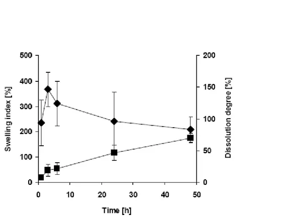
Erosion (degradability) is also a key parameter for materials that are used as implants and/or in tissue regeneration. Swelling and leaching result from diffusion. Swelling involves transport of ions or fluid from the tissue into the biomaterial. As a consequence of swelling, the elastic limit of a material can be reduced leading to static fatigue or crazing [11]. Leaching takes place if, for instance, one component of the biomaterial dissolves into the surrounding fluid phase. This can cause local biological reactions to the released products, reduced fracture strength and elastic modulus of the material. The dissolution varies depending on the nature of the polymer (hydrophilic/hydrophobic). Hydrophobic polymers, for example, dissolve preferably in the amorphous regions, which results in increased area, integrity loss and release of small particles. Bioresorbable polymers are designed to degrade within
the body and be adsorbed naturally when its function has been accomplished [13]. These degradation characteristics differ from polymer to polymer, and can vary from swelling to dissolution by hydrolysis, for instance, when being exposed to body fluids. Bioresorbable materials degrade products that are normal metabolites of the body. Some examples of degradable polymers are polylactide, polyglycolide, polycaprolactone and polyhyaluronic acid esters, but also natural polymers like collagen, chitosan.
2.3.1. Fabrication methods.
A great variety of well-known membrane fabrication techniques are used in tissue engineering applications. Several fabrication methods based on polymer casting are frequently applied to produce tissue engineering membrane, e.g. liquid induced phase separation (LIPS, immersion precipitation)[14-16], thermally induced phase separation (TIPS)[17-20], and evaporation [21-24]. These methods can be used for pure polymers as well as for composites of polymer [25].
Phase inversion is a process whereby a polymer is transformed in a controlled manner from liquid to a solid state. The process of solidification is very often initiated by the transition from one liquid state into two liquids (liquid-liquid demixing). At a certain stage during demixing , one of the liquid phases (the high polymer concentration phase) will solidify so that a solid matrix is formed. By controlling the initial stage of phase transition the membrane morphology can be controlled, i.e. porous as well as nonporous membranes can be prepared.
Evaporation –induced phase separation (EISP)
The most simple technique for preparing phase inversion membranes is precipitation by solvent evaporation. In this method, a polymer is dissolved in a solvent and the polymer solution is cast on a suitable support, e.g. a glass plate or another kind of support, which be porous (e.g. nonwoven polyester) or nonporous (metal, glass or polymer such as polymethylmethacrylate or Teflon). The solvent is allowed to
evaporate in an inert (e.g. nitrogen) atmosphere, in order to exclude water vapour, allowing a dense homogeneous membrane to be obtained. Instead of casting is also possible to deposit the polymer solution on a substrate by dip coating or by spraying, followed by evaporation.
Vapor- induced phase separation (VIPS)
A cast film, consisting of a polymer and a solvent, is placed in a vapour atmosphere where the vapour phase consists of a nonsolvent saturated with a solvent. The high solvent concentration in the vapour phase prevents the evaporation of solvent from the cast film. Membrane formation occurs because of the penetration (diffusion) of non solvent into the cast film.
Thermally induced phase separation (TIPS)
A solution of polymer in a mixed or single solvent is cooled to enable phase separation to occur. Evaporation of the solvent often allows the formation of a skinned membrane.
Non-solvent induced phase separation (NIPS)
Most commercially available membranes are prepared by immersion precipitation: a polymer solution (polymer plus solvent) is cast on a suitable support and immersed in a coagulation bath containing a non solvent. Precipitation occurs because of the exchange of solvent and non solvent. The membrane structure ultimately obtained results from a combination of mass transfer and phase separation.
All phase inversion processes are based on the same thermodynamically stable solution which is subjected to demixing.
The basic parameter describing the miscibility of two or more components is the free enthalpy of mixing
where is the enthalpy of mixing and is the entropy of mixing. Two components (polymer/solvent or polymer/polymer) will mix spontaneously if the free enthalpy of mixing is negative ( ).
The solubility behaviour of polymer solutions differs completely from that of a solution containing low molecular weight components because the entropy of mixing of the long polymeric chains is much lower. Flory and Huggins [26] used a lattice model to describe the entropy of mixing of (polymer) solutions, but it will be not considered here.
2.3.2. Characterisation of membranes
Before describing the membrane characterization methods available and the purpose for which they can be employed, it is important to classify the membranes in two main groups:
porous and non porous membranes.
Characterisation data for porous membranes often give rise to misunderstandings and misinterpretations. It is not unreasonable that it is mainly the size of pore in these membranes that determines which solute can pass or which will be retained. Hence many characterisation methods essentially only determine the pore size and the pore size distributions determined properly, in actual separation processes the membrane performance is mainly controlled by other factors, e.g. concentration, polarisation and fouling. One important, but often not clearly defined variable in the characterisation of porous membranes, is the shape of the pore or its geometry.
Transport through non porous membranes occurs by a solution-diffusion mechanism and separation is achieved either the differences in solubility and/or diffusivity. Hence such membranes cannot be characterised by the methods used for porous membranes.
The determination of the physical properties related to the chemical structure is important for both classes of membranes and it is possible by other methods that allow the determination of surface properties and other physical properties.
Contact angle methods
A drop of liquid sitting on a solid surface represents a powerful, but simple, method to probe surface properties. The phenomenon of the contact angle can be explained as a balance between the force with which the molecules of the liquid (in the drop) are being attracted to each other (a cohesive force) and the attraction of the liquid molecules for the molecules that make up the surface (an adhesive force). An equilibrium is established between these forces, the energy minimum (fig.4). The force balance between the liquid-vapor surface tension of a liquid drop and the interfacial tension between a solid and the drop , manifested through the contact angle of the drop with the surface, can be used quantitatively to characterize the energy of the surface . The basic relationship describing this force balance is : γsv = γsl + γlv cos θ
The energy of the surface, which is directly related to its wettability, is a useful parameter that has often correlated strongly with biological interaction [27-30].
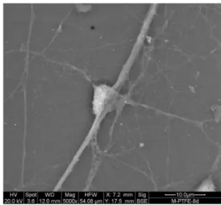
Scanning Electron Microscopy
Scanning electron microscopy (SEM) images of surfaces have great resolution and depth of field, with a three-dimensional quality. SEM functions by focusing and rastering a relatively high-energy electron beam (tpically,5-100 keV) on a specimen. Low-energy secondary electrons are emitted from each spot where the focused electron beam impacts. The measured intensity of the secondary electron emission is a function of the atomic composition of the sample and the geometry of the features under observation. SEM images surfaces by spatially reconstructing on a phosphor screen the intensity of the secondary electron emission. Because of the shallow penetration depth of the low-energy secondary electrons produced by the primary electron beam, only secondary electrons generated near the surface can escape from bulk and be detected. Consequently, SEM is a surface analysis method.
Infrared spectroscopy
Infrared spectroscopy (IR) provides information on the vibrations of atomic and molecular species. It is a standard analytical method that can reveal information on specific chemistry and the orientation of structures. The attenuance total reflectance (ATR) mode of sampling has been used most often in biomaterials studies. The penetration in depth into the sample is 1-5µm. Therefore, ATR is not highly surface sensitive, but observes a broad region near the surface.
Thermal property
Differential scanning calorimetru (DSC) is a method for probing thermal transitions of polymers. A sample cell and a reference cell are supplied energy to varying rates so that the temperatures of two cells remain equal. The temperature is increased, typically at a rate of 10-20 degrees/min over the range of interest, and the energy input required to maintain equality of temperature in two cells is recorded. Plots of energy supplied versus average temperature allow determination of Tg, crystallization
in heat capacity, , has occurred. The areas under the peaks can be quantitatively related to enthalpic changes.
FIGURE 5: DIFFERENTIAL SCANNING CALORIMETRY THERMOGRAM OF A GENERIC POLYMER, SHOWING THE GLASS TANSITION TEMPERATURE, THE CRYSTALLIZATION TEMPERATURE, AND THE MELTING TEMPERATURE OF THE POLYMER SAMPLE.
Bibliografy
[1] Y. Tabata, (2009). "Biomaterial technology for tissue engineering applications." J.R. Soc.Interface.
[2] D.F.Willams (1986). Definition in Biomaterials. Consensus Conference of the European Society for Biomaterials, Chester- England.
[3] B.S. Kim, C. E. Baez, A. Atala (2000). Wordl J. Urol. 18: 2-9.
[4] N.Huebsch, D. J. Mooney (2009). "Inspiration and application in the evolution of biomaterials." Nature 462(26): 426-432.
[5] S.Guelcher, J. Hollinger (2006). An introduction to Biomaterials.
[6] A. S. Kulshrestha, A. Makapatro (2008). Polymers for biomedical applications.
[7] N.Angelova, a. D. Hunkeler (1999). "Rationalizing the design of polymeric biomaterials " Tibtech 17: 409-421.
[8] 8 A.Atala and D. J. Mooney (1997). Synthetic Biodegradable Polymer Scaffolds. Boston.
[9] J. Leuschner, (1992). “Legal requirements for the preclinical toxicological evaluation of biomaterials” Clin. Mater. 10: 51-57.
[10]M. Mulder, (1991). Basic Principles of Membrane Technology. Boston/London.
[11]J. Black, (2006). Biological Performance of Materials: fundaments of Biocompatibility. Boca Raton, Taylor & Francis.
[12]D. F. Stamatialis, B. J. Papenburg, M. Girones, S.Saiful, S. N.M. Bettahalli, S. Schmitmeier, M. Wessling. (2008). "Medical applications of membranes: Drug delivery, artificial organs and tissue engineering." Journal of Membrane Science 308: 1-34.
[13]L.L.Hench; and J. R. Jones (2005). Biomaterials artificial organs and tissue engineering. Boca Raton, CRC press.
[14]S.Y. Kim, T. Kanamori, Y. Noumi, O.C. Wang, T. Shinbo (2004). "Preparation pf porous poly(d,l-lactide) and poly(d,l-lactide-co-glycolide) membranes by a phase inversion process and investigation of their morphological changes as cell culture scaffolds." J.Appl. Polym. Sci 92: 2082-2092.
[15]H.C. Liu, I. C. Lee, J.H. Wang, S.H. Yang, T. H. Young (2004). "Preparation of PLLA membranes with different morphologies for culture of MG-63 cells." Biomaterials 25(18): 4047-4056.
[16]R.A.Zoppi, S. Contant, E.A.R. Duek, F.R. Marques, M.L. Wada, S.P. Nunes (1999). "Porous poly(L-lactide) films obtained by immersion precipitation process:morphology, phase separation and culture of VERO cells." Polymer 40(12): 3275-3289.
[17]J.Guan, K. L. Fujimoto, M.S. Sacks, W.R. Wagner (2005). "Preparation and characterization of highly porous, biodegradable polyurethane scaffolds for soft tissue applications." Biomaterials
26(18): 3961-3971.
[18]S.Li, V. L. Carrubba, S. Piccarolo, D. Sannino, V. Brucato (2004). "Preparation and properties of poly(lactic acid) scaffolda by thermally induced phase separation from a ternary polymer-solvent system." Polym.Int. 53(12): 2079-2085.
[19]F.J. Hua, G. E. Kim, J.D. Lee, Y.K.Son, D.S.Lee (2002). "Macroporous poly(L-lactide)scaffold 1. Preparation of a macroporous scaffold by liquid-liquid phase separation of a PLLA-dioxane-water." J. Biomed. Mater. Res. 63(2): 161-167.
[20]Y.S. Nam, T. G. Park (1999). "Porous biodegradable polymeric scaffolds prepared by thermally induced phase separation." J. Biomed. Mater. Res. 47(1): 8-17.
[21]C. Vaquette, S. Fawzi-Grancher, P. Lavalle, C. Frochot, M.L. Viriot, S. Muller, X. Wang (2006). "In vitro biocompatibility of different polyester membranes." Bio-Med. Mater. Eng. 16(4): S131-S136.
[22]A.S. Htay, S. H. Teoh, D.W. Hutmacher (2004). "Development of perforated microthin poly(epsilon-caprolactone)films as matrices for membrane tissue engineering " J. Biomater. Sci. Polym. Ed. 15(5): 683-700.
[23]M. Tanaka, M. Takebayashi,, M. Miyama, J. Nishida, M. Shimomura (2004). "Design of novel biointerfaces (II). Fabrication of self- organized porous polymer film with highly uniform pores." Bio-Med. Mater. Eng. 14(4): 439-446.
[24]Y.J.Park, N. H. Nam, S.J. Ha, C.M. Pai, C.P. Chung, S.J. Lee (1997). "Porous poly(L-lactide) membranes for guided tissue regeneration and controlled drug delivery: membrane fabrication and characterization." J.Controll. Release 43(2-3): 151-160.
[25]F. Causa, P. A. Netti, L. Ambrosio, G. Ciapetti, N. Baldini, S. Pagani, D. Martini, A. Giunti (2006). "Poly-ε-caprolactone/hydoxyapatite composites for bone regeneration : in vitro characaterization and human osteoblast response." J.Biomed. Mater. Res. Part A 76A(1): 151-162. [26]P. J. Flory, (1953). Principles of Polymer Chemistry Cornell University Ithaca.
[27]J. D. Andrade, (1985). Surface and interfacial aspects of biomedical polymers. New York, Plenum Publisher.
[28]R. J. Good, (1993). Contact angle, wetting, and adhesion :a critical review. The Netherlands, VSP Publisher .
[29]W. A. Zisman (1964). Relation of the quilibrium contact angle to liquid and solid constitution. Contact angle, Wettability and Adhesion. Washington, ACS Advances in Chemistry Series. 43. [30] L. McIntire, V. P. Addonizio, D.L.Coleman, S.G. Eskin, L.A. Harker, J.L.Kardos, B.D.
Ratner, F.J. Schoen, M.V. Sefton, F.A. Pitlick (1985). "Guidelines for Blood-Material Interactions-Devices and Technology Branch, division of Heart and Vascular Disease." National Heart, Lung and Blood Institute NIH pubblication 85: 2185.
Chapter III
Membrane Approaches for Liver and Neuronal Tissue Engineering
3.1 Introduction
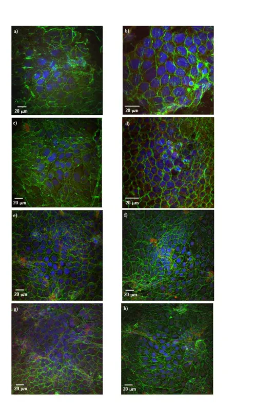
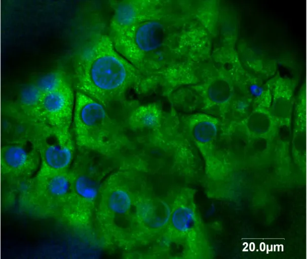
Progress in both membrane and cell culture technology has also greatly contributed to the success of artificially engineered tissue. It was demonstrated that polymeric membranes are attractive for their characteristics of selectivity, stability, and biocompatibility in the use of biohybrid systems for cell culture. In particular, semipermeable membranes act as supports for the adhesion of anchorage-dependent cells and allow the specific transport of metabolites and nutrients to cells and the removal of catabolites and specific products [1–3].
In tissue-engineered constructs, the surface and transport properties of the membranes play an important role in the promotion of cell adhesion, proliferation, and viability. The material surface properties, such as chemical composition, hydrophilicity/ hydrophobicity, charge, free energy, and roughness, affect cell adhesion through the modulation of proteins secreted by cells or contained in the physio- logical liquids. Novel strategies aimed at improving cell– biomaterial interactions have been proposed: the development of new biocompatible and cytocompatible materials and modification of surface chemistry including grafting of functional groups, and immobilization of molecules leaving the bulk properties unaltered.
In this chapter, we review the membrane bioartificial systems developed for tissue engineering. Particular attention is given to the recent achievements in liver and neuronal tissue engineering.
3.2 Membrane for liver tissue regeneration
Each year 30.000 people die of end-stage liver disease in the United States, with an estimated annual cost of 9 billion dollars [4]. Liver transplantation is the only
established successful treatment for end-stage liver disease, and currently there are 100.524 on the waiting list for a donor organ and of those there are 16.005 candidates awaiting a liver transplant (based on United Network for Organ Sharing Organ Procurement and transplantation Network, UNOS OPTN, data as of November 6, 2008). In 2006, there were 6.649 liver transplants performed in United States and 1.935 died while waiting for a liver transplant because of the organ shortage [5]. The European Liver Transplant Registry (ELTR) has cumulated data concerning 75,530 transplantations in 67,848 patients from 136 centres in 23 countries from May 1968 to June 2007 [6]. Cirrhosis is the most frequent indication for transplantation in Europe, followed by cholestatic disease, primary liver tumours and acute hepatic failure [7].
Liver failure is potentially reversible because of liver regeneration [8], so considerable work has been done over many years to develop effective liver support devices. Various non-biological approaches, such as haemodialysis, hemoperfusion, and plasmapheresis have a limited success because of the insufficient replacement of the synthetic and metabolic functions of the liver in these systems [9-10]. On the other hand, extracorporeal biological treatment including whole-liver perfusion, liver slice perfusion and cross haemodialysis, have shown some beneficial results, but they are difficult to implement in a clinical setting [11]. For these reasons, many researchers have developed various extracorporeal bio-hybrid artificial liver (BAL) systems. Generally, a BAL system consists of functional liver cells supported by an artificial cell culture material. In particular, it incorporates hepatocytes into a bioreactor in which the cells are immobilized, cultured and induced to perform the hepatic functions by processing the blood or plasma of liver failure patients. The BAL system acts as a bridge for the patients until a donor organ can available for transplantation or until their liver regeneration [12]. The development of a BAL system involves many design considerations. It must provide: 1) an adhesion support to the cells; 2) an adequate mass transfer of oxygen, nutrients and toxic substances
from blood or plasma of patients to the cell compartments and of proteins, catabolites and other specific compounds produced by cells from the cell compartment to the blood or plasma; 3) immunoprotection of cells and 4) biocompatibility. The BAL devices are classified by the cell source, the type of culture system for the hepatocytes, and the configuration of the bioreactor in Table 1.
References Bioartificial System Bioreactor Configuration Membrane Cell Source
Cell capacity Culture
technique Cell position
Level of development Matsumara et al., 1987 [47] Kiil dyalizer
bioartificial liver Plate Cellulose
(MWCO 20 kDa) Primary rabbit hepatocytes 1x10 10 suspension Dalysate compartment First clinical report Margulis et
al., 1989 [48] Cartridge Polyvinyl chloride
Porcine
hepatocytes 4x10⁷ suspension Shell
Phase II clinical trials Sussman et al.1992 [49] ELAD Amphioxus cell
Technology Hollow Fiber
Cellulose acetate (MWCO=70 kDa)
Human cell line (C3A)
2x10¹¹
Aggregates Shell Phase I clinical
trial Demetriou et
al 1995 [50]
Hepat Assist
Circe Biomedical Hollow Fiber
Polysulphone membranes (Pore size 0.2 µm) Cryopreserved porcine hepatocytes 5x10⁹ Microcarrier attached irregular aggregates
Shell Phase II/III
clinical trial Gerlach et al.
1994
[51] LLS
Charite, Humboldt
Univ Germany Hollow Fiber
Polyamide (MWCO=100 kDa) Polyethersulfone (MWCO=80 kDa) Silastic Polypropylene (0.2 µm pore size) Pig Primary hepatocytes -endothelial cells 2.5x10⁹
Aggregates Shell Phase I
Patzer et al 2002
[52]
BLSS
Excorp Medical Inc. Hollow Fiber Cellulose acetate (MWCO=100kDa) Porcine Primary hepatocytes 70-120g Collagen gel entrapped Shell phase I/II clinical trials Flendrig et al 1997 [53] AMC-BAL
Univ. Amsterdam Spirally wound
Non woven polyester matrix, polypropylene membranes (pore size
0.2 µm)
Pig primary liver cells 1x10¹⁰ Small aggregates Shell/on the non-woven Polyester matrix Phase I Ding et al., 2003 [54]
BAL TECA Corp.
Hollow Fiber Polysulfone
(MWCO=100 kDa) Swine hepatocytes
1x10¹⁰

![Figure 2 Urea synthesis of human hepatocytes cultured in biohybrid PEEK-WC-PU system for 24 days and treated with: (◊) Ursolic Acid (UA) [100 g ml -1 ]; (■) Diclofenac (DIC) [700 M]; ( ) Ursolic Acid-Diclofenac association (UA+DIC)](https://thumb-eu.123doks.com/thumbv2/123dokorg/2875719.9829/57.892.99.784.134.524/synthesis-hepatocytes-cultured-biohybrid-ursolic-diclofenac-diclofenac-association.webp)