S U P P L E M E N T A R T I C L E
Clinical Infectious Diseases
Correspondence: S. A. Madhi, Respiratory and Meningeal Pathogens Research Unit, Chris Hani Baragwanath Academic Hospital, Central-West Wing, 11th Floor, Chris Hani Road, Soweto
2013, South Africa ([email protected]).
Clinical Infectious Diseases® 2019;69(S4):S342–50
© The Author(s) 2019. Published by Oxford University Press for the Infectious Diseases Society of America. This is an Open Access article distributed under the terms of the Creative Commons
Attribution License (http://creativecommons.org/licenses/by/4.0/), which permits unrestricted
reuse, distribution, and reproduction in any medium, provided the original work is properly cited. DOI: 10.1093/cid/ciz573
An Observational Pilot Study Evaluating the Utility of
Minimally Invasive Tissue Sampling to Determine the
Cause of Stillbirths in South African Women
Shabir A. Madhi,1,2Jayani Pathirana,1,2 Vicky Baillie,1,2 Clare Cutland,1,2 Yasmin Adam,3 Alane Izu,1,2 Quique Bassat,4,5,6,7,8 Dianna M. Blau,9
Robert F. Breiman,10 Martin Hale,11 Siobhan Johnstone,1,2 Roosecelis B. Martines,12 Azwifarwi Mathunjwa,1,2 Susan Nzenze,1,2 Jaume Ordi,4,5
Pratima L. Raghunathan,9 Jana M. Ritter,12 Fatima Solomon,1,2 Jeannette Wadula,13 Sherif R. Zaki,12 and Richard Chawana1,2
1Medical Research Council: Respiratory and Meningeal Pathogens Research Unit, University of the Witwatersrand, Faculty of Health Science, Johannesburg, South Africa; 2Department of Science
and Technology/National Research Foundation: Vaccine Preventable Diseases, University of the Witwatersrand, Faculty of Health Sciences, Johannesburg, South Africa; 3Department of Obstetrics
and Gynaecology, Chris Hani Baragwanath Academic Hospital, School of Clinical Medicine, Faculty of Health Sciences, University of the Witwatersrand, Johannesburg, South Africa; 4ISGlobal,
Hospital Clínic, Universitat de Barcelona, Barcelona, Spain; 5Centro de Investigação em Saúde de Manhiça (CISM), Maputo, Mozambique; 6Catalan Institution for Research and Advanced Studies
(ICREA), Barcelona, Spain; 7Pediatric Infectious Diseases Unit, Pediatrics Department, Hospital de Sant Joan de Deu, University of Barcelona, Barcelona, Spain; 8Consorcio de Investigacion
Biomedica en Red de Epidemiologia y Salud, Madrid, Spain; 9Center for Global Health, Centers for Disease Control and Prevention, Atlanta, Georgia, USA; 10Emory Global Health Institute, Emory
University, Atlanta, Georgia, USA; 11National Health Laboratory Service, Department of Anatomical Pathology, School of Pathology, University of the Witwatersrand, Faculty of Health Sciences,
Johannesburg, South Africa; 12Infectious Diseases Pathology Branch, Division of High-Consequence Pathogens and Pathology, National Center for Emerging and Zoonotic Infectious Diseases,
Centers for Disease Control and Prevention, Atlanta, Georgia, USA; and 13National Health Laboratory Service, Department of Microbiology and Infectious Diseases, School of Pathology, Faculty of
Health Sciences, University of the Witwatersrand, Johannesburg, South Africa
Background. Despite approximately 2.6 million stillbirths occurring annually, there is a paucity of systematic biological
investi-gation and consequently knowledge on the causes of these deaths in low- and middle-income countries (LMICs). We investigated the utility of minimally invasive tissue sampling (MITS), placental examination, and clinical history, in attributing the causes of stillbirth in a South African LMIC setting.
Methods. This prospective, observational pilot study undertook sampling of brain, lung, and liver tissue using core biopsy needles,
blood and cerebrospinal fluid collection, and placental examination. Testing included microbial culture and/or molecular testing and tissue histological examination. The cause of death was determined for each case by an international panel of medical specialists and categorized using the World Health Organization’s International Classification of Diseases, Tenth Revision application to perinatal deaths.
Results. A cause of stillbirth was identifiable for 117 of 129 (90.7%) stillbirths, including an underlying maternal cause in 63.4%
(n = 83) and an immediate fetal cause in 79.1% (n = 102) of cases. The leading underlying causes of stillbirth were maternal hyperten-sive disorders (16.3%), placental separation and hemorrhage (14.0%), and chorioamnionitis (10.9%). The leading immediate causes of fetal death were antepartum hypoxia (35.7%) and fetal infection (37.2%), including due to Escherichia coli (16.3%), Enterococcus species (3.9%), and group B Streptococcus (3.1%).
Conclusions. In this pilot, proof-of-concept study, focused investigation of stillbirth provided granular detail on the causes
thereof in an LMIC setting, including provisionally highlighting the largely underrecognized role of fetal sepsis as a dominant cause.
Keywords. MITS; stillbirth and fetal death; maternal hypertension; fetal hypoxia; fetal infection.
In 2015, the estimated number of stillbirths (2.6 million)
ap-proximated neonatal deaths (2.7 million) [1], 98% of which
occurred in low- and middle-income countries (LMICs). Furthermore, the annual rate reduction of stillbirths from 2000 to 2015 was 2%, which was lower than rate reduction in neo-natal mortality (3.1%) and deaths in children aged 1–59 months
(4.7%) [2, 3]. The United Nations Children’s Fund–adopted
“Every Newborn Action Plan” aspires to reduce stillbirth rates from 18.4 per 1000 births in 2015 to 12 per 1000 births by 2030
[4]. The paucity of systematic investigation and consequent
limited understanding of the causes of stillbirth in LMICs is recognized as a major impediment in achieving this goal.
Currently, estimates on causes of stillbirth in LMICs are prem-ised on limited vital registration and verbal autopsy data, coupled
with modeling of known risk-factor prevalence [1]. The
ubiqui-tous absence of biological investigation of stillbirths in LMICs is multifactorial—resources are limited and a significant proportion of cases occur at home and thus outside of the health system— and is compounded by cultural issues related to acceptability of
postmortem autopsies [5]. Detailed investigation on the causes
of stillbirths was undertaken by the Stillbirth Collaborative Research Network (SCRN) in a multicenter study in the United
States between 2006 and 2008, when the stillbirth rate was 6 per 1000 births. The most informative tools in attributing the cause of stillbirth in the SCRN study were maternal medical record re-view, placental examination and histology (52.3%), complete di-agnostic autopsy (CDA, which included bacterial culture of tissue
samples; 31.4%), and karyotyping (9%) [6].
To overcome the obstacles of undertaking CDA in LMICs, a Mozambican study compared the utility of minimally invasive autopsy (MIA) to CDA for attributing the cause of stillbirths
and neonatal deaths [7]. A cause of stillbirth was attributed to
89% of 18 stillbirths investigated by CDA and in 83% based on MIA findings. There was moderate concordance (κ = 0.78 [95% confidence interval, .56–.99]) in the cause of death (CoD) attri-bution between the 2 methods, despite the absence of placental examination and not using any clinical information when evaluating for the CoD.
The aim of our study was to pilot the utility of minimally invasive tissue sampling (MITS, also referred to as MIA), to-gether with maternal medical record review and placental mac-roscopic and histological examination, in attributing the cause of stillbirths in an LMIC setting in South Africa.
METHODS
Study Population and Site
The study population and study methods are described in the companion article on neonatal deaths in this supplement
[8]. In brief, the study was done at Chris Hani Baragwanath
Academic Hospital (CHBAH), Soweto, a secondary-tertiary hospital, where approximately 75% of 28 000 annual births in Soweto occur. CHBAH was the only public hospital at the time of the study in Soweto (unpublished administrative data, Obstetrics Department, CHBAH). While additional deliveries occur at 5 midwife-operated units in Soweto, women who are deemed to be high risk according to national guidelines are re-ferred to a hospital, including women in preterm labor, those presenting with a stillbirth, or who had a previous stillbirth. The stillbirth rate in Soweto was 22.5 per 1000 births in 2016, which is higher than that estimated for South Africa based on
vital registration data (14 per 1000 births in 2015) [2].
Study Design
This prospective, observational study enrolled stillbirths delivered at CHBAH from 20 July 2015 to 8 August 2016. Stillbirths were defined as absence of any signs of life diag-nosed either before delivery by sonography, or at birth by the attending clinician/midwife. In this study, we lim-ited enrollment to stillbirths weighing ≥1000 g based on
World Health Organization (WHO) reporting criteria [4].
Furthermore, we prioritized stillbirths from women who had consented to partake in a parallel observational study as-sessing for serocorrelates of protection against invasive group B Streptococcus (GBS) disease (ClinicalTrials.gov registration
number: NCT02215226), followed by women who consented to study participation after confirmation of an intrauterine death or delivery of a stillborn baby. Assessment of gestational age of the stillbirths was based on the last menstrual period or on ultrasound findings performed as standard of care.
The study staff retrospectively abstracted clinical informa-tion from maternal medical records, including details on the mother, antenatal care, and labor history using a standard template. Stillbirths were categorized by timing of fetal death: antepartum, that is, fetal death confirmed by ultrasonography prior to the onset of labor; or intrapartum if there was evi-dence of fetal life during active labor by physical examination, cardiotocography, or ultrasonography.
Minimally Invasive Tissue Sampling
Trained study staff undertook the MITS procedure. Detail of the methods used for undertaking MITS, including
histolog-ical evaluation, is described elsewhere in this supplement [8].
In brief, this included multiple core tissue biopsies of the brain, left and right lung, and liver. The first tissue sample from each site was sent for culture (lung and liver), the second sample was used for molecular microbiological tests (lung only), and 6 subsequent tissue samples were sent for histopathology (brain, lung, and liver). Microbial culture and molecular testing were done on blood and cerebrospinal fluid (CSF) samples.
Placental Examination
Following delivery, the placenta was retrieved and weighed prior to resecting a wedge of the parenchymal tissue and chorioamniotic membranes, which was placed in a sterile con-tainer for bacterial microscopy and culture. The rest of the placenta was then immersed in 10% buffered formalin and trans-ported to Lancet Laboratories (Johannesburg, South Africa). Placental examination included macroscopic visualization, fol-lowing which the histopathologist selected portions of placenta, which were embedded in paraffin and processed for routine
he-matoxylin and eosin staining using standard protocols [9].
Molecular Tests
Details of the molecular testing for organisms presence in blood, lung, and CSF samples is as described in the companion
manu-script on neonatal deaths [8]. The testing used a commercially
available multiplex polymerase chain reaction (PCR) assays (Fast-Track Diagnostics [FTD], Sliema, Malta), and was done at the Respiratory and Meningeal Pathogens Research Unit labo-ratory. CSF samples were tested using the FTD-Neuro-9 menin-gitis panel, which included probes for cytomegalovirus (CMV), Epstein-Barr virus, adenovirus, herpes simplex virus 1 and 2, varicella zoster virus, enterovirus, parechovirus, human her-pesvirus 6 and 7, and parvovirus B19. Lung and blood sample molecular testing was done using the FTD-Neonatal Sepsis kit, which included probes for CMV, group B Streptococcus,
Listeria monocytogenes, Escherichia coli, Staphylococcus au-reus, Chlamydia trachomatis, and Ureaplasma urealyticum/ parvum. The molecular assays were not specifically designed for investigating infectious causes of stillbirths.
Determination of Cause of Death
The CoD attribution was based on consensus opinion of a mul-tidisciplinary international panel of specialists (listed under the Determination of Cause of Death [DeCoDe] panel, see Acknowledgments), as described in the Supplementary Materials
and elsewhere [8]. The framework for reporting on the CoD was
based on the 2016 WHO perinatal CoD reporting recommenda-tions [10], which includes recording of the “underlying” condition which led to the sequence of events culminating in fetal demise, and the most “immediate” medical event that caused the fetal death.
Statistical Analysis
Demographic and clinical data are summarized using means and standard deviations or medians and range where appro-priate. Student t tests were performed to compare means and the Wilcoxon-rank sum test was used for nonnormal quantita-tive data. Fisher exact test was used for comparing categorical variables. Standardized weight scores were calculated using the International Fetal and Newborn Growth Consortium for the
21st Century standards and small for gestational age was de-fined as a standardized score (z score) < −2 [11].
Ethical Considerations
The study was approved by the Human Research Ethics Committee (HREC 15021) of the University of the Witwatersrand, and regis-tered at the South African National Department of Health Trials Registry (application number 3988). Grief counseling was ad-ministered to bereaved parents and guardians, following which they were informed of the study and provided an opportunity to discuss with other family members prior to consenting. The study staff continued providing grief counseling following the MITS, including when giving feedback on the cause of death.
RESULTS
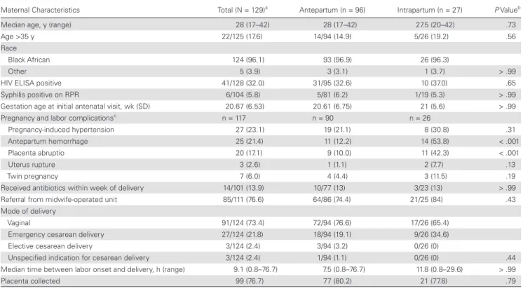
Overall, 129 stillbirths were enrolled, including 96 (74.4%) antepartum and 27 (20.9%) intrapartum cases; the timing of
death was uncertain for 6 cases (Table 1). Seventy-seven
per-cent of the women with stillbirths were referred to CHBAH from a surrounding midwife-obstetric unit. The median age of the women was 28 years, 17.6% were >35 years of age, and 96.1% were black Africans. The prevalence of maternal human immunodeficiency virus (HIV) and syphilis infection was
Table 1. Maternal Demographic and Clinical Features of Antepartum and Intrapartum Stillbirths
Maternal Characteristics Total (N = 129)a Antepartum (n = 96) Intrapartum (n = 27) P Valueb
Median age, y (range) 28 (17–42) 28 (17–42) 27.5 (20–42) .73
Age >35 y 22/125 (17.6) 14/94 (14.9) 5/26 (19.2) .56
Race
Black African 124 (96.1) 93 (96.9) 26 (96.3)
Other 5 (3.9) 3 (3.1) 1 (3.7) > .99
HIV ELISA positive 41/128 (32.0) 31/95 (32.6) 10 (37.0) .65
Syphilis positive on RPR 6/104 (5.8) 5/81 (6.2) 1/19 (5.3) > .99
Gestation age at initial antenatal visit, wk (SD) 20.67 (6.53) 20.61 (6.75) 21 (5.6) > .99
Pregnancy and labor complicationsc n = 117 n = 90 n = 26
Pregnancy-induced hypertension 27 (23.1) 19 (21.1) 8 (30.8) .31
Antepartum hemorrhage 25 (21.4) 11 (12.2) 14 (53.8) < .001
Placenta abruptio 20 (17.1) 9 (10.0) 11 (42.3) < .001
Uterus rupture 3 (2.6) 1 (1.1) 2 (7.7) .13
Twin pregnancy 7 (6.0) 4 (4.4) 3 (11.5) .19
Received antibiotics within week of delivery 14/101 (13.9) 10/77 (13) 3/23 (13) > .99
Referral from midwife-operated unit 85/111 (76.6) 64/86 (74.4) 21/25 (84) .43
Mode of delivery
Vaginal 91/124 (73.4) 72/94 (76.6) 17/26 (65.4)
Emergency cesarean delivery 27/124 (21.8) 18/94 (19.1) 9/26 (34.6)
Elective cesarean delivery 3/124 (2.4) 3/94 (3.2) 0/26 (0)
Unspecified indication for cesarean delivery 3/124 (2.4) 1/94 (1.1) 0/26 (0) .44
Median time between labor onset and delivery, h (range) 9.1 (0.8–76.7) 7.5 (0.8–76.7) 11.8 (0.8–29.6) > .99
Placenta collected 99 (76.7) 77 (80.2) 21 (77.8) .79
Data are presented as No. (%) unless otherwise indicated.
Abbreviations: ELISA, enzyme-linked immunosorbent assay; HIV, human immunodeficiency virus; RPR, rapid plasma reagin; SD, standard deviation.
aIncluded are 6 subjects without sufficient documentation to attribute whether antepartum or intrapartum death.
bComparison of antepartum to intrapartum cases.
cBased on retrospective review of available maternal records with adequate documentation of information.
32.0% and 5.8%, respectively. Maternal pregnancy and labor-related complications included 23.1% with pregnancy-induced hypertension and 21.4% with antepartum hemorrhage. The median time between presentation to the hospital and delivery was 7.5 and 11.8 hours for antepartum and intrapartum still-births, respectively (Table 1).
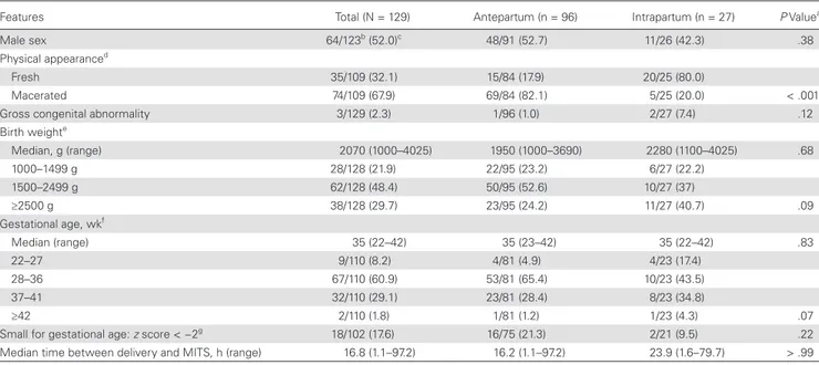
Two-thirds of the stillbirths had a macerated appearance, which expectedly was more common in antepartum (82.1%)
than intrapartum (20.0%) cases (P < .001; Table 2). Gross
con-genital abnormalities were present in 3 stillbirths, including 1 case each of hydrocephalus and anencephaly, and the third being unspecified. The median birth weight and gestational age were 2070 g and 35 weeks, respectively, with 78.1% of stillbirths weighing >1500 g. Small for gestational age (SGA) was present in 17.6% of 102 cases in whom gestational age and birth weight data were available. Two of 41 (4.8%) stillbirths born to HIV-infected mothers had a reactive HIV type 1 PCR test at birth. Testing for HIV by PCR failed in 5 cases of antepartum still-births. Overall, the median time between delivery and MITS was 16.8 hours (Table 2).
Placental Pathology and Tissue Sample Histopathology
Placentas were available for investigation in 99 (76.7%) stillbirths, including 77 (80.2%) antepartum, cases 21 (70.8%) intrapartum cases and one case in whom timing of stillbirth could not be
determined (Supplementary Table 1). Macroscopic evidence of
placenta infarction was evident in 26.3% and retroplacental he-matoma in 31.2% of cases. Histologically, placental infarction was identified in 50.0% of cases. Of those in which the extent of in-farction was quantified (n = 38), 28.9% had ≥20% parenchymal involvement. Chorioamnionitis was histologically evident in 27.6% (n = 27) of placentas, and of those graded, 31.8% had grade I, 40.9% grade II, and 27.3% grade III inflammatory cellular in-filtrates. Umbilical cord abnormalities included presence of cord thrombosis (4.2%), cord knots (4.2%), funisitis (3.1%), and
cho-rionic vasculitis (2.2%) (Supplementary Table 1). There were no
significant differences in placental findings between antepartum and intrapartum stillbirths.
There was a low yield of adequate samples (<2.5% for any organ) for histopathologic investigation of fetal tissue (Supplementary Table 2). The majority of tissue samples were either autolyzed (40.6%–64.8%), had a suboptimal number of core tissues for investigation (0.8%–43.8%), or no targeted tissue was collected at all (13.3%–55.5%).
Underlying Causes of Stillbirth
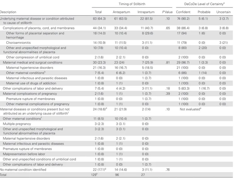
An underlying maternal cause of stillbirth was attributed by the DeCoDe panel for 64.3% (n = 83) of cases; with the
level of certainty graded mainly as “confident” (90%) (Table
3). The DeCoDe panel could not attribute an underlying
Table 2. Demographic and Clinical Characteristics of Antepartum and Intrapartum Stillbirth Investigated by Minimally Invasive Tissue Sampling
Features Total (N = 129) Antepartum (n = 96) Intrapartum (n = 27) P Valuea
Male sex 64/123b (52.0)c 48/91 (52.7) 11/26 (42.3) .38
Physical appearanced
Fresh 35/109 (32.1) 15/84 (17.9) 20/25 (80.0)
Macerated 74/109 (67.9) 69/84 (82.1) 5/25 (20.0) < .001
Gross congenital abnormality 3/129 (2.3) 1/96 (1.0) 2/27 (7.4) .12
Birth weighte Median, g (range) 2070 (1000–4025) 1950 (1000–3690) 2280 (1100–4025) .68 1000–1499 g 28/128 (21.9) 22/95 (23.2) 6/27 (22.2) 1500–2499 g 62/128 (48.4) 50/95 (52.6) 10/27 (37) ≥2500 g 38/128 (29.7) 23/95 (24.2) 11/27 (40.7) .09 Gestational age, wkf Median (range) 35 (22–42) 35 (23–42) 35 (22–42) .83 22–27 9/110 (8.2) 4/81 (4.9) 4/23 (17.4) 28–36 67/110 (60.9) 53/81 (65.4) 10/23 (43.5) 37–41 32/110 (29.1) 23/81 (28.4) 8/23 (34.8) ≥42 2/110 (1.8) 1/81 (1.2) 1/23 (4.3) .07
Small for gestational age: z score < −2g 18/102 (17.6) 16/75 (21.3) 2/21 (9.5) .22
Median time between delivery and MITS, h (range) 16.8 (1.1–97.2) 16.2 (1.1–97.2) 23.9 (1.6–79.7) > .99
Data are presented as No. (%) unless otherwise indicated. Abbreviation: MITS, minimally invasive tissue sampling.
aComparison of antepartum to intrapartum cases.
bSix subjects without sufficient documentation to attribute whether antepartum or intrapartum death.
cAll values in parentheses are a percentage of total observations given in the denominator.
dData available for 109 cases in total, including 84 antepartum and 25 intrapartum cases.
eData available for 128 cases in total, including 95 antepartum and 27 intrapartum cases.
fGestational age available for 110 overall, including 81 antepartum and 23 intrapartum cases.
gData available for 102 subjects with paired gestational age and birth weight, including 75 antepartum and 21 intrapartum cases.
maternal condition in 35.7% of cases, including 24 (18.6%) cases in which although there was a maternal medical con-dition that was considered to be a possible risk factor for stillbirths, the DeCoDe panel adjudicated the condition not to have caused the stillbirth. This included 10 women with underlying HIV infection and 3 with multiple pregnancies. In a further 17.1% (n = 22) of cases, there was no attributable maternal condition identified to have caused the stillbirth (Table 3).
Overall, the leading underlying maternal causes of stillbirth were “maternal hypertensive disorders” (16.3%), “other placental
separation and hemorrhage” (14.0%; all diagnosed as abruptio placenta), chorioamnionitis (10.9%), and “other morphological and functional abnormality of the placenta” (7.8%; 5 of 6 with placental histology showing evidence of infarcts). Of the 46 cases without an identifiable underlying maternal cause of stillbirth, 34 of the fetuses had an immediate fetal cause of stillbirth attributed (Supplementary Table 3). This included “fetal infection” as the underlying and immediate CoD in 14 (10.9%) cases, all of which were antepartum deaths. Cumulatively, an underlying maternal or immediate fetal condition was diagnosed as the CoD in 90.7% (117/129) of cases.
Table 3. Maternal Underlying Conditions and Subcategory Diagnoses Associated With Fetal Death
Description
Timing of Stillbirth
P Value
DeCoDe Level of Certaintya
Total Antepartum Intrapartum Confident Probable Uncertain
Underlying maternal disease or condition attributed
to cause of stillbirth 83 (64.3) 61 (63.5) 22 (81.5) .10 74 (90.2) 5 (6.1) 3 (3.7)
Complications of placenta, cord, and membranes 44 (34.1) 33 (34.4) 11 (40.7) .65 38 (86.4) 3 (6.8) 3 (6.8)
Other forms of placental separation and
hemorrhage 18 (14.0) 10 (10.4) 8 (29.6) 17 (94) 1 (6) 0 (0)
Chorioamnionitis 14 (10.9) 11 (11.5) 3 (11.1) 11 (79) 0 (0) 3 (21)
Other and unspecified morphological and functional abnormalities of placenta
10 (7.8) 10 (10.4) 0 (0) 8 (80) 2 (20) 0 (0)
Other compression of umbilical cord 2 (1.6) 2 (2.1) 0 (0) 2 (100) 0 (0) 0 (0)
Maternal medical and surgical conditions 30 (23.3) 23 (24) 7 (25.9) .81 29 (96.7) 1 (3.3) 0 (0)
Maternal hypertensive disorders 21 (16.3) 16 (16.7) 5 (18.5) 21 (100) 0 (0) 0 (0)
Other maternal conditionsb 7 (5.4) 6 (6.2) 1 (3.7) 6 (86) 1 (14) 0 (0)
Maternal infectious and parasitic diseases 1 (0.8) 0 (0) 1 (3.7) 1 (100) 0 (0) 0 (0)
Maternal use of drugs of addiction 1 (0.8) 1 (1) 0 (0) 1 (100) 0 (0) 0 (0)
Other complications of labor and delivery 7 (5.4) 4 (4.2) 3 (11.1) .18 5 (83.3) 1 (16.7) 0 (0)
Maternal complications of pregnancy 2 (1.6) 1 (1) 1 (3.7) .39 2 (100) 0 (0) 0 (0)
Premature rupture of membranes 1 (0.8) 0 (0) 1 (3.7) 1 (100) 0 (0) 0 (0)
Other maternal complications of pregnancy 1 (0.8) 1 (1) 0 (0) 1 (100) 0 (0) 0 (0)
Maternal diseases or conditions present but not
attributed as an underlying cause of stillbirthc 24 (18.6)
d 21 (21.9) 2 (7.4) .10 Not evaluatede
Other maternal conditionsf 11 (8.5) 10 (10.4) 1 (3.7)
Multiple pregnancy 3 (2.3) 3 (3.1) 0 (0)
Other and unspecified morphological and
functional abnormalities of placenta 3 (2.3) 3 (3.1) 0 (0)
Maternal hypertensive disorders 2 (1.6) 2 (2.1) 0 (0)
Maternal infectious and parasitic diseases 1 (0.8) 1 (1) 0 (0)
Premature rupture of membranes 1 (0.8) 0 (0) 0 (0)
Malpresentation before labor 1 (0.8) 1 (1) 0 (0)
Other and unspecified conditions of umbilical cord 1 (0.8) 1 (1) 0 (0)
Other complications of labor and delivery 1 (0.8) 0 (0) 1 (3.7)
No maternal condition identified 22 (17.1)g 14 (14.6) 3 (11.1) .76
Total 129h 96 27
Data are presented as No. (%) unless otherwise indicated. Abbreviation: DeCoDe, Determination of Cause of Death.
aThe level of confidence to the diagnosis attributed to by the DeCoDE panel.
bOne case each of diabetes mellitus in pregnancy, acute vaginitis, primary thrombophilia, infections of genitourinary tract in pregnancy, anemia complicating pregnancy, elderly primigravida
and multigravida, pure hypercholesterolemia.
cListing of illnesses considered to be possibly associated with pathogenesis of stillbirth among those cases in which no underlying maternal condition was identified by the DeCoDe panel,
and the condition, although present, was not considered to have caused the stillbirth.
dTotal includes 1 case in which the timing of stillbirth was unknown.
eGrading of level of certainty in cause of death attribution not undertaken for possible contributing causes of death.
fHuman immunodeficiency virus disease complicating pregnancy, childbirth, and the puerperium/antiviral drugs (n = 10); infections of genitourinary tract in pregnancy (n = 1).
gTotal includes 5 cases in whom the timing of stillbirth was unknown.
hTotal includes 6 cases in whom the timing of stillbirth was unknown.
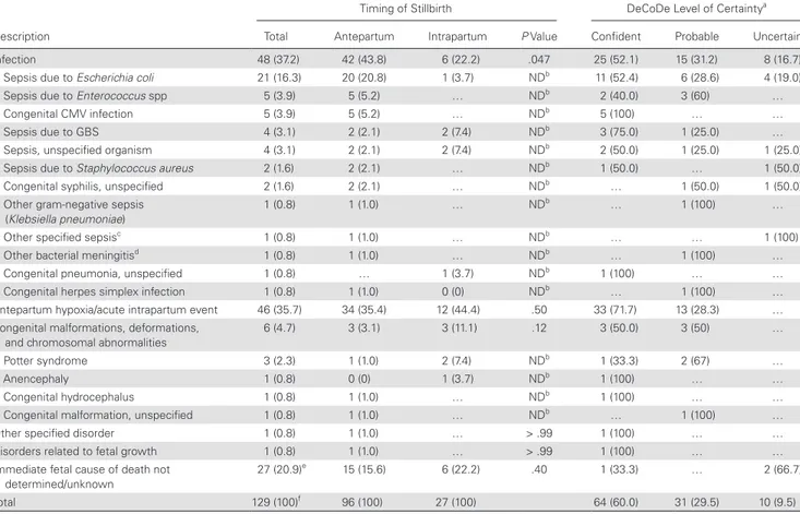
Immediate Causes of Fetal Death
The most common immediate fetal causes of stillbirth were fetal infection (37.2%), “antepartum hypoxia/acute intrapartum event” (35.7%), and “congenital malformations/chromosomal
abnormalities” (4.7%) (Table 4). The level of certainty in
attrib-uting the immediate CoD by the DeCoDe panel (n = 105), was “confident,” “probable,” and “uncertain but possible” in 60.0%,
29.5%, and 9.5%, respectively (Table 4). In 27 (20.9%) cases, the
immediate fetal CoD was not determined.
Thirty-eight of 48 (79.1%) fetal deaths attributed to infec-tion were due to sepsis with the leading implicated organ-isms being E. coli (21/48 [43.8%]), Enterococcus species (5/48
[10.4%]), and GBS (4/48 [8.3%]) (Table 4). The overall
contri-bution of these 3 respective pathogens as the cause of stillbirth
was 16.3%, 3.9%, and 3.1% (Table 4). The levels of confidence
for attributing infection as the cause of fetal death were 52.1%, 31.2%, and 16.7% as “confident,” “probable,” and “uncertain,”
respectively (Table 4). Only 2 (1.6%) stillbirths were attributed
to syphilis (1 probable and 1 uncertain), whereas 5 (3.9%) were attributed to disseminated congenital CMV infection with high level of confidence, and 1 (0.8%) case to herpes simplex virus (Table 4).
Among the 18 (of 102) stillbirths categorized as SGA, there were no cases where disorders of fetal growth was attributed as the underlying CoD. The specific attributed underlying CoD among these 18 cases included 6 (33.3%) due to “maternal hy-pertensive conditions” (5 preeclampsia, 1 gestational hyper-tension without proteinuria); 4 (22.2%) “other and unspecified morphological and functional abnormalities of placenta” (3 placental insufficiency, 1 placental infarction); 1 “other form of placental separation and hemorrhage” (premature separation of the placenta); and 2 “other maternal conditions” (1 unspeci-fied diabetes mellitus in pregnancy and 1 anemia complicating pregnancy). No maternal condition was evident in 5 (27.8%) of the SGA cases, 3 of whom had culture-confirmed fetal infection and 2 of whom had evidence of antepartum hypoxia.
Table 4. Immediate Perinatal Cause of Fetal Death
Description
Timing of Stillbirth
P Value
DeCoDe Level of Certaintya
Total Antepartum Intrapartum Confident Probable Uncertain
Infection 48 (37.2) 42 (43.8) 6 (22.2) .047 25 (52.1) 15 (31.2) 8 (16.7)
Sepsis due to Escherichia coli 21 (16.3) 20 (20.8) 1 (3.7) NDb 11 (52.4) 6 (28.6) 4 (19.0)
Sepsis due to Enterococcus spp 5 (3.9) 5 (5.2) … NDb 2 (40.0) 3 (60) …
Congenital CMV infection 5 (3.9) 5 (5.2) … NDb 5 (100) … …
Sepsis due to GBS 4 (3.1) 2 (2.1) 2 (7.4) NDb 3 (75.0) 1 (25.0) …
Sepsis, unspecified organism 4 (3.1) 2 (2.1) 2 (7.4) NDb 2 (50.0) 1 (25.0) 1 (25.0)
Sepsis due to Staphylococcus aureus 2 (1.6) 2 (2.1) … NDb 1 (50.0) … 1 (50.0)
Congenital syphilis, unspecified 2 (1.6) 2 (2.1) … NDb … 1 (50.0) 1 (50.0)
Other gram-negative sepsis
(Klebsiella pneumoniae) 1 (0.8) 1 (1.0) … ND
b … 1 (100) …
Other specified sepsisc 1 (0.8) 1 (1.0) … NDb … … 1 (100)
Other bacterial meningitisd 1 (0.8) 1 (1.0) … NDb … 1 (100) …
Congenital pneumonia, unspecified 1 (0.8) … 1 (3.7) NDb 1 (100) … …
Congenital herpes simplex infection 1 (0.8) 1 (1.0) 0 (0) NDb … 1 (100) …
Antepartum hypoxia/acute intrapartum event 46 (35.7) 34 (35.4) 12 (44.4) .50 33 (71.7) 13 (28.3) …
Congenital malformations, deformations,
and chromosomal abnormalities 6 (4.7) 3 (3.1) 3 (11.1) .12 3 (50.0) 3 (50) …
Potter syndrome 3 (2.3) 1 (1.0) 2 (7.4) NDb 1 (33.3) 2 (67) …
Anencephaly 1 (0.8) 0 (0) 1 (3.7) NDb 1 (100) … …
Congenital hydrocephalus 1 (0.8) 1 (1.0) … NDb 1 (100) … …
Congenital malformation, unspecified 1 (0.8) 1 (1.0) … NDb … 1 (100) …
Other specified disorder 1 (0.8) 1 (1.0) … > .99 1 (100) … …
Disorders related to fetal growth 1 (0.8) 1 (1.0) … > .99 1 (100) … …
Immediate fetal cause of death not determined/unknown
27 (20.9)e 15 (15.6) 6 (22.2) .40 1 (33.3) … 2 (66.7)
Total 129 (100)f 96 (100) 27 (100) 64 (60.0) 31 (29.5) 10 (9.5)
Data are presented as No. (%) unless otherwise indicated.
Abbreviations: CMV, cytomegalovirus; DeCoDe, Determination of Cause of Death; GBS, group B Streptococcus; ND, not done.
aLevel of confidence to the diagnosis attributed to by the DeCoDE panel.
bDue to low cell counts, only comparisons for main maternal cause of death were done.
cIdentified Ureaplasma urealyticum and Escherichia coli on blood sample.
dCulture of Sphingomonas paucimobilis on cerebrospinal fluid.
eTotal includes 6 cases in which the timing of stillbirth was unknown.
fTotal includes 6 cases in which the timing of stillbirth was unknown.
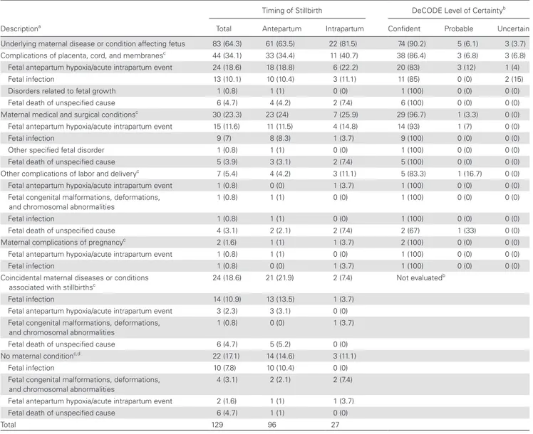
Association Between “Underlying” and “Immediate” CoD Attribution
Forty-one of 46 (89.1%) stillbirths with “antepartum hypoxia” as an immediate CoD had an identifiable underlying condition predisposing to hypoxia, foremost among which were placental
disorders (n = 24/46 [52.2%]) (Table 5 and Supplementary
Table 3). This included 12 cases of “other placental separa-tion and hemorrhage” (26.1%), 8 cases of “other morpholog-ical and functional placenta abnormalities” (17.4%), and 2 cases each (4.3%) related to “umbilical cord compression” and “chorioamnionitis.” Furthermore, maternal hypertensive dis-orders were attributed as the underlying cause of “antepartum hypoxia” in 10 (21.7%) cases.
Among the 48 stillbirths attributed to fetal infection (in-cluding sepsis, pneumonia, or meningitis) as the immediate cause, sepsis was the underlying CoD in 10 (20.8%) cases among whom no maternal condition was identified, whereas “chorioamnionitis” and “maternal hypertensive disorders” were the underlying diagnoses in 12 (25.0%) and 9 (18.8%) cases with fetal infection, respectively. Four of 6 (66.7%) stillbirths with congenital abnormalities as an immediate CoD were as-sessed to have died due to the congenital abnormality in the
absence of any underlying maternal condition (Supplementary
Table 3). Although no immediate CoD was attributable to 27 stillbirths, an underlying condition was present in 15, of which 4 (14.8%) were due to “maternal hypertensive disorders”
Table 5. Maternal Conditions and Associated Immediate Fetal Cause of Stillbirth
Descriptiona
Timing of Stillbirth DeCODE Level of Certaintyb
Total Antepartum Intrapartum Confident Probable Uncertain
Underlying maternal disease or condition affecting fetus 83 (64.3) 61 (63.5) 22 (81.5) 74 (90.2) 5 (6.1) 3 (3.7)
Complications of placenta, cord, and membranesc 44 (34.1) 33 (34.4) 11 (40.7) 38 (86.4) 3 (6.8) 3 (6.8)
Fetal antepartum hypoxia/acute intrapartum event 24 (18.6) 18 (18.8) 6 (22.2) 20 (83) 3 (12) 1 (4)
Fetal infection 13 (10.1) 10 (10.4) 3 (11.1) 11 (85) 0 (0) 2 (15)
Disorders related to fetal growth 1 (0.8) 1 (1) 0 (0) 1 (100) 0 (0) 0 (0)
Fetal death of unspecified cause 6 (4.7) 4 (4.2) 2 (7.4) 6 (100) 0 (0) 0 (0)
Maternal medical and surgical conditionsc 30 (23.3) 23 (24) 7 (25.9) 29 (96.7) 1 (3.3) 0 (0)
Fetal antepartum hypoxia/acute intrapartum event 15 (11.6) 11 (11.5) 4 (14.8) 14 (93) 1 (7) 0 (0)
Fetal infection 9 (7) 8 (8.3) 1 (3.7) 9 (100) 0 (0) 0 (0)
Other specified fetal disorder 1 (0.8) 1 (1) 0 (0) 1 (100) 0 (0) 0 (0)
Fetal death of unspecified cause 5 (3.9) 3 (3.1) 2 (7.4) 5 (100) 0 (0) 0 (0)
Other complications of labor and deliveryc 7 (5.4) 4 (4.2) 3 (11.1) 5 (83.3) 1 (16.7) 0 (0)
Fetal antepartum hypoxia/acute intrapartum event 1 (0.8) 0 (0) 1 (3.7) 1 (100) 0 (0) 0 (0)
Fetal congenital malformations, deformations,
and chromosomal abnormalities 1 (0.8) 1 (1) 0 (0) 1 (100) 0 (0) 0 (0)
Fetal infection 1 (0.8) 1 (1) 0 (0) 1 (100) 0 (0) 0 (0)
Fetal death of unspecified cause 4 (3.1) 2 (2.1) 2 (7.4) 2 (67) 1 (33) 0 (0)
Maternal complications of pregnancyc 2 (1.6) 1 (1) 1 (3.7) 2 (100) 0 (0) 0 (0)
Fetal antepartum hypoxia/acute intrapartum event 1 (0.8) 1 (1) 0 (0) 1 (100) 0 (0) 0 (0)
Fetal infection 1 (0.8) 0 (0) 1 (3.7) 1 (100) 0 (0) 0 (0)
Coincidental maternal diseases or conditions
associated with stillbirthsc 24 (18.6) 21 (21.9) 2 (7.4) Not evaluated
b
Fetal infection 14 (10.9) 13 (13.5) 1 (3.7)
Fetal antepartum hypoxia/acute intrapartum event 3 (2.3) 3 (3.1) 0 (0)
Fetal congenital malformations, deformations, and chromosomal abnormalities
1 (0.8) 0 (0) 1 (3.7)
Fetal death of unspecified cause 6 (4.7) 5 (5.2) 0 (0)
No maternal conditionc,d 22 (17.1) 14 (14.6) 3 (11.1)
Fetal infection 10 (7.8) 10 (10.4) 0 (0)
Fetal congenital malformations, deformations,
and chromosomal abnormalities 4 (3.1) 2 (2.1) 2 (7.4)
Fetal antepartum hypoxia/acute intrapartum event 2 (1.6) 1 (1) 1 (3.7)
Fetal death of unspecified cause 6 (4.7) 1 (1) 0 (0)
Total 129 96 27
Abbreviation: DeCoDe, Determination of Cause of Death.
aListing of illnesses considered to be possibly associated with pathogenesis of stillbirth among those cases in which no underlying maternal condition was identified by the DeCoDe panel
and the condition, although present, was not considered to have caused the stillbirth.
bGrading of level of certainty in cause of death attribution not undertaken for possible contributing causes of death.
cMain row headings are underlying maternal conditions with immediate fetal conditions given below.
dAbsence of an underlying maternal condition associated with stillbirths.
and 6 (22.2%) due to “complications of placenta, cord, and membranes.”
DISCUSSION
This study highlights the potential of blood, lung, and CSF mi-crobial culture and molecular tests (including on organ tissue samples), coupled with maternal clinical medical record review, and macroscopic and histological placenta examination, in as-certaining the causes of stillbirth in an LMIC setting. A cause of stillbirth was identifiable for 90.7% of the 129 stillbirths inves-tigated, including an underlying maternal cause in 63.4% and an immediate fetal cause in 79.1% of cases. The main objective of this pilot study that preceded the multicenter Child Health and Mortality Prevention Surveillance (CHAMPS) program currently under way was to evaluate the utility of MITS as a di-agnostic tool, rather than provide conclusive data on the causes of stillbirths. Nevertheless, in addition to indicating the poten-tial of our approach in ascertaining granular causes of stillbirth in LMIC settings, the findings provide insight into the patho-genesis of stillbirths in settings such as ours. This included the potentially underappreciated dominant role of fetal infection as the cause of stillbirth in 37.2% of cases. The leading bacteria implicated in these deaths were E. coli (16.3%), Enterococcus species (3.9%), and GBS (3.1%). In the majority of stillbirths due to sepsis, pneumonia, or meningitis (n = 40), an underlying maternal condition that likely compromised placental integrity and predisposed to such infection was identified. This included “histologically confirmed chorioamnionitis” (n = 9 [22.5%]) and “maternal hypertensive or other disorders” (n = 6 [15%]).
Although fetal bacterial infection is a recognized cause of stillbirth [1, 12, 13], its relative role and characterization of the specific organisms has largely been ill defined in LMICs due
to the absence of systematic investigation of these deaths [2].
Illustrative of this is a modeling simulation of stillbirth causes in LMICs, which estimated that 32% of deaths were caused by in-fections in sub-Saharan Africa. These inin-fections, however, were attributed to malaria (20%), syphilis (11%), and HIV infection (1%), with no mention of the fetal invasive bacterial infection
[1]. In our study, which was done in a malaria-free and high
maternal HIV prevalence (29%) setting, only 1.6% of stillbirths were attributed to syphilis (despite 5.8% of women screening positive during pregnancy), and none of the cases were attrib-uted to maternal HIV infection.
The recent Mozambican study, which validated MIA to CDA, attributed 22.2% (n = 4) of 18 stillbirths to fetal sepsis; 3 of which
were due to GBS [7]. The absence of considering maternal medical
records or placenta evaluation in the Mozambican study, however, precluded an assessment on whether underlying maternal or pla-cental conditions predisposed to these infections as is evident in our study. Furthermore, intrauterine growth restriction was attributed as the cause of stillbirth in 39% of cases in the Mozambican study, without any explanation for the possible underlying cause thereof.
In contrast, although 17.6% of stillbirths in our study were assessed as SGA, only 1 death was attributed to a disorder of fetal growth as the immediate CoD (in which “placental morphological and func-tional abnormality” was identified as the underlying cause). An identifiable, underlying maternal or placental condition was attrib-uted as the cause of stillbirth in 72.2% of stillbirths with SGA. These findings further emphasize the importance of maternal medical history and need for placenta investigation for an in-depth under-standing on the causes of stillbirth in LMICs. A more granular un-derstanding of the pathway to fetal death would be instrumental in determining which underlying factors might be modifiable during pregnancy and labor, in the absence of which strategies aimed at preventing these deaths might be misdirected.
Although this was a hospital, facility-based study in a rela-tively well-resourced setting compared to most other sub-Sa-haran African countries, antepartum hypoxia (35.7%) was still the leading immediate cause of stillbirths. Our systematic investigation of the cases, however, enabled us to identify the underlying maternal conditions that led to the asphyxia in the majority of the cases. This included intrauterine asphyxia sec-ondary to placenta abnormalities and maternal medical con-ditions such as hypertension. Such data assist in prioritizing interventions that might prevent these deaths.
Our study observed a low yield of information from histopa-thology investigation of core biopsies of various solid tissue organs targeted for investigation; this despite MITS being done at a median of 16.8 hours postdelivery, albeit presentation to the hospital was 9.1 hours beforehand and the exact time since death was unknown for antepartum cases. The high number of autolyzed samples (41%–65%) could possibly be due to the lifeless fetus having been in utero at 37°C for a lengthy period. Furthermore, the MITS was undertaken using a standardized approach and without any sonar guidance, which could have contributed to the high percentage of cases in which tissue sampling was either inadequate or no target tissue sample was obtained at all. As such, these data indicate the limitation of core tissue sampling for histopathology examination, especially of macerated stillbirths. The yield of adequate tissue sam-pling among the stillbirth cases has not been reported on in the
Mozambican study [7]. The “blind” MITS approach used, however,
provided adequate tissue for histopathology examination among the majority of the neonatal deaths that were investigated as part of the broader study (76.8% with adequate samples, 8.4% with subop-timal number of core samples) and among whom only 0.6% of sam-ples were autolyzed as reported elsewhere in this supplement [8].
Other limitations of our study include retrospective review of maternal medical records, and failure to retrieve the placenta for macroscopic and histopathology examination in 23.3% of cases, which could have undermined our attribution on the underlying CoD for some cases. In addition, we opportunis-tically used commercially available molecular assays designed for investigation of viral meningoencephalitis and neonatal sepsis, rather than assays designed specifically to investigate for
organisms associated with stillbirths, although there is some overlap.
Another challenge of this study was the interpretation of the repertoire of data available for CoD ascertainment. We chose to de-termine the causes of stillbirth through deliberation of individual cases by an expert panel. Although this exercise was more intensive than, as an example, that employed in SCRN study in which cases were deliberated upon by 2 obstetricians [6], it does raise issues on how best to interpret and analyze data to enable comparisons be-tween studies. Recent epidemiological studies in LMICs have devel-oped computer-based hierarchal algorithms for assigning cause of stillbirth. The data underpinning these algorithms were, however, limited to maternal medical record review. The algorithm attrib-uted a cause to 82.9% of the stillbirths, with fetal asphyxia (46.6%) associated with prolonged or obstructed labor (19%), preeclampsia (18%), and antepartum hemorrhage (18%) as the leading cause of death. In addition, infection was attributed as the cause of death in 20.8% of the cases. The absence of biological investigations of these cases, however, prevented ascertainment as to the specific causes of these infections, as well as defining whether underlying placenta abnormalities contributed to the pathogenesis of these infections
[14]. Whether such algorithms can be developed with additional
layered information such as placental histopathology and culture and molecular test results from stillbirths warrants further consid-eration. The recently launched CHAMPS project is a case example
through which this could be achieved [15].
In conclusion, this study highlights the potential of maternal medical record review, coupled with targeted MITS and placental macroscopic and histological examination in providing in-depth insight into the underlying and immediate causes of stillbirths in a setting where the stillbirth rate was 23 per 1000 births. Similar data are required from a diversity of settings to better characterize the research agenda and interventions that are required, to achieve the global target of reducing stillbirth rates to 12 per 1000 by 2030 [4].
Supplementary Data
Supplementary materials are available at Clinical Infectious Diseases online. Consisting of data provided by the authors to benefit the reader, the posted materials are not copyedited and are the sole responsibility of the authors, so questions or comments should be addressed to the corresponding author. Notes
Author contributions. Conceptualization and methodology, supervi-sion, funding acquisition, and writing of the first draft: S. A. M. Data cura-tion: S. A. M., R. C., J. P., V. B., A. I., S. N., F. S., A. M., M. H., S. Z., J. W., Y. A., C. C. Data analysis: S. A. M., R. C., A. I., S. J. Laboratory testing: V. B., M. H., S. Z., J. W. Project administration: A. M., S. N., R. C. Training of staff on minimally invasive tissue sampling: Q. B., J. O. Revisions and editing: S. A. M., J. P., V. B., C. C., Y. A., A. I., Q. B., D. M. B., R. F. B., M. H., S. J., R. M., A. M., S. N., J. O., P. L. R., J. M. R., F. S., J. W., S. Z., R. C.
Acknowledgments. The authors acknowledge the important contribu-tions of the Determining Cause of Death (DeCoDe) panel (speciality in pa-renthesis) that was chaired by Scott Dowell (epidemiologist, pediatrician) and Christopher Wilson (infectious diseases, pediatrician). Other DeCoDe panel members were as follows: Quique Bassat (infectious diseases, pediatri-cian), Dianna Blau (microbiology), Robert Breiman (epidemiology, infectious
diseases, physician), Daniel Chandramohan (epidemiology, World Health Organization International Classification of Diseases coding specialist), Richard Chawana (physician), Kevin Clarke (microbiology), Martin Hale (pathology), Penny Heaton (physician), Eric Houpt (microbiology, infectious disease, phy-sician), Aggrey Igunza (study clinician), Grace Irimu (pediatrician), Reinhard Kaiser (epidemiology, physician), Nicholas Kassebum (microbiology), Keith Klugman (microbiology, infectious diseases, physician), Karen Kotloff (infec-tious diseases, pediatrician), Shabir A. Madhi (infec(infec-tious diseases, pediatrician), Kim Mulholland (infectious diseases, epidemiology, pediatrician), Jaume Ordi (pathology), Robert Pattinson (obstetrician), Rebecca Philipsborn (pediatri-cian), Pratima Raghunathan (epidemiology), Anna Seale (infectious diseases, pediatrician), Milagritos Tapia (infectious diseases, pediatrician), Allan Taylor (epidemiology, pediatrician), Sherif Zaki (pathology), and Reena Mohanlal, Sugeshnee Pather, Marie Suleman, Eunice Van Den Berg, and Reubina Wadee (Anatomical Pathology, National Health Laboratory Service of South Africa).
Financial support. This work was supported by the Bill & Melinda Gates Foundation [OPP1101764].
Supplement sponsorship. This supplement is sponsored by the Emory Global Health Institute and the Bill & Melinda Gates Foundation.
Potential conflicts of interest. S. A. M. has received grants from the Bill & Melinda Gates Foundation, Pfizer, Glaxo Smith Kline, Biovac, Novavax, and Sanofi Pasteur; and honoraria from The Bill & Melinda Gates Foundation. All other authors report no potential conflicts of interest. All authors have submitted the ICMJE Form for Disclosure of Potential Conflicts of Interest. Conflicts that the editors consider relevant to the con-tent of the manuscript have been disclosed.
References
1. Lawn JE, Blencowe H, Waiswa P, et al. Stillbirths: rates, risk factors, and accelera-tion towards 2030. Lancet 2016; 387:587–603.
2. Blencowe H, Cousens S, Jassir FB, et al. National, regional, and worldwide es-timates of stillbirth rates in 2015, with trends from 2000: a systematic analysis. Lancet Glob Health 2016; 4:e98–108.
3. Liu L, Oza S, Hogan D, et al. Global, regional, and national causes of under-5 mortality in 2000–15: an updated systematic analysis with implications for the sustainable development goals. Lancet 2016; 388:3027–35.
4. United Nations Children’s Fund. Every newborn: an action plan to end
pre-ventable newborn deaths. 2013. Available at: http://www.everynewborn.org/
Documents/Every_Newborn_Action_Plan-ENGLISH_updated_July2014.pdf. Accessed 29 August 2018.
5. Maixenchs M, Anselmo R, Zielinski-Gutiérrez E, et al. Willingness to know the cause of death and hypothetical acceptability of the minimally invasive autopsy in six diverse African and Asian settings: a mixed methods socio-behavioural study. PLoS Med 2016; 13:e1002172.
6. Bukowski R, Carpenter M, Conway D, et al. Causes of death among stillbirths. JAMA 2011; 306:2459–68.
7. Menendez C, Castillo P, Martínez MJ, et al. Validity of a minimally invasive autopsy for cause of death determination in stillborn babies and neonates in Mozambique: an observational study. PLoS Med 2017; 14:e1002318.
8. Madhi SA, Pathirana J, Baillie V, et al. Unraveling specific causes of neonatal mortality using minimally invasive tissue sampling: an observational study. Clin Infect Dis 2019; 69(Suppl 4):S342–50.
9. Cardiff RD, Miller CH, Munn RJ. Manual hematoxylin and eosin staining of mouse tissue sections. Cold Spring Harb Protoc 2014; 2014:655–8.
10. World Health Organization. The WHO application of ICD-10 to deaths during the
perinatal period. 2016:1–88. Available at: http://www.who.int/reproductivehealth/
publications/monitoring/icd-10-perinatal-deaths/en/. Accessed 29 August 2018. 11. Villar J, Giuliani F, Fenton TR, Ohuma EO, Ismail LC, Kennedy SH;
INTERGROWTH-21st Consortium. INTERGROWTH-21st very preterm size at birth reference charts. Lancet 2016; 387:844–5.
12. Goldenberg RL, Culhane JF. Infection as a cause of preterm birth. Clin Perinatol 2003; 30:677–700.
13. McClure EM, Goldenberg RL. Infection and stillbirth. Semin Fetal Neonatal Med 2009; 14:182–9.
14. McClure EM, Garces A, Saleem S, et al. Global network for women’s and children’s health research: probable causes of stillbirth in low- and middle-income coun-tries using a prospectively defined classification system. BJOG An Int J Obstet Gynaecol 2018; 125:131–38.
15. Farag TH, Koplan JP, Breiman RF, et al. Precisely tracking childhood death. Am J Trop Med Hyg 2017; 97:3–5.