Cells in Motion Viewpoints
Preface
Emerging aspects of leukocyte migration
Reinhold F¨orster1and Silvano Sozzani2,31Institute of Immunology, Hannover Medical School, Hannover, Germany
2Department Molecular and Translational Medicine, University of Brescia, Brescia, Italy
3Humanitas Clinical and Research Center, Rozzano, Italy DOI: 10.1002/eji.201343670
Introduction
The motility of its cellular components is a key feature of the immune system since it allows the selective recruitment of cells to defined places and niches not only to combat invading pathogens but also to pass through tightly controlled programs of immune cell development and matura-tion. In nature, two profoundly different processes contribute to immune cell motil-ity: passive transport in blood and lymph fluid between different lymphoid and non-lymphoid organs, and active migratory processes that control entry and exit to and from lymphoid organs as well as their precise positioning therein. Furthermore, active cell migration is a key process that allows the entry and exit of effector cells to places of infection and inflammation. It is generally accepted that the passive, blood-mediated transport of immune cells to lym-phoid and nonlymlym-phoid organs occurs as a random, noncontrolled process. In con-trast, a plethora of studies in the past two decades have revealed many of the cellular and molecular cues that control immune cell migration to and within defined com-partments. While passive transport allows rapid cellular distribution and dissemina-tion throughout the body, active migratory processes are of paramount importance to tightly control the immune system. Pas-sive transport and active migration provide the basis of all immune responses not only in vertebrates but also in all multicellular organisms that harbor motile immune cells with defined effector functions. This View-points series addresses several emerging aspects of leukocyte migration.
Key
players
in
immune
cell
migration
Leukocyte migration from the blood com-partment into tissues is a process that was first reported almost 200 years ago and molecularly defined as a multistep pro-cess in the early 1990s of the past century [1]. Additional studies have subsequently defined the molecules and the sequence of the events involved in leukocyte-endothelial cell interaction [2–5]. Leuko-cyte extravasation is now envisioned as a process that undergoes the following sequential steps: tethering, rolling, activa-tion, adhesion, crawling, and transmigra-tion, with each step relying on the func-tion of a defined set of molecules. The expression of L-selectin by leukocytes and
P- and E-selectins by activated
endothe-lial cells are mostly responsible for tether-ing and rolltether-ing of leukocytes on the lumi-nal endothelial blood surface. Selectins interact with cognate glycosylated ligands expressed by the interacting cells. On the other hand, leukocyte activation and arrest on endothelium is rapidly induced by the engagement of leukocyte chemotac-tic receptors by chemotacchemotac-tic factors immo-bilized by glycosaminoglycans or heparan sulphates. The “inside-out” signaling gen-erated by activated chemotactic receptors is crucial for the transition of integrins from the low to the high affinity con-formation and for the arrest of leuko-cytes on activated endothelial cells [6]. A more recent contribution to this sce-nario is emerging from the understand-ing of the role of the so-called family of the atypical chemokine receptors (ACKRs).
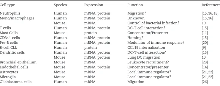
ACKRs are seven-transmembrane span-ning proteins that bind chemotactic fac-tors in a promiscuous manner in the absence of the classic G-protein-coupled signaling that characterizes chemotactic receptors. ACKRs, located at either the haematic or lymphatic endothelial barri-ers, are now believed to shape the chemo-tactic gradient through the degradation of secreted chemokines or the presenta-tion/concentration of chemokines on the surface of activated endothelial cells. In the Viewpoint series in this issue, Del Prete et al. summarize recent insights into an ACKR named CCRL2 [7].
Chemotactic factors include pathogen-associated molecular patterns such as formylated peptides, complement pro-teins (e.g. C5a and C3a), lipids (e.g. PAF and LTB4), and chemokines [8]. The Viewpoint by Yang et al. summa-rizes the role of alarmins, a new hetero-geneous family of chemotactic agonists [9]. Alarmins include different types of molecules, such as antimicrobial peptides, nuclear-binding proteins, heat shock pro-teins, nucleotides/metabolites, and gran-ule proteins released by stressed cells. Alarmins induce the recruitment and acti-vation of antigen presenting cells, as well as other immune cells, often in cooperation with other chemotactic factors through the activation of different types of receptors, many of them still uncharacterized.
Impaired migration in primary
immunodeficiency
The crucial role of leukocyte recruitment in immune responses is witnessed by the
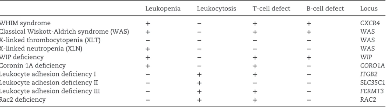
clinical manifestations associated with genetic defects in cell adhesion, actin cytoskeleton reorganization, and chemokine receptor signaling that are reviewed in the Viewpoint by Badolato [10]. Historically, the first such evidence was obtained in patients with mutations in the gene encoding CD18, the β2 inte-grin chain (LAD I; leukocyte adhesion deficiency-1) and in the gene encoding for a GDP-fucose transporter involved in the synthesis of selectin ligands (LAD II). More recently, a third genetic adhesion defect, characterized by mutations in the gene encoding for kindlin-3, a protein involved in integrin activation, has been reported and named LAD III. All these patients present neutrophilia and increased suscep-tibility to bacterial infections. Since LAD III patients present a general defect in integrin activation, they also experience defects in platelet activation and osteoclast func-tions [11]. A defect in actin polymeriza-tion known as Wiskott–Aldrich Syndrome (WAS) is caused by mutations in a pro-tein, the WAS protein (WASP), involved in actin polymerization. Mutations in WASP are responsible for four related diseases characterized by microthrombocytopenia, eczema, and recurrent bacterial infections due to the reduction in the number of na¨ıve T cells, decreased antibody response and impaired migration of innate effector cells and lymphocytes [12]. Finally, the WHIM (warts, hypogammaglobulinemia, infections, and myelokathexis) syndrome represents the first identified example of human disease characterized by a genetic defect of a chemokine receptor. In WHIM patients, the expression of truncated forms of the C-terminal tail of the receptor is associated with a prolonged response to CXCL12, the CXCR4 cognate ligand, which results in neutrophil bone marrow reten-tion. Patients with WHIM syndrome have increased bacterial infections, especially of the respiratory tract; in agreement with the phenotype originally observed in mice deficient for the CXCL12 receptors, CXCR4 and CXCR7 [13, 14], WHIM patients also show a higher rate of congenital heart defects.
Migration
and
induction
of
tolerance
Tolerance to self and innocuous antigens is provided by two main mechanisms, namely central tolerance in primary lym-phoid organs and peripheral tolerance in
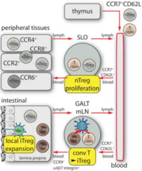
secondary lymphoid organs and peripheral tissues. Chemokines were shown to be pro-foundly involved in both types of toler-ance acting at the level of T-cell and DC recruitment [15]. T-cell central tolerance is induced in the thymus and a nonredun-dant role of chemokines was shown in the trafficking of progenitor cells from the cor-tex to the medulla [16]. Chemokines were also shown to play a crucial role in the recruitment and positioning of thymic DC subsets. In their Viewpoint, Hadeiba and Butcher discuss the role of three main sub-sets of DCs present in the thymus, each of them endowed with specific roles in T-cell clonal deletion and Treg-T-cell induc-tion [17]. The recruitment and correct homing of each of these populations was shown to be dependent on the expression of specific chemokine receptors, such as CCR9 for plasmacytoid DCs, XCR1 for res-ident DCs and CCR2 for migrating con-ventional DCs [18–20]. Similarly, Treg cells, which represent one of the main effector mechanisms of peripheral toler-ance, home, and recirculate through the expression of specific chemokine recep-tors. Thymic-derived natural Treg cells leave the thymus and migrate to sec-ondary lymphoid organs through the expression of CCR7 and CD62L. On the contrary, inducible Treg cells need to express selective homing receptors to recir-culate between the lymph nodes and the extra-lymphatic tissues, such as gut or skin, in order to properly shape their function. In their Viewpoint, Pabst and Bernhardt focus on the mechanisms that control the generation, maintenance, and function of Treg cells in the digestive tract [21].
New
insights
in
cell
migra-tion behavior by novel imaging
techniques
During the past decade, two-photon laser scanning microscopy (2-PM) has been applied with great success to reveal the positioning, movement, and interaction dynamics of immune cells within intact tissues. Compared with conventional epi-fluorescence or confocal microscopy, 2-PM provides better penetration depth in com-bination with little tissue damage while the spatial resolution is of poorer quality. Since 2-PM has been frequently used in intravi-tal experimenintravi-tal settings, the analysis of cellular interactions taking place within living, anaesthetized animals has offered
profound new insights into immune cell motility and function. In contrast to con-ventional fluorescence microscopy, which uses excitation wavelengths within the visible spectrum, in 2-PM a single fluo-rochrome molecule has to be hit not by one but, nearly simultaneously, by two infrared photons. In order to achieve exci-tation, these 2 photons are of approxi-mately double the wavelength and thus half the energy [22]. This approach offers several advantages. First, due to its longer wavelength, the infrared excitation light is scattered less that yields higher pene-tration depth. Second, only within a very small volume within the focal point of the optical system is the probability high enough that near-simultaneous interac-tions of two infrared photons with the very same fluorochrome molecule actually result in its excitation. Thus, generation of “out-of-focus” light as well as fluorochrome bleaching outside of the focal plane is intrinsically eliminated, which helps to conserve the specimen and allows con-tinuous imaging of the very same region for several hours. Tang et al. discuss, in their Viewpoint [23], recent advantages and the so far unmet needs in fluorescence-based optical imaging that are required to further dissect complex cell–cell interac-tions and to visualize the functional con-sequences for immunity and tolerance. Tang et al. [23] discuss five important challenges — imaging depth, multiplex detection, faster imaging, higher resolu-tion, and data capture and processing — that require considerable improvement to gain substantially new insights into the processes that control cell motility and the functional consequences of these processes.
Interfering with cell migration as
novel therapeutic approaches
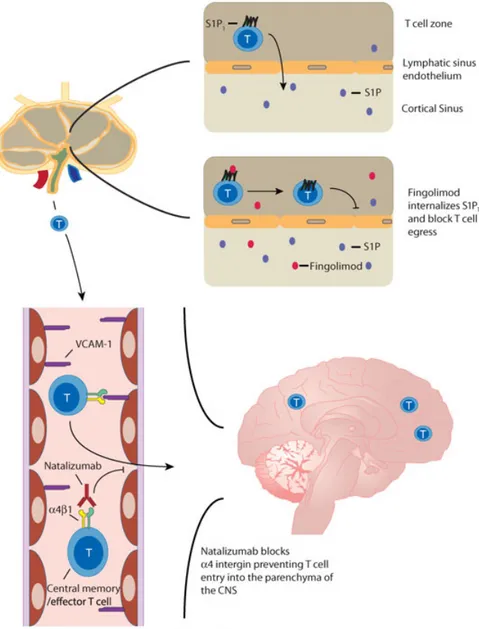
As outlined above, the homing of immune cells to defined organs and subcompart-ments therein is controlled by the orches-trated expression of several classes of molecules required for cell homing. This feature not only allows the controlled access to tissues but also offers new ther-apeutic approaches that interfere with selective steps of the cell homing cas-cade. Griffith and Luster’s Viewpoint gives an overview of the molecules involved in cell homing during inflammatory pro-cesses that are currently targets in clin-ical trials. In particular, they focus ontwo compounds: Natalizumab and Sphin-golimod (FTY720) [24]. Natalizumab is a humanized mAb that blocksα4 integrins and thus interferes with α4β7 integrin-mediated homing of cells to the intes-tine andα4β1 integrin-dependent homing to the brain. This drug has shown very promising results in treating patients with inflammatory bowel disease and multiple sclerosis [25, 26]. However, treatment of patients with this antibody also creates new challenges. In some patients the neu-rotropic JC virus, which is present in more than 50% of the healthy adult population, cannot be controlled, leading to progres-sive multifocal leukoencephalopathy [27]. Sphingolimod is also being tested in clini-cal studies in patients with MS. However, the mode of action is rather different. It binds to four of the five sphingosine-1-phosphate receptors [28] and evidence is accumulating that Sphin-golimod mediates its therapeutic effects by inhibiting the egress of activated T cells from secondary lymphoid organs [29]. Thus, it seems that the trapping of activated cells in lymphoid organs rep-resents the main mode of action of this substance.
Conclusions
Based on fundamental experiments of the past two decades, the mechanisms that regulate egress of immune cells from blood to lymphoid and nonlymphoid tissue are largely understood. In contrast, hardly anything is known regarding the homing of afferent lymph-derived cells to lymphoid organs. This open field requires further investigation since many of the fundamen-tal processes of immunology, such as DC mobilization from the peripheral organs to the draining lymph nodes, rely on this traf-ficking route. New imaging techniques like 2-photon and light-sheet microscopy will be of profound value in gaining a better understanding of the molecular and cellu-lar mechanisms that regulate immune cell trafficking from and to the bloodstream as well as the lymphatic system. The selec-tive interference with immune cell hom-ing or egress to or from defined lym-phoid and nonlymlym-phoid organs offers new therapeutic strategies to dampen chronic inflammatory processes. Several biolog-ics, in particular those directed against
different integrins and selectins, are cur-rently being tested in clinical trials and show very promising results. Although belonging to the large and frequently therapeutically targeted family of G protein-coupled receptors, the therapeutic potential of targeting chemoattractant receptors is still largely unexploited. Clearly, further studies are needed to bet-ter delineate the capacity of this receptor class as drug targets.
Acknowledgments: The work of R.F. is supported by grants of the Deutsche Forschungsgemeinschaft DFG (SFB621-A1; SFB587-B3; SFB738-B5, SFB900-B1; FO334/5–1) and the European Research Council (ERC Advanced Grant 322645, Lymphatics-Homing). S.S. is sup-ported by AIRC (Associazione Italiana per la Ricerca sul Cancro) and Minis-tero dell’Istruzione, dell’Universit`a e della Ricerca (M.I.U.R.), European Project IMI JU-funded project BeTheCure, contract 115142–2.
Conflict of interest: The authors declare no financial or commercial conflict of interest.
References
1 Butcher, E. C., Cell 1991. 67: 1033–1036. 2 Springer, T. A., Annu. Rev. Physiol. 1995. 57:
827–872.
3 Ley, K. et al., Nat. Rev. Immunol. 2007. 7: 678–689.
4 Sanz, M. J. and Kubes, P., Eur. J. Immunol. 2012.
42: 278–283.
5 Salmi, M. and Jalkanen, S., Eur. J. Immunol. 2012. 42: 284–292.
6 Girard, J. P. et al., Nat. Rev. Immunol. 2012. 12: 762–773.
7 Del Prete, A. et al., Eur. J. Immunol. 2013. 43: 1418–1422.
8 Sozzani, S., Cytokine Growth Factor Rev. 2005.
16: 581–592.
9 Yang, D. et al., Eur. J. Immunol. 2013. 43: 1412– 1418.
10 Badolato, R., Eur. J. Immunol. 2013. 43: 1435– 1440.
11 Etzioni, A., Curr. Opin. Immunol. 2009. 21: 481–486.
12 Thrasher, A. J. and Burns, S. O., Nat. Rev.
Immunol. 2010. 10: 182–192.
13 Sierro, F. et al., Proc. Natl. Acad. Sci. USA 2007.
104: 14759–14764.
14 Zou, Y. R. et al., Nature 1998. 393:
595–599.
15 Liu, X. et al., Eur. J. Immunol. 2011. 41: 611–623.
16 Bunting, M. D. et al., Immunol. Cell Biol. 2011.
89: 185–196.
17 Hadeiba, H. and Butcher, E. C., Eur. J. Immunol. 2013. 43: 1425–1429.
18 Baba, T. et al., J. Immunol. 2009. 183: 3053–3063.
19 Hadeiba, H. et al., Immunity 2012. 36: 438–450.
20 Lei, Y. et al., J. Exp. Med. 2011. 208: 383–394. 21 Pabst, O. and Bernhardt, G., Eur. J. Immunol.
2013. 43: 1422–1425.
22 Cahalan, M. D. et al., Nat. Rev. Immunol. 2002.
2: 872–880.
23 Tang, J. et al., Eur. J. Immunol. 2013. 43: 1412– 1418.
24 Griffith, J. W. and Luster, A. D., Eur. J. Immunol. 2013. 43: 1429–1435.
25 Ransohoff, R. M., N Engl. J. Med. 2007. 356: 2622–2629.
26 Ghosh, N. et al., Int. Rev. Immunol. 2012. 31: 410–427.
27 Berger, J. R. and Koralnik, I. J., N. Engl. J. Med. 2005. 353: 414–416.
28 Mandala, S. et al., Science 2002. 296:
346–349.
29 Cyster, J. G. and Schwab, S. R., Annu. Rev.
Immunol. 2012. 30: 69–94.
Correspondence: Prof. Reinhold Forster, Institute of Immunology, Hannover Medical School, Carl-Neuberg-Str. 1, Hannover 30625, Germany
e-mail: [email protected]
Additional correspondence: Prof. Silvano Sozzani; Department Molecular and Translational Medicine, University of Brescia, Italy
e-mail: [email protected] Received: 30/4/2013
Revised: 30/4/2013 Accepted: 7/5/2013
Keywords: Cell migration· Immunodeficien-cies· Immunotherapy
Abbreviations: ACKR: atypical chemokine receptor · WAS: Wiskott–Aldrich syndrome · WHIM: warts, hypogammaglobulinemia, infections, and myelokathexis
The completeCells in Motion Viewpoint series is available at:
http://onlinelibrary.wiley.com/doi/10. 1002/eji.v43.6/issuetoc
Technologies to observe and understand cells in motion
The future of immunoimaging — Deeper, bigger, more precise, and definitively more
colorful
Jianyong Tang, Nicolas van Panhuys, Wolfgang Kastenm¨uller and Ronald N. Germain
Lymphocyte Biology Section, Laboratory of Systems Biology, National Institute of Allergy
and Infectious Diseases, National Institutes of Health, Bethesda, MD, USA DOI: 10.1002/eji.201243119
Immune cells are thoroughbreds, moving farther and faster and survey-ing more diverse tissue space than their nonhematopoietic brethren. Intravital 2-photon microscopy has provided insights into the move-ments and interactions of many immune cell types in diverse tis-sues, but more information is needed to link such analyses of dynamic cell behavior to function. Here, we describe additional methods whose application promises to extend our vision, allowing more complete, mul-tiscale dissection of how immune cell positioning and movement are linked to system state, host defense, and disease.
The immune system is like a fine mechanical watch — there are a large number of parts that must work together to achieve the right result. For the watch, the goal is keeping perfect time, and for the immune system, the goal is optimally protecting the host. If the pieces are not machined and assem-bled properly, the watch (immune sys-tem) can run too slowly (immuno-deficiency) or too fast (autoimmunity/ inflammatory disease). There is a del-icate balance in the interaction of the watch parts — they must move properly, engage for just the right amount of time, then disengage, and move again. Like-wise, the cells of the immune system must circulate and migrate, find the right cel-lular partner at the right time, engage for the proper duration, signal effectively, change gene expression, and then move once again. Dynamics and positioning are crucial aspects of immune function that need to be described and understood if we want to have an accurate picture of
the system and how it carries out its activities.
Like a watchmaker, who uses magnify-ing lenses to peer at the minute parts of a complex timepiece to check its function, investigators have turned to optical imag-ing to gain knowledge about the dynamic properties of immune cells in their in vivo environments. Over the past decade, in particular, intravital 2-photon imaging has provided a wealth of insights into what is now called “immunodynamics” [1–3]. We have seen how na¨ıve T and B cells move within secondary lymphoid tissues and acquire antigenic information [4–14], the intricate dance of T and B cells at the T/B-border and within germinal centers [15–20] and of developing thymocytes in that organ [21, 22], the reactivation of memory T cells [23–25], and osteoclast, platelet, and neutrophil mobilization in the bone marrow [26–28], as well as the movement of innate and adaptive effectors in tissues such as skin [29–31], liver [32–34], central nervous system [35–37], lung [38, 39], and tumors [40, 41] among others. Migration and local probing behavior of dendritic cells in diverse sites has been examined [42– 45]. The role of stromal elements in guiding immune cell migration has been discovered [46], the key contribution of adequate cell–cell adhesion in over-coming the dispersive migratory proper-ties of lymphocytes and permitting effec-tive intercellular cooperation has become clear [47], the restricted anatomical domains in secondary lymphoid organs within which some innate and adap-tive immune cells migrate while await-ing evidence of host invasion have been revealed [48], and the in vivo operation of chemokines with regard to facilitating the encounter between rare cell populations delineated [49, 50].
While these discoveries have “ani-mated” the field for years, we are still far from where we need to be to link informa-tion on molecules, signaling pathways, and
gene regulatory events to these descriptive dynamics. New tools and techniques are required to see more for longer in larger volumes. Methods that allow simultaneous tracking of receptor signaling events and cell movement, of cytokine production and the response to these key mediators and of gene activation are essential for connecting dynamic behavior with function and differ-entiation. Regions of tissues currently inac-cessible to our imaging platforms need to be made visible, tracking needs to occur in larger volumes to avoid loss of cells over the time span involved in their pro-gression from resting cells to a differen-tiated state, many more cell types (includ-ing stromal elements such as mesenchymal cells, nerves, and vessels) need to be dis-tinctively labeled for visualization at one time, and resolution must be increased to permit intracellular elements to be mon-itored. New computational tools must be developed to cope with the vast amount of data that will be generated by imag-ing more colors, with greater resolu-tion, for longer times, and in larger vol-umes, including visualization methods that make such complex data understandable to the experimentalist. In this Viewpoint, we briefly describe the evolving methods (Fig. 1) that will contribute to overcom-ing these limitations and how their imple-mentation will provide essential insights into immune function in health and disease.
Challenge number 1 — imaging
depth
Even in mice, the main experimental ani-mal used for immune system dynamic imaging, many events that need to be visu-alized are hundreds of microns to mil-limeters from the surface of a tissue. Cur-rent fluorescence-based intravital imaging techniques can only probe the region near the surface (up to 200–300μm) in dense
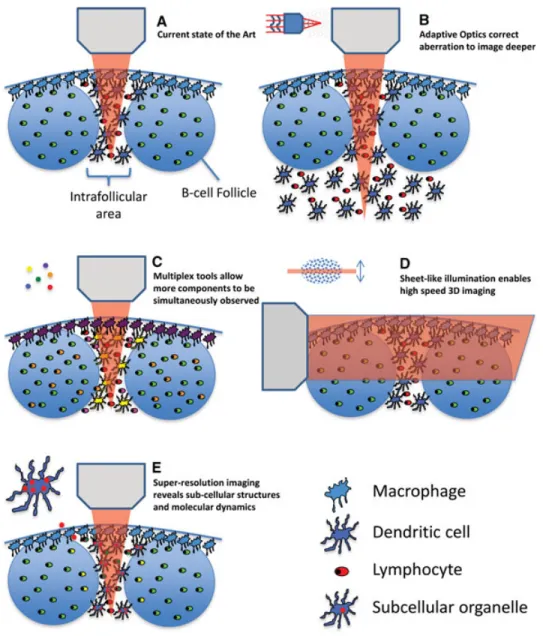
Figure 1. Overview of improved imaging modalities. (A) The current state of the art in imaging the interfollicular area and B-cell fol-licles of a lymph node is shown. (B) Improved imaging depth by means of adaptive optics is shown. (C) Detection of multiple different signals by means of multiplexing tools such as multiple lasers or new chromophores is shown. (D) High-speed 3D imaging using sheet illu-mination is shown. (E) The visual-ization of subcellular compartments using super-resolution imaging tech-niques is shown.
lymphoid and other tissues; this is because of the dispersive optical properties of these biological structures. To gain information deep in live tissue by multiphoton imag-ing, photons carrying input information (excitation) have to be efficiently deliv-ered to the location of interest (focus), and the output information (fluorescence) has to be transmitted back via emitted photons that can be collected and detected by light sensors. Within deeper areas of tissues, scattering and absorption pro-cesses will dramatically attenuate the exci-tation laser intensity that reaches the focal point and the heterogeneous structure of the tissue will distort the light propagation wavefront, so the excitation light cannot be tightly focused. Such optical aberration is a major factor leading to degradation of image quality.
Recognizing this limitation, many groups have been working on solutions. Near term, the simplest approach is the use of far-red or near-infrared (IR) fluorochromes and fluorescent proteins [51–54]. The longer wavelength of the excitation and emission photons involved in imaging such fluorochromes and pro-teins allow for better tissue penetration with less scattering or absorption than shorter wavelength photons. A penetration depth up to 1.6 mm in mouse cortex has been demonstrated with the laser wave-length tuned at 1.28μm and emission in the near-IR range [55].
A very promising method that is not limited by wavelength in this way involves a technique that is termed adaptive optics (AO) (Fig. 1B). Such a method aims to pre-compensate the light wavefront distortion
inside the live tissue and allow a maximum amount of coherent laser light to reach the focal point. This approach can dramat-ically improve imaging contrast [56, 57]. While the speed of the wavefront opti-mization process has been increased sig-nificantly, compatible with typical dynamic multiphoton intravital imaging methods, the improvement of signal quality using AO approaches comes with the price of a smaller field of view. This is because the wavefront distortion has to be mea-sured and compensated at different loca-tions inside the heterogeneous tissue sample, a process that takes time both due to the physical properties and software control of the adaptive mirrors employed to control the beam. These limitations will eventually be overcome through develop-ment of faster hardware and software.
Even with current AO technology, one can now perform highly resolved imag-ing in small fields deep within tissues that are structurally stable (i.e. bone marrow, skin).
Other new optical approaches that extend the penetration depth even further, to beyond millimeter scale, are also in development. One such tech-nique is ultrasound-guided optical imaging [58, 59], which takes advantage of an acoustic wave. The latter is less sensitive to the tissue medium that scatters the light wave and can be used to guide excitation and emission photons. This method can currently achieve imaging resolution up to 12μm at a depth of 2 mm [59], making it potentially useful not only for deep ani-mal imaging but also for application to the dermis in humans, as one example.
These methods not only promise to allow visualization of objects deeper in tissues than presently accessible, but also the imaging over time of larger volumes than can be presently examined. Because of cell motility, capture of information from a larger volume not only reduces sam-pling error (more events can be tracked), but also permits cells to be followed over longer time intervals (because they stay within the imaging volume). In combina-tion with hardware improvements that per-mit faster scanning, more sensitive pho-ton detection, and faster movement of the stage holding the specimen, these advances will provide an enhanced abil-ity to link early and late events during an ongoing response on a per-cell basis throughout a larger range of organ com-partments. Ultimately, this will allow for continuous tracking of cells and their fates after asymmetric division and during dif-ferentiation [60].
Challenge number 2 — multiplex
detection
Immunity is a highly concerted process involving many different cellular and molecular players. For this reason, the present imaging methods that typically involve three to four colors per experi-ment are incapable of revealing many of the elements that play an important role in immune processes. This limitation applies at the macroscale (insufficient diversity of cell types and stromal structures visual-ized), so that the impact of tissue orga-nization and many different types of cell– cell contacts is not well appreciated. It is also true at the nanoscale — without an
ability to multiplex more extensively, we cannot track both cell behavior and the molecules involved in cell interactions and signaling or gene activity. Yet, such co-measurements are critical if we are to link bulk dynamics to the processes of cellular activation and response crucial to immune function. Achieving these goals requires new experimental capability to observe multiple different cellular and molecular players at the same time, in the same sample, and with the same imaging con-figuration.
The current spectral range of fluo-rescence detection is from near ultra-violet (UV) to near IR. Considering the intrinsic fluorescent bandwidth of typical chromophores, the number of different fluorophores that can be distin-guished within this spectral range is lim-ited using conventional filtration methods, even assuming that an ideal set of labels with well-separated fluorescent spectra can be employed experimentally. The fol-lowing four methods promise to overcome these present limitations by facilitating the simultaneous detection of multiple compo-nents (Fig. 1C):
(i) The use of multiple lasers tuned to different excitation wavelengths. Employed in the proper way, this allows optimized excitation of a vari-ety of fluorochromes rather than the severe compromise typical of single-laser instruments, permitting sub-stantial improvement in detection of labels expressed at low levels and the use of more distinct fluorochromes that in combination would not be well excited with single-laser systems [61, 62].
(ii) The development of new chro-mophores (synthetic dyes or fluores-cent proteins) with fluorescence spec-tra beyond the current range. For example, more near-IR or IR fluo-rescent proteins have already been developed in the past few years [51, 63–66], extending the palette avail-able for imaging studies with current microscope systems.
(iii) The application of spectral unmixing strategies. Rather than using filters to isolate distinct (nonoverlapping) regions of emission spectra to iden-tify targets, one can “deconvolve” the entire emitted fluorescent spectrum to identify the target chromophore by its specific emission spectral profile [67]. This requires the chromophore to have a signal strong enough to be
split into the multiple spectral win-dows, each of which captures fewer total emitted photons. One has to find a balance between the spectral preci-sion (more windows) and sensitivity (signal/noise= larger windows), set-ting a limit to the use of this approach when the cell or molecule of interest can only have a limited overall fluo-rescent output. A related approach is to label each target component with multiple chromophores and modu-late the ratio between different chro-mophores to identify various biolog-ical components (spectral painting). This approach has been successfully demonstrated in the application of single molecule mRNA FISH [68] and in Brainbow [69] and similar trans-genic animals. Here, the complex emission spectrum from each differ-ent color combination provides a sig-nature for that target. This approach can be further enhanced by careful choice of chromophores with distinct absorption spectra permitting selec-tive excitation at different laser wave-lengths when using multiline instru-ments.
(iv) The combination of standard wave-length detection methods with other strategies for fluorochrome identifica-tion. One well-established approach in single-cell imaging is fluorescence lifetime measurement [70, 71] and this has recently been incorporated into multiphoton intravital imaging [72]. In this method, special detec-tors and software statistically charac-terize the time interval (on a nano-second scale) between the excitation and emission events for each chro-mophore molecule. Many spectrally overlapped chromophores have very distinct fluorescence lifetimes, so this technique can be used to add another dimension to the “identity space” of these labels. With the newest gener-ation of high sensitivity detectors on commercial microscopes, implemen-tation of fluorescence lifetime mea-surement is becoming an available tool for intravital multiphoton imag-ing.
A final area in which substantial progress is needed in the area of multi-plex detection is in the creation of opti-mized probes for the analysis of intra-cellular signaling, molecular localization, or gene expression. A few intravital stud-ies have characterized such events, most
involving dye-based calcium sensors [13, 73], in vivo staining [74], fluorescent chimeric proteins [75, 76], and fluorescent gene reporters [77–80]. The calcium stud-ies are limited by the leak rate of the sen-sor dyes to just an hour or two after cell transfer, the chimeric proteins are both difficult to detect and the analysis suffers from artifacts of optical resolution limita-tions and signal intensity differences in the axial dimension, and the gene reporters produce cytoplasmic proteins whose life-time greatly exceeds that of cytokine tran-scripts, thus failing to provide a properly time-resolved record of gene activity that can be linked to cell dynamics [81]. To make progress in these areas, these limita-tions must be overcome. New fluorescent proteins have been developed [82] for the generation of optimized FRET sensors that can detect signaling events such as calcium elevation, MAPK pathway activation, and the like and should allow creation of genet-ically labeled cells whose signaling can be tracked without the present posttransfer time limitation. The improved sensitivity of newer instruments will permit better detection of chimeric proteins expressed close to physiologic levels and some of the methods described below will enhance axial resolution that presently limits such analyses. The creation of a new genera-tion of genetic reporters producing desta-bilized fluorescent reporters, or secreted versions of such proteins, will improve the temporal connectivity between appearance of these labeled proteins and the underly-ing genetic activities of the cell of interest [83]. Together, such tools will allow inves-tigators to link molecular events with cell behavior.
Challenge number 3 — faster
imaging
The complexity of biological systems not only exists in the space domain, but also in the time domain. Cellular and molecular dynamics have multiplex temporal features ranging from femtoseconds to hours. Both the sampling speed and length of image collection of present instruments and their linked computers dictate the time resolu-tion that can be achieved.
To date, the predominant scheme for sample illumination is point scanning, which limits information collection rate due to the time needed to move the beam over the entire x-y dimensions of the imag-ing field. A major breakthrough in the past few years allows for parallel illumination
of biological samples, sometimes in three dimensions. In these methods, a modi-fied optical system illuminates a selected plane rather than a single point inside the biological sample, with the fluores-cence from the whole plane detected by a wide field imager (Fig. 1D). For exam-ple, light sheet microscopy [84–86] uses one lens to create a narrow sheet of laser focus inside the tissue, and another objec-tive is used to acquire fluorescent images at the perpendicular direction. Bessel beam-based light sheet microscopy [87] further improves the spatial resolution especially in the axial dimension. Another tempo-ral focusing scheme [88, 89] modulates the excitation laser pulse width so that the multiphoton excitation is confined in a sheet-like region. Although the tempo-ral focusing method is limited to non-linear excitation, it brings the convenience of using only a single objective lens with less spatial restriction for sample arrange-ment.
Challenge number 4 — higher
resolution
In principle, the resolution of an opti-cal microscope is limited by the optiopti-cal diffraction, in the range of a few hundred nanometers with a lens of the highest pos-sible numerical aperture. In practice, one has to find a balance between the working distance of the lens and numerical aper-ture, preventing use of the best resolv-ing lenses for most intravital imagresolv-ing pur-poses. In addition, the optical aberration inside tissues also degrades resolution, so methods to correct such aberrations, like the AO approaches discussed above, are needed to achieve near-diffraction-limit resolution deep inside tissues.
During the past decade, however, there have been a number of exciting break-throughs that allow biological samples to be imaged at a resolution far beyond the optical diffraction limit (Fig. 1E). These novel approaches can be generalized into two types: one type takes advantage of sin-gle molecule location measurement, such as photoactivated localization microscopy, fluorescence photoactivated localization microscopy, and stochastic optical recon-struction microscopy [90–92]; the other creates subdiffraction excitation patterns, such as stimulated emission depletion [93] and structured illumination [94]. These new techniques are poised to reveal detailed molecular and structural informa-tion inside live tissues. Although to date
most applications are with relatively flat cells in culture systems, some exciting recent developments have begun to incor-porate such schemes into intravital imag-ing scenarios [95], permittimag-ing the detec-tion of subcellular events with improved precision, at least close to the surface of various tissues [96].
Challenge number 5 — data
cap-ture and processing
All the above advances will help collect more information on the immune system in situ, but without the proper means for data analysis, we will make little progress in understanding things. A new generation of software tools is needed to better analyze the image data, prefer-ably allowing more to be done automati-cally and in an unbiased manner than is presently possible [97]. Some progress has been made in this arena over the past sev-eral years, both by commercial software vendors and academic centers, but much more is needed. Optimized algorithms are required to handle the much larger num-ber of objects to be tracked when the imaging volume is increased and more cell types are visualized in more colors, for both analytic purposes and for display of the underlying cell movements; embry-ologists have made substantial progress in this direction [86, 98] and their meth-ods need to be adopted (and adapted) by immunological imaging experts. Progress is also needed to address specific issues in molecular imaging, which is highly sensi-tive to depth-related image intensity arti-facts. Some schemes to deal with such problems have already appeared [75, 79] but as the field embraces molecular imag-ing goimag-ing forward, further improvements will be crucial. Other analytic tools that have been introduced to handle the anal-ysis of cell movements into and out of defined volumes such as germinal cen-ters or to measure synaptic dimensions [99, 100], provide a display in two dimen-sions out of four or more parameters such as time, speed, directionality, and distance from a defined site. These parameters greatly aid in understanding chemosens-ing behavior (L¨ammermann et al., sub-mitted manuscript), differentiate random walk from other migratory behavior [101, 102], and evaluate contact times between two cell types [103]. These datasets may then be used to derive mathematical mod-els of complex cellular behavior, which by iterative processes can be refined and
may allow for prediction of biological out-comes that can be tested in in vivo models [102,104–110]. Finally, the field will need to move away from the maximum projec-tion of the 2D movies that are currently used for 3D displays that allow a better appreciation of depth in the full imaging volume.
Conclusion
Here, we have very briefly surveyed some of the emerging techniques that will aid our probing of the dynamic behavior of the immune system going forward. They will allow a larger portion of an organ to be examined, many more elements to be tracked simultaneously, cell analysis to be combined with molecular imaging, and cell function linked to dynamic behavior. An essential point to emphasize, however, is that while such imaging can be revealing in its own right, it is most valuable as a part of the larger fabric of immune investigation; dynamic imaging data need to be properly associated with information gathered by other means, such as static tissue imaging [111, 112], flow cytometry, ex vivo assess-ment of lymphocyte activity and polariza-tion, genetic and epigenetic studies, and overall measurements of systemic immu-nity and host resistance. Only through inte-gration of information garnered using the full range of methods available to the field can we develop a comprehensive model of immune system behavior, in which events on the micro- and macroscale are linked to the mesoscale dynamics of individual cells that are the present focus of imag-ing analysis. With the increased depth and breadth of analysis, we anticipate from rig-orous application of the methods reviewed here, we are confident the future is, not to put too fine a point on it, “bright”. Acknowledgments: This work was sup-ported by the Intramural Research Pro-gram of NIAID, NIH. We apologize to all colleagues whose primary work could not be directly cited due to editorial length and reference limitations.
Conflict of interest: The authors declare no financial or commercial conflict of interest.
References
1 Germain, R. N. et al., Science 2012. 336: 1676– 1681.
2 Victora, G. D. et al., Annu. Rev. Immunol. 2012.
30: 429–457.
3 Pittet, M. J. et al., Cell 2011. 147: 983–991. 4 Miller, M. J. et al., Science 2002. 296: 1869–
1873.
5 Stoll, S. et al., Science 2002. 296: 1873–1876. 6 Lindquist, R. L. et al., Nat. Immunol. 2004. 5:
1243–1250.
7 Suzuki, K. et al., J. Exp. Med. 2009. 206: 1485– 1493.
8 Phan, T. G. et al., Nat. Immunol. 2007. 8: 992– 1000.
9 Carrasco, Y. R. et al., Immunity 2007. 27: 160– 171.
10 Junt, T. et al., Nature 2007. 450: 110–114. 11 Mempel, T. R. et al., Nature 2004. 427: 154–
159.
12 Hickman, H. D. et al., Nat. Immunol. 2008. 9: 155–165.
13 Qi, H. et al., Science 2006. 312: 1672–1676. 14 Bousso, P. et al., Nat. Immunol. 2003. 4: 579–
585.
15 Victora, G. D. et al., Cell 2010. 143: 592–605. 16 Qi, H. et al., Nature 2008. 455: 764–769. 17 Okada, T. et al., PLoS Biol. 2005. 3: 1047–1061. 18 Allen, C. D. et al., Science 2007. 315: 528–531. 19 Schwickert, T. A. et al., Nature 2007. 446: 83–
87.
20 Gunzer, M. et al., Blood 2004. 104: 2801–2809. 21 Bousso, P. et al., Science 2002. 296: 1876–1880. 22 Le Borgne, M. et al., Nat. Immunol. 2009. 10:
823–830.
23 Cavanagh, L. L. et al., Nat. Immunol. 2005. 6: 1029–1037.
24 Sung, J. H. et al., Cell 2012. 150: 1249–1263. 25 Kastenmuller, W. et al., Immunity 2013.
38:502–513.
26 Kohler, A. et al., Blood 2011. 117: 4349–4357. 27 Ishii, M. et al., Nature 2009. 458: 524–528. 28 Junt, T. et al., Science 2007. 317: 1767–1770. 29 Celli, S. et al., Nat. Med. 2011. 17: 744–749. 30 Peters, N. C. et al., Science 2008. 321: 970–974. 31 Gebhardt, T. et al., Nature 2011. 477: 216–219. 32 Egen, J. G. et al., Immunity 2011. 34: 807–819. 33 McDonald, B. et al., Science 2010. 330: 362–
366.
34 Geissmann, F. et al., PLoS Biol. 2005. 3: e113. 35 Wilson, E. H. et al., Immunity 2009. 30: 300–
311.
36 Kawakami, N. et al., J. Exp. Med. 2005. 201: 1805–1814.
37 Kim, J. V. et al., Nature 2009. 457: 191–195. 38 Kreisel, D. et al., Proc. Natl. Acad. Sci. USA
2010. 107: 18073–18078.
39 Bruns, S. et al., PLoS Pathog. 2010. 6: e1000873. 40 Breart, B. et al., J. Clin. Invest. 2008. 118: 1390–
1397.
41 Boissonnas, A. et al., J. Exp. Med. 2007. 204: 345–356.
42 Chieppa, M. et al., J. Exp. Med. 2006. 203: 2841–2852.
43 McDole, J. R. et al., Nature 2012. 483: 345–349. 44 Thornton, E. E. et al., J. Exp. Med. 2012. 209:
1183–1199.
45 Miller, M. J. et al., Proc. Natl. Acad. Sci. USA 2004. 101: 998–1003.
46 Bajenoff, M. et al., J. Immunol. 2008. 181: 3947–3954.
47 Schneider, H. et al., Science 2006. 313: 1972– 1975.
48 Kastenmuller, W. et al., Cell 2012. 150: 1235– 1248.
49 Castellino, F. et al., Nature 2006. 440: 890–895. 50 Hugues, S. et al., Nat. Immunol. 2007. 8: 921–
930.
51 Filonov, G. S. et al., Nat. Biotechnol. 2011. 29: 757–761.
52 Lin, M. Z. et al., Nat. Methods 2011. 8: 726–728. 53 Mojzisova, H. et al., Curr. Opin. Genet. Dev.
2011. 21: 549–557.
54 Shcherbo, D. et al., Nat. Methods 2010. 7: 827– 829.
55 Kobat, D. et al., J. Biomed. Opt. 2011. 16: 106014.
56 Ji, N. et al., Nat. Methods 2010. 7: 141–147. 57 Tang, J. et al., Proc. Natl. Acad. Sci. USA 2012.
109: 8434–8439.
58 Wang, Y. M. et al., Nat. Commun. 2012. 3: 928. 59 Si, K. et al., Sci. Rep. 2012. 2: 748.
60 Chang, J. T. et al., Science 2007. 315: 1687– 1691.
61 Mahou, P. et al., Nat. Methods 2012. 9: 815– 818.
62 Entenberg, D. et al., Nat. Protoc. 2011. 6: 1500– 1520.
63 Hilderbrand, S. A. et al., Curr. Opin. Chem. Biol. 2010. 14: 71–79.
64 Luo, S. et al., Biomaterials 2011. 32: 7127–7138. 65 Andresen, V. et al., Curr. Opin. Biotechnol.
2009. 20: 54–62.
66 Subach, F. V. et al., Nat. Methods 2011. 8: 1019–1026.
67 Zimmermann, T. et al., Adv. Biochem. Eng.
Biotechnol. 2005. 95: 245–265.
68 Femino, A. M. et al., Science 1998. 280: 585– 590.
69 Livet, J. et al., Nature 2007. 450: 56–62. 70 Dong, C. Y. et al., Methods Cell Biol. 2003. 72:
431–464.
71 French, T. et al., Methods Cell Biol. 1998. 56: 277–304.
72 Fruhwirth, G. O. et al., Proc. SPIE 2009. 7183: 71830L-71830L.
73 Bhakta, N. R. et al., Nat. Immunol. 2005. 6: 143–151.
74 Schwendele, B. et al., Eur. J. Immunol. 2012.
42: 2193–2196.
75 Friedman, R. S. et al., J. Exp. Med. 2010. 207: 2733–2749.
76 McCombs, J. E. et al., Methods 2008. 46: 152– 159.
77 Hoffman, R. M. et al., Curr. Pharm. Biotechnol. 2012. 13: 537–544.
78 Giepmans, B. N. et al., Science 2006. 312: 217– 224.
79 Melichar, H. J. et al., Immunol. Cell Biol. 2011.
89: 549–557.
80 Croxford, A. L. et al., Immunology 2011. 132: 1–8.
81 Beuneu, H. et al., Immunity 2010. 33: 412–423.
82 Lam, A. J. et al., Nat. Methods 2012. 9: 1005– 1012.
83 Houser, J. R. et al., Yeast 2012. 29: 519–530. 84 Keller, P. J. et al., Science 2008. 322:
1065–1069.
85 Huisken, J. et al., Science 2004. 305: 1007– 1009.
86 Truong, T. V. et al., Nat. Methods 2011. 8: 757– 760.
87 Planchon, T. A. et al., Nat. Methods 2011. 8: 417–423.
88 Durst, M. E. et al., Opt. Commun. 2008. 281: 1796–1805.
89 Oron, D. et al., Opt. Express 2005. 13: 1468– 1476.
90 Betzig, E. et al., Science 2006. 313:
1642–1645.
91 Hess, S. T. et al., Biophys. J. 2006. 91: 4258– 4272.
92 Rust, M. J. et al., Nat. Methods 2006. 3: 793– 795.
93 Hell, S. W. et al., Opt. Lett. 1994. 19: 780–782.
94 Gustafsson, M. G. et al., Biophys. J. 2008. 94: 4957–4970.
95 York, A. G. et al., Nat. Methods 2011. 8: 327– 333.
96 York, A. G. et al., Nat. Methods 2012. 9: 749– 754.
97 Ludewig, B. et al., Eur. J. Immunol. 2012. 42: 3116–3125.
98 Tomer, R. et al., Nat. Methods 2012. 9: 755– 763.
99 Klauschen, F. et al., Nat. Protoc. 2009. 4: 1305– 1311.
100 Klauschen, F. et al., Nat. Protoc. 2009. 4: 1006– 1012.
101 Harris, T. H. et al., Nature 2012. 486: 545–548.
102 Beltman, J. B. et al., J. Exp. Med. 2007. 204: 771–780.
103 Dustin, M. L. et al., Proc. Natl. Acad. Sci. USA 1997. 94: 3909–3913.
104 Garin, A. et al., Immunity 2010. 33:
84–95.
105 Figge, M. T. et al., J. Exp. Med. 2008. 205: 3019– 3029.
106 Bogle, G. et al., PLoS One 2012. 7: e45258. 107 Beltman, J. B. et al., J. Immunol. Methods 2009.
347: 54–69.
108 Beltman, J. B. et al., Immunol. Cell Biol. 2007.
85: 306–314.
109 Bogle, G. et al., Immunol. Cell Biol. 2010. 88: 172–179.
110 Mandl, J. N. et al., Proc. Natl. Acad. Sci. USA 2012. 109: 18036–18041.
111 Gerner, M. Y. et al., Immunity 2012. 37: 364–376.
112 Moreau, H. D. et al., Immunity 2012. 37: 351–363.
Correspondence:Dr. Ronald N. Germain, Laboratory of Systems Biology, National Institute of Allergy and Infectious Diseases, National Institutes of Health, Bldg. 4, Rm. 126A MSC-0421, 10 Center Drive, Bethesda, MD 20892-1892, USA Fax:+1-301-480-1660 e-mail: [email protected] Received: 5/11/2012 Revised: 1/3/2013 Accepted: 3/4/2013
Keywords: 2-photon rDynamic rImaging r Immune responses rMulticolor
Abbreviations:AO: adaptive optics· IR: infrared
The completeCells in Motion Viewpoint series is available at:
http://onlinelibrary.wiley.com/doi/10. 1002/eji.v43.6/issuetoc
Molecules regulating migration
Alarmin-induced cell migration
De Yang1,2,3, Feng Wei2,3, Poonam Tewary2, O. M. Zack Howard2and Joost J. Oppenheim2
1Basic Research Program, Scientific Application and International Corporation–Frederick, Inc., Frederick, MD, USA
2Laboratory of Molecular Immunoregulation, Cancer and Inflammation Program, National Cancer Institute, Frederick National Laboratories for Cancer Research, Frederick, MD, USA
3Research Center of Basic Medical Sciences, Tianjin Medical University, Tianjin, China DOI: 10.1002/eji.201243138
Alarmins are endogenous, constitu-tively available, damage-associated molecular patterns that upon release can mobilize and activate various leukocytes for the induction of innate and adaptive immune responses. For our immune system to func-tion appropriately, it relies on nav-igating various leukocytes to dis-tinct places at the right time. The direction of cell migration is deter-mined by chemotactic factors that include classical chemoattractants,
chemokines, certain growth
fac-tors, and alarmins. This viewpoint provides an overview of alarmin-induced cell migration. Alarmins are capable of inducing the migra-tion of diverse types of leukocytes and nonleukocytes either directly by triggering specific receptors or indirectly by inducing production of chemokines through the activation of various leukocytes via pattern recog-nition receptors. The receptors used by alarmins to directly induce cell
migration can either be Gαi protein-coupled receptors or receptors such as the receptor for advanced glycation end products; however, the intracel-lular signaling events responsible for the direct chemotactic activities of alarmins are, to date, only par-tially elucidated. Given that alarmins act in concert with chemokines to regulate the recruitment and traf-ficking of leukocytes, these damage-associated molecular patterns are
biological processes as discussed in this viewpoint.
Introduction
The migration of single cells is impor-tant for many physiologic processes such as ontogenic development, organogenesis, hematopoiesis, tissue regeneration, and immune responses. Cell migration is also involved in pathological conditions such as autoimmune disorders, vascular disease, and tumor metastasis. The ability of the immune system to respond appropriately to microbial invasion, tissue damage, and other insults relies to a large extent on the mobilization/recruitment of various leuko-cytes and progenitor cells to the right place at the right time [1,2]. Migration of cells in vivo is controlled by many sequential inter-actions involving adhesion molecules, gly-cosaminoglycans, chemotactic factors, and their receptors [1–5].
For cells to move directionally, they must first acquire a polarized morphol-ogy where F-actin is primarily enriched at the front and myosin II is assembled on
the sides and at the back of the cell [6]. Subsequently, the polarized cells undergo a highly coordinated cycle of protrusions and retractions that are coupled with trac-tion provided by the formatrac-tion and release of adhesive contacts with the extracellu-lar matrices [6]. Cells must be able to determine where and when protrusions, retractions, and adhesions have to occur to migrate to the correct location, which is established by chemotactic gradients. Chemotactic factors are comprised of clas-sical chemoattractants such as formyl peptides and anaphylatoxins (e.g. C5a), chemokines including CXC, CC, CX3C, and XC chemokines, and growth factors such as EGF and vascular endothelial growth fac-tor [1, 4, 7, 8]. Classical chemoattractants and chemokines provide extracellular cues by signaling predominantly through Gαi protein–coupled receptors (GiPCRs), while growth factors do so by signaling through their corresponding receptors [4, 7, 8].
A more recently identified type of chemotactic factor is the alarmin family. Alarmins are structurally distinct endoge-nous mediators that, upon release and gaining access to immune cells, can acti-vate the immune system by inducing
the recruitment and activation of vari-ous leukocytes, particularly APCs, includ-ing DCs [9–11]. Consequently, alarmins are capable of inducing both innate and antigen-specific host immune responses. Most alarmins are constitutively expressed and stored in intracellular compartments such as the nucleus, cytoplasm, or gran-ules. The expression of some alarmins can also be upregulated by microbial products, cytokines, and stress. During microbial infection and/or tissue injury, alarmins rapidly become available extracellularly as a result of degranulation, passive release due to cell necrosis, or active release in response to inducing agents [9–11].
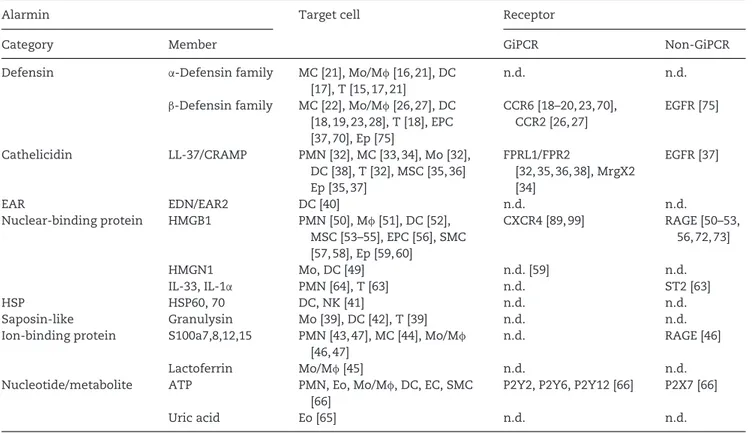
Known alarmins are multifunctional and can be classified into eight distinct molecular categories (Table 1), includ-ing defensins (e.g. α- and β-defensins), cathelicidin (e.g. human LL-37 or mouse cathelicidin-related antimicrobial peptide (CRAMP)), eosinophil-associated ribonu-cleases (e.g. eosinophil-derived neuro-toxin (EDN)), nuclear-binding proteins (e.g. high-mobility group box-1 pro-tein (HMGB1), high-mobility group nucleosome-binding protein 1 (HMGN1), and the cytokines IL-1α and IL-33), HSPs
Table 1. Target cells and receptors of alarmin-induced cell migration.
Alarmin Target cell Receptor
Category Member GiPCR Non-GiPCR
Defensin α-Defensin family MC [21], Mo/Mφ [16,21], DC
[17], T [15, 17, 21]
n.d. n.d.
β-Defensin family MC [22], Mo/Mφ [26,27], DC [18, 19, 23, 28], T [18], EPC [37, 70], Ep [75] CCR6 [18–20, 23, 70], CCR2 [26, 27] EGFR [75] Cathelicidin LL-37/CRAMP PMN [32], MC [33, 34], Mo [32], DC [38], T [32], MSC [35, 36] Ep [35, 37] FPRL1/FPR2 [32, 35, 36, 38], MrgX2 [34] EGFR [37] EAR EDN/EAR2 DC [40] n.d. n.d. Nuclear-binding protein HMGB1 PMN [50], Mφ [51], DC [52], MSC [53–55], EPC [56], SMC [57, 58], Ep [59, 60] CXCR4 [89, 99] RAGE [50–53, 56, 72, 73] HMGN1 Mo, DC [49] n.d. [59] n.d. IL-33, IL-1α PMN [64], T [63] n.d. ST2 [63] HSP HSP60, 70 DC, NK [41] n.d. n.d. Saposin-like Granulysin Mo [39], DC [42], T [39] n.d. n.d.
Ion-binding protein S100a7,8,12,15 PMN [43, 47], MC [44], Mo/Mφ
[46, 47]
n.d. RAGE [46]
Lactoferrin Mo/Mφ [45] n.d. n.d.
Nucleotide/metabolite ATP PMN, Eo, Mo/Mφ, DC, EC, SMC
[66]
P2Y2, P2Y6, P2Y12 [66] P2X7 [66]
Uric acid Eo [65] n.d. n.d.
CCR: CC chemokine receptor; EAR: eosinophil-associated ribonuclease; EC: endothelial cell; Ep: epithelial cell; FPRL1: formyl peptide receptor-like 1 receptor; Mφ: macrophage; Mo: monocyte; MrgX2: Mas-related gene X2; MSC: mesenchymal stem/stromal cell; PMN: polymorphonuclear neutrophil; T, T cell. n.d.= not determined.
(e.g. HSP60, HSP70), saposin-like gran-ulysin, ion-binding proteins (e.g. lacto-ferrin, S100 proteins), and nucleotides/ metabolites (e.g. ATP, uric acid). Several alarmins, including HMGB1, S100A8/9, ATP, and uric acid, were found to not only play a chemotactic role but to also function as damage-associated molec-ular patterns (DAMPs) since they are released as a result of cell injury/death and can perpetuate immune responses [10–12]. The term DAMP was proposed in 2004 to designate hyppos (biological molecules with hydrophobic portions) capable of initiating repair, remodeling, and immune responses [12]. DAMPs are broadly defined and include both endoge-nous molecules engaged in host defense and metabolism (HSPs, uric acid, etc.) and exogenous pathogen-associated molecu-lar patterns such as LPS, flagellin, and bacterial DNA [12].
Given the dual roles of defensins, cathelicidins, EDN, and HMGB1, the term alarmin was coined, also in 2004, to clas-sify a set of endogenous mediators that possess the dual capacities of promoting host defenses against dangers by induc-ing the migration/recruitment and activa-tion of APCs and consequently are capa-ble of initiating/enhancing innate and adaptive host immune responses [9, 13]. Alarmins, like cytokines, can be danger-ous if produced in excess under inappro-priate circumstances such as during severe injury, autoimmunity, and tumor progres-sion. Since alarmins and DAMPs overlap in terms of release and immunostimula-tory effect, alarmins can be considered an endogenous subset of DAMPs [10, 14]. It has also become clear since the initial clas-sification that alarmins are involved in the induction of cell migration, in vivo recruit-ment, and cell activation through multiple mechanisms.
Alamins
directly
induce
cell
migration
The chemotactic effects of alarmins Alarmins are chemotactic for diverse types of leukocytes as well as nonleukocytes (Table 1). Both human and mouse α-andβ-defensins are chemotactic for imma-ture DCs, monocytes/macrophages, mast cells (MCs), and certain subsets of T lym-phocytes [15–27]. The leukocyte chemo-tactic activity of defensins appears to be universal across vertebrate species since defensins from bovine and fish species
are also chemotactic [28,29]. Cathelicidins can chemoattract many subsets of leuko-cytes as well as nonleukoleuko-cytes such as mes-enchymal stromal cells and keratinocytes [30–38]. EDN, HSPs, granulysin, S100 proteins, and lactoferrin are chemotactic for various subsets of leukocytes [39–48]. The recently identified alarmin HMGN1 is important for the recruitment of DCs in vivo [49] and possesses direct chemotac-tic activity for monocytes and DCs (D. Yang et al., unpublished results). HMGB1, which belongs to the nuclear-binding protein category as does HMGN1, is a multi-functional alarmin that has been shown to induce migration of the widest spec-trum of target cells, including neutrophils [50], monocytes/macrophages [51], DCs [52], mesoangioblasts [53], mesenchymal stromal cells [54, 55], endothelial pro-genitor cells (EPCs) [56], smooth mus-cle cells (SMCs) [57, 58], fibroblasts and keratinocytes [59], and certain tumor cells [60]. HMGB1 also promotes the outgrowth of neurites and the motility of neurons [61, 62]. IL-33, a nuclear-binding protein alarmin, is reported to be chemotactic for Th2 T lymphocytes and neutrophils [63, 64]. Nucleotides and their metabolites, including uric acid, are the only type of alarmins that are not protein in nature. Uric acid has thus far only been reported to induce chemotaxis of eosinophils [65], while ATP is chemotatic for many leukocytes and nonleukocytes (reviewed recently in [66]).
The chemotactic effects of alarmins on various target cells are often demonstrated in vitro by a Boyden chamber-based multi-wall chemotaxis assay, a reliable method widely used for investigating the migra-tion of cells in response to many chemo-tactic factors including chemokines. The capacity of various alarmins to induce in vivo cell recruitment can be demon-strated by injecting alarmin(s) into air-pouch, peritoneal cavity, or solid tissue and subsequently quantifying the type and number of leukocytes attracted into the injection sites. For example, the T-cell attracting capacity of human neutrophil-derived α-defensin was confirmed by the accumulation of human CD3-positive T cells at the subcutaneous site 4 h after injection of α-defensin in an experimen-tal model system [15]. The in vivo capac-ity of HMGB1 to induce the recruitment of mesoangioblasts was determined by the migration of intraartery injected mesoan-gioblasts toward HMGB1-loaded heparin-Sepharose beads implanted in the muscle
of experimental mice [53]. The capacity of EDN or cathelicidin to chemoattract APCs in vivo was demonstrated by the recruit-ment of DCs and macrophages into air-pouches after intrapouch administration of the respective alarmins [40, 67]. The most often used in vivo model is the peritonitis model, which has been used to verify the in vivo leukocyte-recruiting capacity of lacto-ferrin, granulysin, HMGB1, and HMGN1 [42, 45, 49, 68].
What appears to be common among most alarmins is the capacity to induce the migration of APCs including monoyctes, macrophages, and DCs (Table 1). It is likely that most alarmins participate in reg-ulating the recruitment and trafficking of DCs. Since the characterization of alarmin-induced cell migration is still incomplete, much more has to be elucidated before a complete picture can be painted with regard to the target cell spectrum of all alarmins.
Receptors that mediate the chemotac-tic effects of alarmins
The chemotactic cell migration induced by most alarmins can be inhibited by pre-treatment of the target cells with pertus-sis toxin, a bacterial toxin capable of pre-venting G proteins from interacting with GiPCRs on the cell membrane by catalyz-ing the ADP-ribosylation of theαisubunits of the heterotrimeric G protein [17, 18, 21, 22, 25, 32, 33, 35, 36, 39, 40, 45, 52, 67]. This indicates that the direct chemotac-tic effects of many alarmins are mediated by GiPCRs (Table 1). Some alarmins use more than one GiPCR, e.g.β-defensin 2 uses both CCR6 and CCR2 [18, 19, 26, 27]. Furthermore, GiPCR usage by alarmins overlaps with that of chemokines; e.g. theβ-defensin 2 and 3 share CCR6 with CCL20 [18, 19, 23, 24, 69–71], while the β-defensin 3 and 14, which were more recently reported to also use CCR2 [26, 27], share CCR2 with a number of CC chemokines such as CCL2, CCL7, CCL8, and CCL12 [1, 4, 7].
The capacity of human and mouse cathelicidin to induce the migration of most target cells is mediated by FPRL1 (FPR2 in mouse) [32, 35, 36, 67] and it has recently been shown that LL-37 induces MC migration and degranulation through MrgX2, a GiPCR belonging to Mas-related gene family [34]. ATP uses several GiPCRs for inducing the migration of various tar-get cells, such as P2Y2 on DCs, eosinophils, and fibroblasts, P2Y6 on monocytes and P2Y12 on microglia and SMCs [66]. The
identity of GiPCRs responsible for mediat-ing the chemotactic effects ofα-defensins, eosinophil-associated ribonucleases, HSPs, HMGN1, granulysin, lactoferrin, and uric acid, remain to be identified (Table 1).
Receptors other than GiPCRs are also used by alarmins to induce chemotac-tic migration (Table 1). HMGB1-induced migration of neutrophils, monocytes, DCs, mesoangioblasts, endothelial progenitors, and glioblastoma cells is dependent on the presence of the receptor for glyca-tion end products (RAGE) [50–53, 56, 72, 73]. While S100A8- and S100A15-induced migration of neutrophils and macrophages is mediated by GiPCRs [46–48], monocyte migration induced by S100A7 is RAGE-mediated [46]. IL-33-induced cell migra-tion appears to be mediated by ST2, an IL-1 receptor like-1 receptor [63, 74]. The EGF receptor may mediate the cell-attracting effect of some alarmins. The migration of keratinocytes in response to LL-37 appears to be mediated by transactivation of EGF receptor [37]. The migration of ker-atinocytes induced by severalβ-defensins is also reported to involve EGF recep-tor transactivation in a pertussis toxin-sensitive manner [75], which suggests that an as-yet-unidentified GiPCR may also be involved. The interaction between LL-37 and insulin-like growth factor 1 receptor on MCF-7, a human breast cancer cell line, also induces cell migration, suggesting that LL-37 may also use this receptor to pro-mote the migration of certain tumor cells [76].
Intracellular signaling pathways Several intracellular signaling cascades driving cell migration are triggered by alarmins. Cathelicidins elevate intracel-lular Ca2+ in neutrophils, monocytes, and MCs [30, 32, 33, 67]. LL-37 induces signaling through insulin-like growth factor 1 receptor to activate ERKs, phosphatidylinositol-3 kinase (PI3K)-Akt pathways, both of which participate in the migration and motility of fibroblasts and breast cancer cells [76]. The migra-tion of intestinal epithelial cells induced by humanβ-defensin 2 is accompanied by an increase of intracellular Ca2+, activation of RhoA, and PI3K [71]. HMGB1-induced migration of glioblastoma cells, endothe-lial cells, and mesoangioblasts depends, at least in part, on the activation of ERKs [73, 77, 78]. HMGB1 triggers cytoskele-ton reorganization in SMCs, which is nec-essary for their migration [57]. On the
other hand, HMGB1-stimulated migration of human chondrosarcoma cells and SMCs requires the activation of the PI3K-Akt signaling pathway [58, 60]. Defensins also induce the activation of the PI3K-Akt sig-naling pathway in intestinal epithelial cells in the course of promoting their migration [71]. Overall, it appears that alarmins, in the process of inducing the migration of diverse types of cells, trigger the activa-tion of PI3K/Akt, ERKs, small GTPases (e.g. Rho, Rac, etc.), PKC, and elevation of intra-cellular Ca2+.
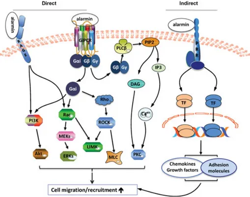
For cell migration induced by alarmins, the intracellular signaling events that con-nect the receptor (s) and intracellular sig-naling messengers (e.g. PI3K/Akt, ERKs, PKC, Rho, Ca2+, etc.) still need to be determined. Since the predominant recep-tors mediating the chemotactic effects of alarmins are GiPCRs and RAGE, and the signaling pathways of chemotactic receptors (e.g. FPRL1, CCR2, CCR6) or RAGE have been elucidated in some detail [1, 4, 7, 8], it is likely that alarmins trigger similar intracellular signaling pathways to those of chemokines and AGEs for the induction of cell migration as shown in Fig-ure 1. Thus binding of an alarmin with its GiPCR presumably results in the activation of heterotrimeric G-protein(s) that dissoci-ate into activdissoci-ated Gαi and Gβγ subunits. Activated Gαi and GβGγ trigger a series of reactions, commencing with the acti-vation of phospholipase C (PLCβ), PI3K, and small GTPases (Rac and Rho) [1, 4, 7, 8]. Ligand engagement of RAGE leads to activation of PI3K [79] and Rac [80]. PLCβ hydrolyses phosphatidylinositol-4, 5-bisphosphate (PIP2), generating inositol 1,4,5-trisphosphate (IP3), and diacylglyc-erol. IP3 induces the release of calcium from intracellular stores, and together with diacylglycerol, activates PKC. Activation of PI3K in turn phosphorylates Akt. Rac and Rho initiates a series of reactions that lead to the activation of ERKs, LIM (named by the initials of the three homeodomain proteins Lin11, Isl-1, and Mec-3 in which it was first discovered) domain kinase (LIMK), and myosin light chain. These pro-tein kinases act cooperatively to rearrange cytoskeleton fibers leading to cell migra-tion (Fig. 1).
Alarmins indirectly promote cell
migration and recruitment
Alarmins can activate many cell types, often by triggering a pattern recognition receptor such as TLRs [9–11, 14, 49, 81].This leads to the production of chemotac-tic factors and adhesion molecules, both of which participate in promoting cell migration and/or recruitment [1–5,8]. For example, treatment of airway epithelial cells with humanα-defensin promotes the production of CXCL8 [82]. LL-37 treat-ment of endothelial cells leads to the production of CCL2 [83]. Several human β-defensins can stimulate keratinocytes to produce a number of chemokines includ-ing CXCL10, CCL2, CCL20, and CCL5 [75]. HSP70 released from heat-shocked tumor cells stimulates the production of chemokines (CXCL10, CCL2, and CCL5) that induces the infiltration of DCs and T cells [84]. Activation of macrophages or DCs by EDN, HMGB1, HMGN1, gran-ulysin, S100a8/9, and uric acid leads to the production of many chemokines such as CXCL5, CXCL7, CXCL8, CXCL9, CXCL10, CXCL12, CCL1, CCL2, CCL3, CCL5, CCL7, and CCL8 [39,42,49,52,65,85–89]. These chemokines, in turn, promote the migra-tion and recruitment of cells positive for the corresponding chemokine receptors including CXCR1, CXCR2, CXCR3, CCR1, CCR2, CCR5, and CCR6. LL-37 is capa-ble of inducing the generation of vascular endothelial growth factor, which, in turn, promotes angiogenesis by facilitating the recruitment of multipotent mesenchymal stromal cells [36].
LL-37-treated EPCs migrate better than nontreated progenitor cells to the injured area due to the upregulation of E-selectin and P-selectin glycoprotein ligand-1 [90]. LL-37 also enhances ICAM-1 expression by endothelial cells [83]. HMGB1 is capa-ble of inducing α5β1 integrin expres-sion by human chondrosarcoma cells [60]. HMGB1 not only upregulates the expres-sion of ICAM-1, and theβ1 and β2 inte-grins on EPCs, but it also increases the affinity of these integrins, contributing to the enhanced adhesion and recruitment of EPCs [56]. Human β-defensin 2 has recently been shown to induce the arrest of Th17 cells on inflamed endothelial cells in an ICAM-1-dependent manner [91]. Therefore, alarmins have the capacity to regulate the expression or activation of var-ious adhesion molecules on both endothe-lial cells and target cells and thus can pro-mote the migration of target cells.
Overall, alarmins can induce cell migra-tion directly through interacting with their GiPCRs or RAGE, or indirectly by stim-ulating the production of chemokines, growth factors, and adhesion molecules often via activating the corresponding pat-tern recognition receptor (Fig. 1). The