Scuola Politecnica e delle Scienze di Base
Dottorato di ricerca in Scienze Chimiche XXX ciclo
Models and Methods to understand ultrafast
dynamics in photochemistry
Ph.D. Student:
Maria Gabriella Chiariello
Supervisor:
Prof. Nadia Rega
Novembre 2017
The present work of thesis aims at the development of a computational protocol for simulating and interpreting the ultrafast phenomena occurring upon the interaction of molecules with light. Nowadays, thanks to the development of high resolution time resolved spectroscopic techniques we are able to follow photoinduced chemi-cal reactions on the time schemi-cale of nuclear motions, and potentially to reveal their coupling with electronic density redistribution.
In this context, time resolved vibrational spectroscopies play a key role; indeed this advanced technique allows for ’watch’ nuclear motions of molecules in real time upon excitation, revealing at atomistic level the reaction mechanism triggered by photoexcitation. The resulting experimental spectra are often very complex, and the disentanglement of the information hidden in the signals is not a simple task. In this sense, the atomistic picture provided by theoretical-computational approaches can be an excellent support, not only to establish a direct link between the experimental signal and the molecular motion producing it, but also to reveal what are actually the electronic/nuclear motions at the origin of the photoreactivity. However, the predictive and interpretative ability of a smart theoretical strategy can be exploited to achieve this challenging goal. On this ground, the present Ph.d project has been developed.
One of the most widespread example of photoreactivity is given by the excited state proton transfer reaction (ESPT), which was also deeply investigated by these advanced time-resolved spectroscopic techniques. Generally, ESPT takes place be-tween a so-called photoacid molecule, acting as proton donor, and a solvent molecule, acting as proton acceptor. Within the class of photoacid molecules, Pyranine (8-hydroxypyrene-1,3,6- trisulfonic acid, HPTS) represents a fascinating and particular case (see figure 1a). Pyranine is a commonly used photoacid, showing in water a pKa value of 7 that drops to 0 upon excitation. Pyranine is also a weak photoacid, showing a slower ESPT kinetics when compared to other photoacids. Specifically, in the excited state pyranine donates a proton to a nearby solvent water molecule with time constants of 3 and 90 ps. Previous studies demonstrated that pyranine photoacidity cannot be simply interpreted and explained in terms of the electronic density redistribution responding to the external perturbation. Time resolved vibra-tional spectroscopy experiments revealed that in the electronic excited state HPTS chromophore undergoes a transient and sequential activation and decay of low
fre-connection has not been given yet. As first stage of the project, we focused on the photoreactivity of pyranine in pure water solution. In particular, we analyzed the vibrational fingerprints of the photoexcited HPTS in a time window of 0-1000 femtoseconds (fs). To this aim, we adopted an integrated computational approach including ab-initio molecular dynamics (AIMD) to simulate the HPTS in aqueous solution in both the ground and the excited state. As a key tool of our compu-tational strategy, we developed and adopted a time resolved vibrational analysis, based on the wavelet tranform, of the AIMD trajectories. With this approach we will be able to understand whether the photoinduced nuclear rearrangement of the chromophore can be considered the actual driving force of the ESPT reaction.
For the simulation of the solute (HPTS)-solvent (water) system an hybrid ex-plicit/implicit solvation method enforcing non-periodic boundary conditions was adopted. By this approach, it is possible to retain the specific interactions be-tween the solute and the solvent and accurately reproduce the solvent dynamics in proximity of the HPTS molecule. Figure 1a shows our model. The HPTS solute is treated at quantum mechanical (QM) level, while the explicit water molecules are modeled by molecular mechanics. Moreover, in the case of photoactivated nu-clear motions of pyranine we deal with collective low frequency modes, probably coupled to the solvent. In this context, the choice of explicit treatment of the sol-vent effect around the pyranine solute is mandatory. The nuclear photorelaxation of pyranine is finely controlled by the sequential activation of transient vibrational modes. Starting from the photoexcitation, the time window of 1 ps is a suitable time to watch the low frequency modes lifetime. All the modes have a quite com-plex composition, involving all the molecular skeleton. The wavelet maps reproduce the sequential activation of the main vibrational modes, in fair agreement with the experimental findings. The method employed is able to provide an accurate picture about the time evolution of the photoactivated vibrational modes, matching in all cases the kinetics time constants of the experimental signals. Moreover, they have the key role of optimizing the structural arrangement with the hydrogen bonded water molecule. In particular, some low frequency modes such as the ring breathing mode (190 cm-1
), ring out of plane wagging (108 cm-1
) or the ring deformation mode (280 cm-1
) are involved in modulation of intermolecular distances and orientation with the water solvent molecule hydrogen bonded to phenolic group of pyranine. A rational analysis of the structural parameters involved in ESPT reaction in both ground and excited state, revealed that the hydrogen bond distance between pyra-nine and the water molecule is optimized in excited state, resulting in a stronger hydrogen bond between the proton-donor acceptor couple. Moreover, the excitation induces the arrangement of the heavy atoms involved in ESPT reaction to oscillate around planarity values, as expressed by the time behavior of two key dihedral angles
acetate concentration, different ESPT mechanisms can be modelled, in particular a direct ESPT reaction from pyranine to acetate is promoted at high concentration of acetate base. Otherwise, one or more intervening water molecules can directly modulate the proton hop from pyranine to acetate. The ESPT reaction involv-ing both pyranine-acetate and pyranine-water-acetate cluster surrounded by several water shells was investigated.
Five excited state trajectory for simulate the direct ESPT reaction from pyranine to acetate have been collected, employing the same QM/MM recipe (see figure 1b). The excited state proton transfer reaction occurs in sub-ps time scale and it strictly depends on the choice of the starting point. In particular, the solvation around the phenolic oxygen of pyranine and relative orientation of pyranine-acetate are the two factors identified as crucial to accelerate the ESPT reaction.
The excited state proton transfer reaction involving the pyranine-water-acetate cluster is an intermediate situation between pyranine in pure water and
pyranine-acetate. In the first case, before the ESPT event the sequential activation of low
frequencies vibrational modes is necessary to induce a structural optimization be-tween the proton donor-acceptor pair. On the other hand for the pyranine-acetate system the reaction is basically barrierless and the nuclear rearrangement is not required. The presence of acetate, for the pyranine-water-acetate cluster, obviously makes the ESPT faster, but the central water suggests that low frequencies skeleton modes have to optimize as well the structural arrangement of the reactive site. Ring breathing and out of plane wagging modes have been identified and recognized again as a key modes coupled to the motion of hydrogen bonded water molecule.
The time resolved analysis was extended and tested to study the anharmonic coupling between vibrational modes. In general, a time resolved vibrational signal can have characteristic intensity and frequency oscillations over the time. These oscillations can be ruled and modulated by low frequency vibrational modes. The time behavior of a vibrational signal provides indication about the interactions be-tween normal modes. The case study is represented by a diarylethenes photochromic molecule (see figure 1d), that in excited state undergoes an opening ring reaction. The characteristic intensity oscillations of some vibrational signals appear clearly in the wavelet maps. This suggests an anharmonic coupling with some low frequency modes. In particular, our findings support the idea that the coupling between high and low frequency modes localized on the central cyclohexadiene ring would promote the opening reaction.
c) pyranine-water-acetate cluster in water solution. The QM region includes in addition a solvent water molecule. d) Structure of 1,2-bis(2,4-dimethyl-5-phenyl-3-thienyl)perfluoro-cyclopentene, a popular diarylethene showing photochromic properties.
Introduction 1
1 Trick of the trade: Methods and Models 8
1.1 Ab-initio molecular dynamics (AIMD) . . . 8
1.2 Vibrational Problem from AIMD . . . 11
1.3 Time resolved analysis: Wavelet Transform . . . 13
2 Following in real time nuclear photorelaxation: pyranine photoacid in aqueous solution 17 2.1 Introduction to photoacids class: pyranine case . . . 17
2.2 Ground state equilibrium sampling . . . 20
2.3 Photoexcited pyranine features . . . 23
2.4 Vibrational fingerprint of pyranine . . . 28
3 Exploring excited state reactivity of pyranine-acetate system in aqueous solution 39 3.1 Introduction to pyranine photoreacitivity in presence of acetate . . . 39
3.2.1 Ground state equilibrium sampling . . . 42
3.2.2 Rationalizing photo-reactivity: direct ESPT reaction . . . 47
3.2.3 Time resolved vibrational analysis . . . 52
3.3 Modeling Pyranine-water-acetate cluster in aqueous solution . . . 56
3.3.1 Ground state equilibrium sampling . . . 56
3.3.2 Rationalizing photo-reactivity: double ESPT reaction . . . 62
3.3.3 Time resolved vibrational analysis . . . 68
4 Unveiling anharmonic coupling by means of excited state ab-initio dynamics: application to diarylethenes photoreactivity 72 4.1 Introduction to diarylethenes photoreactivity . . . 72
4.2 TD-DFT picture of the potential energy surfaces . . . 76
4.3 MC-SCF study of M-CHD . . . 81
4.3.1 Justifying the model . . . 81
4.3.2 CAS-SCF investigation of M-CHD . . . 82 4.4 Excited state AIMD: extending the time resolved vibrational analyisis 86
Concluding remarks 95
Appendix A 100
List of figures 102
The interaction with light can dramatically affect the well-known ground state reac-tivity of molecules, resulting in an unexpected and almost new photoinduced chem-istry [1–8]. That’s one of the reasons why the photochemchem-istry is, nowadays, one of the most fascinating and promising research field in chemistry.
Following the photoexcitation, a chromophore lies on higher electronic potential energy surface and it may undergo different pathways. A popular picture of the possible photophysical processes is presented in Figure 2, starting from the absorp-tion of radiaabsorp-tion until a de-excitaabsorp-tion makes the chromophore drop down to the ground state throughout a radiation or radiationless decay. Indeed, once photoex-cited, a chromophore can relax toward the minimum energy structure on excited state surface, or it can react with other photoexcited molecules. Moreover, crossings between electronic surfaces may take place giving rise to the so-called non-adiabatic reactivity [9–11]. A photochemical process can be considered as the result of the electronic and nuclear dynamics responding to the external perturbation, i.e. the photoirradiation with light. These far-for equilibrium dynamics occur on different time scales: the electronic redistribution happens on attoseconds time scales [12–14]
and the subsequent nuclear rearrangement requires instead picoseconds (ps) scales. Following in real time the photoinduced nuclear dynamics is definitely funda-mental to rationalize the structural driving forces at the origin of photoreactivity. Among the approaches turned suitable to this challenge purpose, we mention: the non-linear vibrational spectroscopy techniques [15–17] and the ab-initio molecular dynamic (AIMD) methods [18–20]. The advent of ultrafast pulsed laser techniques has given a new point of view on the photochemistry. In particular, the time re-solved vibrational spectroscopies, like the Femtosecond Stimulated Raman Spec-troscopy (FSRS) [21–25], allow to watch the time evolution of the vibrational modes upon excitation. FSRS was crucial to elucidate the mechanism of photochemical reaction involving chromophore even in complex environment, such as the excited proton transfer reaction (ESPT) of the green fluorescent protein [26–30]. Moreover, the analysis of the oscillating behavior of the time resolved vibrational bands is of-ten performed to unveil the coupling between modes [31]. Otherwise quantitative measure of correlations between vibrational modes can be available by means of the 2-dimentional infrared spectroscopy (2D-IR) experiments [32–35], another non-linear spectroscopic technique providing structural insights such as anharmonicities and coupling between the normal modes.
On the other hand, the interpretative and predictive skills of a smart theoretical-computational protocol based on AIMD are essential to fulfill information that are not accessible to the experiment. By means of AIMD methods the atomistic
pic-Fig. 2: Schematic representation of the common photophysical processes occruring on excited potential energy surfaces, from [1].
ture is never lost and the molecular response to the photoexcitation is monitored in real time [36–39]. Nevertheless, AIMD has to be combined with a rational vibra-tional analysis having the ability to keep the temporal information contained in the collected trajectories.
On this ground, the target of this thesis is to provide an integrated theoretical-computational procedure to study the nuclear dynamics of photoexcited chromophores in their realistic environment. The method employed is essentially based on ex-cited state AIMD together with a time resolved vibrational analysis. The molecular systems under investigation relax on excited state potential energy surface (PES), defined on-the-fly by means of an ab-initio electronic potential, which is obtained at time dependent density functional theory (TD-DFT) [40–42] level of theory.
The molecular response to the photoexcitation is carried out by the evolution of the vibrational modes. The standard method to solve the vibrational problem,
based on the diagonalization of the hessian matrix, is not suitable in combination with dynamics simulations. Indeed, it is difficult to be used on large system (like chromophores in solution) and the frequencies generally obtained in the harmonic approximation, even on the excited state equilibrium geometry, do not report about the transient nature of the normal modes. We propose a vibrational analysis com-pletely based on the so-called generalized vibrational modes extracted from dynam-ics [43, 44]. The method basically involves the analysis of the velocities fluctuations collected in the ground and excited state trajectories. It can be easily applied to large molecular system also when the molecular system is separated according to a quantum mechanics (QM)/ molecular mechanics (MM) hybrid scheme [45, 46].
Because of the transient nature of the vibrational processes triggered by the pho-toexcitation the computational procedure needs a time resolved analysis tool. To address this issue a multiresolution vibrational analysis based on the Wavelet
Tran-form (WT) [47] was adopted. So far WT has found few but successful applications
in chemistry. In partcular, Wavelet Tranform was already employed in combination with AIMD simulations to disentagle the evolution of a simulated Stokes Shift [48] or the dipole moment in exciton dynamics [49], and to analyze the evolution of struc-tural parameters in the frequency domain. The simulated vibrational signals can be be plotted in 2-dimention maps where the time and frequency information are both reteined. Following this procedure, a direct comparison with the experimental time resolved signals is therefore possible.
As a first case study to test the developed methodology, the pyranine molecule (8-hydroxypyrene-1,3,6-trisulfonic acid, HPTS) was chosen [50,51]. HPTS is a popular photoacid chromophore, that found many applications as pH probe and fluorescent tag [52–54] . This kind of molecules upon excitation increases hugely acidity and an ESPT reaction takes place. More closely, pyranine has a pka of 7 in electronic ground state that drops to 0 in excited state, and in water solution where a solvent molecule acts as proton acceptor, the ESPT reaction occurs in 3 and 90 picoseconds [55, 56]. Pyranine is usually identified as weak photoacid [57] because of the comparison with other photoacids molecules, which show ESPT kinetics on sub-ps time scales. Experimental studies revealed how in excited state a sequential activation of low frequencies skeleton modes take place [58–60] . It was hypothesized that they could be essential in promoting the ESPT reaction toward a water solvent molecule.
The second chapter will focus on the transient vibrational features of photoex-cited pyranine in pure water. In our model, pyranine chromophore is surrounded by several water shells treated explicitly. It will be shown how a characteristic activa-tion of low frequencies modes can induce structural optimizaactiva-tion, involving both the chromophore skeleton and the water of the firs solvation shell, promoting the ESPT reaction. Pyranine chromophore is very sensitive to the environment, and if a base like acetate is present in water solution the reaction became faster (sub-ps) [61]. This will be the subject of the chapter three, where the ESPT reaction between pyranine and acetate is considered. Depending on the acetate concentration, different ESPT
mechanisms can be modelled, in particular a direct ESPT reaction from pyranine to acetate is promoted at high concentration of acetate base. Otherwise, one or more intervening water molecules can directly modulate the proton hop from pyranine to acetate. The ESPT reaction involving both pyranine-acetate and pyranine-water-acetate cluster surrounded by several water shells will be discussed in chapter three. It will be interesting to observe how pyranine vibrational features change when the ESPT reaction become ultrafast.
In last chapter, the time resolved analysis will be extended and tested to study the anharmonic coupling between vibrational modes. In general, a time resolved vi-brational signal can have characteristic intensity and frequency oscillations over the time. These oscillations can be ruled and modulated by low frequency vibrational modes. The time behavior of a vibrational signal provides indication about the in-teractions between normal modes. The case study is represented by a diarylethenes photochromic molecule [62, 63], that in excited state undergo an opening ring re-action. The characteristic intensity oscillations of some vibrational signals appear clearly in the wavelet maps. This suggests an anharmonic coupling with some low frequency modes. It will be discussed how the coupling can promote the open ring re-action. The diarylethenes reactivity is unfortunately recognized to be non-adiabatic and controlled by a crossing between electronic energy surfaces [64]. TD-DFT is not able to provide a complete picture of the whole open ring reaction. The first part of the work was therefore aimed at defining the PES regions, in which the
non-adiabatic coupling is low and the Born-Oppenheimer (BO) approximation still holds. Here, a TD-DFT based dynamics sampling is allowed and employed. For the sake of completeness, a study on a diarylethenes model system, has been conducted with a post Hartree-Fock electronic method [65, 66], in order to characterize the non-adiabatic crossing point between the electronic states.
The next chapter contains the methodological development about the extraction of generalized vibrational modes from dynamics and the wavelet transform based analysis, together with the computational details of the ground and excited state ab-initio dynamics and the hybrid model adopted.
Trick of the trade: Methods and
Models
1.1
Ab-initio molecular dynamics (AIMD)
The molecular dynamics simulations allow for reproducing the behavior of complex chemical and biological system, under equilibrium and far-for equilibrium condi-tions [67–70]. Understanding the mechanism underling a specific chemical process at molecular level would lead to predict the behavior, driving the experimental and applications studies. The big issue at the heart of any molecular dynamics approach is how to describe the interatomic interactions. The classical molecular dynamics are extremely fast, even when large system like proteins, nucleic acids and molecules in solution are treated. The potential energy ruling the dynamics is usu-ally decomposed in terms of parametrized functions [71–74], accounting for bonded interactions, electrostatic and Van der Waals interactions etc. The potential energy based on parametrized force field are unable to describe processes involving bond braking and formation.
On the other hand, the advent of the ab-initio molecular dynamics was a rev-olution in the field of computer simulations. Indeed, AIMD combines the accurate ab initio electronic structure theory with the classical molecular dynamics scheme. Within the validity of the Born-Oppenheimer (BO) approximation, the nuclear and electronic degrees of freedom are separated. The basic idea of the ab-initio dynamics scheme relies on the classical simulation of the nuclear motion. The nuclear degrees of freedom are propagated according to the Newton equation of motion. The en-ergies and the forces acting on the nuclei are computed from electronic structure calculation performed on-the-fly at every step of the dynamics trajectory [75, 76].
For solving the electronic structure problem the most used approach are repre-sented by the Kohn-Sham density functional theory [77–79] and the post Hartree-Fock methods [65, 66].
The Born-Oppenheimer, Ehrenfest and Car-Parrinello approaches belong to fam-ily of ab-initio molecular dynamics methods.
The Ehrenfest dynamics [80–82] involves to solve the time dependent Schrodinger equation on-the-fly as the nuclear trajectory is propagated according to the classical mechanics. Moreover, Ehrenfest approach accounts for non-adiabatic effects and it allows therefore for investigate reactions, involving non-adiabatic process, i.e. mul-tiple potential energy surfaces.
The BO approach [48, 83–85] is based on the assumption that a single surface rules the dynamics. Energies, gradients and molecular properties are computed
on-the-fly solving the time independent, stationary Schrodinger equation at every step of the nuclei classical propagation. The BO dynamics is less demanding that the Ehrenfest approach, allows for simulate reactions happening on longer time scale (ps).
To overcome the problem of the computational expenses due to the calculation of electronic structure at each step of the molecular dynamics trajectories, the CP approach was developed. In the framework of an extended lagrangian formalism, the electrons are considered as active degrees of freedom propagated as classical particles, with the inclusion of suitable constrains. The CP idea had a huge impact on the field of ab-initio dynamics simulation and it inspired generation of scientists [86–89]. An alternative formalism to the CP is represented by the Atom centered density matrix propagation (ADMP) method. ADMP [90–94] employs atom centered Gaussian basis sets and the single particle density matrix within the extended lagrangian formalism, in place of the plane-wave basis sets chosen as dynamics variables in the CP algorithm.
This family of molecular dynamics methods was widely validated and employed to the study of molecular system in their ground electronic state. Nowadays, many efforts are devoted to found the best dynamics scheme to investigate the electronic excited state.
In this work of thesis, we propose a recipe based on BO dynamics scheme to col-lect the excited state trajectories, in combination with the time dependent version
of DFT (TDDFT) in linear response formalism [95–97], for solving the electronic structure problem. According to the linear response theory, the excited state wave-function is not explicitly computed. The energies and the properties of the excited state are obtained by means of the ground reference state to a time dependent per-turbation. TD-DFT was remarkably successful at providing an understanding of molecular excited state, with an acceptable computational cost.
In conclusion, for the simulation of the molecules under investigation in their realistic environment the BO/TDDFT dynamics scheme is combined with an hybrid quantum mechanics (QM)/ molecular mechanics (MM) scheme.
1.2
Vibrational Problem from AIMD
The vibrational analysis here employed is a method for obtaining generalized vibra-tional modes (or quasi-normal or quasi-harmonic) from fluctuations calculated by an AIMD QM/MM simulation [43, 98, 99].
The underling idea is that atomic fluctuations, can be related to an effective force constant matrix relative to the average dynamics structure, from which vibrational modes can be extracted [100, 101]. The effective force constant matrix and the fluc-tuations matrix are inversely proportional, so that they share the same eigenvectors, i.e. the quasi-harmonic vibrational modes. Moreover, these modes can be different from the standard normal modes, because the anharmonicity effects are naturally taken into account. It is so not required the computation of force constant matrix, but it is sufficient to diagonalize the mass weighted fluctuation matrix to obtain
modes and frequency.
A similar analysis can be carried out in momenta subspace [44, 102, 103] instead of the configuration space. It has been shown that normal mode directions obtained from velocities and coordinate fluctuations are very similar. The assumption is that at any temperature, the 3N generalized modes Q correspond to uncorrelated momenta, and can be obtained by diagonalizing the K matrix of the mass weighted atomic velocities ˙q with elements:
Kij =
1
2 < ˙qi˙qj > (1.1) where i and j run over the 3N atomic coordinates and < .. > indicates an average over the time. Modes velocities can be therefore calculated by projecting atomic velocities along normal-like directions:
˙
Q(t) = L†˙q(t) (1.2)
Finally, the fourier transform (FT) of the ˙Q time correlation functions provides the associated frequencies:
P(ω) = Z
dteiωt < ˙Qi(t) ˙Qj(t) > (1.3)
So far we described what is usually done for an equilibrium analysis. Actually, the aim is investigate far from equilibrium events, specifically the transient vibra-tional signals appearing upon electronic excitation. We assume that, at least in the
ultrafast part, the L matrix is an acceptable approximation of the vibrational modes composition. Therefore, once the photorelaxation process has been simulated by ex-cited state (ES) trajectories, evolution in the time of QES modes are obtained from
extracted mass weighted atomic velocities ˙qES, according to the transformation
˙
QES(t) = L†˙qES(t) (1.4)
In order to obtain the vibrational frequency values along the time, we adopted a multiresolution vibrational analysis based on the Wavelet Transform (WT) [104]. Here, for the first time, we use WT to obtain transient vibrational signals corre-sponding to the ˙QES(t) modes extracted from AIMD. We adopt the continuous WT
expression
W(a, b) = Z
˙
QES(t)ψa,b(t)dt (1.5)
The time dependent signal ˙QES(t) is analyzed and decomposed in terms of
wavelet basis ψa,b. The details of the wavelet transform are given in next section.
1.3
Time resolved analysis: Wavelet Transform
The Fourier transform performed on a time dependent signal can be seen as a signal decomposition in terms of sine and cosine functions. On the other hand, according to the wavelet transform [47, 104–106], the time dependent signal is decomposed into a set of another kind of basis functions, the wavelets. Recalling the previous
equation:
W(a, b) = Z
C(t)ψa,b(t)dt (1.6)
where C(t) is a generic time dependent function and ψa,b are the wavelet basis
obtained from a prototype wavelet (called mother wavelet) from dilatation, contrac-tion and translacontrac-tion. As mother wavelet we used the Morlet wavelet, having the form:
ψ(t) = π−14eiω0te− t2
2 (1.7)
It is basically a plane wave modulated by a gaussian function and ω0 is the
nondi-mensional frequency, usually taken to be 6 to satisfy the admissibility condition. In figure 1.1, the Morlet wavelet together with the dilated and translated wavelet are shown. The scale parameter a, proportional to the inverse of frequency, regulates the dilatation and contraction of the mother wavelet and extracts the different fre-quencies hidden in the time-dependent signal. While the translation of the wavelet basis, ruled by the b parameter, ensure the localization of the frequencies in time domain.
The application of the wavelet transform to chemical problem, in particular to far-for equilibrium [48, 49, 107, 108] process is an appealing choice. The far-for equi-librium process, such as the nuclear response to the photoexcitation, are typically transient events, having a lifetime. The far-for equilibrium events give rise to
non-Fig. 1.1: Morlet mother wavelet, with dilated wavelet version.
stationary signals. A non-stationary signal may appear only in a short time interval. Using the FT, this local information is separated into a large number of bases and these signals are averaged over the whole time domain. The FT is not an appro-priate tool for the time analysis of the stationary signals. The analysis of non-stationary time series should be done localizing the signals in both frequency and time domain. The Short Time Fourier Transform (STFT) [109] could be, in prin-ciple, used to this aim. Nevertheless, the STFT employs a time-frequency window, having a fixed width. A time dependent signal can oscillating according to high and low frequencies. The low frequency oscillations should be analyzed by a wide time window, while the high frequency oscillations require a narrow time window. STFT, using a window with fixed width, could be unable to simultaneously detect the high and low frequency of a complex time dependent signal. On the other hand, the WT is characterized by a no-fixed time-frequency window. The time dependent signal, instead, is decomposed into components with different scales [109, 110]. The flexible
time window allows for a simultaneous analysis of the high and low frequencies oscil-lation, with an acceptable resolution. Thanks to wavelet transform, a complex time dependent signal, even of non-stationary nature can be analyzed and decomposed to obtain the corresponding frequencies. The frequencies are resolved in time, so that it is possible to recognize when the frequency rises and decays. The application to the vibrational modes of photoexcied pyranine, allows us to follow and watch the lifetime of the vibrational modes.
Following in real time nuclear
photorelaxation: pyranine
photoacid in aqueous solution
2.1
Introduction to photoacids class: pyranine case
The excited state proton transfer reaction (ESPT) is one of the most widespread ex-ample of photoreactivity [111–113]. The absorption of radiation can hugely increase the acidity of particular molecules, called photoacid, which dissociate at the excited state transferring a proton to a nearby solvent molecule or to a strong base present in solution. The understanding of the intermolecular ESPT reaction requires the in-vestigation of several elementary steps composing the overall process. The ultrafast electronic redistribution upon the photoexcitation, the nuclear relaxation, the hy-drogen bonds rearrangement and the solvation dynamics finely control the kinetics and thermodynamics of the ESPT event [114,115]. Despite the detailed information provided by the advanced spectroscopic [116–118] techniques, the understanding of the ESPT reactivity at molecular level remains difficult to extrapolated, especially
at the fast and the ultrafast timescale. As a matter of fact, the kinetics of the ESPT toward a solvent molecule covers a wide range of timescales, going from femto- to nanoseconds. Beneath different kinetics there are, of course, different ESPT mech-anisms. A classification of the photoacid molecules into four regimes, according to their pKa* values and ESPT rate, was recently proposed [57]. When going from regime I to regime IV the strength of the photoacid and the ESPT increase, while the pKa* value diminishes.
The 8-hydroxy-1,3,6-pyrenetrisulfonate (HPTS or pyranine) is a widely used pho-toacid [119,120], that found many applications as pH probe and fluorescent tag even in complex biological matrix. Pyranine is usually classified as weak photoacid and it belongs therefore to the first regime. In water, it has a pKa of 7 that drops to 0 upon excitation. Pyranine has a slower ESPT kinetic, compared with other photoacid molecules where the ESPT happens in sub-ps time scales [121, 122].
Specifically, in the excited state pyranine donates a proton to a nearby solvent water molecule with time constants of 3 and 90 ps. Moreover, the photoinduced behavior of HPTS chromophore is very sensitive to change of the environment. In particular, the ESPT reaction is absent when for example methanol is used as solvent [123]. On the other hand, if a base, like acetate, is present in water solution the ESPT kinetic becomes faster (sub-ps time scale) [61]. Acetic acid has a dissociation constant, pKa of 4.76 in the middle of the pKa value of pyranine in ground (pKa 7) and excited state (pKa* 0). At the ground state a spontaneous proton transfer from
pyranine to acetate does not occur, while the electronic excitation should promote a barrierless proton transfer reaction toward acetate.
Pyranine was the subject of several spectroscopic studies, aiming at elucidate the electronic and nuclear mechanisms driving its photoreactivity. In particular, Femtosecond stimulated Raman Spectroscopy [58, 61, 123, 124] was employed to in-vestigate the mechanism of pyranine ESPT in different environmental conditions. The time resolved FSRS spectra in water revealed that in the excited state HPTS chromophore undergoes a transient and sequential activation and decay of low fre-quency (< 1200) skeleton modes. This peculiar vibrational activity precedes and accompanies the ESPT reactive event, indeed, these modes seem to play a key role in activating and promoting the ESPT.
In this chapter we deal with the nuclear relaxation of photoexcited pyranine in water solution, in particular the time resolved vibrational procedure, based on generalized modes extracted from dynamics and wavelet transform, will be adopted to simulate and interpret the vibrational fingerprint of pyranine in water.
For the simulation of the solute(HPTS)-solvent(water) system, an explicit/implicit solvation method was employed [125–127]. This approach allows to retain the spe-cific interactions between the solute and the solvent and accurately reproduce the solvent dynamics in proximity of the HPTS molecule. Moreover, many of the vibra-tional modes of interest have a collective nature involving all the four ring aromatic system and they are probably coupled to the solvent molecules directly bound to
phenolic group of pyranine. Modes extracted from dynamics come from the sam-pling of the molecule in its realistic environment, so these modes naturally take into account of the coupling with solvent explicitelly treated.
The photoacidity weakness of the pyranine can be explain supposing that the electronic re-arrangement over the HPTS chromophore is unable alone to provide the conditions for proton transferring to a water solvent. The nuclear motions respond-ing to the new electronic arrangement has to be considered essential to promote the ESPT reaction. In the nest sections, we will see how the composition and the sequential activation of the characteristic HPTS low frequency skeleton modes trig-ger a structural optimization with the solvent water molecules (proton acceptor) hydrogen bound to phenolic group of pyranine (proton donor).
2.2
Ground state equilibrium sampling
In order to take into account solvent effects on the photorelaxation of the pyranine chromophore, we adopted an hybrid explicit/implicit solvent scheme within non periodic boundary conditions (NPBC). [125–127] The explicit description of the water solvent molecules is indeed necessary to account for solute-solvent specific interactions, especially those involving the polar (phenolic) and charged (sulfonic) groups of pyranine. Moreover, it has been hypothesized that some low frequencies skeleton modes of photoexcited pyranine play a key role in promoting the excited state proton transfer reaction toward the water molecule that is hydrogen bonded to the phenolic acid group of pyranine. Therefore, the explicit description allows for
the analysis of pyranine vibrational modes that are naturally coupled to the water molecule motions (vide infra).
We start our analysis with the characterization of the solvent microsolvation around the acid group of the pyranine solute, at equilibrium in the ground state. In Figure 2.1 radial distribution functions (RDFs) of the pyranine acid oxygen-water oxygen (Opyr-Ow) and the pyranine acid hydrogen-water oxygen (Hpyr-Ow) distances
are reported, respectively. In both the cases a well defined peak, corresponding to the first solvation shell around the phenolic acid group, is recognizable. Peaks are centered at around 1.7 and 2.5 ˚A for Hpyr-Ow and Opyr-Ow RDFs, respectively,
according to the typical hydrogen bond distances. The first peak is limited by a pronounced minimum in both the RDFs, reaching a zero value for the (Hpyr-Ow)
RDF. This is a clear indication that there is no exchange of water molecules from and toward the first solvation shell during the sampling period (10 ps). It is noteworthy that the integration number of this peak (black dashed line in Figure 2.1) indicates that one water molecule is hydrogen bonded to the pyranine acid group during the simulation time.
In Figure 2.1 we report the time evolution of the main hydrogen bond struc-tural parameters between the phenolic O-H group of pyranine and the H-bonded water molecule, namely the intermolecular distances Hpyr-Ow, Hpyr-Ow and the
in-termolecular angle Hpyr-Opyr-Ow. Intermolecular Hpyr-Ow and Opyr-Ow distances
Fig. 2.1: a) Hpyr-Ow (black) and Opyr-Ow (red) radial distribution functions b) the pyranine
chromophore surrounded by water molecules c) Ground state time evolution of Hpyr-Ow (blue)
and Opyr-Ow (red) intermolecular distances. d) Ground state time evolution of Hpyr-Opyr-Ow
the orientation of the hydrogen bond, also assumes small values, below 30◦. A strong hydrogen bond between pyranine and water is therefore retained during the whole simulation time.
The resulting solvation layout represents the equilibrium conditions in ground state. A representative configuration of this equilibrium was chosen as starting point for the simulation in the excited state.
2.3
Photoexcited pyranine features
The S1 excited state of pyranine is obtained from S0 by a single HOMO-LUMO
excitation, with π-π∗ character. Contours of the involved frontier orbitals are shown in Figure 2.2, as obtained by a TD-DFT calculation on the ground state energy minimum of the HPTS molecule, including solvent effects in implicit way according the polarizable continuum model in its conductor-like version.
From a simple inspection of HOMO and LUMO contours, we note that the charge transfer character of the transition is low, also in proximity of the acid group. That means, the depletion of electronic density on the acid group associated to the electronic transition is not able alone to induce an immediate dissociation of the proton, as it can be expected from the weak photoacidity of pyranine. Instead, upon the excitation an electronic redistribution occurs over the whole nuclear skeleton, involving an exchange of alternating bond order among C-C bonds. In particular, the overall reorganization mainly corresponds to the π electronic density depletion on C-C bonds aligned with the red arrows shown in Figure 2.2, while the π electronic
density on C-C bonds that are almost perpendicular to the red arrows increases when going from the ground to excited state.
Therefore, we can expect a C elongation (lowering of the bond order) and a C-C shortening (increasing of the bond order) in the first and second case, respectively, when going from the ground to excited state. Overall, the excitation leads to the lengthening of the whole molecule along the axis approximately aligned with OH bond.
We investigated the photoinduced relaxation of the pyranine rings by comparing the evolution in S0 and S1 of the same initial configuration (coordinates and
mo-menta) for about 1 ps. We then analyzed the time relaxation of C-C bonds in the excited state in the frequency domain by means of the wavelet analysis.
In Table 2.1 we report average values of C-C distances calculated from AIMD trajectories in the ground and the excited state. As expected, upon the excitation we observe elongation of up to 0.04 ˚A for C-C bonds 1, 3, 5, 6, 8 and 10, and a shortening of about 0.02 ˚A for the remaining ones.
As an example, in Figures 2.3a and 2.3c we show the time evolution in both the ground and the excited state of 1 and 2 C-C distances, which in the excited state become longer and shorter respectively. We can recognize how the C-C 1 distance in the excited state oscillates around greater values when compared to the corresponding evolution in the ground state. An opposite behavior is instead
Fig. 2.2: a) HOMO-LUMO contours of pyranine computed at B3LYP/6-31g(d,p)/C-PCM level of theory. b) Labels used to tag C-C bonds analyzed in the present work.
C-C bond S0 average distance S1 average distance
1 1.41 1.44 2 1.43 1.41 3 1.36 1.40 4 1.44 1.42 5 1.42 1.45 6 1.42 1.44 7 1.44 1.42 8 1.36 1.39 9 1.44 1.42 10 1.41 1.44
Table 2.1: Average C-C distances (˚A) obtained from AIMD simulations of pyranine in aqueous solution in the ground and the first singlet excited state. Standard deviations are 0.02 ˚A and 0.03 ˚A for S0and S1trajectories, respectively. See Figure 4.2 for the numbering of C-C bonds.
Fig. 2.3: a) Ground (blue) and excited (red) state time evolution of the C-C 1 distance. b) 2D wavelet map of the C-C 1 distance in excited state. c) Ground (blue) and excited (red) state time evolution of the C-C 2 distance. d) 2D wavelet map of the C-C 2 distance in excited state.
observed for the C-C 2 distance.
Time resolved vibrational analysis in the excited state of C-C bonds 1 and 2 have been perfomed by computing corresponding wavelet spectra, which are reported as time-frequency 2-dimentional maps in Figures 2.3b and 2.3d, respectively.
From inspection of Figure 2.3 several interesting features of the pyranine photoin-duced vibrational dynamics can be inferred. In the ultrafast part of the relaxation, namely for times below 100 fs after the excitation, signals associated to the C-C distances 1 and 2 are centered around 1500 and 1200 cm−1 respectively, i.e. around
values close to those adopted in the ground state. Then, clear blue and red shifts are observed in the two cases, testifying an exchange of force constants associated to the two bonds. Notably, the overall relaxation toward values adopted in the excited state takes about 500 fs to complete. This time is in nice agreement with what ob-served by FSRS experiments, indicating about 600 fs as the time necessary to carry out most of the pyranine relaxation on the excited potential energy surface. [58,124] Another important observation regards the evolution of the intensity of both signals. Indeed, the vibrational dynamics shows a clear oscillatory behaviour. In particular, periods between intensity maxima strongly suggest that the pyranine skeleton relaxation is modulated by low frequency modes (below 200 cm−1), possibly
collective ring modes, which accompany the electronic redistribution of the molecule, and the consequent rearrangement of forces among nuclei.
Moreover, the C-C 1 and 2 vibrational bands are almost out of phase, indicating that rearrangement of forces in bonds with alternating order are anticorrelated in the time. A similar oscillatory and out of phase trend has been already experi-mentally observed in the photorelaxation of C-O and C-N stretching modes of the Green Fluorescent Protein chromophore. [26] Therefore, our results also suggest that the anticorrelated relaxation modulated by collective low frequency modes could be a common motif in photoinduced vibrational dynamics of aromatic or conjugate systems.
2.4
Vibrational fingerprint of pyranine
The pyranine ESPT reaction involves the motion of the proton from the photoacid molecule to a solvent molecule of the first solvation shell, and from here to the bulk through the diffusion process along hydrogen bond networks. The first and essential step, even before the proton hopping, is the formation of a tight proton donor-acceptor couple, i.e. the photoexcited pyranine and an hydrogen bonded water molecule.
Upon excitation, the pyranine molecule undergoes a remarkable vibrational dy-namics. FSRS results highlighted a characteristic and transient Raman activity in the excited state, associated to the sequential activation of low frequency vibra-tional modes. Several efforts have been made in order to individuate, among the complex sequence of activation/decay of vibrational signals, possible driving forces for the pyranine photoacidity. It has been hypothesized that low frequencies skele-ton modes of photoexcited pyranine play a key role in promoting the excited state proton transfer reaction toward the water molecule hydrogen bonded to the phenolic acid group.
Here, we analyzed the structural rearrangement involving the pyranine and the water molecule hydrogen bonded to the acid O-H group. Moreover, we individu-ated the generalized normal modes that are mainly involved in the pyranine-water relaxation, and analyzed their vibrational dynamics in the excited state.
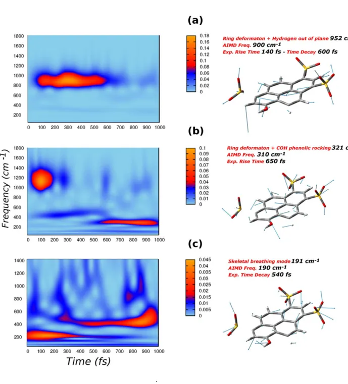
obtained by the equilibrium sampling, according to equation 1.2 and they are shwon in figure 2.4 and 2.5. The time dependent signal that account for the vibrational photorelaxation on which the wavelet transform is performed, is instead obtained from equation 1.4. The left colum of figure 2.4 and 2.5 depict the corresponding 2D wavelet maps.
The three vibrational modes shown in figure 2.4, have a collective nature, in-volving the motion of the whole four ring aromatic system. In particular, the mode associated to the frequency of 900 cm-1
is an in plane ring deformation with a strong hydrogen out of plane component (see figure 2.4). In figure 2.4 b is depicted the 310 cm-1
mode, which it’s again an in plane ring deformation, but it’s also composed by a phenolic -COH rocking component. Lastly, the lowest frequency mode (figure 2.4 c) is described as a symmetric skeletal breathing mode, experimentally found at 191 cm-1
. This is an in plane collective mode engaging all the 4-aromatic rings. The breathing mode basically entails the lengthening of the whole molecule, pushing the phenolic acid group toward the proton acceptor, i.e. the hydrogen bonded water molecule.
The wavelet maps, obtained according to equation 1.4, are plotted in a frequen-cies ranges of 0-1800 cm-1
and 0-1400 cm-1
, respectively for the 900 and 310 cm-1
modes and for 191 cm-1
mode, because at higher frequencies there are not signals of considerable intensity.
The deformation mode at 900 cm-1
.
Fig. 2.4: Q modes (right panels) and corresponding 2D wavelet power spectra (left panels). a) Ring deformation mode combined with hydrogens out of plane motion, with an AIMD frequency of 900 cm-1
; b) Ring deformation modes associated to a strong -COH phenolic rocking, the AIMD frequency is 310 cm-1
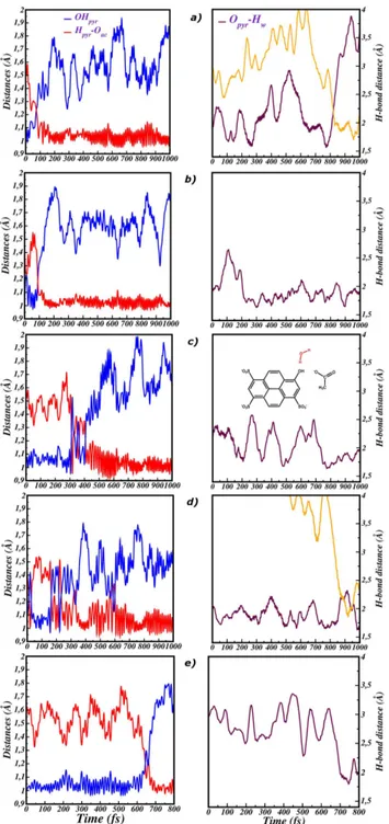
excitation, at about 100 fs and the wavelet spectrum shows a decay at the time of 600 fs. This behaviour is in perfect agreement with the experimental evidences of kinetics costants of 140 fs (rise) and 600 fs (decay) [58].
The wavelet spectrum of the 310 cm-1
deformation mode, depicted in figure 2.4 , also shows interesting features. The signal at 310 cm-1
, associated to the deformation mode with experimental frequency of 321 cm-1
, appears at the time of 600 fs, well reproducing again the experimental rise time of 650 fs [58]. Another main signal is instead centered around 1100 cm-1
. That’s basically the -COH phenolic rocking mode, experimentally found at 1138 cm-1
[58]. The 310 cm-1
mode , is overall composed by a four rings collective deformation, associated to a strong phenolic -COH motion. Nevertheless, the -COH rocking mode alone is located at 1138 cm-1
. In other words, the 310 cm-1
mode is also composed by phenolic -COH rocking and if one considers this motion alone, it has a frequency of 1138 cm-1
. During the dynamical simulation, the sampling of the 310 cm-1
mode involves naturally also the -COH rocking motion, so that the wavelet spectrum ends up to show both the frequencies at 310 cm-1
and 1138 cm-1
.
Lastly, in figure 2.4 , the wavelet spectrum of the lower frequency ring breathing mode is shown. It’s quite easy to recognize a component below 200 cm-1
, associated to the breathing mode. Following the electronic excitation, this mode quickly starts to rise and decays before 600 fs. This feature is in excellent agreement with the spectroscopic experimental signal, showing a decay time constant for this mode of
540 fs [58]. In addition the spectrum shows another important band appearing at about 500 fs and centered around 450 cm-1
. This latter signal can be associated to a mode experimentally found at 460 cm-1
, with a risetime of 600 fs [58]. The mode at 456 cm-1
is similar to the breathing mode in terms of collective motion of the 4-aromatic rings system. The dynamical simulation made possible to sample and isolate the 191 cm-1
breathing mode, that is naturally and sequentially coupled to the 460 cm-1
. In the excited state wavelet spectrum the frequencies are resolved in time, and we can see how the 460 cm-1
mode appears simultaneously to the breathing decay, qualitatively reproducing the experimenatal risetime of 600 fs. The method employed is able to provide an accurate picture about the time evolution of the photoactivated vibrational modes, matching in all cases the kinetics time constants of the experimental signals. Beyond that, it’s noteworthy that the sequential trend with which the modes vanish and appear, is in perfect agreement with the experimental one. In case of photoexcited pyranine, the time window of 0-600 fs is clearly a key time in the context of the overall relaxation process. Indeed, that’s the time in which the decay of the 190 and 900 cm-1
modes and the rise of 310 and 460 cm-1
modes, happens simultaneously.
A deeper analisys of the composition of these three modes shows how all of them have important contribution localized to the phenolic acid group. In particular while for the lower frequency modes (190 and 310 cm-1
), these are basically -COH phenolic rocking motion, the 900 cm-1
plane contribution. As vibrational modes are exctracted from dynamics trajectory, where the water solvent molecules are explicitly take into account, it’s possible that they are coupled to the motion of the solvent in first solvation shell. More closely, during the simulation time we found that just one water molecule remains close, hydrogen bonded, to the acid group of pyranine, i.e. there is no exchange of water molecules between the solvation shells around the pyranine phenolic group.
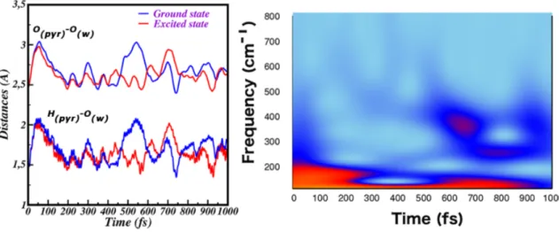
In figure 2.6 we compare the time evolution in the ground and the excited state of the intermolecular distances between the pyranine phenolic oxygen and the H-bonded water oxygen (Opyr-Owat), and between the pyranine acid group hydrogen
the water oxygen (Hpyr-Owat). It can be noted that in the excited state these
dis-tances are shorter since about 50 fs, indicating that the phototriggered relaxation induces a tighter arrangement of the proton donor-acceptor couple. It is interesting also to observe in the frequency domain the trend shown by the Hpyr-Owat distance
as extracted from the excited state trajectory. Indeed, the wavelet spectrum in Fig-ure 2.6 of the Hpyr-Owat distance shows a band around 180 cm
-1
that is activated in the ultrafast part of the spectrum, with a time decay of about 400 fs.
A pyranine low frequency mode photoactivated at about 280 cm−1 has been
extensively investigated by FSRS techniques, and an important role played in pro-moting the pyranine photoacidity has been pointed out. [58,124] Here, we extracted a collective deformation mode involving all the 4-rings of the molecule with an im-portant contribution from the phenolic O-H stretching. It is reasonable to expect,
.
Fig. 2.5: Q modes (right panels) and corresponding 2D wavelet power spectra (left panels). a) Ring out of plane wagging mode, with an AIMD frequency of 113 cm-1
; b) Combination of wagging and breathing mode r, the AIMD frequency is 613 cm-1
; c) Ring deformation associated to a phenolic OH stretching component, with AIMD frequency of 280 cm-1
therefore, that the activation of this band is mainly responsible of the early tight-ening of the pyranine-water couple. Composition of the mode and corresponding wavelet spectra in the excited state are reported in Figure 2.5 c.
The vibrational dynamics shown by this mode is quite involved. We observe a signal at about 280 cm−1 appearing in the ultrafast time of the pyranine
relax-ation, showing several raise and decay episodes with constant times of about 200 fs. Moreover, another strong band very close in frequency appears at about 500 fs.
In Figure 2.7 we show the time evolution in both ground and excited state of di-hedral angles involving the O-H phenolic group of pyranine and the H-bonded water molecules, namely the CCOH and the CCOO angles shown. These structural pa-rameters describe the relative orientation between the photoacid and the H-bonded water molecule.
We observe that in the excited state (red lines in Figure 2.7) both angles oscillate around values lower than in the ground state. In both cases, average values become close to 180◦. Therefore, the excitation induces a more planar arrangement of the
pyranine-water molecule, which is more suited for the proton transfer. Importantly, at variance of the ground state evolution, oscillations of these dihedral angles in the excited state are modulated by a low frequency mode, which we can individuate at about 108 cm−1 from time periods between maximum values.
From AIMD of pyranine in water we extracted a collective four ring out of plane mode of the pyranine, involving in particular the out of plane motion of the O-H
group. In Figure 2.5 a we report the composition of the mode and the corresponding wavelet spectra calculated in the excited state. Also in this case the spectrum is quite complex. The lowest frequency component is indeed at 108 cm-1
, and it is active along the time window of 1 ps. Along the time, however, coupling to bands at about 190, 300 and 600 cm−1 can be observed. Notably, a similar mode is experimentally
found at 108 cm-1
, with a decay time constant of about one ps.
The wavelet map in Figure 2.5 a shows an other important contribution coming from the band centered at about 600 cm-1
. It was associated to a mode that has in plane and out of plane features, experimentally described as ’combination of wagging and breathing’ [58]. The combination mode was isolated alone and its composition is shown in figure 2.5 b , with the corresponding wavelet map on the left panel. The AIMD frequency of 613 cm-1
is in nice agreement with the experimental one at 630 cm-1
and the experimental rise time of 300 fs is also well reproduced in 2D wavelet map.
In summary, we observe a structural reorganization of the couple formed by the pyranine and the H-bonded water molecule in the first picosecond of the photoin-duced relaxation. Both intermolecular distances and relative orientation are more favorable for the future proton transfer. From our analysis, this rearrangement is mainly modulated by two low frequency skeleton modes at 108 cm-1
and 280 cm-1
, affecting the water orientation and proximity, respectively. The importance of the mode at 280 cm−1 has been already experimentally revealed, while the role played
Fig. 2.6: Left panel: Time evolution in both the ground (blue) and excited state (red) of the intermolecular distances between pyranine and water of the first solvation shell, Hpyr-Ow and
Opyr-Ow. Right panel: 2D wavelet map of the intermolecular Hpyr-Ow stretching in excited state.
Fig. 2.7: Comparison between the ground (blue lines) and excited (red lines) state behaviour of the CCOH (left panel) and the CCOO (right panel) dihedral angles.
by the vibrational mode at 108 cm−1 is proposed here for the first time. Indeed, this
mode appears to be essential in the proton donor acceptor couple rearrangement that is preparatory for the photoreactivity.
Within the class of photoacid molecules, pyranine is classified as weak. Indeed, the origin of pyranine photoreactivity cannot be simply interpreted and explained in terms of the electronic density redistribution responding to the external perturba-tion. On the other hand, nuclear dynamics turns out to be preparatory and essential in promoting the ESPT reaction.
Our findings support the hypothesis that the nuclear photorelaxation of pyranine is finely controlled by the activation of transient vibrational modes at low frequency. The most important stage of the skeleton relaxation is completed in about 500 fs, in agreement with FSRS data. In particular, the exchange of the order among C-C bonds occurs according to a complex vibrational dynamics, showing oscillatory patterns that are out of phase and modulated by modes below 200 cm−1.
We also individuated modes playing a role in optimizing the structural rearrange-ment of the pyranine and water molecule hydrogen bonded to the O-H group. In particular, a ring out of plane wagging (108 cm-1
) and a ring deformation mode (280 cm-1
) support the rearrangement of the intermolecular orientation and distances. As a consequence of the ultrafast relaxation, the pyranine-water couple is tighter and better oriented for the ESPT, occurring at later times.
Exploring excited state reactivity
of pyranine-acetate system in
aqueous solution
3.1
Introduction to pyranine photoreacitivity in
presence of acetate
The pyranine molecule is classified as weak photoacid. It is acknowledged that in water solution HPTS chromophore transfers a proton to a water solvent molecule at the excited state, with time constants of 3 and 90 ps [58]. Analysis of the frontiers orbitals involved in the interested electronic transition, HOMO-LUMO, showed how the whole charge transfer character is not significant, especially around the phenolic acid group. This means that the main driving force of the ESPT process should not be ascribed to the electronic redistribution triggered by photoirradiation, but rather the nuclear response to the new electronic arrangement must be considered fundamental. In the previous chapter we showed how some photoactivated normal modes are crucial to induce a structural optimization with the water molecule of the
first solvation shell. The pyranine ESPT rate is very sensitive to the environment. For example in methanol the ESPT is inhibited [123]. Nevertheless, if a base, like acetate, is present in water solution the ESPT kinetic becomes faster (sub-ps time scale) [61]. Acetic acid has a dissociation constant pKa of 4.76 between the pKa value of pyranine in the ground (pKa 7) and the excited state (pKa 0). Modeling this kind of reaction is not a simple task, because different ESPT mechanism could be hypothesized depending on the acetate concentration. In particular, a direct ESPT from HPTS to acetate molecule could be considered at high acetate concentration. Otherwise, the ESPT could involve one or more intervening water molecules between the proton donor (pyranine) and the final proton acceptor (acetate). Both the direct and indirect (modulated by one water molecule) ESPT have been considered here. The absorption spectra (404 nm) of the neutral HPTS does not change in presence of acetate, and the electronic transition has the same π − π∗ character of the HPTS chromophore in water solution. In order to get a first insight about the PES(s) involved in ESPT process, constrained energetic profiles were computed in both ground and excited state to model the following reactions:
HPTS + CH3COO− −→ PTS− + CH3COOH
HPTS + H2O + CH3COO− −→ PTS− + H2O + CH3COOH
In the first case the OH distance of pyranine phenolic group was chosen as reaction coordinate; the distance was scanned with a step 0.05 ˚A in the 1.05-1.55 ˚
Fig. 3.1: a)Ground (blue) and excited (red) constrained energy profiles calculated for the
pyranine-acetateESPT reaction at B3LYP/6-31g(d,p)/C-PCM level of theory. b)Ground (blue) and excited (red) constrained energy profiles calculated for the pyranine-water-acetate ESPT reaction at the same level of theory.
ESPT with one intervening water molecule required a double and concerted scan of the OHpyr and OHw distances. In both cases the energies of the first points,
corresponding to a neutral HPTS and a deprotonated acetate, were set to zero. PT and ESPT energy profiles for pyranine-acetate and pyranine-water-acetate systems in water solution are shown in figure 3.1a,b.
At the ground state , it’s possible to observe a monotonic increase of the relative energy of about 2.5 kcal/mol along the OHpyr distance, in case of a direct PT from
pyranine to acetate. At the excited state a very different reactivity is observed, with the anionic product stabilized of about 2.5 kcal/mol with respect to the reagent. Namely, at the ground state the PT does not spontaneously occur, while the ESPT
proceeds barrierless. The excited state potential energy profiles associated to the non-direct ESPT mechanism (see Figure 3.1b ) show a little barrier of 2 kcal/mol before reaching the anionic ESPT product stabilized of about 3.5 kcal/mol with respect to the neutral reactant. In this case the reaction is not barrierless, according to the experimental evidences [61] that ascribe the ultrafast component of the ESPT (340 fs) to the direct proton shuttle and the slower but more probable component (1 ps) to the mechanism in which there is one or more intervening water molecules. A first glance can be obtained by this minimal molecular representation of the species involved in the reactivity, with an averaged account of the bulk solvent. Nevertheless, in order to rationalize the ultrafast kinetics involved in different mechanisms and to found a structural origin of the pyranine photoreactivity in presence of acetate, a dynamical approach is mandatory. That will be the subject of the next sections.
3.2
Modeling Pyranine-acetate cluster in aqueous
solution
3.2.1
Ground state equilibrium sampling
An explicit treatment of the water solvent, according to a molecular mechanics force field is here adopted, just as in the case of the pyranine in pure water. The solvent molecules are not mainly involved in a direct ESPT reaction between pyranine and acetate, but the proton donor-acceptor couple, defining the QM region, has polar and charged groups establishing specific interactions with the surrounding water molecules, modelled according to the MM force field. The atomistic description
Fig. 3.2: Time evolution of OHpyr (blue), Hpyr-Oac (red) and Opyr-Oac (indigo) extracted from
the ground state trajectory.
of the solvent is mandatory to get its correct structuration around the pyranine-acetate cluster, in particular around the oxygen atoms of pyranine-acetate and pyranine. We performed a ground state ab-inito dynamics of the couple pyranine-acetate, treated at B3LYP/6-31g(d,p) level of theory, surrounded by 1020 water solevent molecules trated by TIP3P model. The trajectory was 10 ps long, with a time step of 0.2 fs. In figure 3.2 the time evolution of the bonds and the intermolecular distances involved in the PT reaction are shown, namely OHpyr, Hpyr-Oac and Opyr-Oac.
Brief proton hopping are observed in some time ranges, around 1000, 5000, 7500 fs. More closely, the proton is here shared between the two heavy atoms of pyranine and acetate. Nevertheless, a proton transfer event from pyranine to acetate does not occur during the simulation time of 10 ps. The dynamical simulation basically
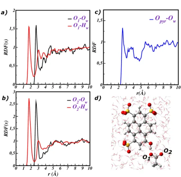
confirms that in the ground state, under equilibrium condition, the proton shuttle does not spontaneously occur, according to the aforementioned constrained energetic profiles and to the pKa values of acetate (4.5) and pyranine (7) in the ground state. In order to unveil the structuration of the solvent around the pyranine-acetate QM cluster, the radial distribution functions (RDF) between the charged and the polar groups of the QM cluster and the water molecules are calculated and depicted in figure 3.3 . In particular figure 3.3 a,b shows the RDF between the oxygen atoms of acetate and the solvent molecules, the RDF of the phenolic oxygen of pyranine is instead presented in figure 3.3 c.
In figure 3.3 a, we show RDFs of the distances between the acetate oxygen that is hydrogen bonded to pyranine (O1) and the water oxygen (Ow) and hydrogen (Hw),
respectively.
The first solvation shell around O1 corresponds to the first well-defined peaks,
centered at 1.77 ˚A and 2.77 ˚A for the O1-Hw and O1-Ow distances , respectively.
After the first peak the RDF has a no zero value, indicating that an exchange of water molecules occurs between the first and the second solvation shell during the simulation time of 10 ps. The integration of the first peaks suggests that the first solvation shell is on average composed by 1.5 water molecules establishing hydrogen bonds with O1. The RDF of the acetate oxygen exposed to the solvent (O2) (see
fig-ure 3.3 b) shows similar featfig-ures. The peaks corresponding to the first solvation shell are centered at 1.77 ˚A and 2.74 ˚A and exchange of solvent molecules from the first
Fig. 3.3: a) RDFs between O1 and the water molecules, O1-Owat (black) and O1-Hw (red)
dis-tances are shown. b) RDFs between O2 and the water molecules. c) RDF between Opyr and the
to the other solvation shells are monitored as well. The O2 does not interact directly
with pyranine molecule, so that it’s completely surrounded by water molecules. The higher peaks of the first solvation shell report about an average number of 2.5 water molecules around the O2. Lastly, RDF in figure 3.3c provides information about the
solvation of the phenolic oxygen of pyranine Opyr . The peak at 2.58 ˚A
correspond-ing to the Opyr-Ow distance suggests that on average one water molecule is strongly
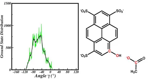
H-bound to this atom. This water plays a key role in the framework of the ESPT reaction, because it would be involved in a stabilization of the negative charge on the phenolic oxygen, once the proton moves to the acetate. The equilibrium picture points out the arrangement of the solvent molecules around the reactive cluster com-posed by the negative charged acetate and protonated pyranine. The arrangement is obviously subject to change during and after the ESPT reaction as response to the new charge distribution on the reactive cluster pyranine-acetate. Representa-tive points of that equilibrium are chosen as starting points for the propagation of the excited state dynamics. Because the ESPT direct reaction of pyranine-acetate couple is very fast (sub-ps time), the starting point of the excited state trajectory can strongly affect the ESPT kinetic. In particular, the relative orientation of ac-etate and pyranine will be shown to be fundamental as structural parameter of the starting structure. In this context, the intermolecular dihedral angle COpyr-COac
(γ) depicted in figure 3.4 was found suitable to describe the orientation of acetate. The pyranine is basically planar and the O1 of acetate is H-bound to its phenolic
Fig. 3.4: Ground state average distribution of the γ dihedral angle.
group. Regardless of that constrained , the other acetate moiety is free to move around the solvent molecules and to assume different conformations with respect to the pyranine plane. In figure 3.4 is shown the average distribution of the values that the dihedral angle assumes in the ground state simulation. The sample range covers the values 0-113◦. Values around zero correspond to a situation of planarity between
the four atoms defining the dihedral angle, but the points in -40-80 range are the most representative of the ground state equilibrium. The γ dihedral angle COpyr
-COac is defined by the heavy atoms that are directly involved in ESPT reaction. In
the next section, it will be shown that the initial value of the γ dihedral angle will be one of the key factor affecting the ESPT kinetic.
![Fig. 2: Schematic representation of the common photophysical processes occruring on excited potential energy surfaces, from [1].](https://thumb-eu.123doks.com/thumbv2/123dokorg/2756646.926/11.892.232.708.223.459/schematic-representation-photophysical-processes-occruring-excited-potential-surfaces.webp)