Dipartimento di Biologia, Ecologia e Scienze
della Terra
Scuola di Dottorato: Scienze della Vita
Curriculum: Biologia Vegetale
Ciclo – XXIX
Settore Scientifico Disciplinare BIO/01
Impact of DNA methylation on plant
growth and development: a study on a
methylation-defective mutant of
Arabidopsis thaliana
Direttore della Scuola:
Prof. Marcello Canonaco
Supervisore:
Co-Supervisore:
Dott. Leonardo Bruno
Prof.ssa Mieke Van Lijsebettens
Dottorando:
Dott. Forgione Ivano
I
Table of Contents
Pag.
ABSTRACT
IIITable of abbreviations VII
CHAPTER 1: INTRODUCTION
1.1 Epigenetic 1
1.1.1 Histone modification 2
1.1.2 Small RNA 5
1.1.3 DNA methylation 6
1.2 DNA methylation in plants 10
1.2.1 Sequence context and methylation pathways 10 1.2.2 DNA Methylation landscape of the Arabidopsis genome 12 1.2.3 Methods to identify DNA methylation 13 1.2.4 DNA methylation in the context of plant development 15 1.2.5 DNA methylation-defective mutants of Arabidopsis thaliana 19
1.3 Aim of the work 23
CHAPTER 2: MATERIALS AND METHODS
2.1 Plant lines 24
2.2 Seeds sterilization and In vitro plant growth conditions 24 2.3 Molecular characterization of T-DNA insertion mutant in
the DRM1, DRM2 and CMT3 genes in the triple mutant 25
2.4 Root analysis 26
2.5 Morphometric analysis of the primary root 27
2.6 RNA extraction 28
2.7 Single strand cDNA synthesis 29
2.8 Quantitative Reverse Transcriptase Polymerase
Chain Reaction (qRT-PCR) 29
2.9 Rosette area and leaf series 33
2.10 Epidermal cell analysis 33
2.11 Confocal microscopy analysis 34
2.12 Methylated DNA Immunoprecipitation (MeDIP) 35
II
CHAPTER 3: RESULTS
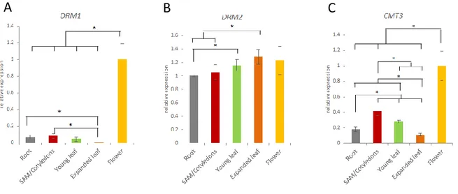
3.1 Organ-specific expression of DRM1, DRM2 and CMT3
genes in Arabidopsis wild type plant 43
3.2 In silico methyltransferases gene expression 44 3.3 DRM1, DRM2 and CMT3 expression during flower
development in Arabidopsis wild type plant 47
3.4 Molecular characterization of T-DNA insertion in the DRM1, DRM2 and CMT3 genes in the Arabidopsis drm1 drm2 cmt3 triple mutant 49 3.5 Expression levels of DRM1, DRM2 and CMT3 in
drm1 drm2 cmt3 T-DNA insertion line of Arabidopsis 52 3.6 Phenotypic analysis and growth parameters of drm1 drm2 cmt3 54
3.6.1 Seed germination and root growth 54
3.6.2 Shoot vegetative growth 57
3.6.3 Reproductive growth 59
3.7 Auxin distribution in Arabidopsis drm1 drm2 cmt3 61 3.8 Expression levels of auxin related genes in the whole plants
of Arabidopsis drm1 drm2 cmt3 mutant 64
3.9 Expression levels of auxin related genes in the primary root
of Arabidopsis drm1 drm2 cmt3 mutant 65
3.10 Expression levels of auxin- and growth-related genes in leaves
of drm1 drm2 cmt3 Arabidopsis mutant 66
3.11 Methylation levels of up-regulated auxin genes in Arabidopsis
drm1 drm2 cmt3 mutant through MeDIP analysis 70
3.12 ChIP analysis of CLF target genes 75
CHAPTER 4: DISCUSSION
79CONCLUSION
83Supplemental material 86
Aknowledgements 89
III
Abstract
Epigenetic modifications of DNA contribute to chromatin remodeling process and gene expression regulation playing a relevant role on the development of eukaryotic organisms. DNA methylation is an important epigenetic mark consisting in the addition of a methyl group on cytosine bases, which is observed in most of the organisms at the different evolution levels. In plants, DNA methylation is controlled by several genetic pathways, encoding different methyltransferases which act on different sequence contexts. Targets for cytosine DNA methylation in plant genomes are CG, CHG and CHH (H is A, T, C) sequences. The plant DNMT1-homolog METHYLTRANSFERASE1 (MET1) maintains DNA methylation at CG sites, whereas the DNMT3 homolog DOMAINS REARRANGED METHYLASE 1 and 2 (DRM1 and DRM2) are responsible for the de novo methylation in all sequence contexts. In addition, the plant-specific CHROMOMETHYLASE3 (CMT3) is responsible for DNA maintenance methylation at CHG sites, as well as at a subset of CHH sites. In plants DNA methylation is involved in diverse biological processes.
Loss of methylation in the Arabidopsis thaliana mutants met1 and ddm1 (decrease in DNA methylation 1) causes several developmental abnormalities. Similarly, combined mutations in the DRMs and CMT3 genes induce pleiotropic defects in plants. Here, we used the Arabidopsis thaliana triple mutant drm1 drm2 cmt3, defective in DNA methylation to get deeper insight into the correlation between DNA methylation and plant growth. We identified novel developmental defects of the triple mutant dealing with the agravitropic response of the root and an altered differentiation pattern of the leaf which also exhibits a curly shape. Confocal microscopy of mutant transgenic lines expressing DR5:GFP reporter gene allowed us to verify that the loss of DNA methylation impacts on the accumulation and distribution of auxin from embryo to adult plant. The expression of auxin-related genes has been also found to be altered in drm1 drm2 cmt3 mutant. Furthermore, through an optimized and implemented protocol of comparative analysis of genomic methylated regions based on MeDIP-qPCR, we provide evidence about the direct and organ-specific modulation of auxin-related genes through DNA methylation process.
The epigenetic mechanisms interplay with each other rather than work independently to modulate gene function. Accordingly, in our study we provide a novel evidence of the crosstalk between DNA methylation status and histone modification.
IV Indeed, in the drm1 drm2 cmt3 mutant the overexpression of CLF gene, a component of PCR2 complex that performs trimethylation of histone H3 lysine 27, was accompanied by a high level of histone methylation, as evaluated through ChIP-qPCR analysis, and by a concomitant down-regulation of genes target of PRC2 complex action. Thus, the results obtained in these three years of PhD course are encouraging and may open new perspectives in the study of the DNA methylation in plants.
V
Abstract
Le modificazioni epigenetiche del DNA contribuiscono al processo di rimodellamento della cromatina ed alla regolazione dell’espressione genica giocando un ruolo rilevante negli eventi di sviluppo degli organismi eucarioti. La metilazione del DNA è un importante tratto epigenetico che consiste nell’aggiunta di un gruppo metilico attraverso la formazione di un legame covalente al carbonio 5 della citosina. Tale meccanismo è stato osservato nella maggior parte degli organismi a diversi livelli della scala evolutiva. In particolare, i contesti di sequenza della metilazione del DNA nelle piante sono: CG, CHG, e CHH (H è A, T, C). I diversi enzimi responsabili del meccanismo di metilazione del DNA sono: i) METHYLTRANSFERASE1 (MET1) omologo di DNMT1 mantiene la metilazione del DNA nei siti CG; ii) DOMAINS REARRANGED METHYLASE 1 and 2 (DRM1 and DRM2), omologhi di DNMT3, sono responsabili della metilazione de novo in tutti i contesti di sequenza; iii) CHROMOMETHYLASE3 (CMT3), unica del regno delle piante, è responsabile della metilazione di mantenimento dei siti CHG e di un subset di siti CHH. Nelle piante la metilazione del DNA è coinvolta in diversi processi biologici.
La perdita di metilazione nei mutanti met1 e ddm1 (decrease in DNA methylation 1) di Arabidopsis thaliana causa diverse anomalie nello sviluppo, così come mutazioni combinate dei geni DRMs e CMT3 inducono difetti pleiotropici nelle piante. Nel presente lavoro di Dottorato, è stato utilizzato il triplo mutante drm1 drm2 cmt3 di Arabidopsis thaliana, deficiente nel processo di metilazione del DNA (de novo e di mantenimento) per studiare la relazione tra il meccanismo di metilazione e la crescita della pianta. Sono stati identificati nuove alterazioni nello sviluppo del triplo mutante drm1 drm2 cmt3 come la risposta agravitropica della radice ed un alterato pattern di differenziazione della foglia che esibiva un fenotipo aberrante definito “curly leaf”. L’analisi di linee transgeniche, che esprimono il gene reporter DR5:GFP, condotta mediante microscopia confocale, ha consentito di verificare che la perdita di metilazione del DNA ha un impatto sull’accumulo e distribuzione dell’auxina dalle prime fasi embrionali fino allo stadio adulto della pianta. Tramite analisi di qRT-PCR è stato dimostrato che nel triplo mutante drm1 drm2 cmt3 è presente, rispetto alle linee di controllo wild type, un’alterazione dell’espressione dei geni correlati al pathway dell’auxina. Inoltre, attraverso un protocollo ottimizzato di analisi comparativa delle regioni metilate, basato sulla tecnica della MeDIP-qPCR, è stato dimostrato che la
VI metilazione del DNA esercita una modulazione diretta ed organo-specifica sui geni correlati alla biosintesi dell’auxina.
Nel modulare l’espressione genica i meccanismi epigenetici interagiscono tra di loro piuttosto che agire in modo indipendente. Questo lavoro ha permesso di scoprire una nuova interazione tra lo stato di metilazione del DNA e le modificazioni istoniche. Infatti, nel mutante drm1 drm2 cmt3 l’over-espressione del gene CLF, un componente del complesso PRC2 che effettua tri-metilazione della lisina 27 dell’istone H3, è accompagnata da un alto livello di metilazione istonica, come è stato valutato dall’analisi ChIP-qPCR, e da una concomitante down-regolazione di geni target del complesso PRC2. Pertanto, i risultati ottenuti in questi tre anni di Dottorato sono incoraggianti e aprono nuove prospettive di studio della metilazione del DNA in piante.
VII
LIST OF ABBREVIATIONS
ARP ASYMMETRIC LEAF1/ROUGH SHEATH2/PHANTASTICA
BR Brassinosteroids
CDGS Chromatin-dependent gene silencing
cDNA Complementary DNA
ChIP Chromatin DNA Immunoprecipitation
ChIP Chromatin Immunoprecipitation
Col-0 Columbia-0
CZ Central Zone
DNA Deoxyribonucleic acid
dsRNA Double-stranded RNA
GFP Green Fluorescent Protein
H3K27me3 Histone 3 Lysine 27 trimethylation
HGI Horizontal Growth Index
KNOX1 KNOTTED-LIKE HOMEOBOX Class I
LBD LATERAL ORGAN BOUNDARIES DOMAIN
m5C 5-methylcytosine
MeDIP Methylated DNA Immunoprecipitation MeDIP Methylated DNA Immunoprecipitation
miRNA MicroRNAs
mRNA Messenger RNA
MS Murashige and Skoog
NAA 1-Naphthaleneacetic acid
NPA 1-N-Naphthylphthalamic acid
piRNA PIWI-interacting RNA
PTGS Post-transcriptional gene silencing PTM Post-translational modifications
PZ Peripheral Zone
QC Quiescent Center
qRT-PCR Quantitative Reverse Transcriptase Polymerase Chain Reaction
RAM Root Apical Meristem
RdDM RNA-directed DNA methylation
VIII
RNAi RNA interference
RNAseq RNA sequencing
SAM Shoot Apical Meristem
SD Standard Deviation
siRNA Short interfering RNA
smRNA Small RNA
ssRNA Single-stranded RNA
T-DNA Transfer DNA
TE Transposable Element
TGS Transcriptional gene silencing TSS Transcription Starting Site
UTR Untranslated Region
1
CHAPTER 1: INTRODUCTION
1.1 Epigenetics.
The term epigenetics was introduced in the early 1940s by Conrad Waddington (Waddington, 1942). In the original definition, epigenetics referred to all molecular pathways modulating the gene expression that do not involve the DNA sequence itself.
While the field of genetics focuses on the study of inherited genes, the epigenetics is the branch of the biology which studies changes in chromatin structure without alterations in the DNA sequence resulting in stable heritable phenotype (Berger et al., 2009). The epigenetic modifications described in current literature generally comprise histone variants, post-translational modifications of amino acids on the amino-terminal tail of histones, covalent modifications of DNA bases and non-coding RNAs including long and short non-coding RNAs (Fig. 1).
Figure 1. Chromosomes are composed of chromatin, consisting of DNA wrapped around eight histone protein units. Histone tails protruding from histone proteins are decorated with modifications, including phosphorylation (Ph), methylation (Me), and acetylation (Ac). DNA molecules are methylated by the addition of a methyl group to carbon position 5 on cytosine bases. Transcription involves the conversion of DNA to messenger RNA (mRNA). mRNA is translated into a protein product (Relton and Davey Smith, 2010).
2 Epigenetic marks affect gene expression and have tissue-specific patterns (Eckhardt et al., 2006) underlying tissue-specific gene expression (Musco and Peterson, 2008).
Epigenetic processes are essential for development and differentiation, but they can also arise by random change or under the influence of the environment (Issa, 2000). Indeed, in many studies, environmentally induced changes in gene expression are associated with epigenetic mechanisms. Moreover the epigenetic mechanisms interplay with each other rather than work independently to modulate gene function.
1.1.1 Histone modifications.
As above mentioned, the regulation of gene expression involves an intertwined complex of chromatin modifiers that is connected to post-translational histone modifications (PTM). The chromatin consists of DNA wrapped around a protein complex core that forms chromosomes. The single unit of the chromatin is the nucleosome which is based on a histone octamer that is surrounded by 146 base pairs of DNA. The major proteins of chromatin are the histones, small proteins containing a high proportion of basic amino acids (arginine and lysine) that facilitate binding to the negatively charged DNA molecule. The histones are classified into five types: H1, H2A, H2B, H3 and H4 (Kornberg and Lorch, 1999) and their variants (Ausiò, 2006). Detailed analysis of the nucleosome has shown that the DNA is wrapped 1.65 times around a histone core consisting of two molecules each of H2A, H2B, H3, and H4. To the core histones, there is the linker histone, H1, which contacts the exit/entry of the DNA strand on the nucleosome. Finally, 15-30 residues at the amino terminal of all the histones are unstructured and commonly referred to as tails (Kornberg and Lorch, 1999).
3 Figure 2. Schematic representation of the organization of the DNA wrapped around the histones to form the
nucleosome, which in turn give rise to the chromatin and then the chromosomes.
The chromatin shows a highly dynamic structure with different levels of condensation during the life cycle of the cell. It is commonly divided into euchromatin and heterochromatin. In euchromatic regions, genes are actively transcribed, whereas heterochromatic regions are transcriptionally inactive. Nucleosomes must dynamically change so that DNA binding complexes can access their binding sites. These dynamic changes involve the formation or disruption of interactions within the interfaces between the DNA and histones and are strictly dependent on post-translational modifications of the histone tails. The histone tails are subject to a vast array of post-translational modifications that include: ubiquitination, sumoylation, methylation, acetylation, phosphorylation, ribosylation, glycation, and carbonylation.
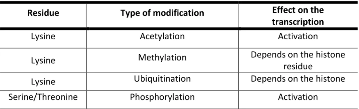
Ubiquitin is a polypeptide often associated with proteolysis. Ubiquitination occurs at the histones H2A and H2B where is attached to the lysine residues. H2A ubiquitination is more frequently correlated with gene silencing (Cao and Yan, 2012), while the H2B ubiquitination induces transcriptional activation by promoting other epigenetic marks related to histone methylation (Shukla et al., 2006). While DNA methylation is mainly linked to gene silencing, histone methylation represents a mark for transcription activation (Kouzarides, 2002).
A genome-wide analysis of histone methylation marks in plants revealed that trimethylation of histone 3 lysine 4 and di-/trimethylation of histone 3 lysine 36 (H3K4me3 and H3K36me2/me3, respectively) are enriched in actively transcribed gene sequences, whereas H3K27me3 and H3K9me2 are the main gene silencing markers (Zhang et al., 2007, Wang et al., 2009). A few histone methylations such as H3K27me1, H3K27me2 and H4K20me1 have been found in both transposon regions and heterochromatic regions (Roudier, et al., 2011).
Acetylation of core histones have been shown to positively affect gene transcription (Nelissen et al., 2007). Histones are acetylated at the lysine residues of the N-terminal tail. The addition of an acetyl group on the lysine residues neutralizes the positive charge of the histone tails and decreases its affinity for DNA. The new conformation facilitates the access of transcriptional regulatory proteins to the chromatin resulting in an increased transcriptional activity (Nelissen et al., 2010).
All the histone proteins of the core in the nucleosome are phosphorylated at specific serine and threonine residues. The phosphorylated histones are correlated with
4 transcriptional activation and often linked to acetylation of H3K9 and H3K14 (Turner, 2000).
Table 1. Post-translational histone modifications in plants.
Residue Type of modification Effect on the
transcription
Lysine Acetylation Activation
Lysine Methylation Depends on the histone
residue
Lysine Ubiquitination Depends on the histone
Serine/Threonine Phosphorylation Activation
In the past decade there were many genetic and biochemical studies that explored the relationship between DNA methylation and histone modification, particularly focusing on methylation of histones. It is known that DNA methylation and histone methylation are led by different sets of enzymes in different chemical reactions, but there are evidences that these pathways can be dependent on one another. Histone methylation can help to direct DNA methylation, and DNA methylation might be used as template for some histone modifications (Du et al., 2012). In particular, according to Ooi et al. (2007) DNA methylation might be mediated through histone modification: methylation of H3K4 (mono, di and trimethylation) might be formed in the embryo before de novo DNA methylation.
In addition, a model for heterochromatin assembly that links DNA methylation with histone methylation has been proposed by Soppe et al. (2002). By using two hypomethyleted mutants ddm1 and met1 they investigated on the relationship between DNA methylation and chromatin organization in Arabidopsis thaliana. The decrease in DNA methylation in both hypomethylated mutants occurred in parallel with the reduced methylation of H3K9, so the main cause of reduction of H3K9 methylation should be the reduction in DNA methylation.
Several other studies reported potential functional relationships between DNA methylation and H3K27me3 suggesting that these two epigenetic marks represent major epigenetic silencing mechanisms in plants and in many animal systems. For example H3K27me3 has been suggested to directly target DNA methylation in mammalian cells (Viré et al., 2006). By contrast, Zhang et al. (2007) suggest that the patterning and function of H3K27me3 in Arabidopsis are independent of DNA methylation.
5
1.1.2 Small RNA.
Diverse types of RNA ranging from small to long non-coding RNAs are regulators of gene expression beside play a role in genome stability and defence against foreign genetic elements. Indeed, these RNAs are able to modify chromatin and target gene expression via RNA interference (RNAi) pathways and prevent translational process by degradation at post-transcriptional level. These RNAs also play a role in chromatin remodelling and structure through pathways that do not involve RNAi; they seem to contain signals that recruit chromatin-modifying complexes (Rinn and Chang, 2012).
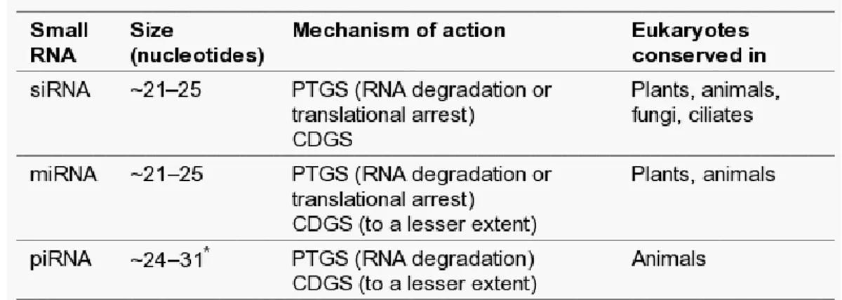
Three classes of small RNA have been identified in eukaryotes. The first two classes, short interfering RNAs (siRNAs) and microRNAs (miRNAs), are 21-25 nucleotides and are generated from longer dsRNA precursors by DICER, a ribonuclease III (RNaseIII) enzyme (Verdel et al., 2004). The third class of small RNAs, called PIWI-interacting RNAs (piRNAs), with a larger average size (24-31 nucleotides) than siRNAs and miRNAs, are involved in defence against parasitic DNA elements (Das et al., 2008).
Table 2. Conservation of small-RNA silencing pathways in eukaryotes. All three of the major RNA silencing
pathways identified seem to act in both post-transcriptional gene silencing (PTGS) and chromatin-dependent gene silencing (CDGS) pathways (Moazed, 2009).
There are many reports on how RNAi mediate histone or DNA methylation events that repress transcription (Moazed, 2009; Matzke et al., 2001). Studies of the flowering plant Arabidopsis thaliana demonstrated that one of the post-transcriptional gene silencing pathway was due to the production of small interfering RNAs (siRNAs) and its interaction with DNA methylation through RNA-directed DNA methylation (RdDM) of target loci (Matzke et al., 2001).
6 The pathway in which small RNAs (smRNAs) modulate gene activity is related to the type of ARGONAUTE (AGO) protein, which represents the catalytic component of the RISC (RNA induced silencing complex) which either prevents translational process or degrade target transcript. The association of siRNAs with one of the ten AGO proteins depends on its length and 5΄ terminal nucleotide. For instance, AGO1 is associated to the majority smRNAs that are 21 or 22 nucleotide long and carry 5΄ uracil, while most of the smRNAs associated with AGO2 are 21 nucleotide long and have 5΄adenine (Mallory and Vaucheret 2010). Since smRNAs are of different sizes and contain distinct 5΄ terminal nucleotides, they can repress gene expression either at the post-transcriptional level at the chromatin level through TGS depending on which AGO protein they interact.
1.1.3 DNA Methylation.
Nowadays it is known that among the epigenetic modifications DNA methylation, which is a covalent addition of a methyl group to the fifth carbon of the cytosine, has a direct impact on the regulation of gene expression.
Covalent modification of DNA have been described since 1948 (Hotchkiss, 1948), but it was only in 1969 that Griffith and Mahler proposed that gene expression (Fig. 3) may be modulated by these modifications (Griffith and Mahler, 1969).
Figure 3. The addition of methyl groups to CG is one mechanism for suppressing (or silencing) gene transcription (Fry, 2011).
This model was supported by additional studies over the next years, targeting the DNA methylation as the responsible for the stable maintenance of a particular gene expression pattern through mitotic cell division (Holliday and Pugh, 1975). The lack of methylation in the promoter region of the genes is usually associated with the
7 chromatin pattern of actively transcribed genes, as characterized by an opened nucleosome configuration (Tazi and Bird, 1990).
DNA methylation basically is a post-replication event. There are specific enzymes called methyltransferases which catalyze the transfer of methyl groups from S-adenosyl-L-methionine to DNA bases (Fujimoto et al., 1965). Although DNA methylation was found in both adenine and cytosine in bacteria and eukaryotes, it has been shown that cytosine methylation is more impactful on regulation in eukaryotes while adenine methylation seems to have mainly regulatory role in bacteria (Bickle and Kruger, 1993).
In animal genomes, cytosine methylation was thought to be restricted largely to the CG dinucleotide. In mammals, DNA methylation is predominantly found in cytosines of the dinucleotide sequence CG. Studies in the past decade have shown that CH (H = A, T, or C) methylation is present in cultured pluripotent stem cells, including embryonic stem cells (Ramsahoye et al., 2000), and recently CH methylation was found in the adult mouse cortex and human brain (Lister et al., 2013).
There are two basic types of DNA methylation processes known in eukaryotic cells. First is de novo methylation which is involved in the rearrangement of methylation pattern during embryogenesis or differentiation processes in adult cells (Razin and Cedar, 1993). The methyltransferases create hemimethylated CG dinucleotides after replication where methylation is found only on the original strand and is absent from the newly synthesized strand (Fig. 4). The second methylation activity in eukaryotic cells is the so-called maintenance methylation which is necessary to preserve DNA
methylation after every cellular DNA replication cycle. Hemimethylated CGs attract the
maintenance methyltransferases, which methylate the unmethylated strand to restore symmetric DNA methylation (Fig. 4).
Figure 4. The two types of DNA methylation. Schematic representation of de novo methylation and maintenance methylation of DNA. The methyl group is indicated by a red lollipop.
Methyltransferase enzymes involved in DNA methylation belong to different families and subfamilies whose members act on different sequence context and
8 different processes (i.e. maintenance VS de novo). For example, in mammalian genomes DNA methylation is established and maintained by two DNA METHYLTRANSFERASES (DNMTs) families. In particular DNMT1 acts in DNA methylation maintenance (Cheng and Blumenthal, 2008), while members of the DNMT3 family are responsible for de novo methylation (Okano et al., 1999).
Relevant roles of DNA methylation have been demonstrated in mammalian genomes. One of these is the maintenance of the inactive state of one of the female X chromosomes. X-inactivation is a process by which one of the two X chromosomes present in female mammals is inactivated. The inactive X chromosome is silenced by packaging in a transcriptionally inactive structure, the heterochromatin. By comparing the epigenetic profile of the heterochromatin of the inactive X chromosome with the euchromatin of the active X chromosome, it has been demonstrated that the inactive X chromosome has high levels of DNA methylation. Note that together with increased levels of DNA methylation, X chromosome inactivation shows low levels of histone acetylation, low levels of histone H3 lysine-4 methylation, and high levels of histone H3 lysine-9 and H3 lysine-27 methylation, all of which are strictly linked with gene silencing (Chow at al., 2005).
It was also found that DNA methylation is a key molecular mechanism of genomic imprinting. In this epigenetic phenomenon genes could be differently expressed depending on whether they came from the mother or the father (Surani et
al., 1984). Forms of genomic imprinting have been demonstrated first in mammals and
later in plants. There are evidences that, in plants, genes are imprinted primarily in the endosperm and in tissues that surround and nourish embryo during its development. An example is represented by the Arabidopsis thaliana gene MEDEA (MEA) that regulates cell proliferation by exerting a gametophytic maternal control during seed development (Grossniklaus et al., 1998) Several studies report that the majority imprinted genes examined show differences in DNA methylation. Consistently, the maintenance of the genomic imprint at the MEA locus is related to its methylation status (Vielle-Calzada et
al., 1999).
Another relevant role of DNA methylation deals with the inactivation of Transposable Elements (TEs) present in the genomes of higher eukaryotes. Active TEs also known as “jumping genes”, move from one location on the genome to another.
Much of what a TE does depends on where it lands. TE landing inside a gene can result in a mutation. This behavior of TEs might play a causal role in disease development, generate new genes and alter gene regulation and consequently cause
9 new phenotypes. Not all transposition results in negative effects. In fact, transposons can drive the evolution of genomes by facilitating the translocation of genomic sequences, and the repair of double-stranded breaks (Pray, 2008). TEs have the propensity to increase their representation from one generation to the next by replicating themselves at a higher rate than other genes. Moreover, there is evidence that transposons in all species examined appear to be largely quiescent (Yoder et al., 1997). This lack of activity is due to an epigenetic control. Molecular analysis of these elements revealed that inactivation was associated with DNA methylation where addition of a methyl group to cytosine was often associated with silencing.
Finally, it has been proposed that DNA methylation also represents one of the ways for controlling telomere length. Telomere is the region present at each end of a chromosome, formed by repetitive nucleotide sequences, that protects the chromosome end from degradation or fusion with neighboring chromosomes (Chan and Blackburn, 2002). Most adult cells progressively lose telomeres during cell division and tissue renewal, and it has been proposed that this telomere shortening contributes to the development of age-related pathologies, limiting human lifespan (Flores et al., 2005). Recent studies have shown that mammalian telomeres and subtelomeric regions are also enriched in epigenetic marks that are characteristic of heterochromatin. Mammalian telomere repeats (TTAGGG) cannot be methylated because they lack CG sequences. However decreases in DNA methylation, at subtelomeric regions, are accompanied by dramatically elongated telomeres (Gonzalo et. al., 2006).
In human, disruption of DNA methylation and other epigenetic marks give rise to many diseases. Changes in both DNA methylation and histone modifications have been
discovered in many cancer types. There are findings on the association of DNA
hypomethylation with tumors suggesting activation of oncogenes as a possible
consequence of decrease in DNA methylation (Feinberg and Vogelstein, 1983).Also in
mouse alteration in DNA methylation status related the total homozygous knockout of
DNMT1 homologous with its ortholog in human and chicken (Yen et al., 1992), was
lethal for the embryo (Li et al., 1992) suggesting a relevant role in embryo development. However, despite all this information, the dynamic status of DNA methylation during development and across generations, as well as the related role, remains poorly understood and mainly in plants (Lisch, 2009).
10
1.2 DNA methylation in plants.
The occurrence of DNA methylation in plants was firstly assessed in 1950s by
establishing that higher plant DNA contains 5-methylcytosine (m5C) in addition to four
ordinary bases (G, A, T, C) (Wyatt, 1950).
Since then, significant progress has been made and in the past decade plant research has improved our knowledge on the distribution and function of plant DNA methylation at a genome-wide scale. A very recent study provides a comparative DNA methylation profile of 34 different flowering plants (Niederhuth et al., 2016).
1.2.1 Sequence context and methylation pathways.
In plant genomes DNA methylation is more extensive and affects a wider sequence diversity than in animals. Plants have relatively high concentrations of
5-methylcytosine (m5C) compared to non-plant species because cytosine methylation
occurs in three sequence contexts: symmetric CG, CHG, and asymmetric CHH (in which H= A, T or C) (Henderson and Jacobsen, 2007).
Like in animals, different families of DNA methyltransferases with distinct substrate specificity and different modes of action account for DNA methylation in
plants: METHYLTRANSFERASE (MET) family, DOMAINS REARRANGED
METHYLTRANSFERASES (DRMs) family and the CHROMOMETHYLASES (CMTs) plant specific family (Table 3).
In details, DNA methylation in CG context is maintained by MET1, the homolog
of mammalian DNMT1, which is recruited to hemi-methylated CG sites and methylates
the opposing strand (Finnegan et al., 1996), whereas CHG methylation is maintained by
CMT3 (Lindroth et al., 2001).
The maintenance of methylation at CHG sites does not seem to depend on the palindromic symmetry of the sequence. The CMT3 activity is strongly associated with dimethylation of lysine 9 on histone 3 where the dual recognition of H3K9me2 by BAH (Bromo Adjacent Homology) and CHROMO domains of CMT3 leads the methylation of
CHG sites (Du et al., 2012). In more detail, it has been demonstrated thatKRYPTONITE
(KYP) is capable of binding methylated mCHH or mCHG of the DNA through its SRA (SET and RING-associated) domain (Jackson et al., 2002). CMT3 is recruited by H3K9me and
11 further methylates CHG of the DNA to create binding sites for KYP, resulting in a self-reinforcing feedback loop (Law and Jacobsen, 2010).
In addition to CMT3, other members of the CMT family are present in
Arabidopsis thaliana genome. CMT1 is expressed at low levels and is truncated in many
Arabidopsis ecotypes (Henikoff and Comai, 1998). CMT2, establishes and maintains asymmetrical methylation of CHH sites (Stroud et al., 2014). Recent studies on whole-genome methylation profile of cmt2 mutant showed loss of CHH methylation predominantly at TEs in the heterochromatic fraction (Zemach et al. 2013).
DRM1 and DRM2, homologue of the mammalian de novo DNA methyltransferase DNMT3, mediate de novo methylation of cytosines in all classes of sequence contexts (CG, CHG and CHH) (Law et al., 2010) (Table 3). Both enzymes are targeted by small interfering RNAs through a pathway termed RNA-directed DNA methylation (RdDM),
Table 3. Plant DNA methyltransferases.
Gene Name Target Sequence Effect on Chromatin/Transcription Effects of Mutation METHYLTRANSFERASE1 (MET1) CG
Maintains the global methylation Inability to establish CG methylation CHROMOMETHYLASE2 (CMT2) CHH Maintains the DNA methylation Loss of CHH methylation CHROMOMETHYLASE3 (CMT3) CHG Maintains the DNA methylation Loss of CHG methylation DOMAIN REARRANGED METHYLTRANSFERASES (DRM1, DRM2) CG, CHG and CHH De novo methylation of asymmetric sites; DRM2 is involved in de novo methylation in the RdDM pathway Loss of de novo DNA methylation
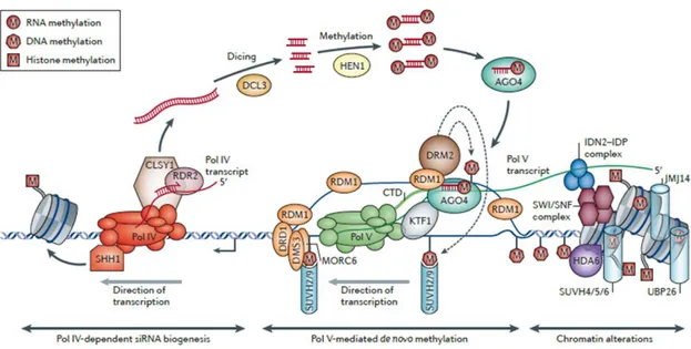
RdDM pathway is under the control of a specialized mechanism which depends on two plant-specific RNA polymerase called Pol IV and Pol V. Pol IV transcribes a
single-stranded RNA (ssRNA)at its target loci.The ssRNA is copied into a double-stranded RNA
(dsRNA) by the RNA-Dependent RNA polymerase 2 (RDR2) with the assistance of the
chromatin remodeller CLASSY 1 (CLSY1).The dsRNA is processed by DICER-LIKE 3 (DCL3)
12
(HEN1) and incorporated into ARGONAUTE 4 (AGO4). Pol V transcribes a scaffold RNA
that base-pairs with AGO4‑bound siRNAs.A key role is played by RNA-DIRECTED DNA
METHYLATION 1 (RDM1) the only protein that interacts with both AGO4 and DRM2 thus creating a bridge between them (Gao et al., 2010). These nucleoprotein complexes target chromatin-associated scaffold transcripts in a sequence-specific manner. The
chromatin-bound complexes then recruit DRM1 and DRM2 which methylate DNAin CG,
CHG, and CHH sequence contexts (Haag et al., 2011)(Fig. 5).
Figure 5.Canonical RNA-directed DNA methylation (RdDM) pathway (Matzke and Mosher, 2014).
1.2.2 DNA Methylation landscape of the Arabidopsis genome.
Most of the studies on DNA methylation in plants have been performed on the model plant Arabidopsis thaliana due to the high-quality sequence of its genome.
Several studies report DNA methylation to be conspicuously dense in the pericentromeric regions of nuclear chromosomes (Zhang et al., 2006; Zilberman et al., 2007; Lister et al., 2009) where CHG methylation results in higher levels likely due to its preference for transposon-related sequences. In contrast, to the dense methylation in pericentromeric regions, CG and CHH methylated sequences are widely distributed
13 distributed in distinct profiles over the Arabidopsis genome showing highest fraction in CG context. Indeed the CG methylation represents more than half of the methylated fraction of the DNA, whereas CHG and CHH represent an equally lower fraction (Lister et
al., 2008).
Methylation of genes has been extensively studied in Arabidopsis (Chan et al., 2005). Genome-wide analysis of DNA methylation revealed that more than 60% of the 27000 Arabidopsis thaliana genes are entirely unmethylated. A large number of genes (about 30%) are methylated within transcribed regions but not within their promoter, and a restricted 5% of the Arabidopsis expressed genes is methylated upstream of the transcription start site (Zhang et al., 2006).
DNA methylation of promoters is known to block transcription initiation, gene-body methylation has been hypothesized to interfere with elongation within active genes in Arabidopsis, especially if the methylation is close to the 5’ end (Hohn et al., 1996).
DNA methylation can inhibit transcriptional activity or make silent chromatin in two different ways. First, DNA methylation can physically impede the binding of transcription factors to the gene making them inaccessible to the transcription machinery. Second, methylated DNA may recruit methyl-cytosine binding proteins which in turn may recruit additional protein complexes, such as histone deacetylases and other chromatin remodeling protein that can modify the structure, resulting in inactive and silent chromatin (Boyes and Bird, 1991).
1.2.3 Methods to identify DNA methylation.
DNA samples are usually derived from a collection of cells, which might be variable in their methylation pattern. For this reason analysis of DNA methylation is complicated. Conventional techniques such as cytosine hybridization-based are not able to distinguish 5-methyl-cytosine from unmethylated cytosine. Furthermore DNA methyltransferases are not present during PCR or in biological cloning systems so DNA methylation information is lost during amplification. To maintain DNA methylation during PCR it would require a thermostable enzyme with no de novo methyltransferase activity, but it has not been discovered to date (Laird, 2010). Currently, methylation dependent pre-treatments of DNA are employed to reveal the presence or absence of
14 the methyl group at cytosine residues. There are three main approaches used to identify DNA methylation.
First, restriction endonucleases are such powerful tools in molecular biology. They are dependent on the presence of specific recognition sequences and some of
them are inhibited by the methyl group on the cytosine.Endonuclease activity followed
by hybridization can provide information on DNA methylation pattern. The major issue with this technique is about the location of the methyl-cytosines and therefore the precise sequence of the methylation is not identifiable. The method of methylation-sensitive restriction digestion followed by PCR amplification across the restriction site is a very sensitive technique that is still used today. However, it can give false-positive results caused by incomplete digestion for reasons not connected to DNA methylation (Tompa et al., 2002).
Similarly to chromatin immunoprecipitation (ChIP) assay used in determination
of enriched histone modifications, methylated genomic regions can be detected using
antibodies specific for 5 methyl-cytosines or using methyl-binding proteinswith affinity
for methylated DNA. A specific antibody for methylated cytosine is employed to detect enrichment of methylated regions by immunoprecipitation of denatured genomic DNA, followed by hybridization (MeDIP-chip), sequencing (MeDIP-seq) or amplification (MeDIP-qPCR).
These techniques have been widely used to explore the methylomes of plant
(Zhang et al., 2006), mouse (Mukhopadhyay et al., 2004
)
and human (Irizarry et al.,2008). However, these techniques are subject to limitations such as low resolution of detection.
Bisulfite conversion provides high-resolution detection of DNA methylation. Chemical treatment of genomic DNA with sodium bisulfite converts cytosines residues, much more rapidly than methylated cytosines, into uracil via a sulfonation, deamination, desulfonation reaction (Hayatsu, 2008). Subsequently the use of direct sequencing determines the locations of unmethylated cytosines and 5-methyl-cytosines at single-nucleotide resolution. Over the past decade, Sanger sequencing after bisulfite modification has represented a key role in the study of DNA methylation. Today, by coupling bisulfite conversion to NGS (Next Generation Sequencing) technology it is possible to map the sites of DNA methylation at single-base resolution throughout an entire genome with very high quality and sensitivity.
15
1.2.4 DNA methylation in the context of plant development.
The development of a multicellular organism requires the spatially coordinated acquisition of numerous cell identities. Therefore a spatio-temporal regulation of gene expression is required. On the other hand to generate the body pattern the cells of the developing organism have to exchange and respond to information about their relative position. In such positional signaling endogenous hormones play a relevant role (Swarup et al., 2001; Davies, 2010; Zažímalová et al., 2014).
In higher plants, the apical-basal axis that determine the future growth direction of the organism is precociously established during embryogenesis and is driven by the shoot and root apical meristems (SAM and RAM) positioning. The radial pattern is made of concentric layers of the main tissue types. The normal apical-basal axis formation in embryo is determined by the correct distribution of auxin which is under the control of the PIN family proteins that are expressed in embryo in different places and at different time during development. PIN7 and PIN1 are the earliest PIN genes expressed in the embryo. PIN7 protein is found at the apical membrane of the basal cell in the two-cell embryo and at the apical membrane in suspensor cells until the 32 cell stage suggesting a role for PIN7 in transporting auxin from the basal cell to the apical cell. PIN1 protein is localized throughout the embryo, from the single-cell stage until the 32-cell stage. PIN4 protein begins to accumulate in the hypophyseal lineage and suspensor. PIN3 mRNA expression is detected at the root pole of the heart-stage embryo (Friml et al., 2002; Friml et al., 2003).
Both SAM and RAM are maintained during the postembryonic phase and are essential for plant growth and development because they features as stem cell niche where cells continuously divide and produce progeny cells which undergo different fate, while self-renewing (Bitonti and Chiappetta, 2010; Petricka et al., 2012; Zažímalová et al., 2014; Pfeiffer et al., 2016; Soyars et al., 2016).
In such a way, SAM continuously produces stems and lateral organs (leaves and floral organs). Leaf initiation occurs at specific positions at the periphery of the SAM (Reinhardt et al., 2000) which is organized in zones with different rates of cell division and different functions (Medford et al., 1992). The dome-shaped structure can be generally divided into three distinct zones: the Central Zone (CZ) at the surface of the dome, surrounding its apex which maintains indeterminate growth and produce daughter cells for the neighboring peripheral and rib zones; the Peripheral Zone (PZ), also on the surface contributes to the formation of new organs; the Rib Zone (Rib),
16 deeper within the dome, below the central zone. The majority of cell division occurs in the peripheral zone and rib meristem, with slower division occurring in the central zone. SAM is also subdivided into distinct cell layers termed L1, L2, and L3 (Satina et al., 1940). The L1 and L2 each form single cell sheet because of anticlinal cell division patterns. When the cells in the L1 layer differentiate they produce pavement cells, guard cells and trichomes. The cells of the L2 layer give rise to mesophyll cells (photosynthetic tissue) in the leaf. In developing anthers L2 layer cells differentiate into microsporocytes and tapetum, the latter nourishes the developing pollens. Similarly, in ovules the L2 cell layer is responsible for megasporocytes development. The cells in the L3 undergo both anticlinal and periclinal cell division patterns to form the inner core of the meristems where cells later on differentiate into vascular bundles and pith cells and form the stem tissue.
The SAM has properties of a self-regulatory system in which the interactions between the genes WUSCHEL (WUS) and CLAVATA (CLV) establish a feedback loop between the stem cells and the underlying organizing center. The WUSCHEL (WUS) gene is required for stem cell identity (Pfeiffer et al., 2016; Soyars et al., 2016). The CLAVATA (CLV1, CLV2, CLV3) genes, which encode for components of a signaling pathway that limits the size of the SAM, promote the progression of meristem cells toward organ initiation. The function of the CLV genes is antagonized by the SHOOTMERISTEMLESS (STM) gene, a transcription factors belonging to the class 1 KNOTTED-LIKE HOMEOBOX (KNOX1) gene family (Clark et al., 1996). STM, indeed, promotes meristem formation and maintenance. The repression of the gene STM and the activation of the gene ASYMMETRIC LEAVES1 (AS1) are also crucial for leaf initiation (Byrne et al., 2000). Notably, an interaction between these genetic network and different hormone classes has been largely demonstrated (Gray, 2004).
The RAM assures the growth and development of root system (Bitonti and Chiappetta, 2010; Petricka et al., 2012). The primary root, or radicle, is the first organ to appear when a seed germinates. It grows downward into the soil, anchoring the seedling and enhancing water uptake. The RAM dome is located subterminally and is covered by the root cap, which protects the apical meristem, produces mucilage to facilitate a passage for the growing root, and serves as a gravity perceiving tissue. The RAM pattern consists of concentrically arrayed stem cells that extend the radial pattern in the growing root, the four cells of the quiescent center (QC), which divide only infrequently, and most distally, the initials of the central root cap (columella). Several network of genes are involved in the establishment and maintenance of the RAM. One of them
17 deals with the PLETHORA (PLT) gene family that plays a key role in the acquisition of the QC fate (Aida et al.,2004). These genes are required for stem cell specification and maintenance in the RAM and act in parallel with SHORT-ROOT (SHR) and SCARECROW (SCR) to define QC and stem cell position. SHR is transcribed exclusively in the provascular tissue from embryogenesis onward, but the protein moves outwards to the surrounding cell layers including the QC and promotes SCR expression in these cells (Nakajima et al., 2001). SCR is required for QC identity, which in turn promotes the activity of surrounding stem cells (Sabatini et al., 2003).
The proper expression of PLT1 and PLT2 relies on the establishment of an auxin response maximum (Blilou et al., 2005). It is known that auxin is transported in a directional manner by membrane spanning proteins that mediate the influx and efflux of this signaling molecule into and out of cells. The localization of members of the PINFORMED (PIN) family of auxin efflux facilitators within cells reflects the direction of auxin transport (Petrásek et al., 2006). In particular PIN1 is localized at the basal (root apex-facing) side of the root vasculature; PIN2 at the basal side of the cortical cells and at the apical (shoot apex-facing) side of the protoderm and root cap cells; PIN3 in the columella cells of the root; PIN4 at the basal side of cells in the central root meristem and in the cells of the quiescent center; and PIN7 at the basal side of the stele cells and in the columella cells (Feraru and Friml, 2008). Moreover, the auxin effects on the transcription of genes involved in root patterning are mediated by members of the AUXIN RESPONSE FACTORS (ARF) family (Aida et al.,2004). The MONOPTEROS (MP) gene encodes a member of the ARFs that can bind to promoter elements of auxin-inducible genes which is required for embryonic root formation and RAM maintenance.
The development of plant leaves follows a common basic program. Leaves initiate at the flanks of the SAM and then develop into a flat blade of variable size and form. This process is regulated by several plant hormones and transcriptional regulators (Bar and Ori, 2014). Leaf development can be divided in two three main processes: i) initiation of the leaf primordium, ii) establishment of dorsiventrality and iii) development of a marginal meristem which lead to lamina growth. So far, several aspects of the hormonal and genetic network that control leaf development have been clarified in recent developmental and molecular genetic studies of Arabidopsis (Bar and Ori, 2014). In more details, it has been demonstrated that the hormone auxin is a central regulator of leaf initiation and points of auxin accumulation have been found to precede organ initiation. These are created by auxin biosynthesis in the SAM and by directional auxin transport generated by PIN1 auxin transporter (Bar and Ori, 2014). Moreover leaf
18 initiation is tightly correlated with vasculature development which involves changes in PINs polarization, from a polarization towards the outermost cell layer (L1) of primordium to a basal localization towards the future vasculature (Scarpella et al., 2006).
Specification of the leaf primordium domain depends also on differential expression of genes that regulate the balance between undeterminated vs committed cell fates. In particular this balance is mainly controlled by (i) KNOX1 transcription factors, which are expressed in the SAM central dome, where promote meristematic state, and are downregulated at the site of organ initiation (Hay and Tsiantis,2010); (ii) ARP [ASYMMETRIC LEAVES1 (AS1), ROUGH SHEATH2 (RS2), PHANTASTICA] transcription factors and the LBD (LATERAL ORGAN BOUNDARIES DOMAIN) protein AS2 which are expressed at the site of leaf initiation and, together with LBD protein AS2 repress KNOX1 thus specifying leaf initiation domain (Barkoulas et al., 2007; Bar and Ori 2014). To more KNOX genes KNAT2 and KNAT6 are also negatively regulated by AS1 but their inter-relationship is less understood. Interestingly, recent studies demonstrated a role for chromatin remodeling factors in the repression of KNOX1 genes by AS1-AS2 in Arabidopsis. For example, AS1 interacts with the histone deacetylase HDA6 and increased acetylation of KNOX1 genes has been detected in hda6 mutants (Luo et al., 2012). In addition, it has recently been shown that the AS1-AS2 complex recruits POLYCOMB-REPRESSIVE COMPLEX 2 (PRC2), a complex involved in chromatin structure modification, to the promoters of two KNOX1 genes, likely determining their repression at later stages of leaf development (Lodha et al., 2013).
Following initiation, the leaf primordium undergoes lamina growth, morphogenesis and differentiation associated to a precocious establishment of leaf dorsiventrality (i.e. the juxtaposition of abaxial and adaxial tissues). The establishment of leaf dorsiventrality which involves the commitment of abaxial and adaxial cell fate and underpins functional specialization of the upper and lower side of the leaf. Many genes are involved in this commitment working in both cooperative and antagonistic way. For example, a relevant role is played by AS1 and AS2 genes that are expressed through the leaf primordium at early stage when the leaf develops. AS2 localizes to the adaxial side while AS1 is confined to the inner regions between the adaxial and abaxial domains. Both promote adaxial fate by repressing the abaxial promoting factors. Moreover, a balance between transient cell proliferation, which underlies growth and morphogenesis and differentiation, which requires repression of meristematic status, is
19 essential during leaf blade formation. An overlapping set of genes and antagonistic transcription factors are involved in controlling also these processes (Bar and Ori, 2014). Therefore, very complex genetic frameworks and signaling machinery are at the base of plant development which requires a precise temporal and spatial regulation of regulatory genes. Moreover, the plants are sessile organisms, constantly exposed to the environmental cues. Therefore, a rapid modulation of gene expression is also essential to adapt themselves to environmental changes. In this context, epigenetic mechanisms which act at the transcriptional or post-transcriptional level can play a relevant role.
Accordingly, there are evidences that DNA methylation, together with other epigenetic marks, is involved in the regulation of gene transcriptional activity in response to both endogenous developmental factors and external stimuli (Finnegan et al., 1993 ; Dennis et al., 1998; Bitonti et al., 2002; Fojtova et al., 2003; Greco et al., 2012). Moreover, through novel dissection methods, such as laser-capture microscopy (Kerk et al., 2003; Nakazono et al., 2003) combined with highly sensitive detection of DNA or chromatin modifications at target loci, it has been demonstrated that the root and shoot tissues of Arabidopsis accumulate significant epigenetic changes which accompany the transcriptional reprogramming associated with the development of roots and shoots. A recent comparative analysis about DNA methylation in shoot and root revealed that sites that are differentially methylated are preferentially hypermethylated in shoots compared with roots (Widman et al., 2013). Hence, epigenetic differences between shoots and roots in Arabidopsis reveals tissue-specific regulation.
1.2.5 DNA methylation-defective mutants of Arabidopsis thaliana.
During the past decade, the most widely used and easy experimental organism for studies in molecular genetics, has been the small Arabidopsis thaliana. Despite, Arabidopsis is not an economically important plant, it has been a model to study a broad range of problems in development, metabolism, genetics, environmental adaptation, pathogen interactions, and many other areas.
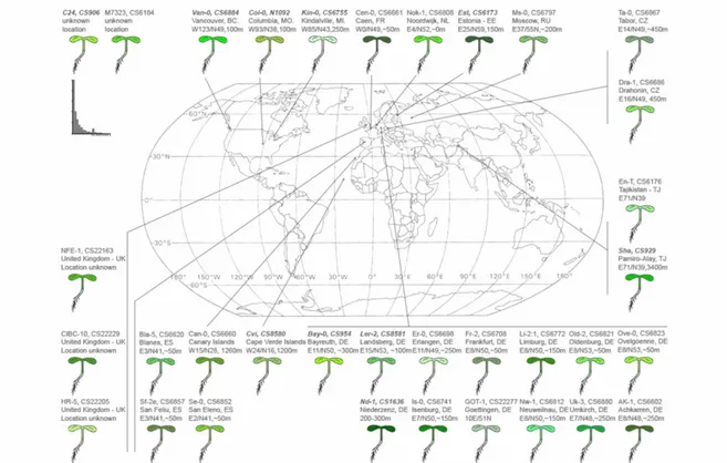
Arabidopsis thaliana is a member of the Cruciferae (family Brassicaceae, Capparales). The genus Arabidopsis contains about ten species that are native to
20 Eurasia, North Africa and North America (Mitchell-Olds, 2001). World map showing the geographical distribution (longitude, latitude, elevation) of more than 30 Arabidopsis ecotypes (Fig. 6).
Figure 6.Image of worldwide distribution of Arabidopsis thaliana (Jonathan Clarke, 1993).
Arabidopsis thaliana has been for many plant scientist the organism of choice as model system presenting many advantages: it is easily manipulated indeed, it can be grown in confined laboratory environments; it has a rapid life cycle of about 6 weeks from germination to mature seed and a very prolific seed production. Furthermore one advantage offered to the plant researcher by Arabidopsis is its relatively small genome size (135 Mb approximately divided in five chromosomes). It is know that the large crop genomes pose challenges to the researcher, including difficulty in sequencing as well as in isolation and cloning of mutant loci. The entire Arabidopsis genome has been completely sequenced and it represents a powerful tool for studying the function and the identity of more than 27000 protein coding genes. Arabidopsis plants are found to
be easily transformable with the current technologies. Finally, the large collections of
Arabidopsis thaliana mutant alleles are the most important resource for in vivo characterization of gene function.
21 This large collection includes mutants affected in the establishment, maintenance or removal of DNA methylation. The plant research community has employed, for long time, these mutants in order to provide information about the functional aspect of this epigenetic modification in the plant genome. One of these mutant is the loss-of-function met1 mutant. As above mentioned, MET1 protein is responsible for maintaining cytosine methylation throughout the Arabidopsis genome. In the met1 mutants a reduction of global cytosine methylation levels, particularly at CG sites, was observed (Finnegan et al. 1996; Ronemus et al. 1996) together with developmental abnormalities in both vegetative and reproductive structures. Seedlings from the T2 and subsequent generations had decreased stature, smaller rounded leaves, leaves with margins curled toward the upper leaf surface, decreased fertility, reduced apical dominance and shorter roots (Finnegan et al. 1996). A large class of endogenous genes has been described with CG methylation in their ORFs and promoter region. The promoter of FLOWERING WAGENINGEN (FWA) is normally methylated within two direct repeats causing FWA expression to be silenced. It has been verified that the met1 mutation causes FWA demethylation and missexpression resulting in a dominant late-flowering phenotype (Soppe et al., 2000). Similar abnormalities were observed in methylation-deficient Arabidopsis lines with defects in either the SWI2/SNF2chromatin remodeling factor-related gene DDM1 (Kakutani et al., 1996) or the Dnmt1-related MET1 gene (Finnegan et al., 1996; Ronemus et al., 1996).
Other loss-of-function mutants, drm1 and drm2, deals with DRM1 and DRM2 genes whose encoded product may be responsible for methylation of cytosines in inverted repeat transgenes at both CHG and CHH sites (Cao and Jacobsen, 2002a) and together with CMT3 maintains the non-CG methylation in Arabidopsis. Additional mutant are cmt2 and cmt3. Actually, the Arabidopsis genome contains three CMT-encoding genes. CMT1 is preferentially expressed in flowers and the extraordinarily low presence of its mRNA in Arabidopsis thaliana plants made it impractical to obtain full-length cDNA using standard protocols. Molecular characterization of CMT1 genomic and cDNA sequences revealed that CMT1 protein is truncated in several Arabidopsis ecotypes (Henikoff and Comai, 1998). CMT2 is expressed and is a putative DNA methyltransferase. Recently a whole-genome methylation profiling in cmt2 mutant showed loss of CHH methylation predominantly at large TEs that were heterochromatic (Stroud et al., 2014). CMT3 is the main methyltransferase of the CMT family and is involved in CHG DNA methylation. Cmt3 loss-of function mutants showed a genome-wide loss of CHG methylation (Lindroth et al., 2001).
22 The drm1, drm2 and cmt3 single mutants did not show any apparent phenotypes, as well as drm1 drm2 double homozygotes showed a morphology similar to the wild-type WS strain, even after five generations of inbreeding (Cao and Jacobsen, 2002a, 2002b). Whereas the triple drm1 drm2 cmt3 mutant evidenced pleiotropic phenotypic abnormalities, dealing with plant size, leaf shape and seed production (Cao and Jacobsen, 2002a; Chan et al., 2006; Bruno et al., unpublished data)
23
1.3 Aim of the work.
The overall aim of this thesis was to extend the knowledge about the DNA methylation and its role in the modulation of the plant growth and development.
As already mentioned plant development relies on very complex signaling machinery and genetic networks which requires a temporal and spatial regulation of regulatory genes. Moreover, since plants are sessile organisms, they developed a surprising growth plasticity to achieve adaptive traits under environmental pressure.
In this context, DNA methylation, which controls gene expression together with other epigenetic events, may represent a dynamic mechanism of adaptation, through a rapid and simultaneous modulation of genetic pathways.
The main objective of the present PhD project was to identify genetic networks and metabolic pathways that are affected as consequence of the altered DNA methylation pattern taking advantage DNA methylation-defective mutants of Arabidopsis thaliana.
Preliminary data, obtained through visual and microscopic observations carried out by the Plant Biology group at the University of Calabria, indicated the drm1 drm2 cmt3 triple DNA methylation mutant, defective in three genes that encode for methyltransferases, was the most interesting subject to bring our attention because conversely than the single mutants drm1, drm2, cmt3 and the double mutant drm1 drm2 it showed visible developmental defects compared to the wild type.
Starting from these assumptions, in this project we wanted to investigate about novel growth disorders in DNA methylation triple mutant and elucidate the correlation between DNA methylation status and specific genetic and/or hormonal pathways.
24
CHAPTER 2: MATERIALS AND METHODS
2.1 Plant lines.
A triple DNA methylation mutant of Arabidopsis thaliana, created by crossing (as reported in Henderson and Jacobsen, 2008) single T-DNA homozygous mutant lines of drm1-2 (SALK_021316; At5g15380), drm2-2 (SALK_150863; At5g14620) and cmt3-11 (SALK_148381; At1g69770), was purchased from Nottingham Arabidopsis Seed collection (http://arabidopsis.info) and it was a pure mutant line for each locus where the T-DNA was inserted as reported in the paragraph 3.4 of the result chapter. drm1 drm2 cmt3 drm1 drm2 cmt3 triple mutant is in Columbia (Col-0) background. Seedlings of Col-0 were used as control for the comparison.
2.2 Seeds sterilization and In vitro plant growth conditions.
Arabidopsis thaliana ecotype Columbia-0 and the mutant seeds were sterilized separately in 70% Ethanol (EtOH) for 2 minutes then in 5% bleach and 0.05% Tween20 for 10 minutes and washed in water for five times for 5 minutes. After sterilization seeds were sown in Petri dishes containing growth medium. In this study MS medium (Murashige and Skoog, 1962) was used including vitamins (Glycine 2 mg/l, myo-Inositol 100 mg/l, Nicotinic acid 0,5 mg/l, Pyridoxine HCl 0,5 mg/l, Thiamine HCl 0,1 g/l), 1% sucrose, 0,1 g/l myo-inositol, 0,5g/l 2-N-morpholine ethane sulphonic acid (MES), 8 g/l agar. The pH was set to 5,7 with KOH 1N.
After 2 days of stratification at 4 °C Petri dishes were transferred in the growth chamber in 16h/8 (day/night) with white light (neon fluorescent tubes “Radium NL Spectralux, cool white”), 100 μmoles m-1 s-1 light intensity, and 21°C, and 50% relative humidity.
For germination experiments the seeds were sterilized, sown in Petri dishes on MS medium, stratified and transferred in the growth chamber as described above. One-hundred seeds of Col-0 and one-One-hundred seeds of drm1 drm2 cmt3 from different batches were sown in the same Petri dish. Observations were performed every day to check the germination status of the seeds. The results are from three independent repeats.
25 For exogenous IAA treatment seeds of Col-0 and mutant were sterilized and sown in Petri dishes containing growth medium (see materials and methods paragraph 2.2) supplemented with 0.02, 0.2, 1 µM IAA concentration. After 2 days of stratification at 4 °C Petri dishes were transferred in the growth chamber in 16h/8 (day/night) with white light (neon fluorescent tubes, cool white), 100 μmoles m-1 s-1 light intensity, and 21°C, and 50% relative humidity. Six days old seedlings were photographed and imaging analysis was carried out by Image J software (https://imagej.nih.gov/ij/). The obtained data of three biological replicates (n=30) were used for statistical analysis of the mean and standard deviation and Student’s t-test.
The study of aerial part of the plant was carried out on 15 days old seedlings transferred from MS medium to Jiffy-7 Pellets and grown in growth chamber in the same conditions as above. Observations were performed periodically and every stage was photographed in order to catch difference between wild type and mutant.
2.3 Molecular characterization of T-DNA insertion mutant in the DRM1, DRM2
and CMT3 genes in the triple mutant.
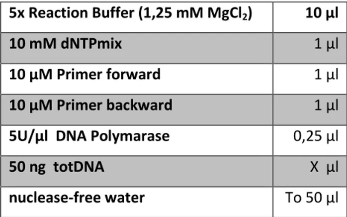
Wild type ecotype Columbia-0 and drm1 drm2 cmt3 insertion T-DNA line were obtained from the European Arabidopsis Stock Centre (NASC). Triple mutant generated by crossing drm1 (SALK_021316), drm2 (SALK_150863) and cmt3 (SALK_148381) plants; individual mutants are confirmed lines isolated from original SALK lines and are homozygous for the insertion. To screen for homozygous insertion alleles in drm1 drm2 cmt3 triple mutant, primers were designed following the instructions of the Salk institute genomic analysis laboratory (http://signal.salk.edu/tdnaprimers.2.html). PCR were performed by using complementary primers to the region flanking the T-DNA insertions site in DRM1, DRM2 and CMT3 genes and a primer (LBb1.3) complementary to the T-DNA sequence (Tab. 1) in order to detect the insertion within the gene. For general PCR the Taq DNA polymerase (Promega) was used. The reaction mixes were prepared in a sterile nuclease-free 0,2 ml microcentrifuge tubes with following components:
26 5x Reaction Buffer (1,25 mM MgCl2) 10 µl
10 mM dNTPmix 1 µl
10 µM Primer forward 1 µl
10 µM Primer backward 1 µl
5U/µl DNA Polymarase 0,25 µl
50 ng totDNA X µl
nuclease-free water To 50 µl
PCR was performed on a Primus 96 Plus Thermal Cycler (MWG Biotech) using the following program:
Step 1 Step 2 Step 3 Step 4
Temperature 95°C 95°C 55-60°C 72°C 72°C 4°C Time 2 min 40 sec 40 sec 1-3 min 5 min ∞
Cycles 1x 35x 1x
Table 4: List of primers and PCR conditions used for genotyping.
Locus Name Sequence
LBb1.3 5'-ATTTTGCCGATTTCGGAAC-3'
AT5G15380 SALK_021316(DRM1)LP 5'-GAGCCGTCTCATCAAACTGAC-3' AT5G15380 SALK_021316(DRM1)RP 5'-TTGCAGGAGCAAATATGGAAC-3' AT5G14620 SALK_150863(DRM2)LP 5'-AGATCGCTTCCAGAGTTAGCC-3' AT5G14620 SALK_150863(DRM2)RP 5'-TTGTCGCAAAAAGCAAAAGAG-3' AT1G69770 SALK_148381(CMT3)LP 5'-CCCTCAACAATTAACTGACGC-3' AT1G69770 SALK_148381(CMT3)RP 5'-ATAAGAGAAGGAGCTGCTGCC-3'
2.4 Root analysis.
For root analysis seeds of wild type (Col-0) and drm1 drm2 cmt3 triple mutant were sterilized and sown together (4 seeds Col-0 and 4 seeds triple mutant) on MS medium in square plates (12 x 12 cm). After 2 days of stratification the plates were placed in vertical position in the growth chamber under continuous light condition with