Genomic blueprint of a relapsing fever pathogen in
15th century Scandinavia
Meriam Guellila,1, Oliver Kerstena, Amine Namouchia, Egil L. Bauerb, Michael Derrickb, Anne Ø. Jensenb, Nils C. Stensetha,1, and Barbara Bramantia,c,1
aCentre for Ecological and Evolutionary Synthesis, Department of Biosciences, University of Oslo, N-0316 Oslo, Norway;bNorwegian Institute for Cultural Heritage Research, N-0155 Oslo, Norway; andcDepartment of Biomedical and Specialty Surgical Sciences, Faculty of Medicine, Pharmacy and Prevention, University of Ferrara, 35-441221 Ferrara, Italy
Contributed by N. C. Stenseth, August 13, 2018 (sent for review May 4, 2018; reviewed by Sally J. Cutler and Albert R. Zink)
Louse-borne relapsing fever (LBRF) is known to have killed millions of people over the course of European history and remains a major cause of mortality in parts of the world. Its pathogen,Borrelia recurrentis, shares a common vector with global killers such as typhus and plague and is known for its involvement in devastating historical epidemics such as the Irish potato famine. Here, we de-scribe a European and historical genome ofB. recurrentis, recov-ered from a 15th century skeleton from Oslo. Our distinct European lineage has a discrete genomic makeup, displaying an ancestral oppA-1 gene and gene loss in antigenic variation sites. Our results illustrate the potential of ancient DNA research to elucidate dynamics of re-ductive evolution in a specialized human pathogen and to uncover aspects of human health usually invisible to the archaeological record. ancient genomics
|
relapsing fever|
vector-borne pathogen|
immune evasion
|
aDNAL
ouse-borne relapsing fever (LBRF), once one of many fevers ravaging Europe, has disappeared from the Western world and is now endemic to only eastern Africa. The disease is part of a group of well-known deadly louse-borne pathogens, which spe-cialize in vector–human transmission. Its causative pathogen is the spirochete Borrelia recurrentis, whose only known vector is the human body louse, Pediculus humanus. This sets it apart from other tick-borne relapsing fever (TBRF) pathogens, such as its most closely related strains Borrelia duttonii and Borrelia crocidurae (1, 2). Genomes of the genus Borrelia are unique. They are composed of up to 24 circular and linear plasmids with covalently closed hairpin telomeres showcasing AT-rich genomes of very small sizes. While the chromosomes, generally around 900–930 kbp, are very conserved across species, a high level of DNA rear-rangements can be found among the plasmids (3–5), with some carrying essential genes, such as the telomere resolvase gene resT on plasmid pl23 of B. recurrentis and B. duttonii.The B. recurrentis genome (1.24 Mbp) is composed of one linear chromosome and seven linear plasmids and is character-ized by its low GC content (mean 28.1%). Genomic and eco-logical data on the pathogen are scarce, since it is challenging to cultivate and studies lack an animal model (6, 7). Research on the pathogen has therefore been limited to clinical samples, resulting thus far in the publication of one deposited reference sequence, strain A1 (5), and six additional datasets from eastern African strains (8).
LBRF is fatal in 10–40% of untreated cases (9) and is trans-mitted from vector to host when the hemocoel of crushed lice comes into contact with intact mucosa or skin (9). Studies (10) have also suggested a possible transmission of LBRF via lice feces. Like all relapsing fevers (RF), it is characterized by mul-tiple febrile episodes separated by short periods of remission. Defining symptoms of LBRF are epistaxis and jaundice (9).
The disease was first mentioned in medical texts by Hippo-crates from the fourth century BCE, in which he describes a series of fevers afflicting the populations of Thasos after a harsh winter (11). Further references to LBRF can be found throughout
European history with prominent examples being outbreaks dur-ing the Great Irish Famine of 1846–1852 (11–13) and the post-World War I pandemic (1919–1923), which is estimated to have killed more than five million people in central Europe and Russia alone (14, 15). It has also been hypothesized that the so-called “pestis flava” or “Buidhe Chonaill” of sixth century CE Ireland was an LBRF epidemic (16). LBRF frequently emerged with ty-phus, which is also louse-borne and caused by the bacterium Rickettsia prowazekii (12).
Today, outbreaks of the disease can be found sporadically in Ethiopia, Eritrea, Somalia, and Sudan, where it is still endemic and until recently was the fifth most common cause of death (17). In addition to recent European reports of the disease among refugees (18), which clearly highlight how quickly a de-cline in hygiene and living conditions can lead to the spread of LBRF, a study by Brouqui et al. (19) detected a clear increase of individuals with IgG antibodies for LBRF in 2000 and 2002. The study therefore suggests that small, undetected outbreaks of the disease still exist to this day in European populations exposed to body lice infestation.
While LBRF is the only known RF capable of reaching epi-demic proportions, it was never possible to confirm the presence
Significance
Louse-borne relapsing fever was one of the major diseases affecting Western human populations, with its last major pandemic killing millions after World War I. Despite the major role fevers have played in epidemic events throughout history, molecular evidence for the presence of their etiological agent has been extremely scarce in historical samples worldwide. By
comparing our medieval Borrelia recurrentis genome with
modern representatives of the species, we offer an historical snapshot of genomic changes in an immune-evasion system and of reductive evolution in a specialized vector-borne human pathogen. This shotgun sequencing project highlights the po-tential for ancient DNA research to uncover pathogens which are undetectable to osteological analysis but are known to have played major roles in European health historically.
Author contributions: M.G. designed research; M.G. and O.K. collected data; M.G., O.K., and A.N. performed research; M.G. analyzed data; E.L.B, M.D., and A.Ø.J. provided archaeological/osteological information; and M.G., O.K., E.L.B., M.D., A.Ø.J., N.C.S., and B.B. wrote the paper.
Reviewers: S.J.C., University of East London; and A.R.Z., EURAC Research. The authors declare no conflict of interest.
This open access article is distributed underCreative Commons
Attribution-NonCommercial-NoDerivatives License 4.0 (CC BY-NC-ND).
Data deposition: Sequencing data have been deposited at the European Nucleotide Archive (accession no.PRJEB24501).
1To whom correspondence may be addressed. Email: [email protected], n.c.
[email protected], or [email protected].
This article contains supporting information online atwww.pnas.org/lookup/suppl/doi:10.
1073/pnas.1807266115/-/DCSupplemental.
MIC
of the pathogen in archaeological samples. Previous attempts at detecting B. recurrentis in archaeological samples have been unsuccessful (20, 21) but have succeeded in detecting other louse-borne pathogens such as the typhus agent R. prowazekii. The only previously reported ancient DNA (aDNA) sequences of the genus Borrelia came from the Tyrolean Iceman Ötzi (22), who carried sequences matching the Lyme disease pathogen Borrelia burgdorferi.
Results
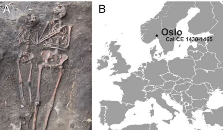
Skeleton OSL9/SZ50522 (Fig. 1A) was found during the exca-vation of a graveyard south of St. Nicolay’s Church, in Oslo (Fig. 1B,SI Appendix, andSI Appendix, Fig. S1) (23). It was part of a double burial (SA50521) situated close to the southern boundary of the graveyard and was identified as a female individual (age 28–35 y) who had been buried with a child (age 7–9 y) (24). A rib fragment was radiocarbon dated to Cal CE 1430–1465 (SI Ap-pendix, Fig. S2 and Table S1).
The initial metagenomic analysis of shotgun sequencing data from individual OSL9/SZ50522 revealed hits matching the spi-rochete B. recurrentis. After mapping the data to the B. recurrentis A1 genome assembly, we recovered 16.9% of the chromosome with a mean depth of coverage below 1 and deamination patterns matching aDNA.
To assemble the medieval B. recurrentis strain at an adequate depth and to cover more of the reference sequence, we se-quenced 13 additional genomic libraries from two teeth (A+B) (SI Appendix, Fig. S3) over four lanes on a HiSeq 2500 Illumina system and generated ca. 1.2 billion raw DNA sequences (Dataset S8). Overall, each library contained less than 0.04% reads mapping to B. recurrentis (Dataset S4).
We were able to assemble a B. recurrentis genome at a mean depth of 6.4× with 95.2% of the genome being covered at least twice (Dataset S7), with a total of 152,490 reads mapping to B. recurrentis (Dataset S4). Ultimately, we were able to assemble 98.2% of the chromosome at a mean depth of coverage of 8.3× (Fig. 2A).
The read-length distribution (mean: 69 bp) (SI Appendix, Fig. S9) of all datasets showed that the DNA was in a highly frag-mented state. Consistent with aDNA, we could also detect sig-nificant DNA damage patterns for the reads mapping to the B. recurrentis A1 assembly (Fig. 2B and SI Appendix, Fig. S5). This was further supported by the identification of a northern European mtDNA haplogroup and the DNA damage profile of the reads mapping to the revised Cambridge reference sequence (rCRS) build of the human mitochondrion (SI AppendixandSI Appendix, Fig. S6).
The unique genomic structure of these highly specialized bacteria allows high mapping specificity across the B. recurrentis genome (4, 5). This, in turn, allows us to infer the presence and absence of genomic regions via the level of coverage observed after mapping the raw datasets to Borrelia references. Addi-tionally, the use of a shotgun dataset, as opposed to a target-enrichment sequencing strategy, did not restrict or shift the available data to known preselected modern sequences.
To ascertain that the organism represented in our meta-genomic output was in fact a B. recurrentis strain and not one of its most closely related species, and potentially to detect signs of plasmid rearrangements, we mapped all datasets against avail-able reference sequences for B. duttonii Ly and B. crocidurae Achema independently (Fig. 2C andSI Appendix, Fig. S4). The number of reads mapping to the chromosomal assemblies varies only slightly from one species to another, but we observe dis-tinctly higher numbers of reads mapping with edit distance 0 to the B. recurrentis genome than to any of the other genomes (Fig. 2D andSI Appendix, Fig. S7andDataset S5).
Most B. recurrentis plasmids are colinear to B. duttonii plas-mids with the exception of pl6, which is colinear to a 5-kbp plasmid in B. crocidurae. While pl6 is covered at a mean depth of coverage of 12.6× in our genome mapping, we clearly ob-served that noncolinear plasmids of B. duttonii and B. crocidurae are not present in our medieval strain (SI Appendix, Fig. S4).
Six newly published de novo genomes by Marosevic et al. (8) stemming from Eastern African refugees treated for relapsing fever seem to have reinforced previously raised doubts (25) re-garding the length of plasmids pl124 and pl6 (NC_011263.1). The new strains all exhibit a ca. 40-kbp extension at the 5′ end of the previously reported pl124 and a ca. 1-kbp reduction at the 3′ end of pl6, much like their respective colinear plasmids pl165 in B. duttonii and a 5-kbp-long plasmid in B. crocidurae. Interestingly, our ancient B. recurrentis genome also displays signs of a longer pl124 and a shorter pl6 (SI Appendix, Figs. S4A and S10) com-pared with the reference strain A1. Our mapping to pl6 was completely missing 1 kbp at the 3′ end of the plasmid. Similarly, while aligning our data to pl165 of B. duttonii, we observed uni-form mapping at a mean depth of coverage of 3× across the entire length of the plasmid, including the missing 40-kbp extension at the 5′ end. It is highly likely that these discrepancies can be linked to the deposition of an incomplete assembly of the original ref-erence genome strain A1, since both our medieval European strain and the modern African strains show the same variations. This was further validated by pulse-field gel electrophoresis and the amplification of a gene ortholog to cihC in B. duttonii at 27,465–28,535 bp (26).
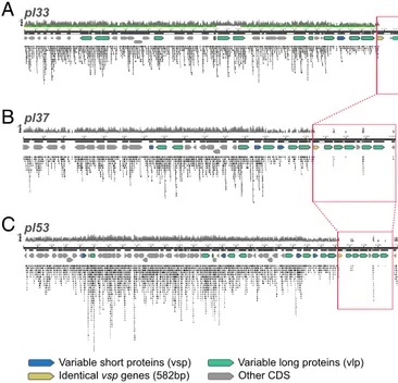
Upon further inspection of the plasmids, we found that three identical 582-bp-long variable short proteins (vsp) genes are missing at the 3′ end of plasmids pl33, pl37, and pl53 (Fig. 3). Strikingly, these genes mark the end of mapping for plasmid pl33 and pl37, while pl53 retains a hypothetical protein at its 3′ end. The first ca. 130 bp at the 5′ end of the vsp gene are covered by nonunique reads restricted to the gene across all three plasmids. This indicates that pl33, pl37, and pl53 are shorter in strain OSL9 and that we could be seeing signs of undetected plasmid rearrangements involving a similar vsp gene. The genes missing after the 582-bp vsp genes are copies of five variable long pro-teins (vlp) genes and multiple hypothetical propro-teins. Identical copies of a 785-bp-long vlp pseudogene are missing across all three plasmids, with the copy on pl33 being only 475 bp long. While the deposited assembly of this plasmid is known to be missing parts of its telomeric sequence, it might also be missing the end of the 785-bp vlp pseudogene and one or more of the four additional identical vlp pseudogenes missing across pl37 and pl53 (Fig. 3 andDataset S2).
Additionally, plasmids pl37 and pl53 have a decreased coverage in the intervals 23,822–28,471 and 39,440–44,087 bp, respectively.
Os
O
lo
lo
Call C CE 14430-30-1461465
A
B
Fig. 1. Sample origin and site location. (A) In situ picture of the double burial SA50521 with individual OSL9/SZ50522 to the right. (B) Location of the archaeological site and C14 date of the burial displayed in A.
The sequences are both 4,648 bp long and share 99% identity over their entire length. These intervals distinguish themselves by a patchy and low coverage, with most vlp and vsp genes in the region being covered ca. 50–80% (mean coverage 75.2%), while the covered vlp and vsp genes outside these intervals show a mean coverage of 96.9%, hinting at potential sequence degradation or plasmid rearrangements. Overall, 11 vlp pseudogenes and three vsp genes are missing, with one additional vsp pseudogene and three vlp genes showing signs of potential degradation.
We investigated the presence of known frameshift and stop-gain mutations throughout our ancient genome and found that, with the exception of oppA-1, all pseudogenes, which B. recurrentis A1 acquired during its reductive evolution and divergence from B. duttonii Ly, were present in the medieval strain. Our alignments to both B. recurrentis A1 and B. duttonii Ly support (depth of coverage 5×) that the oppA-1 gene could still be active in the OSL9 strain by retaining its ancestral glutamine (residue 59) (SI Appendix, Fig. S8).
After aligning all OSL9A-B reads to the chromosome of the reference strain, B. recurrentis A1, we detected 321 SNPs (Dataset S3). These SNPs were then combined with all other SNPs found in RF Borrelia strains included in this study to build a phylogenetic tree using the maximum likelihood method. Be-fore building the phylogeny, we checked for the presence of recombination using a phi test and ClonalFrameML software but could not detect any sign of recombination, as these approaches yielded a P value of 0.8287 and a likelihood of−612.47, respectively. The midpoint-rooted tree was generated using PhyML. The
phylogeny clearly shows two clusters represented by LBRF and TBRF species (Fig. 4), and, as previously reported, the African RF isolates cluster together (8). Interestingly, the ancient ge-nome recovered in Oslo is clustered between B. duttonii and the African B. recurrentis strains in the phylogeny. Compared with all other Borrelia in this study, 164 SNPs are specific to the OSL9 strain. These SNPs are distributed as 77 nonsynonymous SNPs, 77 synonymous SNPs, and 10 intergenic SNPs.
Discussion
LBRF is characterized by multiple relapses of fever, which are believed to be caused by bacterial immune evasion systems. RF Borreliae use plasmid-encoded antigenic phase variation within a clonal population as a mechanism of immune evasion. It utilizes the sequential expression of different variants of an antigenic surface protein to evade the host immunity, prolong the in-fection, and promote transmission. Hence, each new wave of infection, in this case febrile relapse, is characterized by a new serotype (5, 6, 27–29). In B. recurrentis, these proteins are the surface lipoproteins vlp and vsp, which are also known to be its main proinflammatory proteins (30). The genes expressing these proteins are arranged across plasmids in silent and expressed copies, and the interaction between the host immunity and the phase-variation mechanism encoded in the pathogen’s genome are believed to be responsible for the febrile relapses of LBRF (29). The number of phase-variation loci or loci families dictates the number of possible serotypes and, thus, the number of ge-nome variants theoretically available to evade the host immunity,
A
Chromosome 10 Kbp 20 Kbp 30 Kbp40 Kbp 60 Kbp 70 Kbp 80 Kbp 90 Kbp 110 Kbp 120 Kbp 130 Kbp 140 Kbp 160 Kbp 170 Kbp 180 Kbp 190 Kbp 210 Kbp 220 Kbp 230 Kbp 240 Kbp 260 Kbp 270 Kbp 280 Kbp 290 Kbp 310 Kbp 320 Kbp 330 Kbp 340 Kbp 360 Kbp 370 Kbp 380 Kbp 390 Kbp 410 Kbp 420 Kbp 430 Kbp 440 Kbp 460 Kbp 470 Kbp 480 Kbp 490 Kbp 510 Kbp 520 Kbp 530 Kbp 540 Kbp 560 Kbp 570 Kbp 580 Kbp 590 Kbp 610 Kbp 620 Kbp 630 Kbp 640 Kbp 660 Kbp 670 Kbp 680 Kbp 690 Kbp 710 Kbp 720 Kbp 730 Kbp 740 Kbp 760 Kbp 770 Kbp 780 Kbp 790 Kbp 810 Kbp 820 Kbp 830 Kbp 840 Kbp 860 Kbp 870 Kbp 880 Kbp 890 Kbp 910 Kbp 920 Kbp pl124 10 Kbp 20 Kbp 30 Kbp 40 Kbp 60 Kbp 70 Kbp 80 Kbp 90 Kbp 110 Kbp pl23 10 Kbp pl33 10 Kbp 20 Kbp pl35 10 Kbp 20 Kbp 30 Kbp pl37 10 Kbp 20 Kbp 30 Kbp pl53 10 Kbp 20 Kbp 30 Kbp 40 Kbp pl6 0 Kbp 50 Kbp 100 Kbp 150 Kbp 200 Kbp 250 Kbp 300 Kbp 350 Kbp 400 Kbp 450 Kbp 500 Kbp 550 Kbp 600 Kbp 650 Kbp 700 Kbp 750 Kbp 800 Kbp 850 Kbp 900 Kbp 930 Kbp 0 Kbp 50 Kbp 100 Kbp 123 Kbp 0 Kbp 22 Kbp 0 Kbp 33 Kbp 0 Kbp 35 Kbp0 Kbp 36 Kbp 0 Kbp 52 Kbp 0 Kbp 6 Kbp 10X 2X 5X 50% 0 Kbp 10 Kbp 20 Kbp 30 Kbp 40 Kbp 50 Kbp 60 Kbp 70 Kbp 80 Kbp 90 Kbp 100 Kbp 110 Kbp 120 Kbp 130 Kbp 140 Kbp 150 Kbp 160 Kbp 170 Kbp 180 Kbp 190 Kbp 200 Kbp 210 Kbp 220 Kbp 230 Kbp 240 Kbp 250 Kbp 260 Kbp 270 Kbp 280 Kbp 290 Kbp 300 Kbp 310 Kbp 320 Kbp 330 Kbp 340 Kbp 350 Kbp 360 Kbp 370 Kbp 380 Kbp 390 Kbp 400 Kbp 410 Kbp 420 Kbp 430 Kbp 440 Kbp 450 Kbp 460 Kbp 470 Kbp 480 Kbp 490 Kbp 500 Kbp 510 Kbp 520 Kbp 530 Kbp 540 Kbp 550 Kbp 560 Kbp 570 Kbp 580 Kbp 590 Kbp 600 Kbp 610 Kbp 620 Kbp 630 Kbp 640 Kbp 650 Kbp660 Kbp 670 Kbp 680 Kbp 690 Kbp 700 Kbp 710 Kbp 720 Kbp 730 Kbp 740 Kbp 750 Kbp 760 Kbp 770 Kbp 780 Kbp 790 Kbp 800 Kbp 810 Kbp 820 Kbp 830 Kbp 840 Kbp 850 Kbp 860 Kbp 870 Kbp 880 Kbp 890 Kbp 900 Kbp 910 Kbp 920 Kbp 931 Kbp B. recurrentis A1 Chromosome 931 Kbp B. duttonii L y Chromosome 931 Kbp B. crocidurae Achema Chromosome 919 Kbp CobQ/ParA Absent in B. recurrentis 1 Kbp Missing in B. recurrentis(Hyp. Proteins, vlp genes) 10X 5X 10X 10X 5X 5X
C
D
B. recurrentis B. duttonii B. crocidurae
pl23 pl6/Clone22 Chromsome 0 20 40 60 80 100 0 1 2 3 4 5 0 1 2 3 4 5 0 1 2 3 4 5 % M apped Reads 0 20 40 60 80 100 0 20 40 60 80 100
B
Fig. 2. (A) Coverage plots for mapping of OSL9A-B reads to the B. recurrentis A1 reference sequence. Rings (from outer ring to inner ring) show coverage, GC skew, and GC content. GC content is further highlighted as being above (black) or below (gray) 26%. (B) Damage frequency for the mapping displayed in A with 10% of reads showing a clear deamination signature consistent with aDNA. (C) Comparison of OSL9A-B noncompetitive mappings to the chromosomes of B. recurrentis A1, B. duttonii Ly, and B. crocidurae Achema. It can be seen here that strain OSL9 does not have a CobQ/ParA gene and is also missing a 1-kbp-long sequence at the 5′ end of the B. duttonii chromosome, much like B. recurrentis A1. (D) Distribution of edit distances (plotted on the x axis) of OSL9A-B reads mapping to chromosomal assemblies of B. recurrentis A1, B. duttonii Ly, and B. crocidurae Achema (Left), to colinear pl23 plasmids of B. recurrentis A1 and B. duttonii Ly (Center), and to colinear pl6 plasmids of B. recurrentis A1 and B. crocidurae Achema (Right).
MIC
with some estimating up to 210 different states for every 20 phase-variation sites (28, 29, 31). As a result, although LBRF can be treated with antibiotics, no vaccine against the disease exists today. The only effective antibodies against LBRF seem to be directed at vlp and vsp genes and thus only can be manufactured to fight one randomly appearing serotype at a time (32).
We observed that a number of vlp and vsp genes are absent or potentially degraded in our medieval strain (Fig. 3 andDataset S2), but are present in modern-day African strains and have orthologs in other RF genomes. Some are frameshifted or in-complete in A1 and might have been pseudogenes, but others, like the vsp copies found at the beginning of each missing in-terval, were fully functional genes. We can hypothesize, based on the data at hand, that the observed loss of antigenic phase-variation loci could have led to a difference in phenotype compared with modern strains and hypothetically could have influenced the num-ber of febrile relapses, i.e., the numnum-ber of serotypes that the medieval strain of the pathogen might have been capable of generating. While experimental infections by LBRF have yielded up to 10 relapses (9, 32), most sources cite up to five relapses for the pathogen (33). Similarly, most untreated non-European cases recorded during the World War I/World War II pandemics reached only up to five relapses (11, 34). Historically, Creighton (12) recorded varying numbers of observed relapses across known LBRF outbreaks. An outbreak in 18th century Dublin cites that patients were prone to relapses“even sometimes to the third” (35). Overall, most outbreaks, for which the number of observed relapses was supplied in historical texts, saw one or two or“one or more” re-lapses. However, it should be noted that data on modern untreated cases of LBRF are rare, and most available historical sources only refer to“multiple relapses” or “prone to relapse” (12, 36), consid-erably reducing the amount of data to be evaluated (36).
Compared with B. duttonii Ly, the modern reference strain B. recurrentis A1 has lost a large number of intact vlps and vsps and causes fewer relapses in human patients (5, 33), but this differ-ence is even more pronounced in the medieval strain. The missing genes make up all archival copies of six phase-variation
loci. Overall, this translates to a genome reduction of 1.2% of the pan-genome and 5.1–21% of the affected plasmids, which, in combination with a decrease in GC content (Dataset S4), is in line with observed reductive genome evolution of specialized and highly pathogenic bacteria (37, 38). The presence of additional unknown plasmids seems unlikely, as the number of plasmids tends to follow the same reductive trend as the overall genome (37). Instead, we can detect additional gene loss at the 3′ end of some plasmids.
This reductive evolution characterizes B. recurrentis’ epidemic potential and its increased virulence compared with other RFs. Similar to Bartonella quintana and Rickettsia prowazekii (38, 39), other well-known body lice-to-human transmission specialists, B. recurrentis shows accelerated rates of genome degradation caused by adaptation to host-restricted vectors and functional trade-offs, resulting in a degraded genome, reduced genome size, low coding content, and increased virulence (5, 31, 37, 38).
However, this type of evolution usually involves the loss of regulatory genes (37), and while this is also the case for our medieval strain, there is one exception. The oppA operon, which encodes for an ABC transporter, is significantly involved in the uptake of oligopeptides in many bacterial species (40, 41). OppA-1, which likely plays a critical role in host environment adaptation and essential metabolic functions (42), is a pseudogene in B. recurrentis A1 due to an in-frame stop codon. However, our medieval strain retains the ancestral glutamine much like the TBRF pathogen B. duttonii. While we can only speculate about the effect of this mu-tation on the ecological life cycle of the disease, it is interesting to note that the inactivation of oppA-1 seems to be much more recent than the rest of the pseudogenes in B. recurrentis. These pseudo-genes are degraded to the same extent in both B. recurrentis line-ages, with the exception of some antigenic phase-variation loci, which the medieval strain seems to have lost altogether. Therefore, we could hypothesize that, while both lineages have continued their reductive evolution, they have done so in different ways.
The full ecological dynamics of LBRF remain unclear (43). The European lineage presented in this study probably evolved in a distinctly different environment than known modern African strains. The extent to which anthropogenic pressure on pathogen and vector impacted this evolution is difficult to assess based on a single representative in the medieval lineage. As the only ep-idemic and louse-borne RF, LBRF has been assumed to be re-sponsible for all RF epidemics recorded throughout European
0.02
LBRF
Borrelia recurrentis A1 (1994)
Borrelia recurrentis PBeK (2004) Borrelia recurrentis PAbJ (2015) Borrelia recurrentis PUfA (2015)
Borrelia recurrentis PMaC (2015) Borrelia recurrentis A17 (1997)
Borrelia recurrentis OSL9 (1420-65 CE)
Borrelia recurrentis PAbN (2015)
TBRF
Borrelia duttonii Ly Borrelia hermsii HS1
Borrelia hermsii YOR
Borrelia anserina Es Borrelia parkeri SLO
Borrelia parkeri HR1
Borrelia crocidurae Achema Borrelia hispanica CRI Borrelia hermsii YBT
Borrelia coriaceae Co53
Borrelia crocidurae DOU Borrelia hermsii MTW
Borrelia persica No12 Borrelia hermsii DAH
Borrelia hermsii CC1
Borrelia miyamotoi LB-2001 Borrelia miyamotoi FR64b Borrelia turicatae 91E135
Fig. 4. RL phylogeny based on chromosomal alignments (Datasets S3
andS9). The European LBRF strain (this study) is shown in red, and African LBRF strains are shown in blue. The legend displays the branch length.
A
B
C
Identical vsp genes (582bp)
Variable short proteins (vsp) Variable long proteins (vlp)
Other CDS
pl53 pl37 pl33
Fig. 3. Alignment of OSL9A-B reads to B. recurrentis plasmids pl33 (A), pl37 (B), and pl53 (C). Across all plasmids a section at the 3′ end of the linear plasmids is missing starting with 80% of a vsp gene. The regions boxed in red share identities of 99–100%. Tiled reads seen within the boxed regions are nonunique reads mapping to sequences conserved across multiple plasmids.
history. It was known to have reached epidemic proportions in susceptible populations around key events, such as wars and famines. LBRF is a disease that probably would only in-frequently have resulted in the type of mass mortality that would warrant the planning of mass graves, and, historically, scattered cases of LBRF have been recorded between epidemics, usually among the poor (12). Although at the time of the burial of in-dividual OSL9/SZ50522 the town was still affected by the eco-nomic decline caused by the Black Death in the mid-1300s (44, 45), which probably left parts of the population vulnerable to disease and malnourishment, the results reported in this study represent an isolated case of the disease. Given the available data, we cannot speculate on the presence or size of an outbreak of LBRF in 15th century Oslo, especially since the number of studied samples from the same time period is limited (Dataset S1). However, our results can tentatively corroborate the in-volvement of B. recurrentis in the European epidemics reported in historical text because of its being the only known epidemic RF, the specificity of the reported symptoms, the identification of the spirochete during epidemics in 1868, and the discovery of its re-lation to lice in 1907 (33, 46). Finally, the studied individual probably died at the height of bacteremia, allowing the detection of the pathogen in relatively high quantities within aDNA shotgun datasets.
Previously assembled ancient pathogens have generally been limited to diseases that could leave visible marks on the skeletons or mummified tissues of affected individuals (e.g., leprosy, tu-berculosis, smallpox) (47–49). However, recent studies on path-ogens that are invisible in the archaeological record have started to emerge (48, 50, 51). These studies are of particular relevance, as they clearly illustrate the osteological paradox (52, 53). It is hoped that, as one of the few epidemic fevers believed to have played a major role in the disease landscape of historical Europe, the addition of LBRF will lead the way for the detection of diseases which might not have much relevance to Western health today and therefore are less represented in the genomic data-bases and literature. The unique genome of B. recurrentis has provided a rare opportunity in aDNA to study the genomic make-up of an ancient pathogen and catch a glimpse of the evolutionary process that accompanies the environmental ad-aptation and pathogenesis of specialized human pathogens. Furthermore, the results detailed in our study illustrate the im-portance of human body lice as a vector throughout European history, which has recently also been suggested for plague (54).
Future research into the ecological dynamics of LBRF and surveillance of populations affected by lice infestation in in-dustrialized countries is needed to better understand the per-sistence mechanism of the disease and the dangers of spread in susceptible Western communities. Furthermore, more insights into potential outbreaks of the disease in lice-infested Western populations, as seen in Brouqui et al. (19), and genomic se-quences from affected patients would allow us to determine if LBRF persisted in Europe and thus is phylogenetically related to the lineage recovered in this study or was imported on one or more occasions from outside Europe. Additional genomes, Eu-ropean or otherwise, may further elucidate the evolution and pathogenicity of LBRF.
Methods
Samples. We sampled two well-preserved molars from nine individuals (Dataset S1) recovered from the site of St. Clement’s/St. Nicolay’s church graveyard in Oslo, under clean conditions at the Norwegian Institute for Cultural Heritage Research, Oslo. One exception was tooth OSL6B, which was an incisor. The individuals all stem from different periods of the graveyard, which spans the 11th to the 15th century (Dataset S1). Upon discovery of B. recurrentis reads in OSL9, all sampled individuals were screened for the presence of the pathogen via qPCR but were negative (SI Appendix, Fig. S3), including the only potentially contemporary individual OSL6/SZ19834.
Laboratory Work. Full experimental procedures are provided inSI Appendix.
Data Preparation and Quality Filtering. Following sequencing, the datasets were demultiplexed at the Norwegian Sequencing Centre, and quality control was performed using FastQC (55). Adapters and indices were trim-med using cutadapt2.0 (56). Sequences shorter than 30 bp and below a quality score of 10 were discarded. Trimmed reads were merged using FLASH (57).
Metagenomic Analysis. The datasets were explored using the taxonomic classifier Kraken (58). Upon discovery of B. recurrentis reads, we further investigated the metagenomic data using MetaPhlAn2 (59) and the protein-level classification tool Kaiju (60). All tools indicated the presence of the bacterium in all datasets stemming from individual OSL9/SZ50522. No hits for B. recurrentis were found in any other samples or blank controls.
RF Borreliae Mappings. The merged reads were mapped noncompetitively to the Borrelia recurrentis A1 strain (ASM1970v1), Borrelia duttonii Ly strain (ASM1968v1), and Borrelia crocidurae Achema strain (ASM25934v1) refer-ence sequrefer-ences using BWA-ALN (-n 0·01 -l 16500) and BWA Samse (61). The generated data were converted to bam format and sorted using SAMtools (62, 63). Duplicates were marked using Picard MarkDuplicates module (64). Indels were realigned using GATK RealignerTargetCreator and Indel-Realigner modules (65, 66). Using mapDamage2.0 (67), we quantified aDNA damage patterns for each dataset and recalibrated the quality scores of likely damaged bases. Libraries from tooth B yielded significantly more se-quences mapping to B. recurrentis than libraries from tooth A, with an av-erage 11,346 reads per dataset mapping to the reference compared with an average of 3,402 reads per dataset for tooth A.
Phylogeny. Species for which only contigs were available were assembled with Multi-CAR (68) using the reference sequences for B. recurrentis A1 and Borrelia hermsii HS1. For six recently published B. recurrentis strains (8), we mapped available Illumina reads to the B. recurrentis A1 chromosome as-sembly using BWA-MEM, sorted the data with SAMtools (62), marked du-plicates using Picard (64), and realigned around indels with GATK (65, 66). For all BAM files, SNPs were called with SAMtools mpileup (-R -ugf -B) and BCFtools call (-vm) and filter (-s LOWQUAL -i‘%QUAL > 19’). SNPs were annotated using snpToolkit (69). Complete genomes were compared using Parsnp (70), and polymorphic sites were extracted using gingr. Two meth-ods, Phi test (71) and ClonalFrameML (72), were used to assess the presence of recombination in our dataset. The midpoint-rooted tree was generated using phyML.
ACKNOWLEDGMENTS. Data analysis was performed on the Abel Cluster owned by the University of Oslo and the Norwegian Metacenter for High Performance Computing and operated by the Department for Research Computing at USIT, the University of Oslo information technology de-partment. Skeletal material was provided courtesy of the Museum of Cultural History (University of Oslo) after application to the museum and the Norwegian National Committee for Research Ethics on Human Remains. This project was funded by the European Research Council under FP7-IDEAS-ERC Program Grant 324249 MedPlag.
1. Elbir H, et al. (2012) Complete genome sequence of Borrelia crocidurae. J Bacteriol 194:3723–3724.
2. Fotso Fotso A, et al. (2014) Genome sequence of Borrelia crocidurae strain 03-02, a clinical isolate from Senegal. Genome Announc 2:e01150-14.
3. Gupta RS, Mahmood S, Adeolu M (2013) A phylogenomic and molecular signature based approach for characterization of the phylum spirochaetes and its major clades: Proposal for a taxonomic revision of the phylum. Front Microbiol 4:217.
4. Norris SJ, Lin T (2011) Out of the woods: The remarkable genomes of the genus Borrelia. J Bacteriol 193:6812–6814.
5. Lescot M, et al. (2008) The genome of Borrelia recurrentis, the agent of deadly louse-borne relapsing fever, is a degraded subset of tick-louse-borne Borrelia duttonii. PLoS Genet 4:e1000185.
6. Vidal V, Cutler S, Scragg IG, Wright DJM, Kwiatkowski D (2002) Characterisation of silent and active genes for a variable large protein of Borrelia recurrentis. BMC Infect Dis 2:25.
7. Cutler SJ, et al. (1997) Borrelia recurrentis characterization and comparison with relapsing-fever, Lyme-associated, and other Borrelia spp. Int J Syst Bacteriol 47: 958–968.
MIC
8. Marosevic D, et al. (2017) First insights in the variability of Borrelia recurrentis ge-nomes. PLoS Negl Trop Dis 11:e0005865.
9. European Centre for Disease Prevention and Control (2017) Facts about louse-borne relapsing fever ecdc. Available at https://ecdc.europa.eu/en/louse-borne-relapsing-fever/facts. Accessed August 8, 2017.
10. Houhamdi L, Raoult D (2005) Excretion of living Borrelia recurrentis in feces of in-fected human body lice. J Infect Dis 191:1898–1906.
11. Bryceson A, et al. (1969) Louse-borne relapsing fever; a clinical and laboratory study of 62 cases in Ethiopia and a reconsideration of the literature. Naval Medical Research Unit No 3 APO New York 09319 Field Facility. Available at www.dtic.mil/docs/citations/ AD0722494. Accessed July 12, 2017.
12. Creighton C (1894) A History of Epidemics in Britain: From the Extinction of Plague to the Present Time (Cambridge Univ Press, Cambridge, UK).
13. Hays JN (2005)“Fevers” and the Great famine in Ireland, 1846–1850. Epidemics and Pandemics: Their Impacts on Human History, ed Hays JN (ABC CLIO Santa Barbara, CA), pp 439–448.
14. Sparrow H (1958) Etude du foyer éthiopien de fièvre récurrente. Bull World Health Organ 19:673–710.
15. Sparrow H (1962) Tropical Health: A Report on a Study of Needs and Resources. (National Academies, Washington, DC).
16. Macarthur W (1948) Famine fevers in England and Ireland. Ulster Med J 17:28–33. 17. Cutler SJ (2010) Relapsing fever–A forgotten disease revealed. J Appl Microbiol 108:
1115–1122.
18. European Centre for Disease Prevention and Control (2015) Rapid Risk Assessment: Louse-borne relapsing fever in the EU– 17 November 2015 (ECDC Stockholm). Available at https://ecdc.europa.eu/sites/portal/files/media/en/publications/Publications/ louse-borne-relapsing-fever-in-eu-rapid-risk-assessment-17-nov-15.pdf. Accessed June 30, 2017.
19. Brouqui P, et al. (2005) Ectoparasitism and vector-borne diseases in 930 homeless people from Marseilles. Medicine (Baltimore) 84:61–68.
20. Tran T-N-N, et al. (2011) High throughput, multiplexed pathogen detection authen-ticates plague waves in medieval Venice, Italy. PLoS One 6:e16735.
21. Nguyen-Hieu T, et al. (2010) Evidence of a louse-borne outbreak involving typhus in Douai, 1710-1712 during the war of Spanish succession. PLoS One 5:e15405. 22. Keller A, et al. (2012) New insights into the Tyrolean Iceman’s origin and phenotype
as inferred by whole-genome sequencing. Nat Commun 3:698.
23. Derrick M (2018) Follobaneprosjektet F04 Klypen Øst og Saxegaardsgata 15. Arkeologisk Utgravning Mellom Bispegata og Loenga. Middelalderparken Og Saxegaardsgata 15 & 17, Oslo. NIKU Oppdragsrapport 40/2015 (Norsk institutt for kulturminneforskning, Oslo). 24. Jensen AØ (2018) Osteologisk analyse av skjelettmaterialet fra Nikolaikirkens kirkegård. Follobaneprosjektet F04 Klypen Øst/Saxegaardsgata 15. NIKU Oppdragsrapport 160/2016 (Norsk institutt for kulturminneforskning, Oslo).
25. Miller SC, Porcella SF, Raffel SJ, Schwan TG, Barbour AG (2013) Large linear plasmids of Borrelia species that cause relapsing fever. J Bacteriol 195:3629–3639. 26. Grosskinsky S, et al. (2010) Human complement regulators C4b-binding protein and
C1 esterase inhibitor interact with a novel outer surface protein of Borrelia re-currentis. PLoS Negl Trop Dis 4:e698.
27. Palmer GH, Bankhead T, Seifert HS (2016) Antigenic variation in bacterial pathogens. Microbiol Spectr 4.
28. Norris SJ (2006) Antigenic variation with a twist–The Borrelia story. Mol Microbiol 60: 1319–1322.
29. Foley J (2015) Mini-review: Strategies for variation and evolution of bacterial anti-gens. Comput Struct Biotechnol J 13:407–416.
30. Barbour AG, Restrepo BI (2000) Antigenic variation in vector-borne pathogens. Emerg Infect Dis 6:449–457.
31. Pallen MJ, Wren BW (2007) Bacterial pathogenomics. Nature 449:835–842. 32. Barbour AG, Guo BP (2010) Pathogenesis of relapsing fever. Borrelia: Molecular
Biology, Host Interaction and Pathogenesis, eds Radolf JD, Scott Samuels D (Horizon Scientific, Norfolk, UK), pp 333–357.
33. Cutler SJ (2015) Relapsing fever Borreliae: A global review. Clin Lab Med 35:847–865. 34. Chung H-L, Chang FC (1939) Relapsing fever. clinical and statistical study of 337 cases.
Chin Med J 55, 6–33.
35. Rutty J (1770) A Chronological History of the Weather and Seasons, and of the Prevailing Diseases in Dublin: With Their Various Periods, Successions, and Revolutions, During the Space of Forty Years. With a Comparative View of the Difference of the Irish Climate and Diseases, and Those of England and Other Countries (Robinson and Roberts, Lon-don).
36. Lyons RT (1872) A Treatise on Relapsing or Famine Fever (Henry S. King, London). 37. Diop A, Raoult D, Fournier P-E (December 27, 2017) Rickettsial genomics and the
paradigm of genome reduction associated with increased virulence. Microbes Infect, 10.1016/j.micinf.2017.11.009.
38. Alsmark CM, et al. (2004) The louse-borne human pathogen Bartonella quintana is a genomic derivative of the zoonotic agent Bartonella henselae. Proc Natl Acad Sci USA 101:9716–9721.
39. Ogata H, et al. (2001) Mechanisms of evolution in Rickettsia conorii and R. prowa-zekii. Science 293:2093–2098.
40. Kornacki JA, Oliver DB (1998) Lyme disease-causing Borrelia species encode multiple lipoproteins homologous to peptide-binding proteins of ABC-type transporters. Infect Immun 66:4115–4122.
41. Wang X-G, Lin B, Kidder JM, Telford S, Hu LT (2002) Effects of environmental changes on expression of the oligopeptide permease (opp) genes of Borrelia burgdorferi. J Bacteriol 184:6198–6206.
42. Wang X-G, et al. (2004) Analysis of differences in the functional properties of the substrate binding proteins of the Borrelia burgdorferi oligopeptide permease (Opp) operon. J Bacteriol 186:51–60.
43. Amanzougaghene N, et al. (2017) Detection of bacterial pathogens including po-tential new species in human head lice from Mali. PLoS One 12:e0184621. 44. Nedkvitne A, Norseng PG (2000) Middelalderbyen Ved Bjørvika. Oslo 1000-1536
(Cappelen, Oslo).
45. Unger CR, Huitfeldt-Kaas HJ, eds (1847) Diplomatarium Norvegicum: Oldbreve Til Kundskab Om Norges Indre Og Ydre Forhold, Sprog, Slægter, Sæder, Lovgivning Og Rettergang I Middelalderen. Samling 11 (PT Mallings boghandels forlag, Christiania, Norway), Vol 5, p 835.
46. Southern PM, Sanford JP (1969) Relapsing fever: A clinical and microbiological review. Medicine (Baltimore) 48:129.
47. Kay GL, et al. (2015) Eighteenth-century genomes show that mixed infections were common at time of peak tuberculosis in Europe. Nat Commun 6:6717.
48. Patterson Ross Z, et al. (2018) The paradox of HBV evolution as revealed from a 16th century mummy. PLoS Pathog 14:e1006750.
49. Schuenemann VJ, et al. (2013) Genome-wide comparison of medieval and modern Mycobacterium leprae. Science 341:179–183.
50. Vågene ÅJ, et al. (2018) Salmonella enterica genomes from victims of a major sixteenth-century epidemic in Mexico. Nat Ecol Evol 2:520–528.
51. Kay GL, et al. (2014) Recovery of a medieval Brucella melitensis genome using shot-gun metagenomics. MBio 5:e01337–e14.
52. Siek T (2013) The osteological paradox and issues of interpretation in paleopathol-ogy. vis-à-vis: Explor Anthropol 12:92–101.
53. Wood JW, et al. (1992) The osteological paradox: Problems of inferring prehistoric health from skeletal samples. Curr Anthropol 33:343–370.
54. Dean KR, et al. (2018) Human ectoparasites and the spread of plague in Europe during the Second Pandemic. Proc Natl Acad Sci USA 115:1304–1309.
55. Andrews S (2010) A quality control tool for high throughput sequence data. Available at https://www.bioinformatics.babraham.ac.uk/projects/fastqc/. Accessed August 14, 2017. 56. Martin M (2011) Cutadapt removes adapter sequences from high-throughput
se-quencing reads. EMBnet J 17:10–12.
57. Magoˇc T, Salzberg SL (2011) FLASH: Fast length adjustment of short reads to improve genome assemblies. Bioinformatics 27:2957–2963.
58. Wood DE, Salzberg SL (2014) Kraken: Ultrafast metagenomic sequence classification using exact alignments. Genome Biol 15:R46.
59. Segata N, et al. (2012) Metagenomic microbial community profiling using unique clade-specific marker genes. Nat Methods 9:811–814.
60. Menzel P, Ng KL, Krogh A (2016) Fast and sensitive taxonomic classification for metagenomics with Kaiju. Nat Commun 7:11257.
61. Li H, Durbin R (2009) Fast and accurate short read alignment with Burrows-Wheeler transform. Bioinformatics 25:1754–1760.
62. Li H, et al.; 1000 Genome Project Data Processing Subgroup (2009) The sequence alignment/map format and SAMtools. Bioinformatics 25:2078–2079.
63. Li H (2011) A statistical framework for SNP calling, mutation discovery, association mapping and population genetical parameter estimation from sequencing data. Bioinformatics 27:2987–2993.
64. Picard (2017) Available at broadinstitute.github.io/picard/. Accessed August 14, 2017. 65. DePristo MA, et al. (2011) A framework for variation discovery and genotyping using
next-generation DNA sequencing data. Nat Genet 43:491–498.
66. Van der Auwera GA, et al. (2013) From FastQ data to high confidence variant calls: The Genome Analysis Toolkit best practices pipeline. Curr Protoc Bioinformatics 43: 11.10.1–11.10.33.
67. Jónsson H, Ginolhac A, Schubert M, Johnson PLF, Orlando L (2013) mapDamage2.0: Fast approximate Bayesian estimates of ancient DNA damage parameters. Bioinformatics 29:1682–1684.
68. Chen K-T, et al. (2016) Multi-CAR: A tool of contig scaffolding using multiple refer-ences. BMC Bioinformatics 17:469.
69. Namouchi A (2018) snpToolkit. Available at https://bitbucket.org/Amine-Namouchi/ snptoolkit. Accessed June 26, 2018.
70. Treangen TJ, Ondov BD, Koren S, Phillippy AM (2014) The Harvest suite for rapid core-genome alignment and visualization of thousands of intraspecific microbial core-genomes. Genome Biol 15:524.
71. Bruen TC, Philippe H, Bryant D (2006) A simple and robust statistical test for detecting the presence of recombination. Genetics 172:2665–2681.
72. Didelot X, Wilson DJ (2015) ClonalFrameML: Efficient inference of recombination in whole bacterial genomes. PLOS Comput Biol 11:e1004041.