delivery of peptide nucleic acids by
an argininocalix[4]arene
Jessica Gasparello
1, Alex Manicardi
2,3, Alessandro Casnati
2, Roberto Corradini
2,
Roberto Gambari
1, Alessia Finotti
1& Francesco Sansone
2The application of Peptide Nucleic Acids (PNAs), mimics of DNA lacking the sugar-phosphate backbone, for antisense/anti-gene therapy and gene editing is limited by their low uptake by cells. Currently, no simple and efficient delivery systems and methods are available to solve this open issue. One of the most promising approach is the modification of the PNA structure through the covalent linkage of poliarginine tails, but this means that every PNA intended to be internalized must be modified. Herein we report the results relative to the delivery ability of a macrocyclic multivalent tetraargininocalix[4] arene (1) used as non-covalent vector for anti-miR-221-3p PNAs. High delivery efficiency, low cytotoxicity, maintenance of the PNA biological activity and ease preparation of the transfection formulation, simply attained by mixing PNA and calixarene, candidate this vector as universal delivery system for this class of nucleic acid analogues.
Peptide nucleic acids (PNAs) are DNA analogues in which the sugar-phosphate backbone is replaced by
N-(2-aminoethyl)glycine units1–6. These molecules efficiently hybridize with complementary DNA and RNA,
forming both double helices through Watson-Crick base pairing6 and triplexes through Watson-Crick and
Hoogsteen hydrogen bonds, the latter being able to perform strand invasion7,8. PNAs are also very useful for
in cells and in vivo DNA and RNA recognition, since they are resistant to DNases and proteases9,10, and show
higher specificity for the target DNA and RNA sequences with respect to nucleic acids1–6. For these reasons, they
have been proposed for antisense6,11,12 and anti-gene13–15 therapy in a number of studies and lately for precise
gene editing16–18. Recently, the use of PNAs as molecules targeting microRNAs (miR/miRNA) has been firmly
demonstrated19–26.
One of the critical challenges in PNA technology is their delivery to cells27, in particular their low uptake
by eukaryotic cells28–32. In order to solve this drawback, several approaches have been considered. One of these
is conjugation with carrier peptides33–36, in particular those sensitive to microenvironment changes24; antimiR
activity was indeed observed for instance by conjugation of PNAs to polyarginine (poly-R) tails21,25, or by
modi-fication of the PNA backbone with cationic amino acid side chains37,38.
As alternative to the chemical modification, for PNA delivery particularly convenient is the use of carriers able to interact with the cargo in a non-covalent and reversible way. This strategy would allow in principle to make available a system effective with all native PNA sequences that are intended to be transported into cells. In this context, it was actually already explored the delivery of PNAs and their derivatives or analogs with liposomes39,
polymer nanoparticles40 and pseudovirions41, and by co-transfection with partially complementary DNA42.
Inorganic nanocarriers, such as nanozeolites29 or mesoporous silica nanoparticles (MSNPs)28 have been also
used for cellular delivery of PNAs; for MSNPs–mediated PNA delivery an anti-miR activity was demonstrated28.
However, the preparation of all these systems and the PNA incorporation generally require special and often time consuming procedures.
Calixarenes functionalized with ammonium groups were shown to be suitable for the efficient delivery of nucleic acids. Some of us recently reported43–46 on the delivery of plasmid DNA using the macrocyclic calix[4]
arene in cone geometry as scaffold for vector molecules. This geometry makes possible the display of two spatially 1Department of Life Sciences and Biotechnology, Section of Biochemistry and Molecular Biology, University of ferrara, ferrara, italy. 2Department of chemistry, Life Sciences and environmental Sustainability, University of Parma, Parma, italy. 3Present address: Department of Organic and Macromolecular chemistry, Ghent University, Ghent, Belgium. correspondence and requests for materials should be addressed to A.f. (email: alessia.finotti@ unife.it) or f.S. (email: [email protected])
Received: 24 October 2018 Accepted: 3 January 2019 Published: xx xx xxxx
well-defined regions, one hydrophobic and the other hydrophylic, overall resulting in a pronounced amphiphilic character of the vector. The clustering on the macrocycle of only four guanidinium groups or basic amino acids, such as arginine and lysine, was enough to give rise to new non-viral vectors for cell transfection of DNA, more potent than commercial transfecting agents. The exploited parallel arrangement of the amino acid units makes available, with respect to more classical polyarginine peptides, the primary α-amino groups that might favor the protection of the vector–DNA complex from the lysosomal degradation and facilitate the release of DNA from the endosomes into the cytosol through a proton sponge effect. This would, in part, contribute to the high trans-fection efficiency observed in particular for argininocalix[4]arene 146 (Fig. 1).
The expected interactions between these cationic calixarenes and DNA can be not only charge-charge inter-actions between ammonium and phosphates groups, but also hydrogen bonds with nucleobases as previously proposed and shown for analogous positively charged calixarene derivatives47,48. Therefore, despite the neutral
backbone of PNAs, their similarity with DNA as a consequence of the presence of nucleobases made us reasona-bly confident that these macrocyclic delivery systems could be also used for the transfection of these mimics. In this work, we herein report that neutral PNAs can be indeed efficiently delivered inside cells using the arginino-calix[4]arene 1, chosen as first example of cationic calixarene to be investigated because of its highest ability to vehicle DNA among a panel of calixarene-based vectors43–46. As cellular model system, the human glioma U251
cells were employed. The PNAs used (Fig. 1) were previously demonstrated to be able to target miR-221-3p, thereby altering its biological activity and providing a clear-cut end-point for evaluation of their biological effect using the delivery system under investigation.
Methods
Synthesis and characterization of argininocalix[4]arene 1.
The structure of the calixarene-based molecule 1 is depicted in Fig. 1. Its synthesis and characterization have been previously reported46. Derivative1, employed for PNA delivery experiments, was re-suspended in a solution of EtOH/H2O/DMSO (2/2/1 v/v/v) under sterile condition.
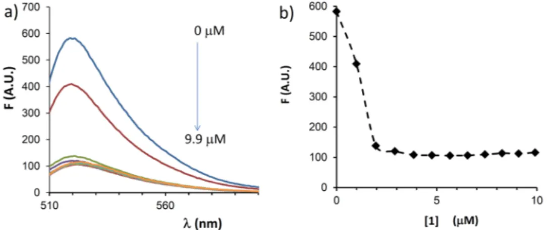
For a fast evaluation of the interaction with PNA, fluorescently labelled Fl-a221PNA (Fl: Fluorescein) was diluted to a final concentration of 2 μM in buffer (HEPES 16 mM, NaCl 40 mM, pH 7.4) and placed in a flu-orescence cuvette; to this, a concentrated solution of the argininocalix[4]arene 1 (100 μM in EtOH/H2O/ DMSO = 2/2/1 v/v/v) was added stepwise (up to a final concentration of 9.9 μM of calixarene), and changes in the fluorescein emission intensity (with λex = 494 nm) were measured after each addition. Correction for lamp fluc-tuation was performed by measuring the fluorescence of a standard 2 μM fluorescein solution after each spectra; this correction and that for dilution were performed using the formula: Fi, corr = Fi,obs × (Fin /Fi) × (Vi /Vin), where Fi: intensity at the ith point and Fin: initial intensity of the fluorescein standard, Vi: total volume at the ith point and Vin: initial volume.
Synthesis and Characterization of a221PNAs.
The synthesis of anti-miR-221 PNAs (a221PNAs) was performed using standard manual Boc procedure as previously reported49. Purification was performed byHPLC and the PNAs were characterized by HPLC-MS as described previously49. PNA features and sequences are
reported in Fig. 1.
Culture Conditions and transfection procedure.
The human glioma U251 cells50 were cultured inhumidified atmosphere of 5% CO2/air in RPMI-1640 medium (Gibco, Life Technologies, Monza, Italy) supple-mented with 10% (v/v) fetal bovine serum (FBS, Biowest, Nuaillé, France), 100 units/mL penicillin and 100 µg/mL streptomycin (Pen-Strep, Sigma-Aldrich, Saint Louis, Missouri, USA). All PNA transfection procedures were performed under the conditions described here below. Briefly, a mixture constituted by RPMI-1640 medium containing calixarene 1 at 2.5 µM final concentration, and 2 µM PNA was prepared and incubated for 20 minutes at room temperature, without serum. After the incubation, 10% (v/v) of FBS was added. Cell culture medium was
Figure 1. Structure of calix[4]arene 1 and sequences of the PNAs used in the present study. The PNA sequences
are complementary to a tract of miR-221-3p as shown up on the right. Modifications at the N-terminus of
a221PNA were performed linking the report unit and/or the octaarginine sequence. AEEA: 2-(2-aminoethoxy)
Lakes, New Jersey, USA). Cells were washed twice with DPBS 1X, re-suspended in 150 µL of DPBS 1X and ana-lyzed by FACS analysis for FITC fluorescence. For each sample 30.000 events were acquired and data analysis was performed using CellQuest Pro software (BD, Becton Dickinson, Franklin Lakes, New Jersey, USA).
Cell imaging acquisition.
Cell internalization of fluorescent PNAs was evaluated using BioStation IM (Nikon, Minato, Tokyo, Japan). Until image recording, cells were pre-treated with Hoechst 33342 dye, at final concentration of 0.1 µg/mL, to identify nucleus position into cells. Cells images were taken after 24 hours of incu-bation using DAPI filter (461 nm) to visualize nuclei and 530 nm filter to visualize FITC conjugate molecules. Two different magnifications were employed x40 and x80.RNA Extraction.
Cultured cells were trypsinized and collected by centrifugation at 1,200 rpm for 10 minutes at 4 °C, washed with cold DPBS 1X and lysed with Tri-Reagent (Sigma Aldrich, St. Louis, Missouri, USA), accord-ing to manufacturer’s instructions. Isolated RNA was washed once with cold 75% ethanol, air-dried and dissolved in nuclease free water before use.miRNAs reverse transcription and RT-qPCR.
For miRNAs quantification, obtained RNA was reverse transcribed using TaqMan MicroRNA Reverse Transcription Kit (Applied Biosystems, Foster City, CA, USA) and miRNA specific stem-loop primer (96-5p, ID: 000186; 155-5p, ID: 002623; hsa-miR-210-3p, ID: 000512; hsa-miR-221-3p, ID: 000524). Reverse transcription quantitative polymerase-chain reac-tion (RT-qPCR) was performed according to the manufacturer’s protocols and as indicated elsewhere50. All RTreactions, including RT-minus controls and no-template controls were run in duplicate using the CFX96 Touch Real-Time PCR Detection System (BioRad, Hercules, CA, USA). The relative expression was calculated using the comparative cycle threshold method (ΔΔCT) and U6 snRNA (hsa U6 snRNA, ID:001973) and hsa-let-7c (hsa-let-7c, ID:000379) as endogenous controls.
Analysis of Apoptosis.
Analysis of apoptotic profile on U251 cells, transfected with PNAs, was performed using Muse Annexin V & Dead Cell Kit (Millipore, Billerica, MA, USA) according to the instructions supplied by the manufacturer. Briefly, 100 µL of suspension cells were added to 100 µL of Muse Annexin V & Dead Cell reagent, incubated at room temperature, protected from the light for 20 minutes and analysed using Muse Cell Analyzer (Millipore, Billerica, MA, USA).Statistical Analysis.
Results are expressed as mean ± standard error of the mean (SEM). Comparisons between groups were made by using paired Student’s t test. Statistical significance was defined with p < 0.05 (*, significant) and p < 0.01 (**; highly significant).Results
Analysis of toxicity of 1 on glioma U251 cells.
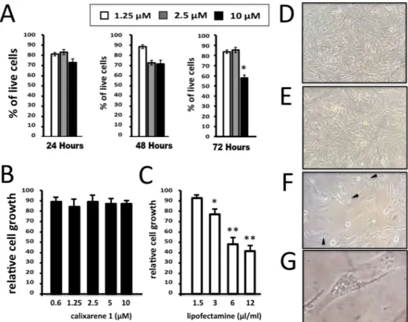
One of the major issues associated with the use of com-mercial available vehicles is the high toxicity of these compounds. For such reason 1 was first evaluated for its effect on cell viability when used to transfect molecules. A viability assay was performed on U251 cell line to detect the rate of live cells, after the transfection with incremental concentrations of transfection agent. Viability profile was determined at three different time points, 24, 48 and 72 hours after transfection, with three differ-ent concdiffer-entrations of 1, 1.25, 2.5, and 10 μM. Solutions of 1 at these three concdiffer-entrations were maintained in contact with cells for all the treatment time. As shown in Fig. 2, 24 hours after the transfection no differences in percentage of total live cells were shown when the two lower concentrations (1.25 and 2.5 µM) of 1 were used, while only slight decrease of live cells percentage was detected at 10 µM dosage. The decrease of the proportion of living U251 cells is not unexpected, since it is well established that most of the transfection reagents are to some extent cytotoxic to cells. Interestingly, however, cytotoxic effects of 1 were found only at 10 µM, while for lower concentrations limited effects in cell viability were detected. The effects of 1 on cell growth was determined and shown in Fig. 2B,C.Comparison was done with the effects of lipofectamine analyzed after 72 hours of cell culture. As clearly evi-dent, lipofectamine causes inhibition of cell proliferation (Fig. 2C), while the anti-proliferative effects of 1 are minor and significant only when the calixarene 1 is used at 10 µM concentration. These findings are in agreement with the cell morphology profile shown in Fig. 2(D–F) and determined by microscopic inspection after 72 hours of cell culture in the presence of 2.5 µM calixarene 1 (Fig. 2E), 3μL/mL lipofectamine (Fig. 2F) or in the absence of both the delivery systems (Fig. 2D). These data support the concept that treatment of the U251 cells with
calixarene 1 does not affect cell morphology. On the contrary deep alteration of cell morphology (including shape and shrinking, appearance of vacuolization and methuosis-like phenotypes, toxic granulation) occurs when U251 cells were treated with lipofectamine. The effects of lipofectamine is not unexpected, since this transfection agent has been shown to exhibit cytotoxicity both in vitro and in vivo51–55.
On the basis of the results shown in Fig. 2, all the experiments presented in this study were conducted at 2.5 µM of vector.
Effects of argininocalixarene 1 on the uptake of calixarene-vehicled PNA by glioma U251
cells.
In order to obtain a proof-of-principle that 1 mediates uptake of PNAs by target cells, a PNA against the microRNA miR-221-3p (a221PNA) was employed. The main reason is that a221PNA has been extensively stud-ied by some of us and is able to confer to the cells an easily detectable biological end-point, i.e. the induction of apoptosis. In the experiments shown in Fig. 3, a fluorescein labeled version of a221PNA, Fl-a221PNA, was used to readily visualize the internalization. First, 1 was loaded with the PNA, in order to obtain a working mixture of1/Fl-a221PNA at 2.5 and 2 µM, respectively. U251 cells were cultured for 48 hours with the 1/Fl-a221PNA mix-ture and FACS analysis performed using FACScan. The results obtained are shown in Fig. 3A and indicate that
Fl-a221PNA delivery mediated by 1 is associated with a complete different distribution of fluorescein-positive
U251 cells. In fact, while the naked Fl-a221PNA does not exhibit cellular uptake, when it is delivered with
1 almost 100% of the cells were found to be fluorescein-positive. The intensity of the fluorescence signal obtained
transfecting Fl-a221PNA with 1 approaches that obtained using the free fluorescein labelled Fl-R8-a221PNA, including in its structure an octaarginine peptide known to strongly facilitate cellular uptake50. The
microphoto-graphs depicted in Fig. 3B evidence a cytoplasmic distribution compatible to the data collected by FACS analysis. A further FACS analysis of fluorescence in cells upon treatment with a mixture of 1 and Fl-R8-a221PNA evi-denced values between those relative to the 1/Fl-a221PNA mixture and R8-a221PNA alone.
In order to rule out that the observed delivery was solely due to permeabilization of cells caused by the argin-inocalixarene, without interaction between the carrier and the PNA, Fl-a221PNA was titrated in vitro in buffer by adding 1 and measuring the changes in fluorescence emission (Fig. 4). A clear quenching effect was observed, with saturation reached at 2.5 μM concentration of the carrier, after which no further decrease in fluorescence was
Figure 2. Effects of calixarene 1 on viability, cell proliferation and morphology of U251 cells. (A) Viability
profile in U251 cells was reported for three incremental concentrations of 1: 1.25 µM (white bars), 2.5 µM (grey bars) and 10 µM (black bars). Three different time points: 24, 48 and 72 hours after the transfection were considered. (B–G) Comparison between the effects of calixarene 1 (B,E) and Lipofectamine RNAiMAX (C,F,G) on U251 cell growth (B,C) and morphology (D–G), determined after 72 hours cell culture. D = control untreated U251 cells. Appearance of vacuolization and methuosis-like patterns are underlined by the
observed, thus indicating the formation of a complex between carrier and PNA. A slight shift of the fluorescence maximum (from 519 to 523 nm) was also observed, consistent with a more polar microenvironment generated by the cationic calixarene.
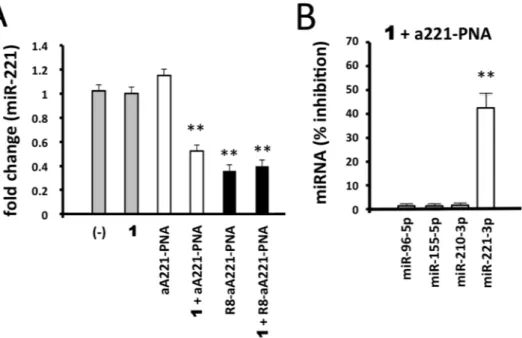
Effects of 1/a221PNA formulation on miR-221.
The data presented in Fig. 3 prompted us to verify whether the biological activity of the a221PNA was maintained when delivered with 1. To this aim we treated glioma U251 cells with 1, a221PNA and 1/ a221PNA formulation. We also considered free R8-a221PNA andFigure 3. PNA cellular uptake. (A) FACS analysis showing the uptake by glioma U251 cells of fluorescein
labeled a221PNAs. U251 cells were cultured for 48 hours with the Fl-a221PNA, 1/Fl-a221PNA formulation,
Fl-R8-a221PNA or 1/Fl-R8-a221PNA formulation, as indicated. (B) Microphotographs showing no
accumulation (left), or cytoplasmic accumulation of the delivered PNAs after 24 hours of cell culture.
Figure 4. Interaction of PNA with calixarene 1. Changes in fluorescence intensity of fluorescein labelled Fl-a221PNA (2 µM) upon addition of increasing concentration (0, 1.0, 2.0, 2.9, 3.9, 4.8, 5.7, 6.5, 7.4, 8.3, 9.1, 9.9 μM) of calixarene 1 in buffer solution (HEPES 16 mM, NaCl 40 mM, pH 7.4); (A) emission spectra of
1/R8-a221PNA formulation in order to verify possible synergic effects between vector 1 and R8
functionaliza-tion. The results of the experiments performed are shown in Fig. 5A and clearly demonstrate that no effects on miR-221-3p hybridization signal was observed in U251 cells treated with 1 alone or with naked a221PNA alone. This was expected, since (a) no major changes in gene expression (including miR-221-3p expression) caused by cytotoxicity occurs when 2.5 μM vector 1 concentration is employed, which was demonstrated to be unable to induce alteration of cell viability, morphology and rate of cell growth (see Fig. 2) and (b) a221PNA does not have effects on cultured cells, since uptake of this and similar molecules lacking the polyarginine tail is not efficient (see Fig. 3) as also previously reported50. On the contrary, significant inhibition of miR-221-3p hybridization was
found when U251 glioma cells were treated with 1/a221PNA formulation. Interestingly (a) the efficiency of this inhibition approached that of R8-a221PNA and (b) no negative effect of 1 were found in U251 cells transfected with 1/R8-a221PNA formulation.
These data conclusively demonstrate that a221PNA delivered by argininocalixarene 1 retains the inhibitory activity on miR-221-3p. The PNA-mediated effects were found to maintain the sequence specificity and selec-tivity. Despite transcriptomic and proteomic studies will fully clarify possible off-target effects of the proposed delivery system, our results firmly demonstrated that a221PNA delivered by argininocalixarene 1 displayed only minor inhibitory effects on other miRNAs expressed in the glioma U251 cells (such as miR-96-5p, miR-155-5p and miR-210-3p) (Fig. 5B and Supplementary Fig. 1).
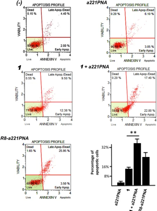
Effects of 1/a221PNA formulation on U251 apoptosis.
In order to verify whether a221PNA delivered by 1 maintains the known ability of PNA-based molecules targeting miR-221-3p to induce pro-apoptotic effects, U251 cells were cultured for 72 hours as already described in Fig. 5 and the Annexin-V assay was performed. The results are shown in Fig. 6 and clearly demonstrate that a221PNA can induce apoptosis only when delivered by 1 or by the covalently linked R8-oligopeptide (as we published elsewhere)50. When the values of % apoptosis(including both early apoptotic and late apoptotic cells) of the untreated cells or cells treated only with 1 are sub-tracted to the values obtained after U251 cell treatment with R8-a221PNA, or the 1/R8-a221PNA formulation, respectively, the increase of apoptosis (which, in the representative experiment shown in Fig. 6, was found 23.05% in the case of R8-a221PNA and 18.12% in the case of the 1/R8-a221PNA) is fully consistent with the FACS (Fig. 3A) and RT-qPCR (Fig. 5) analyses.
Discussion
In spite of being among the most interesting and performing oligonucleotide analogues1–26, peptide nucleic acids
(PNAs) are still of relatively limited use mainly because of the difficulties of internalization into target cells and the lack of a general and simple method for transfection27–32. Conjugation with cell penetrating peptides is one
of the most used approaches21,25,33–38, and several other strategies using nanocarriers28,29, polymer particles40 and
liposomal formulations39 have been proposed. To boost research and application of these compounds, the
avail-ability of a carrier molecule that can be simply mixed in the incubation medium with unmodified PNAs would be of great relevance.
Figure 5. Effects of 1/a221PNA formulation on miR-221. Glioma U251 cells were treated with 1, a221PNA, 1/a221PNA formulation, R8-a221PNA, 1/R8-a221PNA formulation. After 48 hours, RNA was isolated and
the hybridization to a miR-221-3p probe determined by RT-qPCR. Calixarene 1 was used at 2.5 μM; PNAs were used at 2 μM.
The data here reported indeed indicate that argininocalix[4]arene 146 can represent this type of delivery
sys-tem. It is able to efficiently transport neutral PNA molecules to target cells while preserving their biological activ-ity, after a very simple formulation procedure. The following conclusions can be drawn from the obtained results: (a) since the toxicity of 1 is low (see Fig. 2), it can be proposed for long-term treatment of target cells, being this feature a pre-requisite for the development of therapeutic protocols; (b) the delivery of PNAs is efficient, being comparable to that of PNAs functionalized with cell penetrating peptides (see Fig. 3); (c) the biological activity of PNA delivered using 1 is maintained (see Figs 5 and 6).
Of great relevance in our opinion is that the efficiency of the increase of the proportion of apoptotic cells when U251 cells are treated with 1/a221PNA formulation is comparable to that obtained using R8-a221PNA.
The localization inside the cells of the fluorescently labelled Fl-a221PNA delivered by 1 is more diffused com-pared to that of the peptide-conjugated Fl-R8-a221PNA, suggesting that endosomal escape is more efficient for the calixarene-delivered PNA and thus enhancing the effectiveness of the PNAs in cellular systems.
Figure 6. Effects of 1/a221PNA formulation on U251 apoptosis. U251 cells were cultured with 1, a221PNA, 1/a221PNA formulation, R8-a221PNA and 1/R8-a221PNA formulation. After 72 hours the cells were isolated
and Annexin-V assay was performed. Percentage of apoptotic cells in bottom graph refers to the sum of early apoptotic and late apoptotic cells.
No synergy on the transfection process were observed by mixing 1 with R8 containing a221PNA. The FACScan test evidenced a fluorescence similar to that of Fl-R8-a221PNA alone (Fig. 2) and the effect on miR-221-3p resulted comparable with that of R8-a221PNA transfected alone. This could be due to repulsive interac-tions between the arginines present both in 1 and R8-a221PNA that do not allow the binding between the two molecules and at the same time disfavor the delivery of R8-a221PNA alone.
The significant effect of the calixarene on the uptake of a221PNAs is not merely attributable to a cell perme-abilization effect, but it can be related to a strong binding to the PNA, as revealed by the titration experiments depicted in Fig. 4. This supports the initial hypothesis that argininocalixarene 1 can establish other types of interactions with oligonucleotides and oligonucleotide analogues different from the electrostatic ones with phos-phate groups, as actually verified for other cationic calixarene derivatives with nucleic acids47,48. Moreover, it is
known that in protein-DNA complexes, positively charged amino acid side chains not only interact with negative charges, but give rise to other specific interactions such as hydrogen bonding, π-π and cation-π interactions with nucleobases56,57. Analogously, in view of the neutral nature of the PNA backbone hydrogen bonding and cation-π
interactions could realistically occur to a different extent between guanidinium and/or ammonium groups of vector 1 and different nucleobases possibly being more intense with electron-rich purines, in particular guanine. It is likely that multiple different interactions concur to the formation of the PNA:calixarene adduct; thus the exact stoichiometry and strength of binding of other PNAs cannot be extrapolated at this stage. These interactions would constitute the first step of the mechanism of transport of PNA by calixarene 1. On the basis of the mode of binding to and condensation of DNA proposed for guanidinocalixarenes43 and of the experiments in this study,
it is in fact reasonable to think that subsequently, besides these specific interactions, a series of intermolecular hydrophobic interactions among the alkyl chains of different calixarenes bound to PNA molecules occur during the transport into cytosol across the cellular membrane. In any case, though studies are still necessary to shed light on this aspect, it is evident that the PNA:calixarene interactions take place rapidly and efficiently, thus greatly simplifying the transfection formulation protocol.
These findings indicate argininocalix[4]arene 1 as unique molecule for efficient delivery of PNAs to target cells. Its peculiarities are the ease of preparation, the relative structural simplicity associated to high delivery efficiency and negligible toxicity. Although the efficiency of uptake by cells for PNA delivered by 1 is not higher but comparable with that of PNA functionalized with R8 peptide, its use results much more convenient because avoids any modification on the PNA molecules intended to be delivered and the transfection formulation is sim-ply attained by mixing vector and PNA. Calixarene 1 therefore could represent a universal vector for all unmodi-fied PNAs that can simplify and boost their study and use as antisense, and possibly anti-gene, derivatives.
References
1. Nielsen, P. E., Egholm, M., Berg, R. H. & Buchardt, O. Sequence-selective recognition of DNA by strand displacement with a thymine-substituted polyamide. Science 254, 1497–1500 (1991).
2. Gambari, R. Peptide-nucleic acids (PNAs): a tool for the development of gene expression modifiers. Curr. Pharm. Des. 7, 1839–1862 (2001).
3. Gupta, A., Mishra, A. & Puri, N. Peptide nucleic acids: Advanced tools for biomedical applications. J. Biotechnol. 259, 148–159 (2017).
4. Nielsen, P. E., Egholm, M. & Buchardt, O. Peptide nucleic acid (PNA). A DNA mimic with a peptide backbone. Bioconjugate Chem.
5, 3–7 (1994).
5. Buchardt, O., Egholm, M., Berg, R. H. & Nielsen, P. E. Peptide nucleic acids and their potential applications in biotechnology. Trends
Biotechnol. 11, 384–386 (1993).
6. Leijon, M. et al. Structural characterization of PNA-DNA duplexes by NMR. Evidence for DNA in a B-like conformation.
Biochemistry 33, 9820–9825 (1994).
7. Bohländer, P. R., Vilaivan, T. & Wagenknecht, H. A. Strand displacement and duplex invasion into double-stranded DNA by pyrrolidinyl peptide nucleic acids. Org. Biomol. Chem. 13, 9223–9230 (2015).
8. Peffer, N. J. et al. Strand-invasion of duplex DNA by peptide nucleic acid oligomers. Proc. Natl. Acad. Sci. USA 90, 10648–10652 (1993).
9. Gambari, R. Peptide nucleic acids: a review on recent patents and technology transfer. Exp. Opin. Ther. Pat. 24, 267–294 (2014). 10. Demidov, V. V. et al. Stability of peptide nucleic acids in human serum and cellular extracts. Biochem. Pharmacol. 48, 1310–1313
(1994).
11. Nielsen, P. E. Peptide nucleic acids (PNA) in chemical biology and drug discovery. Chem. Biodivers. 7, 786–804 (2010).
12. Larsen, H. J., Bentin, T. & Nielsen, P. E. Antisense properties of peptide nucleic acid. Biochim. Biophys. Acta 1489, 159–166 (1999). 13. Nielsen, P. E. Gene targeting and expression modulation by peptide nucleic acids (PNA). Curr. Pharm. Des. 16, 3118–3123 (2010). 14. Vickers, T. A., Griffith, M. C., Ramasamy, K., Risen, L. M. & Freier, S. M. Inhibition of NF-kappa B specific transcriptional activation
by PNA strand invasion. Nucleic Acids Res. 23, 3003–3008 (1995).
15. Marin, V. L., Roy, S. & Armitage, B. A. Recent advances in the development of peptide nucleic acid as a gene-targeted drug. Exp.
Opin Biol Ther. 4, 337–348 (2004).
16. McNeer, N. A. et al. Nanoparticles that deliver triplex-forming peptide nucleic acid molecules correct F508del CFTR in airway epithelium. Nat. Commun. 6, 6952 (2015).
17. Bahal, R. et al. In vivo correction of anaemia in β-thalassemic mice by γPNA-mediated gene editing with nanoparticle delivery. Nat.
Commun. 7, 13304 (2016).
18. Ricciardi, A. S., Quijano, E., Putman, R., Saltzman, W. M. & Glazer, P. M. Peptide Nucleic Acids as a tool for site-specific gene editing. Molecules 23, 632 (2018).
19. Fabbri, E. et al. A Peptide Nucleic Acid against microRNA miR-145-5p enhances the expression of the cystic fibrosis transmembrane conductance regulator (CFTR) in calu-3 cells. Molecules 23, 71 (2017).
20. Gupta, A. et al. Anti-tumor activity of miniPEG-γ-modified PNAs to inhibit microRNA-210 for cancer therapy. Mol. Ther. Nucleic
Acids 9, 111–119 (2017).
21. Fabbri, E. et al. Modulation of the biological activity of microRNA-210 with peptide nucleic acids (PNAs). ChemMedChem 6, 2192–2202 (2011).
22. Fabani, M. M. et al. Efficient inhibition of miR-155 function in vivo by peptide nucleic acids. Nucleic Acids Res. 38, 4466–4475 (2010).
23. Gambari, R. et al. Targeting microRNAs involved in human diseases: a novel approach for modification of gene expression and drug development. Biochem Pharm. 82, 1416–1429 (2011).
acid oligonucleotides. Bioconjugate Chem. 21, 1902–1911 (2010).
34. Mondhe, M., Chessher, A., Goh, S., Good, L. & Stach, J. E. M. Species-selective killing of bacteria by antimicrobial peptide-PNAs.
PLOSOne 9, e89082 (2014).
35. Cheng, C. J. et al. Cellular uptake and biological activity of peptide nucleic acids conjugated with peptides with and without cell-penetrating ability. J. Pept. Sci. 16, 71–80 (2010).
36. Hu, J. & Corey, D. R. Inhibiting gene expression with peptide nucleic acid (PNA)-peptide conjugates that target chromosomal DNA.
Biochemistry 46, 7581–7589 (2007).
37. Manicardi, A. et al. Cellular uptakes, biostabilities and anti-miR-210 activities of chiral arginine-PNAs in leukaemic K562 cells.
ChemBioChem 13, 1327–1337 (2012).
38. Hnedzko, D., McGee, D. W., Karamitas, Y. A. & Rozners, E. Sequence-selective recognition of double-stranded RNA and enhanced cellular uptake of cationic nucleobase and backbone-modified peptide nucleic acids. RNA 23, 58–69 (2017).
39. Shiraishi, T., Hamzavi, R. & Nielsen, P. E. Subnanomolar antisense activity of phosphonate-peptide nucleic acid (PNA) conjugates delivered by cationic lipids to HeLa cells. Nucleic Acids Res. 36, 4424–4432 (2008).
40. McNeer, N. A. et al. Nanoparticles deliver triplex-forming PNAs for site–specific genomic recombination in CD34+ human hematopoietic progenitors. Mol. Ther. 19, 172–180 (2011).
41. Macadangdang, B. et al. Inhibition of multidrug resistance by SV40 pseudovirion delivery of an antigene peptide nucleic acid (PNA) in cultured cells. PLoS ONE 6, e17981 (2011).
42. Hamilton, S. E., Simmons, C. G., Kathiriya, I. S. & Corey, D. R. Cellular delivery of peptide nucleic acids and inhibition of human telomerase. Chem. Biol. 6, 343–351 (1999).
43. Sansone, F. et al. DNA condensation and cell transfection properties of guanidinium calixarenes: dependence on macrocycle lipophilicity, size, and conformation. J. Am. Chem. Soc. 128, 14528–14536 (2006).
44. Bagnacani, V. et al. Macrocyclic non-viral vectors: high cell transfection efficiency and low toxicity in a lower rim guanidinium calix[4]arene. Org. Lett. 10, 3953–3956 (2008).
45. Bagnacani, V. et al. Lower rim guanidinocalix[4]arenes: macrocyclic nonviral vectors for cell transfection. Bioconjugate Chem. 23, 993–1002 (2012).
46. Bagnacani, V. et al. Arginine clustering on calix[4]arene macrocycles for improved cell penetration and DNA delivery. Nat.
Commun. 4, 1721 (2013).
47. Zadmard, R. & Schrader, T. DNA Recognition with large calixarene dimers. Angew. Chem. Int. Ed. 45, 2703–2706 (2006). 48. Shi, Y. H. & Schneider, H.-J. Interactions between aminocalixarenes and nucleotides or nucleic acids. J. Chem. Soc. Perkin Trans. 2,
1797–1803 (1999).
49. Brognara, E. et al. Peptide nucleic acids targeting miR-221 modulate P27Kip1 expression in breast cancer MDA-231 cells. Int. J. Oncol.
41, 2119–2127 (2012).
50. Brognara, E. et al. Uptake by human glioma cell lines and biological effects of a peptide-nucleic acids targeting miR-221. J. Neuro
Oncol. 118, 19–28 (2014).
51. Pan, G., Shawer, M., Oie, S. & Lu, D. R. In vitro gene transfection in human glioma cells using a novel and less cytotoxic artificial lipoprotein delivery system. Pharm. Res. 20, 738–44 (2003).
52. Uchida, E., Mizuguchi, H., Ishii-Watabe, A. & Hayakawa, T. Comparison of the efficiency and safety of non-viral vector-mediated gene transfer into a wide range of human cells. Biol. Pharm. Bull. 25, 891–897 (2002).
53. Armeanu, S. et al. Optimization of nonviral gene transfer of vascular smooth muscle cells in vitro and in vivo. Mol. Ther. 1, 366–375 (2000).
54. Dokka, S., Toledo, D., Shi, X., Castranova, V. & Rojanasakul, Y. Oxygen radical-mediated pulmonary toxicity induced by some cationic liposomes. Pharm. Res. 17, 521–525 (2000).
55. Bell, H., Kimber, W. L., Li, M. & Whittle, I. R. Liposomal transfection efficiency and toxicity on glioma cell lines: in vitro and in vivo studies. Neuroreport. 9, 793–798 (1998).
56. Wilson, K. A. & Wetmore, S. D. Combining crystallographic and quantum chemical data to understand DNA-protein π-interactions in nature. Struct. Chem. 28, 1487–1500 (2017).
57. Gromiha, M. M., Santhosh, C. & Ahmad, S. Structural analysis of cation–π interactions in DNA binding proteins. Int. J. Biol.
Macromol 34, 203–211 (2004).
Acknowledgements
This work was supported by Consorzio Interuniversitario di Biotecnologie, by Ministero dell’Istruzione, dell’Università e della Ricerca [PRIN-2010 project 2010JMAZML_005, MultiNanoIta; PRIN-2009 project 20093N774P, Riconoscimento molecolare di micro-RNA (miR) mediante PNA modificati: dalla struttura alla attività], by Associazione Italiana per la Ricerca sul Cancro [project IG 13575, Peptide nucleic acids targeting oncomiR and tumor-suppressor miRNAs: cancer diagnosis and therapy], by EU-FP7 [THALAMOSS Project - Thalassemia Modular Stratification System for Personalized Therapy of B-Thalassemia; n.306201-FP7-HEALTH-2012-INNOVATION-1], by Wellcome Trust, by AIFA and by Fondazione Fibrosi Cistica [Project “Revealing the microRNAs-transcription factors network in cystic fibrosis: from microRNA therapeutics to precision medicine (CF-miRNA-THER)”, FFC#7/2018].
Author Contributions
J.G. and A.F. performed the characterization of calixarene 1 formulations with cells transfection, MUSE, Biostation IM and RT-qPCR; J.G. performed the FACS analyses; R.C. and A.M. provided the PNAs and performed and supervised the study of PNA-1 interactions; A.C. and F.S. supervised the synthesis of and provided vector 1; A.C., R.C., R.G., A.F. and F.S. wrote the manuscript; F.S. and A.F. supervised the whole project.
Additional Information
Supplementary information accompanies this paper at https://doi.org/10.1038/s41598-019-39211-4.
Competing Interests: The authors declare no competing interests.
Publisher’s note: Springer Nature remains neutral with regard to jurisdictional claims in published maps and
institutional affiliations.
Open Access This article is licensed under a Creative Commons Attribution 4.0 International
License, which permits use, sharing, adaptation, distribution and reproduction in any medium or format, as long as you give appropriate credit to the original author(s) and the source, provide a link to the Cre-ative Commons license, and indicate if changes were made. The images or other third party material in this article are included in the article’s Creative Commons license, unless indicated otherwise in a credit line to the material. If material is not included in the article’s Creative Commons license and your intended use is not per-mitted by statutory regulation or exceeds the perper-mitted use, you will need to obtain permission directly from the copyright holder. To view a copy of this license, visit http://creativecommons.org/licenses/by/4.0/.
![Figure 1. Structure of calix[4]arene 1 and sequences of the PNAs used in the present study](https://thumb-eu.123doks.com/thumbv2/123dokorg/4754311.47123/2.892.234.823.69.287/figure-structure-calix-arene-sequences-pnas-present-study.webp)