E-Mail [email protected]
What Is Your Diagnosis?
Respiration 2017;93:296–300DOI: 10.1159/000457804
Coexistent Sarcoidosis and Tuberculosis:
A Case Report
Cristiano Carbonelli
a,b
Ernesto Giuffreda
a
Antonio Palmiotti
a
Domenico Loizzi
c
Filippo Lococo
d
Elisiana Carpagnano
a
Donato Lacedonia
a
Francesco Sollitto
c
Maria Pia Foschino
a
a
Department of Medical and Surgical Sciences, Institute of Respiratory Diseases, Azienda Ospedaliero Universitaria
Foggia, Foggia , b
Department of Medical Sciences, Scientific Institute “Casa Sollievo della Sofferenza”, San Giovanni
Rotondo , c
Department of Thoracic Surgery, University of Foggia, Foggia , and d Unit of Thoracic Surgery, Arcispedale
Santa Maria Nuova-IRCCS, Reggio Emilia , Italy
Received: August 10, 2016
Accepted after revision: January 20, 2017 Published online: February 9, 2017
Cristiano Carbonelli, MD
Department of Medical and Surgical Sciences, Institute of Respiratory Diseases Azienda Ospedaliero Universitaria Foggia, Viale degli Aviatori 1
IT–71100 Foggia (Italy) E-Mail kkarbo @ hotmail.com
© 2017 S. Karger AG, Basel
www.karger.com/res
Case Presentation
A 45-year-old black male with a medical history of chronic hepatitis C virus treated with pegylated interfer-on and ribavirin until 4 minterfer-onths before hospitalizatiinterfer-on was admitted for a bacterial pneumonitis. The examina-tions revealed neck, mediastinal, and abdominal adenop-athies. Histologically, a Zielh-Neelsen-negative necrotiz-ing granulomatous reaction was documented in a later cervical lymph node analysis. The Mantoux test was neg-ative, and bronchoalveolar lavage showed a lymphocytic alveolitis 60% of the total with a CD4/CD8 ratio of 4. An-giotensin-converting enzyme was elevated, and there was no sign of uveal involvement or hypercalciuria. As the microbiological examinations for mycobacteria were re-peatedly negative, even upon examination of a culture on respiratory and peripheral lymph node tissue, a provi-sional diagnosis of sarcoidosis was made, and the patient
was discharged with a follow-up schedule without thera-py apart from oral antiviral drugs (entecavir).
About 3 months later, the patient came to the Depart-ment of Infectious Disease of our hospital, complaining of high-grade septic fever and bilateral pleural effusions. A thoracic and abdominal CT confirmed few nodular el-ements in both lungs along with the confirmation of a pericardial and bilateral pleural effusion and multiple spleen nodules.
The analysis of the pleural fluid was unremarkable, apart from prevalence of lymphocytes upon cytological evaluation. As the patient was prostrated for the long-lasting febrile manifestation, the introduction of steroid therapy led to the resolution of fever.
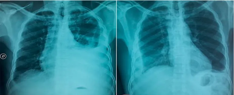
The patient’s conditions ameliorated, and a chest X-ray documented an improvement of the left pleural effu-sion. However, a worsening on the right side was seen with echo signs of multiloculation ( Fig. 1 ).
Coexistent Sarcoidosis and Tuberculosis Respiration 2017;93:296–300
DOI: 10.1159/000457804 297
Fig. 1. Radiological and radiometabolic scenario. Chest X-rays showed the different evolution of pleural effusion
Carbonelli et al.
Respiration 2017;93:296–300 DOI: 10.1159/000457804
298
proved the fever and overall clinical scenario. Lympho-cytic CD4 increased to 391 cells/μL 2 weeks after steroid introduction.
The positivity of cultures on pleural specimens for My-cobacterium tuberculosis was reported 45 days after tho-racoscopy, leading to the confirmation of pleural TB in the framework of a concomitant systemic sarcoid mani-festation.
Discussion
Nevertheless, the words “necrosis” and “pleural in-volvement” in relation with a granulomatous disease are usually interpreted as highly suggestive of TB. Differen-tial diagnosis has to include well-described series of sar-coid pleural involvement. Pleural involvement is frequent in necrotizing sarcoid granulomatosis (NSG), and the treatment with steroids appears to improve the clinical findings [1] .
Diagnosis: Pleural Tuberculosis in the Framework of a Concomitant Systemic Sarcoid Manifestation
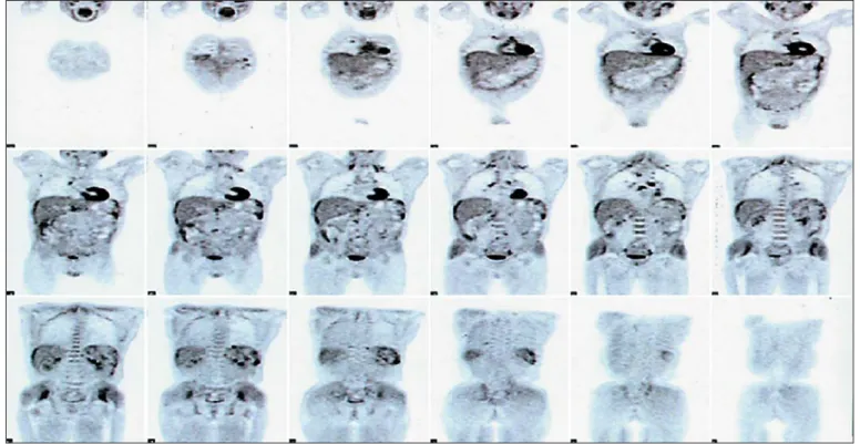
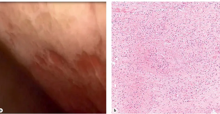
A positron emission tomography/CT examination confirmed multiple areas of a high 18-fluorodeoxyglu-cose uptake at pleural surfaces, systemic lymphonodes, and the spleen ( Fig. 2 ). The patient was referred to the interventional pulmonology service with the aim of per-forming a medical thoracoscopy for a definitive diagno-sis. Geographic pleural thickening ( Fig. 3 a) with multiple multiloculations and a granulomatous necrotizing dis-ease at the level of the pleural tissue were present ( Fig. 3 b). However, Zielh-Neelsen colorations were again negative, as were polymerase chain reaction analyses for tubercu-losis (TB).
Quantiferon positivity, together with the endoscopic scenario and the histological pattern, motivated the intro-duction of antimicrobacterial treatment (isoniazid, ri-fampicin, ethambutol, and pyrazinamide) and the with-drawal of steroids. High-grade fever reappeared, and an increase of GGT, nausea, and abdominal pain were re-ported. Circulating CD4 were 173 cells/μL, and the HIV test was negative. Twenty days later, steroid reintro-duction (methylprednisolone 1 mg/kg) dramatically
Fig. 2. Radiological and radiometabolic scenario. 18 F-FDG positron emission tomography/CT scan with multiple
Coexistent Sarcoidosis and Tuberculosis Respiration 2017;93:296–300
DOI: 10.1159/000457804 299
Sarcoidosis represents a stereotyped, antigen-driven immune disorder with diverse etiologies [2] . A century of epidemiologic, molecular, and immunologic literature emphasizes the hypothesis about a causal role of myco-bacteria antigens in some sarcoidosis cases [3, 4] .
The unspecificity of the clinical findings in these cases is well recognized. The classic manifestations of sarcoid-osis and TB have an overlapping range for which it is sometimes difficult to make a definitive diagnosis in the absence of a microbiological identification [5] . Hepatitis C virus may be a cofactor in the pathogenesis of sarcoid-osis, and a close association between interferon therapy and sarcoidosis has been described [6] .
Sarcoid granulomatosis and TB are sometimes entities difficult to separate, even on a pathological basis in the absence of a cultural microbiological confirmation. In-deed, there is a continuous spectrum of necrosis in sar-coidosis, ranging from minimal to massive grade. The so-called sarcoidosis with the NSG pattern requires a reason-able exclusion of an infectious etiology [7] . Failure to culture mycobacteria from sarcoidosis specimens may be secondary to difficulties in obtaining tissue from critical-ly involved sites, microorganism peculiarity, bacteria burden, or inadequate characteristics of culture assays
b a
Fig. 3. Endoscopic view and pathological examination. Thoracoscopic view ( a ) of the right pleural cavity,
show-ing disseminated fibrinous pleural thickenshow-ings with multiple multiloculations. Pathological examination of the pleural biopsy ( b ), revealing a granulomatous caseating necrotizing pattern. HE. ×200.
[3] . Positron emission tomography imaging can be useful in order to guide the surgical biopsy in NSG [8] .
Coexistence of TB and sarcoidosis is a rarely described [9] and likely a highly underrecognized entity [10] . Bron-choalveolar lavage does not accurately discriminate be-tween sarcoidosis and other causes of lymphocytic alveo-litis such as TB [11] . The case herein supports the hypoth-esis that systemic manifestation of sarcoidosis and its improvement with steroid treatment can occur coinci-dently with low-antigen, pleural TB.
Nuclear medicine techniques and thoracoscopy find-ings for tissue acquisition in the described diagnostic pro-cess must be considered to ascertain a mycobacterial in-fection as the causative agent of a concomitant sarcoid manifestation. The choice to continue both steroid and anti-TB therapy proved to be correct for the late evidence of TB mycobacterial growth only on pleural specimens. In most cases, the diagnosis of necrotizing sarcoidosis and TB with a low antigen load cannot be formulated, and physicians should still manage to protect pharmacologi-cally the less likely clinical hypothesis and, at the same time, closely follow the clinical course and the outcome of the investigations carried out, avoiding irreparable damage in case of misdiagnosis.
Carbonelli et al.
Respiration 2017;93:296–300 DOI: 10.1159/000457804
300
Keywords
Pleural biopsy · Thoracoscopy · Sarcoidosis · Tuberculosis
Abstract
Necrotizing granulomatous diseases of the lungs are usu-ally dependent on a narrow range of differential diagnoses. Tuberculosis (TB) is responsible for the largest number of cases, while necrotizing sarcoidosis is generally considered a rare and easily distinguishable disease substantially based on histological features. However, this entity has become a viable diagnosis in the absence of mycobacteria isolation or when a remarkable clinical improvement cannot be achieved with the combination of anti-TB drugs at full dos-age. The classic manifestations of TB and sarcoidosis have an overlapping range for which it is sometimes difficult to make a clinical diagnosis. Furthermore, the role of myco-bacteria as a trigger antigen capable of evoking the clinical expression of sarcoidosis is a hypothesis supported by evi-dence from some cases. We report a case of bilateral tuber-culous pleurisy in a 45-year-old male native of a North-Afri-can region with an atypical severe multisystem disease characterized by a fever resistant to anti-TB therapy and re-spondent to corticosteroid treatment. The choice to con-tinue both steroid and anti-TB therapy proved to be correct for the late evidence of TB mycobacterial growth only on pleural specimens. The case described is suggestive of a co-existent systemic sarcoid manifestation and low-antigen TB, which is an underrecognized entity in the medical lit-erature. © 2017 S. Karger AG, Basel
Acknowledgement
Thanks to the Pleural Hub group on Facebook for their assis-tance in the multidisciplinary discussion of the case.
Thanks to Ms. Anna T. Valencia, MPH, MBEMH, for language support.
Statement of Ethics
We obtained informed patient consent for the participation and publication of individual patient data.
Disclosure Statement
The authors declare that they have no competing interests to disclose.
Author Contribution
C. Carbonelli performed the interventional procedures, con-ceived the study, participated in its design and coordination, and drafted the manuscript.
E. Giuffreda performed the transthoracic echography and par-ticipated in the design of the study and in the drafting of the man-uscript.
A. Palmiotti performed the transthoracic echography and par-ticipated in the design of the study.
D. Loizzi performed the interventional procedures and partic-ipated in the design of the study.
E. Carpagnano, D. Lacedonia, and F. Lococo participated in the design of the study and in the drafting of the manuscript.
F. Sollitto performed the interventional procedures and par-ticipated in the design of the study.
M.P. Foschino participated in the design of the study and its coordination as well as in the drafting of the manuscript.
All authors have read and approved the final manuscript.
References
1 Chittock DR, Joseph MG, Paterson NA, Mc-Fadden RG: Necrotizing sarcoid granuloma-tosis with pleural involvement. Clinical and
radiographic features. Chest 1994; 106: 672–
676.
2 Moller DR: Treatment of sarcoidosis – from a basic science point of view. J Intern Med 2003;
253: 31–40.
3 Drake WP, Newman LS: Mycobacterial anti-gens may be important in sarcoidosis
patho-genesis. Curr Opin Pulm Med 2006; 12: 359–
363.
4 Fité E, Fernández-Figueras MT, Prats R, Va-quero M, Morera J: High prevalence of Myco-bacterium tuberculosis DNA in biopsies from sarcoidosis patients from Catalonia, Spain.
Respiration 2006; 73: 20–26.
5 Agarwal R, Gupta D: Tuberculous sarcoid-osis: is it a separate entity? Lung India 2009;
26: 61–62.
6 Ramos-Casals M, Mañà J, Nardi N, Brito-Zeròn P, Xaubet A, Sãnchez-Tapias JM, Cer-vera R, Font J; HISPAMEC Study Group: Sar-coidosis in patients with chronic hepatitis C virus infection: analysis of 68 cases. Medicine
(Baltimore) 2005; 84: 69–80.
7 Rosen Y: Four decades of necrotizing sarcoid granulomatosis. What do we know? Arch
Pathol Lab Med 2015; 139: 252–262.
8 Arfi J, Kerrou K, Traore S, Huchet V, Bolly A, Antoine M, Delaunois L, Vander Borght T, Talbot JN: F-18 FDG PET/CT findings in pul-monary necrotizing sarcoid granulomatosis.
Clin Nucl Med 2010; 35: 697–700.
9 Wong CF, Yew WW, Wong PC, Lee J: A case of concomitant tuberculosis and sarcoidosis with mycobacterial DNA present in the
sar-coid lesion. Chest 1998; 114: 626–629.
10 Lin JY, Sheu SJ: Ocular sarcoidosis and tuber-culous lymphadenopathy: coincidence or real
association. J Ophthal Inflamm Infect 2011; 1:
137–140.
11 Bretagne L, Diatta I-D, Faouzi M, Nobile A, Bongiovanni M, Nicod LP, Lazor R: Diagnos-tic value of the CD103+CD4+/CD4+ ratio to differentiate sarcoidosis from other causes of
lymphocytic alveolitis. Respiration 2016; 91:
486–496.