Università degli Studi di Ferrara
DOTTORATO DI RICERCA IN
FARMACOLOGIA E ONCOLOGIA MOLECOLARE
CICLO XXI
COORDINATORE Prof. Pier Andrea Borea
Voltage-dependent anion channels:
different isoforms for different
functions
Settore Scientifico Disciplinare MED/04
Dottorando Tutore
Dott. Diego De Stefani Prof. Rosario Rizzuto
Contents
ABSTRACT ... 1
ABSTRACT (ITALIANO) ... 2
INTRODUCTION ... 3
Mitochondria: the basics ... 4
Calcium signaling: the general framework ... 6
ER/mitochondria crosstalk: local microdomains support mitochondrial Ca2+ uptake ... 8
Mitochondrial Ca2+ channels of the inner mitochondrial membrane ... 15
Calcium release from cellular store: structure and function of the IP3R ... 18
Enhancing ATP production or killing the cell: the yin/yang of mitochondrial calcium ... 21
Voltage-dependent anion channels (VDAC) ... 23
Autophagy ... 27
The mammalian Target of Rapamycin (mTOR) ... 30
Autophagy and cell death ... 32
AIMS ... 34
MATERIALS AND METHODS ... 35
Yeast two-hybrid screening... 35
Subcellular fractionation and proteomic analysis ... 36
IP3R and grp75 expression constructs ... 37
Aequorin as a Ca2+ indicator ... 38
RESULTS ... 48
Chaperone mediated coupling of endoplasmic reticulum and mitochondrial Ca2+ channels ... 48
VDAC1, grp75 and IP3Rs are present in a macromolecular complex at the ER mitochondria interface ... 50
Direct regulation of mitochondrial Ca2+ uptake by the IP 3R ligand binding domain ... 53
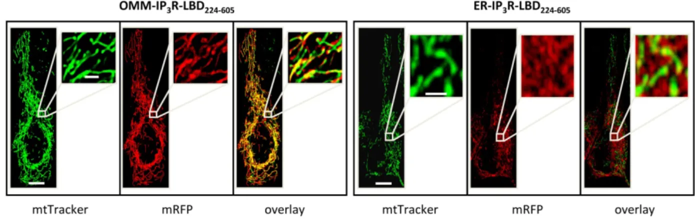
Regulation of mitochondrial uptake by the IP3R-LBD is a result of specific protein interactions at the ER-OMM interface ... 55
Down-regulation of grp75 abolishes the functional coupling between the IP3R and mitochondria ... 59
Effect of different VDAC isoforms on mitochondrial Ca2+ homeostasis ... 63
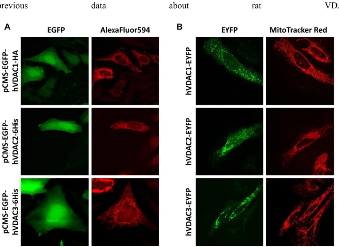
Intracellular localization of VDAC constructs ... 65
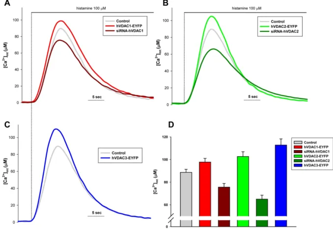
All VDAC isoforms enhance mitochondrial Ca2+ uptake ... 68
VDAC isoforms do not affect cytosolic Ca2+ transients ... 70
VDAC has no effect on ER Ca2+ content and IP3 induced Ca2+ release ... 71
VDAC isoforms differentially regulate cellular sensitivity to apoptotic stimuli ... 72
VDAC1 specific coupling to ER Ca2+ releasing channels ... 74
VDAC isoforms in the control of autophagy ... 77
VDAC2 is selectively required for mTOR dependent autophagy ... 78
VDAC2 controls mTOR association to mitochondria ... 83
DISCUSSION ... 86
1
Abstract
The Voltage-dependent anion channel (VDAC) is the most abundant protein of the outer mitochondrial membrane (OMM) and mediates the flow of ions and metabolites between the cytoplasm and the mitochondrial network. Here we reveal novel and unexpected roles of this protein in the regulation of Ca2+ signaling, cell death and autophagy, throwing light on the differential contribution of the three mammalian isoforms in these cellular processes. In particular, we show that: i) VDAC is physically linked to the endoplasmic reticulum Ca2+ release channel inositol-1,4,5-trisphosphate receptor (IP3R), through the molecular chaperone grp75 and the
functional coupling of these channel directly enhances Ca2+ accumulation in mitochondria; ii) the different VDAC isoforms share common Ca2+ channelling properties in living cells but VDAC1 is the only isotype selectively coupled to the ER Ca2+ releasing machinery, thus laying the foundations for a preferential route specifically transmitting Ca2+-mediated cell death signals between the two organelles; iii) VDAC2 is selectively required for the induction of the autophagic process through the establishment of specific protein-protein interactions and the consequent assembly of macromolecular complexes at the OMM level involved in nutrient sensing mediated by the mammalian Target Of Rapamycin (mTOR) signaling pathway. These data highlight the pleiotropic functions of VDAC and its role as central regulator of cell patho-physiology.
2
Abstract (italiano)
Il Voltage-dependent Anion Channel (VDAC) è la proteina più espressa a livello della membrana mitocondriale esterna dove regola il flusso di ioni e piccoli metaboliti fra il citoplasma e lo spazio intermembrana. In questo lavoro vengono svelati nuovi ed inattesi ruoli di questa proteina nella regolazione del segnale Ca2+, della morte cellulare e dell’ autofagia, ed in particolare sul ruolo specifico che le tre diverse isoforme di questo canale giocano in questi processi. In particolare abbiamo dimostrato che: i) VDAC è in grado di interagire fisicamente con il canale di rilascio per il Ca2+ sensibile all’inositolo-1,4,5-trifosfato (IP
3R) del reticolo endoplasmatico attraverso la proteina
adattatrice grp75 e che l’accoppiamento funzionale fra questi due canali stimola direttamente l’accumulo di Ca2+ nella matrice mitocondriale; ii) nonostante tutte le diverse isoforme di VDAC condividano simili proprietà canale nei confronti dello ione Ca2+, VDAC1 è l’unica isoforma associata in modo specifico ai siti di rilascio del reticolo endoplasmatico e che questa interazione è probabilmente alla base della trasmissione di segnali specifici fra un organello e l’altro che mediano la morte cellulare; iii) VDAC2 è selettivamente coinvolto nell’induzione del processo autofagico grazie a specifiche interazioni proteiche e all’assemblaggio di complessi molecolari a livello della membrana mitocondriale esterna coinvolti nella dalla via di segnalazione del sensore di nutrienti mTOR (mammalian Target Of Rapamycin). Questi dati evidenziano le molteplici funzioni della proteina VDAC e il suo ruolo di fondamentale regolatore di processi sia patologici che fisiologici.
3
Introduction
The mitochondrion represents a unique organelle within the complex endomembrane systems that characterize any eukaryotic cell. Complex life on earth has been made possible through the “acquisition” of mitochondria which provide an adequate supply of substrates for energy-expensive tasks. Higher multicellular organisms have indeed high energy requirements necessary to carry out complex functions, such as muscle contraction, hormones and neurotransmitters synthesis and secretion, in addition to basal cellular metabolism (biomolecules synthesis and transformation, maintenance of ionic gradients across membrane, cell division). Mitochondria can fulfill this huge energy demand thanks to their extraordinary biosynthetic capacities: every day, mitochondria of a single human being can recycle up to 50 Kg of ATP. To further underline the relevance of these subcellular structures, one can also consider how these organelles have affected the physiology of the whole organism: lungs, heart and circulatory system have evolved essentially to provide molecular oxygen to mitochondria, which consume about 98% of the total O2 we breathe. However,
beyond the pivotal role they play in ATP production, a whole new mitochondrial biology has emerged in the last few decades: mitochondria have been shown to participate in many other aspects of cell physiology such as amino-acid synthesis, iron-sulphur clusters assembly, lipid metabolism, Ca2+ signaling, reactive oxygen species (ROS) production and cell death regulation. Hence, it is consequent that any mitochondrial dysfunction will inevitably lead to disease. Indeed, many pathological conditions are associated with organelle failure, including neurodegenerative diseases (Alzheimer’s, Parkinson’s, Hungtinton’s), motoneuron disorders (amyotrophic lateral sclerosis, type 2A Charcot-Marie-Tooth neuropathy), autosomal dominant optic atrophy, ischemia-reperfusion injury, diabetes, ageing and cancer.
4
Understanding how mitochondria can sense, handle and decode various signals from the cytosol and other subcellular compartments represents a new exciting challenge in biomedical sciences.
Mitochondria: the basics
The mitochondrion is a double membrane-bounded organelle thought to be derived from an -proteobacterium-like ancestor, presumably due to a single ancient invasion occurred more than 1.5 billion years ago. The basic evidence of this endosymbiont theory (Dyall et al., 2004) is the existence of the mitochondrial DNA (mtDNA), a 16.6 Kb circular double-stranded DNA molecule with structural and functional analogies to bacterial genomes (gene structure, ribosome). This mitochondrial genome encodes only 13 proteins (in addition to 22 tRNAs and 2 rRNAs necessary for their translation), all of which are components of the electron transport chain (mETC) complexes (I, III and IV), while the whole mitochondrial proteome consists of more than 1000 gene products. Thus, one critical step in the transition from autonomous endosymbiont to organelle has been the transfer of genes from the mtDNA to the nuclear genome. At the same time, eukaryotes had to evolve an efficient transport system to deliver nuclear-encoded peptides inside mitochondria: TIM (Transporters of the Inner Membrane), TOM (Transporters of the Outer Membrane) and mitochondrial chaperones (such as hsp60 and mthsp70) build up the molecular machinery that allows the newly-synthesized unfolded proteins to enter mitochondrial matrix (Mokranjac and Neupert, 2005).
Mitochondria are defined by two structurally and functionally different membranes: the plain outer membrane, mostly soluble to ions and metabolites up to 5000 Da, and the highly selective inner membrane, characterized by invaginations called cristae which enclose the mitochondria matrix. The space between these two structures is traditionally called intermembrane
5
space (IMS), but recent advances in electron microscopy techniques shed new light on the complex topology of the inner membrane. Cristae indeed are not simply random folds but rather internal compartments formed by profound invaginations originating from very tiny “point-like structures” in the inner membrane (Mannella, 2006). These narrow tubular structures, called cristae junctions, can limit the diffusion of molecule from the intra-cristae space towards the IMS, thus creating a micro-environment where mETC complexes (as well as other proteins) are hosted and protected from random diffusion.
As mentioned before, mitochondria are the main site of ATP production. When glucose is converted to pyruvate by glycolysis, only a small fraction of the available chemical energy has been stored in ATP molecules: mitochondria can “release” the remaining amount of energy with an outstanding efficiency (from a single glucose molecule mitochondria produce 15 times more ATP than glycolysis). The main enzymatic systems involved in this process are the tricarboxylic acid (TCA) cycle and the mETC. Products from glycolysis and fatty acid metabolism are converted to acetyl-CoA which enters the TCA cycle where it is fully degraded to CO2. More importantly, these
enzymatic reactions generate NADH and FADH2 which provide reducing equivalents and trigger
the electron transport chain. mETC consists of five different protein complexes: complex I (NADH dehydrogenase), complex II (succinate dehydrogenase), complex III (ubiquinol cytochrome c reductase), complex IV (cytochrome c oxidase) and complex V that constitutes the F1F0-ATP
synthase. Electrons are transferred from NADH and FADH2 through these complexes in a stepwise
fashion: as electrons move along the respiratory chain, energy is stored as an electrochemical H+ gradient across the inner membrane, thus creating a negative mitochondrial membrane potential (estimated around -180 mV against the cytosol). H+ are forced to reenter the matrix mainly through complex V which couples this proton driving force to the phosphorylation of ADP into ATP, according to the chemiosmotic principle. ATP is then released to IMS through the electrogenic Adenine Nucleotide Translocase (ANT) which exchange ATP with ADP to provide new substrate
6
for ATP synthesis. Finally, ATP can easily escape the IMS thanks to the mitochondrial porin of the outer membrane, VDAC (voltage dependent anion channel) (Duchen, 2004). However, in the last two decades interest in mitochondrial biology has literally revamped, since the discovery of their prominent role in triggering cell death through apoptosis (Liu et al., 1996).
Calcium signaling: the general framework
In all eukaryotic cells, the cytosolic concentration of Ca2+ ([Ca2+]c) is tightly controlled by
complex interactions among pumps, channels, exchangers and binding proteins, and relatively small and/or local changes in its concentration modulate a wide range of intracellular actions. [Ca2+]
c in
resting condition is maintained around the value of 100nM, significantly lower than extracellular [Ca2+] (1mM). This condition is guaranteed by the low permeability of the plasma membrane to ions and by the activity of the Plasma Membrane Ca2+-ATPase (PMCA, which pumps Ca2+ outside the cells) and of the Na+/Ca2+ exchanger (NCX). This fine regulation of [Ca2+] allows this ion to act as one of the most important second messenger in signal transduction pathways. (Hajnoczky et al., 2000; Hajnoczky et al., 2002)
The increase of intracellular [Ca2+] can be elicited through two fundamental mechanisms: i) the Ca2+ mobilization from intracellular stores, mainly the endoplasmic reticulum (ER) and Golgi apparatus, or ii) the entry from the extracellular milieu. The main route inducing Ca2+ release from intracellular stores involves the IP3 Receptor (IP3R), a transmembrane protein located on the ER
and Golgi membrane, which exposes on the cytosolic face the IP3 binding site, while it forms a Ca2+
channel in the transmembrane domain. When extracellular soluble agonists binds a G-coupled protein receptor, different isoforms of phospholipase C (PLC) are activated producing inositol-1,4,5-trisphosphate (IP3) from the hydrolysis of phosphatidylinositol 4,5 bisphosphate (PIP2). The
binding of IP3 to its receptor induces its opening and the release of Ca2+ from ER and Golgi. IP3R is
7
transmembrane protein located on the ER membrane and it is activated by the alchaloid ryanodine and by Ca2+ itself, while Sphingolipid Ca2+ release-mediating protein of the ER (SCaMPER) is activated by sphingosine-1-phosphate (Mao et al., 1996). Intracellular store depletion consequent to the opening of the IP3R triggers the activation of an inward rectifying Ca2+ current from the
extracellular spece named capacitative Ca2+ entry (CCE). The molecular determinants of CCE have
been identified in the very last few years and include an ER Ca2+ sensing protein (STIM) and specialized Ca2+ channels in the plasma membrane (Ora, for a recent review (Oh-hora and Rao, 2008)). The second mechanism inducing intracellular Ca2+ increases involves the opening of the plasma membrane Ca2+ channels, which are traditionally grouped into three classes: the Voltage Operated Ca2+ channels (VOCs) which open following a decrease of membrane potential (Bertolino and Llinas, 1992), the Receptor Operated Ca2+ channels (ROCs), also called ligand gated channels, which open following the binding of an external ligand (McFadzean and Gibson, 2002) and the Second Messenger Operated Channels (SMOCs) which open following the binding of a second messenger on the inner surface of the membrane (Meldolesi and Pozzan, 1987). Once activated its downstream targets, Ca2+ has to be rapidly removed from cytosol to restore the resting conditions. So, the Ca2+ signal is terminated by the combined activity of Ca2+ extrusion mechanisms, such as PMCA and NCX, and mechanisms refilling the intracellular stores, like Sarco-Endoplasmic Reticulum Ca2+ ATPases (SERCAs). It has long been known that mitochondria can rapidly accumulate Ca2+ down the electrochemical gradient established by the translocation of protons across the inner mitochondrial membrane (IMM), which is expressed as a membrane potential difference (m) of -180mV (negative inside) under physiological conditions (Mitchell, 1966).
However, the accurate measurements of [Ca2+] in resting cells revealed values well below the affinity of the mitochondrial transporters. Thus, the role of mitochondria in Ca2+ homeostasis was considered marginal (i.e. limited to conditions of cellular Ca2+ overload), till the development of specific and reliable probes directly reported major swings of mitochondrial [Ca2+] (Rizzuto et al.,
8
1992b). While enlivening the interest in mitochondrial Ca2+ homeostasis, these data raised an apparent contradiction between the prompt response of the organelle and the low affinity of the transporter. Based on a large body of experimental evidence, it is now generally accepted that the key to the rapid Ca2+ accumulation rests in the strategic location of a subset of mitochondria, close to the opening Ca2+ channel. While, based on cell morphology, such proximity is expected, and
indeed often observed, in neuronal prolongings, a close proximity between ER-resident Ca2+ channels and mitochondria in non-excitable cells implies the assembly of a dedicated signaling unit at the organelle interphase.
ER/mitochondria crosstalk: local microdomains support mitochondrial Ca
2+uptake
Mitochondrial Ca2+ transport: general concepts
The capacity of isolated mitochondria to rapidly accumulate Ca2+ across their membranes was a relatively early notion in bioenergetics and cell biology, already established in the 1960s (deluca vasington) when research carried out by various groups demonstrated that energized mitochondria can rapidly take up Ca2+ from the medium (for reviews, see (Saris and Carafoli, 2005)). Indeed, based on the chemiosmotic theory, the translocation by protein complexes of H+ across an ion-impermeable inner membrane generates a very large H+ electrochemical gradient and mitochondria employ the dissipation of this proton gradient not only to run the endoergonic reaction of ATP synthesis by the H+-ATPase but also to accumulate cations into the matrix. Ca2+ fluxes across the ion-impermeable inner membrane is, in fact, not mediated by pumps or exchangers, but by a “uniporter” (possibly a gated channel, although the molecular identity and nature of the uniporter remains unknown) that provides a pathway for the accumulation of Ca2+ into the mitochondrial matrix, driven by the electrochemical potential gradient across the inner mitochondrial membrane, usually estimated at ~180 mV negative to the cytosol (Gunter et al.,
9
1998). If Ca2+ accumulation were governed solely by thermodynamic parameters, equilibrium, according to the Nernst equation, would be reached only when Ca2+ in the matrix reaches values 106 higher than in the extramitochondrial space, i.e. in the cytosol. As a consequence, most researchers by the end of 70’s were convinced that these organelles comprised the key intracellular Ca2+ stores in living cells. The scenario changed dramatically at the beginning of the 1980s, when it was not only discovered that the total Ca2+ content of mitochondria in situ was negligibly low, but also that the Ca2+ mobilization from internal compartments elicited by receptor activation involved another cellular organelle, the endoplasmic reticulum (ER) (Streb et al., 1983).
In those years, while Ca2+ emerged as the ubiquitous, fundamental second messenger known to every biology student, it became immediately evident that mitochondria were not the active store in these signaling pathways. Indeed, the messenger shown to be produced upon stimulation of G protein coupled or growth factor receptors, IP3, acts on ion channels located in the ER (Prentki et
al., 1984). Moreover, the latter organelle (and not mitochondria) was shown to contain the molecular elements of a Ca2+ store: a pump (to accumulate Ca2+ against electrochemical gradient), a channel (to rapidly release it), and buffering proteins (to increase the total amount of ion that can be stored). For this reason, the concept of mitochondria as cellular Ca2+ stores was largely dismissed. In support of the notion that mitochondria cannot act as intracellular Ca2+ store, it should be noted that both resting and stimulated values of [Ca2+]c appeared to be well below the affinity of the
mitochondrial uniporter for Ca2+ (an apparent Kd of 20 to 30 M under conditions thought to mimic
the cytoplasm, estimated in the earlier work with isolated organelles). Indeed, the availability of indicators that could be easily loaded into most cell types and calibrated into accurate [Ca2+]
estimates allowed us to verify that in living cells not only resting values (~0.1 M), but also those briefly reached after physiological stimulation (1–3 M), mitochondria could not, at least in principle, accumulate significant amounts of Ca2+. The general consensus thus became that the well
10
established capacity of mitochondria to accumulate Ca2+ would be significant only in conditions of high-amplitude, prolonged [Ca2+]c increases, i.e. in the Ca2+ overload that is observed in various
pathological conditions (such as, for example, excitotoxic glutamate stimulation of neurons), convinced the majority of specialists that these organelles had little to do with physiological Ca2+ handling (Schinder et al., 1996). In contrast with this view, biochemical work demonstrated that three important mitochondrial enzymes intervening in key steps of intermediate metabolism (the pyruvate-, -ketoglutarate- and isocitrate-dehydrogenases) are regulated by Ca2+, a notion that
would imply that [Ca2+]m should vary in the physiological life cycle of a cell (Hansford, 1994;
McCormack et al., 1990). However, given that no direct experimental evidence could support the notion that the Ca2+ concentration in the mitochondrial matrix rapidly changes upon cell stimulation, this observation did not modify the general perception of mitochondria as relatively inactive bystanders in the complex scene of cellular Ca2+ homeostasis.
This situation was completely reversed by the direct demonstration, at the beginning of the last decade, that mitochondria can rapidly accumulate Ca2+ under physiological conditions in living cells (Rizzuto et al., 1992a), and that Ca2+ accumulation modulates mitochondrial metabolic efficiency (Jouaville et al., 1999b), affects calcium signaling (Tinel et al., 1999), and can be a key factor in the activation o programmed cell death, matter of course a revitalized interest in this process.
Measurement of [Ca2+]m in living cells lay the foundation of the “Hotspot Hypothesis”
The concept that mitochondria undergo major changes in matrix [Ca2+] also in physiological conditions awaited the direct, reliable measurement of this parameter in intact living cells. This was first achieved in the 1990s, by targeting to mitochondria a Ca2+-sensitive photoprotein, aequorin (Rizzuto et al., 1992a), and allowed us to demonstrate that in a broad variety of systems that rely on different Ca2+ signalling machineries (i.e. IP3Rs for HeLa cells, hepatocytes or astrocytes, RyRs for
11
[Ca2+]c rises evoked by physiological stimulations are always paralleled by rapid [Ca2+]m increases,
which reach values well above those of the bulk cytosol (up to the millimolar range in chromaffin cells (Montero et al., 2000)). These studies showed that mitochondria in situ are much more efficient at taking up Ca2+ than predicted from their apparently low Ca2+ affinity, a notion that now is widely accepted. Similar conclusions could be reached also with fluorescent indicators, such as the positively charged Ca2+ indicator rhod-2 (that accumulates within the organelle) (Csordas et al., 1999) and the more recently developed GFP-based fluorescent indicators. With the latter probes, endowed with a much stronger signal than the photoprotein, single-cell imaging of organelle Ca2+ can be carried out. Thus it is possible to match the accurate estimates of [Ca2+]m values, obtained
with the photoprotein, with detailed spatiotemporal analyses of [Ca2+]m transients. With these tools
in hands, not only the notion was confirmed that mitochondria promptly respond to cytosolic [Ca2+] rises, but also that the [Ca2+]c oscillations, the typical response to agonists of many cell types, are
paralleled by rapid spiking of [Ca2+]m, thus providing a frequency-mediated signal specifically
decoded within the mitochondria, as clearly shown in hepatocytes (Thomas et al., 1995), cardiomyocytes (Robert et al., 2001), and HeLa cells.
The obvious discrepancy between the low affinity of mitochondrial Ca2+ uptake mechanisms (expected based on the properties of their Ca2+ transporters established in vitro) together with the low concentration of global Ca2+ signals observed in cytoplasm (where Ca2+ elevations rarely exceed 2–3 M), and the efficiency of mitochondrial Ca2+ uptake in intact cells (where[Ca2+] rise,
in a few seconds, to values above 10 M, and in some cell types up to 500 M) led to the formulation of the “hotspot hypothesis”. This hypothesis proposes that mitochondria preferentially accumulate Ca2+ at microdomains of high [Ca2+] that largely exceed the values reported in the bulk cytosol and meet the low affinity of the uniporter. This is achieved through a close interaction between the mitochondria and the ER, the intracellular Ca2+ store.
12
The key experiment that gave rise to the Ca2+ “hotspot” hypothesis was published in 1993 (Rizzuto et al., 1993). This experiment showed that perfusion of permeabilized cells with buffered [Ca2+] similar to those measured in the cytoplasm of stimulated (through activation of G protein-coupled receptors) intact cells induced a relatively inefficient Ca2+ loading of mitochondria, thus confirming the notion of a low-affinity uptake system. Conversely, discharge of Ca2+ from the ER
triggered by the direct perfusion of IP3 (thus causing the opening of the physiological Ca2+ release
pathway also in permeabilized cells) induce mitochondrial Ca2+ uptake almost as efficiently as in intact stimulated cells. Because IP3 itself had no effect on isolated mitochondria, this experiment
(repeated later in many cell types and with different protocols) suggested that release of Ca2+ through the IP3-gated ER channel created a microenvironment of [Ca2+] close to the mitochondria
that was much higher than that measured in the bulk cytosol and high enough to activate the low affinity Ca2+ uniporter. In other words, “privileged,” local signaling between the Ca2+ store (the ER) and mitochondria appeared to be the key to the participation of this organelle in intracellular Ca2+ homeostasis. It is important to underlines that the rapid dissipation of the local gradients by simple diffusion ensures a decrease in the rate of mitochondrial Ca2+ uptake and thus prevents excessive Ca2+ accumulation and mitochondrial damage. Additional strong evidence in support of the hotspot model is the relative insensitivity of the mitochondrial Ca2+ uptake rate, at least in some cell types, to cytoplasmic Ca2+ buffering. In cardiac cells, for example, concentrations of the Ca2+ chelator EGTA sufficient to practically abolish the cytosolic Ca2+ transient in response to caffeine are much less effective at inhibiting Ca2+ increases within mitochondria (Griffiths et al., 1998), which suggests that the distance is so small that Ca2+ diffuses from ER release channels into the mitochondria more rapidly than it can be buffered by EGTA.
The capacity of mitochondria to sense the microenvironment at the mouth of the IP3
-sensitive channel, and thus the high [Ca2+] generated by their opening upon cell stimulation is achieved through a close interaction between the mitochondria and the ER, that could be directly
13
demonstrated using targeted chimeras of green fluorescent recombinant protein (GFP) and a high-resolution imaging system. By imaging green fluorescent protein (GFP) constructs targeted to the it was estimated that 5-20% of the mitochondrial surface is in close apposition to ER in living HeLa cells. In keeping with the idea of “local” Ca2+ cross-talk between the mitochondria and the ER, it was recently demonstrated, through fast single-cell imaging of mitochondrial [Ca2+]m with targeted
Ca2+-sensitive GFPs (pericams and cameleons), that [Ca2+]m increases originate from a discrete
number of sites and rapidly diffuse through the mitochondrial network (Szabadkai et al., 2004). This concept, originally put forward through experiments carried out in HeLa cells, was confirmed in many cell systems, ranging from hepatocytes (in which this morpho-functional arrangement was shown to participate in the regulation of Ca2+ release through the IP3Rs) to neurons, in which
mitochondria were shown to be strategically placed to sense, and modulate, defined plasma membrane Ca2+ microdomains (e.g. those generated in synaptic regions).
The molecular and cellular definition of the ER/mitochondria contacts
Close appositions between ER and mitochondria have been observed in electron micrographs (EM) of fixed samples in many different cell types while experiments performed by our group had eventually confirmed the physical and functional coupling of these two organelles in living cells, by labelling the two organelles with targeted spectral variants of GFP (mtBFP and erGFP) (Rizzuto et al., 1998a). These experiments revealed the presence of overlapping regions of the two organelles (thus establishing an upper limit of 100 nm for their distance) and allowed to estimate the area of the contact sites as 5-20% of total mitochondrial surface. More recently, electron tomography techniques allowed to estimate an even smaller distance (10-25 nm) as well as the presence of trypsin-sensitive (hence proteinaceous) tethers between the two membranes (Csordas et al., 2006).
Unfortunately, very few of the relevant scaffolding or signaling proteins of the ER/mitochondria contacts have been identified, despite the growing interest on the topic.
14
Nevertheless, novel candidates are rapidly being isolated and it can be envisaged that the molecular characterization will rapidly proceed, thanks to the validation of biochemical approaches in the isolation of a subcellular fraction containing the putative ER/mitochondria contacts. Indeed one of the major apparent known technical pitfall of subcellular fractionation, i.e. the “contamination” of the mitochondrial fraction with ER vesicles, has been demonstrated to be due to the actual co-segregation of stably associated mitochondrial and ER membranes. This has led to a more accurate separation, through density gradient centrifugations, of pure mitochondria from the so-called “mitochondria-associated membrane” (MAM), which have been originally shown to be enriched in enzymes involved in lipid transfer between ER and mitochondrial membranes (e.g. the import of phosphatidylserine (PS) into mitochondria) (Stone et al., 2008; Vance, 1990, 2008). The shaping of the ER-mitochondrial network can be affected by binding proteins and physiological ligands; recently Hajnoczky and coworkers demonstrated that exposure to TGFβ affects Ca2+ transfer to the mitochondria through an impairment of the ER-mitochondrial coupling, thus supporting the notion of a highly dynamic regulation of inter-organelle communication (Pacher et al., 2008).
This actively adapting interconnection stems also from the observation that these organelles are intrinsically highly dynamic structures continuously moving (Saotome et al., 2008; Yi et al., 2004a) and remodelling in their shape. As a consequence, the molecular determinants of this dynamism, such as for example, the family of “mitochondria-shaping proteins” (Drp1, mitofusins, Opa1 etc.) constitute potential modulators of ER/mitochondria crosstalk. Along this line, Scorrano and coworkers have recently pointed out the crucial role of the mitofusin (MFN 1 and 2 ), in particular the isoform 2 is thought to be important for ER-mitochondrial interactions engaging them in both homo and etero-complexes(de Brito and Scorrano, 2008). They also showed that genetic ablation of MFN2 causes an increase in the distance between the two organelles with a consequent impairment of mitochondrial Ca2+ uptake, thus further supporting the high [Ca2+] microdomains theory. Moreover the ER-mitochondrial apposition performed by MFN 2 predispose
15
mitochondria to high Ca2+ microdomains and to the consequent overloading, leading eventually to apoptosis by excessive Ca2+ transfer.
Mitochondrial Ca2+ channels of the inner mitochondrial membrane
The molecular machinery of mitochondrial Ca2+ transport is still largely obscure. Indeed,
accumulation into the matrix, and consequent release, occur via the activity of transport mechanisms that were functionally characterized in the 70s but were never molecularly identified despite the extensive efforts in this direction. We and other groups extensively worked on this topic and what emerged was that the outer mitochondrial membrane membrane (OMM, although traditionally considered freely permeable) is a critical determinant of the mitochondrial Ca2+
accumulation (Csordas et al., 2002; Rapizzi et al., 2002a). Thus, the mitochondrial Ca2+ uptake machinery will be brefly discussed, concentrating on the influx and efflux mechanism of the IMM.
The Mitochondrial Calcium Uniporter (MCU)
Mitochondrial Ca2+ uptake plays a key role in the regulation of many cell functions, ranging from ATP production to cell death. However, the molecular mechanism underlying this phenomenon has not yet been completely explaned, indeed, while the contribution of OMM Ca2+ channels (VDAC) has been well characterized, little is known about the so called Mitochondrial Ca2+ Uniporter (MCU). MCU is an highly selective ion channel located in the mitochondrial inner membrane, with a dissociation constant ≤ 2nM over monovalent cations, reaching saturation only at supraphysiological [Ca2+]c. Ca2+ crosses the inner mitochondrial membrane through the MCU
thanks to the considerable driving force represented by the negative transmembrane potential. Also Sr2+ and Mn2+ are conducted by MCU and the relative ion conductance can be resumed as follows: Ca2+ ≈ Sr2+ ≥ Mn2+ ≈ Ba2+ . Studies performed on isolated mitochondria allowed the identification of some regulatory molecules acting on MCU, in particular the most effective inhibitors are the
16
hexavalent cation Rutenium Red (RuR) and its related compound RuR360; MCU is also modulated by aliphatic polyamines, such as spermine and aminoglycosides, and by the adenine nucleotides, in the order of effectiveness ATP>ADP>AMP (whereas the nucleoside adenosine is ineffective) (Litsky and Pfeiffer, 1997) as well as several plant-derived flavonoids (Montero et al., 2004). RuR could represent a potentially important tool for the MCU identification, but it showed some major drawbacks: indeed, it binds a broad array of glycoproteins and it is completely cell-impermeant so that, even at high concentrations (50mM), it is almost ineffective in reducing the mitochondrial Ca2+ transients elicited by cell stimulation. Another important regulator of MCU is Ca2+ itself. As demonstrated by Moreau and its group, in fact, MCU has a biphasic dependence on cytosolic Ca2+ concentration ([Ca2+]c): [Ca2+]c increase can both activate or inactivate mitochondrial Ca2+ uptake.
MCU activation by Ca2+ is mediated by the Ca2+-dependent Calmodulin activation and by the following activation of its effector, Calmodulin-dependent Protein KinaseII (CaM kinaseII), as demonstrated by the impairment of mitochondrial Ca2+ uptake induced by KN-62, an inhibitor of CaM kinaseII. On the other hand, [Ca2+]c increase then inactivates the uptake pathway. These two
processes follow an accurate kinetic: the uptake induction occurs with a time constant of 6 seconds, while the inactivation occurs with a time constant of 17 seconds. This mechanism allows the mitochondrial Ca2+ oscillation, but it prevents an excessive mitochondrial Ca2+ accumulation when intracellular Ca2+ elevation is prolonged (Moreau et al., 2006). Further studies performed to clarify the mechanisms regulating Ca2+ homeostasis, suggest a role of the kinase- mediated network in the regulation of Ca2+ uptake: in particular, the different isoforms of protein kinase C (PKC), when overexpressed in HeLa cells, showed different effects on global Ca2+ signaling (e.g. PKC, possibly through the previously reported PKC-mediated phosphorylation of IP3Rs, reduces ER Ca2+ release
(Ferris et al., 1991),while the other PKC isoforms act on mitochondrial homeostasis: PKC reduces mitochondrial Ca2+ transients, whereas PKC potentiates them) (Pinton et al., 2004). A recent paper
17
by Graier and coworkers suggested that the uncoupling proteins 2 and 3 (UCP2 and UCP3) of the IMM are essential for mitochondrial Ca2+ uptake since isolated liver mitochondria from UCP2 KO mice show no RuR-sensitive Ca2+ uniporter acivity (Trenker et al., 2007). However, attempts from other groups to reproduce these data failed (Brookes et al., 2008), thus the role of UCPs in mitochondrial Ca2+ uptake should be regarded with caution. Finally, circumventing the low affinity
of the MCU, another mode of Ca2+ influx into mitochondria was described by Sparagna et al. (Sparagna et al., 1995), defined rapid mode of uptake or RaM. This route should allow mitochondria to uptake large amounts of Ca2+ in short pulses, at least 300 times faster than through the MCU. It should transport Ca2+ only for a brief period during the initial part of the pulse and then be inactivated by Ca2+ binding to an external binding site. These prerogatives thus imply a marginal role in the total uptake of Ca2+ in the matrix, but could possibly generate local [Ca2+]m
microdomains near the site of the transporter, that could represent hotspots for the regulation of Ca2+-sensitive matrix processes. This work was not followed up, and thus the molecular identity, and even the existence, of RaM is even more elusive than MCU. Indeed, it is sensitive to the same regulatory mechanisms (e.g. it is also inhibited by RuR), so the possibility remains open that it is simply a different functional state of the MCU (Gunter et al., 2000).
Calcium extrusion pathways
The efflux pathways were extensively studied in isolated organelles, and their functional properties are fairly well characterized. The mitochondrial Na+/ Ca2+ exchanger (mNCX) is similar to that found in the plasma membrane; it allows Ca2+ efflux and it is inhibited by Sr2+, Ba2+, Mg2+ or Mn2+, and by a variety of compounds of pharmacological interest such as diltiazem, verapamil and other blockers of the voltage-dependent calcium channels, and more specifically by CGP37157 [30]. As to the stechiometry of the exchange, Ca2+ was reported to be transported out of mitochondria against values greater than those predicted for passive leak, and thus a Ca2+ /3Na+ was postulated (Baysal et al., 1994). The H+/ Ca2+ exchanger (mHCX) is prevalent in non-excitable
18
cells, and it extrudes Ca2+ against a gradient that is much higher than what thermodynamic parameters permit for an electroneutral H+/Ca2+ exchanger (Jung et al., 1996; Pfeiffer et al., 2001). These efflux pathways can become saturated with high matrix Ca2+ load, such that sustained and rapid Ca2+ influx can still lead to mitochondrial Ca2+ overload.
Calcium release from cellular store: structure and function of the IP3R
Many extracellular stimuli, such as hormones, growth factors, neurotransmitters, neutrophins, odorants, and light, function generating IP3 through the phospholipase C isoforms,
activated in different manners: G-protein coupled receptors (PLCβ), tyrosine-kinase coupled receptors (PLCγ), an increase in Ca2+ concentration (PLCδ) or activated by Ras (PLCε) (Litjens et
al., 2007; Rebecchi and Pentyala, 2000). The final effector is the inositol 1,4,5 trisphosphate IP3
-sensitive receptor, a member of a superfamily of ion channels with six transmembrane domains, residing on the ER membrane. The opening of the channel is under dual control, by IP3 and by Ca2+
itself, as will be discussed in more detail later.
From the structural point of view, several domains are recognized in the protein sequence, with different functions. These include the IP3-binding domain (IP3BD), i.e. the minimal sequence
sufficient for IP3 binding, located near the N-terminus of the protein (aa 226-578). Interestingly, this
protein domain contains armadillo-repeat protein structures that are engaged in protein-protein interactions, and mediates intramolecular interactions with other IP3R domains as well as the
association with other regulatory proteins. N-terminally to the IP3BD, i.e. within aa 1-222, a
suppressor region is located that inhibits ligand binding and thus lowers the global receptor IP3
affinity in the physiological range. In the terminal portion, hydrophobic residues form the C-terminal transmembrane/channel-forming domain (Furuichi et al., 1989; Mignery and Sudhof, 1990), and, between them, an internal coupling domain assures the signal of IP3 binding is
19
in their coupling/suppressor domains, the IP3Rs possess consensus sequences for phosphorylation
by numerous kinases, including Protein Kinase A (cAMP-dependent) (Bugrim, 1999), Protein Kinase B (Akt/PKB) (Khan et al., 2006), Protein Kinase G (cGMP-dependent) (Murthy and Zhou, 2003), calmodulin-dependent protein kinase II (CaMKII) (Bagni et al., 2000), protein kinase C (PKC) (Vermassen et al., 2004), and various protein tyrosine kinases (PTK) (Jayaraman et al., 1996).
Three isoforms of IP3R encoded by different genes have been identified with different
agonist affinities and tissue distribution (Furuichi et al., 1994; Iwai et al., 2005; Wojcikiewicz, 1995). Given that the affinity of the IP3-binding core to its ligand is similar for the three isoforms,
the tuning of the whole receptor’s affinity appears to be due to the isotype-specific properties of the N-terminal suppressor domain (Iwai et al., 2007).
Ca2+ regulates channel activity in a biphasic manner, depending on Ca2+ concentration: at [Ca2+] < 300nM, the ion exerts an activatory role, while it has an opposite inhibitory effect at [Ca2+] > 300nM [44], thus allowing a fine dynamic feedback regulation during Ca2+ release (Iino and Endo, 1992). This biphasic regulation is particular evident for isoform 1, while the IP3R-2 has a
moderate Ca2+ sensitivity, and IP3R-3 works at low cytosolic Ca2+ levels and it is not inhibited by
high Ca2+ concentrations. In addition, also the ER Ca2+ content retains the capability to regulate the channel opening: in permeabilized hepatocytes, an increase in [Ca2+]er enhances the sensitivity of
IP3R for its ligand, promoting also spontaneous Ca2+ release, but the nature of this direct regulation
and the protein involved are still a matter of debate (Missiaen et al., 1992; Nunn and Taylor, 1992). In this context, the tight spatial relationship between ER and mitochondria, and the capacity of the latter to rapidly clear the high [Ca2+] microdomain generated at the mouth of the IP3R, makes
mitochondria an active player in the control of IP3R function. The first clear demonstration of this
concept came from the fine work of Lechleiter and coworkers, who demonstrated that energized mitochondria, by regulating the kinetics of ER Ca2+ release, finely tune the spatio-temporal
20
patterning of Ca2+ waves in Xenopus oocytes (Jouaville et al., 1999a). Then, the observation that Ca2+ uptake by mitochondria controls the [Ca2+] microdomain at the ER/mitochondrial contacts and thus the kinetics of IP3R activation/inactivation was extended to a variety of mammalian cell lines,
e.g. hepatocytes (Hajnoczky et al., 1999), astrocytes (Boitier et al., 1999) and BHK-21 cells (Landolfi et al., 1998), thus highlighting its general relevance.
Whereas IP3 and Ca2+ are essential for IP3R channel activation, other physiological ligands,
such as ATP, are not necessary but can finely modulate the Ca2+-sensitivity of the channel (Smith et al., 1985). As for Ca2+, the modulation of IP3R by ATP is biphasic: at micromolar concentrations,
ATP exerts a stimulatory effect, while inhibiting channel opening in the millimolar range (Bezprozvanny and Ehrlich, 1993; Iino, 1991).
Upon IP3 production, IP3Rs have been shown to cluster at the ER membranes: the size and
composition of these clusters depend on the isoform involved, while the global IP3 binding affinity
is shared among the different isoforms (Iwai et al., 2005; Tateishi et al., 2005). Spontaneous clustering of IP3Rs (in particular of IP3R-2, due to its higher IP3 affinity) have been proposed to be
the underlying mechanism responsible for Ca2+ puffs observed in the cytoplasm (Mikoshiba, 2007). The merging of discrete and localized [Ca2+]c increases, due to the opening of clustered IP3Rs
(Parker and Ivorra, 1990), are called “Ca2+ puffs” (Yao et al., 1995). Recruitment of neighboring IP3Rs and combination of Ca2+ puffs results in Ca2+ waves, ensuring that the Ca2+ signal propagates
to the entire cell (Rooney and Thomas, 1993), or limited to specific subcellular regions (Allbritton and Meyer, 1993). The effects triggered by tightly controlling the diffusion of a [Ca2+]c signal
elicited by IP3R opening is well illustrated by pancreatic acinar cells. In this polarized cell type, the
Ca2+ wave originates in the apical pole and may spread through the entire cell reaching the nucleus, determining gene transcription or cell death. Alternatively, the [Ca2+]c signal can remain localized
near the source, i.e. the apical pole, activating short-term effects such as secretion of enzyme-containing granules (Lee et al., 1997a; Lee et al., 1997b; Petersen et al., 1999; Thorn et al., 1993).
21
The fate of the [Ca2+] signal (remaining localized in the apical region, or reaching the basal region) depends on, at first, the effectiveness of the stimulus and the further action of different second messengers, and secondly, on the “firewall” effect given by mitochondria. Indeed, in this case mitochondria were shown to cluster between the apical and basal pole of the cell, thus forming a fixed “Ca2+ buffer” that must be overwhelmed by robust Ca2+ wave in order to reach the
basolateral area. This occurs in case of supramaximal stimulations, but also when pathological challenges (e.g. alcohol or bile acids) synergistically act on the cells..
Enhancing ATP production or killing the cell: the yin/yang of mitochondrial calcium
The main physiological role of Ca2+ uptake was assessed to be the control of metabolic activity of the mitochondria, in terms of ATP production rate. Indeed, important metabolic enzymes localized in the matrix, the pyruvate-, α-ketoglutarate- and isocitrate-dehydrogenases (collectively called the Ca2+-sensitive mitochondrial dehydrogenases, CSMDHs) are activated by Ca2+, with different mechanisms: the first through a Ca2+-dependent dephosphorylation step, the others via direct binding to a regulatory site (McCormack et al., 1990). Those three enzymes represent rate-limiting steps of the Krebs cycle thus controlling the feeding of electrons into the respiratory chain and the generation of the proton gradient across the inner membrane, in turn necessary for Ca2+ uptake and ATP production. These events were directly visualized in intact, living cells using a molecularly engineered luciferase probe (a chimeric photoprotein including the mitochondrial targeting sequence derived from subunit VIII of citochrome c oxidase). The probe revealed an increase in the [ATP] of the mitochondrial matrix following agonist stimulation and mitochondrial Ca2+ uptake (Jouaville et al., 1999a). Subsequent work revealed that this important example is only one of the mechanism controlling mitochondrial metabolism. Indeed, metabolite carriers of the inner membrane, such as aralar1 and citrin, possess a Ca2+ binding site in the portion of the protein
22
protruding in the intermembrane space, which is responsible for stimulation-dependent enhancement of substrate accumulation into the matrix (Lasorsa et al., 2003). This effect is lost if the Ca2+-binding site is deleted from the carrier. Overall, these data indicate that a complex Ca2+ -sensing machinery, localized in different mitochondrial domains, underlies the coupling of aerobic metabolism to Ca2+-mediated signals in the cytosol.
The interest in the process of mitochondrial Ca2+ homeostasis dramatically increased when it became apparent that also cell death is causally linked to organelle Ca2+ loading. On the one hand, it was clear that cellular Ca2+ overload, such as that caused by hyperstimulation of ionotropic glutamate receptors, leads to Ca2+ cycling across the mitochondrial membranes, collapse of the proton gradient and bioenergetic catastrophe, thus leading to cell death by necrosis. On the other hand, Ca2+ proved to sensitize cells to apoptotic challenges, acting on the mitochondrial checkpoint. This notion, subsequently confirmed by the study of other anti- and pro-apoptotic proteins, emerged from the analysis of the effect of Bcl-2 on Ca2+ signaling, as discussed later in this review. As discussed above Ca2+ binding to cyclophilin D positively regulates PTP opening (Basso et al., 2005a) and in turn cell death (Krieger and Duchen, 2002). Once opened, PTP allows the release in the cytosol of intermembrane-residing apoptotic factors, such as cytochrome c, AIF (apoptosis-inducing factor) and Smac/DIABLO, which can trigger apoptosis by both a caspase-dependent and a caspase-independent pathway (Giorgi et al., 2008). Physiological [Ca2+]m oscillations do not
induce PTP opening, but become effective with the synergistic action of pro-apoptotic challenges (such as ceramide or staurosporin) (Pinton et al., 2001b; Szalai et al., 1999).
As to differential effects of specific molecular effort, a deeper insight has been obtained for the IP3R. The involvement of IP3R in triggering apoptosis has been demonstrated in different cell
types through IP3R isoform-specific silencing in response to many apoptotic stimuli. In this intense
research effort, type I and III isoforms were preferentially studied, while the role of IP3R-2 in
23
CHO cells, that expressed all three IP3R isoforms, IP3R-3 was shown to strongly co-localize with
mitochondria and its silencing depressed agonist-dependent mitochondrial Ca2+ signals and apoptosis, triggered by different activators of the extrinsic or intrinsic pathway. Altogether, these data suggested that, at least in this cell type, this isoform could be primarily involved in transferring Ca2+ to mitochondria in apoptosis (Mendes et al., 2005). In other cell types, the experimental
evidence calls for a preferential role of type I IP3R. In Jurkat T lymphoma cells, ablation of IP3R-1
protects cells from apoptosis induced by different apoptotic stimuli (Jayaraman and Marks, 1997). Moreover, in this cell type the death ligand Fas-dependent killing by SW620 colon cancer cells, requires Ca2+ transmission from IP3R to mitochondria: silencing of IP3R-1 completely inhibited
lymphocyte apoptosis, blocking apoptotic Ca2+ release (Steinmann et al., 2008). Thus, it appears reasonable to conclude that, while IP3R-mediated release of Ca2+ from ER appears a key sensitizing
step in various apoptotic routes, the precise molecular definition of this process awaits the fine clarification of the macromolecular complex assembled at the interphase between the two organelles, since significant differences may occur in various cell types and/or physiological conditions.
Voltage-dependent anion channels (VDAC)
Voltage-dependent anion channels (also known as mitochondrial porins) are the most abundant proteins of the outer mitochondrial membrane, and they are thus key players in many cellular processes, ranging from metabolism regulation to cell death. Indeed, every eukaryotic cell requires an efficient exchange of ions and metabolites between cytoplasm and mitochondria, and porins are the key molecular components that mediate this trafficking. VDAC is traditionally considered as a large, high-conductance, weakly anion-selective channel, fully opened at low potential (<20-30 mV), but switching to cation selectivity and lower conductance at higher potentials (the so called “closed” state) (Schein et al., 1976). Over the years, the physiological role
24
of the voltage-gating of VDAC has been quite controversial. How could a membrane that is so permeable as the outer mitochondrial membrane, maintain a membrane potential? Some have assumed that such a potential was not possible. Others have proposed a variety of ways to generate a potential. One of these, the existence of a Donnan potential, is a natural consequence of the presence of charged macromolecules in the intermembrane space and cytosol (Colombini, 1979). Since VDAC is not permeable to these, the free motion of their counterions will result in a potential across the outer membrane that depends on the ionic strength of the medium and the concentration of net charge carried by the macromolecules in the two compartments. Another proposal takes into account the motion of charged substrates associated with mitochondrial metabolism. Differential permeability of VDAC to metabolites would result in a transmembrane potential. Theoretically, sizable potentials could be generated (tens of millivolts) and this would depend on the level of mitochondrial metabolism. This would be a negative feedback process as an increase in metabolic rate would increase the potential resulting in channel closure and decrease access to metabolites. The fundamental question is: is there experimental evidence for such a potential? Cortese et al. measured the pH of the intermembrane space in isolated mitochondria and found that it was more acidic than the medium (Cortese et al., 1992). The difference was 0.4–0.5 pH units in the condensed form (large intermembrane space) and 0–0.2 pH units in the orthodox state (small intermembrane space). These values did not vary much with medium pH. Since protons are highly mobile a pH gradient could only be maintained by a potential across the outer membrane. A pH difference of 0.4–0.5 corresponds to a 20–30 mV potential negative in the intermembrane space. Our group have used pH-sensitive Yellow Fluorescent protein (YFP) targeted to mitochondrial intermembrane space in order to measure the pH within this region and in the cytosol of intact cultured mammalian cells (Porcelli et al., 2005). They find a pH difference: the cytosol was pH 7.4 and the intermembrane space 7.1. A pH difference of 0.3 corresponds to a 15–20 mV potential negative in the intermembrane space. These estimates are within the switching region of the channels when
25
reconstituted in phospholipid membranes (V0 is about 25 mV). Moreover, a number of reports show
that numerous cytosolic components can significantly modulate VDAC gating properties, including NADH (Lee et al., 1996), members of Bcl-2 protein family (Vander Heiden et al., 2001), metabolic enzymes (Pastorino and Hoek, 2008), chaperones (Schwarzer et al., 2002a) and cytoskeletal elements (Rostovtseva et al., 2008). An important question is whether this potential varies in vivo with changes in metabolic conditions. Jonas et al. measured the state of the VDAC channel in living cells: they found in the nerve terminal very little conductance in patch recordings likely made on the outer membrane of mitochondria (Jonas et al., 1999). This indicates that the channels are mainly closed and is consistent with measurements made of the permeability of the outer membrane of isolated mitochondria. The effects they report for the action of NADH and Bcl-xL on these patches shows that the conductances are sensitive to these agents as are VDAC channels (Lee et al., 1994; Malia and Wagner, 2007; Shimizu et al., 2000).
Yeast possesses only one channel forming isoform (but has also another VDAC gene that correctly inserts into OMM showing no channel activity), while higher multicellular organisms and mammals have three distinct VDAC genes (VDAC1, VDAC2 and VDAC3), with VDAC1 representing the best characterized one. These three isoforms show a good sequence homology (about 65 to 75% in similarity) and similar structure, with the only exception of VDAC2 that has a longer (11 aminoacids) N-terminal tail. Yeasts lacking VDAC gene cannot grow on non-fermentable medium, thus highlighting the relevance of this channel in mitochondrial function. Reintroduction of any of the mammalian VDAC genes in this yeast strain can promptly restore growth defects (Xu et al., 1999). Moreover, when reconstituted into liposomes, each isoform induced a permeability with a similar molecular weight cutoff (between 3,400 and 6,800 daltons based on permeability to polyethylene glycol). However, electrophysiological studies on purified proteins showed slight differences in channel properties. VDAC1 is the “prototypic” version whose properties are highly conserved among other species. VDAC2 also has normal gating activity but
26
may exist in 2 forms, one with a lower conductance and selectivity. VDAC3 can also form channels in planar phospholipid membranes but inserts very difficult in artificial membranes.
Very recently three different groups independently solved the 3D-structure of VDAC1 through X-ray crystallography or NMR studies. These data indicate that VDAC1 is -barrel membrane protein composed of 19 -strands with an a-helix N-terminal domain residing inside the pore (≈ 3 nm): this segment most likely represents the voltage sensor since it is ideally positioned to regulate the conductance of ions and metabolites passing through the VDAC pore (Bayrhuber et al., 2008; Hiller et al., 2008; Ujwal et al., 2008). VDAC can potentially regulate every aspect of mitochondrial physiopathology, since all metabolites entering and leaving mitochondria have to cross the OMM through this channel. Indeed, over the years VDAC has been shown to participate in a huge amount of cellular processes, ranging from the regulation of cellular metabolism, through its physical interaction with different hexokinase isoforms (HKI and HKII), a molecular event thought to underpin the aerobic glycolysis phenomenon in cancer cells (Pastorino and Hoek, 2008). Moreover, VDAC has been considered a master regulator of the apoptotic process: on one hand it was thought to be one of the main component of the mitochondrial Permeability Transition Pore (mPTP), the megachannel mediating the collapse of mitochondrial membrane potential (m)
during apoptosis; on the other side it has long been believed a key mediator of Bax-mediated release of cytochrome c. However, despite the huge amount of work carried out on this protein, several recent papers have raised serious doubt about our functional understandings of this channel. Indeed, new approaches mainly based on mice knockout models failed to clearly confirm any of the above mentioned functions (Baines et al., 2007; Basso et al., 2005b; Bellot et al., 2007; Chiara et al., 2008; Galluzzi and Kroemer, 2007; Krauskopf et al., 2006; Rostovtseva et al., 2004) and rather suggest that a whole rethinking of VDAC roles is needed.
27
Autophagy
Autophagy, or cellular self-degradation, is a cellular pathway involved in protein and organelle degradation. This phenomenon was firstly described by Christian de Duve in the late 60’s (Deter and De Duve, 1967), who also initiated the first experiments that provided the clear biochemical proof of the involvement of lysosomes in this process. However, a clear molecular understanding of this cellular event remained unresolved until the isolation of the first autophagy-deficient yeast mutants (Tsukada and Ohsumi, 1993) and the consequent genetic dissection of the pool of regulatory genes (the so-called ATG genes), thus ascribing autophagy among the tightly regulated and genetically programmed cellular processes. There are three primary forms of autophagy: chaperone-mediated autophagy (CMA),microautophagy and macroautophagy. CMAis a secondary response to starvation and, unlike the othertwo processes, involves direct translocation of the targetedproteins across the lysosomal membrane (Massey et al., 2006). Microautophagy is the least-characterized process but is usedto sequester cytoplasm by invagination and/or septation of thelysosomal/vacuolar membrane (Wang and Klionsky, 2003). By contrast,the most prevalent form, macroautophagy, involves the formation of cytosolic double-membrane vesicles that sequester portions of the cytoplasm (Klionsky and Ohsumi, 1999). During macroautophagy, the sequestering vesicles, termed autophagosomes, are not derived from the lysosome/vacuole membrane. Fusion of the completed autophagosome with the lysosome or vacuole results in the delivery of an inner vesicle (autophagic body) into the lumen of the degradative compartment. Subsequent breakdown of the vesicle membrane allows the degradation of its cargo andeventual recycling of the amino acids and other nutrients. Although autophagy and autophagy-related processes are dynamic, they can be broken down into several discrete steps for the purpose of discussion: (1) induction; (2) cargo selection andpackaging; (3) nucleation of vesicle formation; (4) vesicleexpansion and completion; (5) retrieval; (6) targeting, dockingand fusion of the completed
28
vesicle with the lysosome; and (7) breakdown of the intralumenal vesicle and its cargo and recycling of themacromolecular constituents. Briefly, one of the major regulatory components for sensing the extracellular milieu and transducing an appropriate signal to elements that allow autophagy to be induced is the mammalian Target of Rapamycin (mTOR), a highly conserved serine/threonine kinase that causes hyper phosphorylation of the Atg13 protein (Funakoshi et al., 1997). This form of Atg13has a lower affinity for the kinase with which it interacts,Atg1, and the reduced interaction might inhibit autophagy (Kamada et al., 2000). Inhibition of Tor through starvation or treatment with rapamycin results in partial dephosphorylation of Atg13 and allows autophagic induction (Noda and Ohsumi, 1998). The mTOR signaling pathway regulation will be discussed in more details later. Then, bulk cytoplasm is randomly sequestered into the cytosolic autophagosomes, even if some reports show a specificity in the cargo selection (Onodera and Ohsumi, 2004). The subsequent vesicles nucleation process represents probably the least understood step in autophagy, but likely originates from a pre-autophagosomal structure (PAS) already present in the cytoplasm. Vesicle expansion and completion require an ubiquitin-like system mediating protein lipidation through the Atg8 protein (also known as LC3) (Ichimura et al., 2000). Only two proteins are known to remain associated with the completed autophagosomes, the specific receptor Atg19 and Atg8; other proteins that are involved in vesicle formation presumably recycle from the PAS or the vesicles during formation, thus enabling the retrieval of autophagy components. Of course, the timing of vesicle fusion with the lysosome must be tightly regulated, otherwise if the fusion process begins prior to completion of the double-membrane vesicle, the cargo will remain in thecytosol. The molecular machinery mediating this complex process however remains in part still obscure, even if several members of the SNARE protein family have been demonstrate to be necessary (Darsow et al., 1997). Lastly, the whole process must break down the single-membrane subvacuolar vesicles that result from fusion of the autophagosome with the lysosome, a step that mainly depends on the acidic pH of the organelle (Nakamura et al., 1997).
29
Autophagy covers several physiological functions, ranging from a basal housekeeping role to response to metabolic stress and regulation of cell death. Moreover, the relevance of this cellular process at whole organism level is underlined by the observation that the genetic ablation of many Atg genes often lead to organism death due to impaired cell differentiation (Sandoval et al., 2008), embryonic lethality or reduction in survival during neonatal starvation (Kuma et al., 2004). The repertoire of routine housekeeping functions performed by autophagy includes the elimination of defective or damaged proteins and organelles, the prevention of abnormal protein aggregate accumulation, and the removal of intracellular pathogens (Mizushima and Klionsky, 2007). Such functions are likely critical for autophagy-mediated protection against aging, cancer, neurodegenerative diseases, and infection. Although some of these functions overlap with those of the ubiquitin-proteosome system (the other major cellular proteolytic system) the autophagy pathway is uniquely capable of degrading entire organelles such as mitochondria, peroxisomes, and ER as well as intact intracellular microorganisms (Kim et al., 2007; Zhang et al., 2007). Further, the relative role of the autophagy-lysosome system in protein quality control (i.e. the prevention of the intracellular accumulation of altered and misfolded proteins) may be greater than previously thought. Moreover, autophagy is activated as an adaptive catabolic process in response to different forms of metabolic stress, including nutrient deprivation, growth factor depletion, and hypoxia. This bulk form of degradation generates free amino and fatty acids that can be recycled in a cell-autonomous fashion or delivered systemically to distant sites within the organism. Presumably, the amino acids generated are used for the de novo synthesis of proteins that are essential for stress adaptation. It is presumed that the recycling function of autophagy is conserved in mammals and other higher organisms, although direct data proving this concept are lacking. The amino acids liberated from autophagic degradation can be further processed and, together with the fatty acids, used by the tricarboxylic acid cycle (TCA) to maintain cellular ATP production. The importance of autophagy in fueling the TCA cycle is supported by studies showing that certain phenotypes of
30
autophagy-deficient cells can be reversed by supplying them with a TCA substrate such as pyruvate (or its membrane-permeable derivative methylpyruvate). For example, methylpyruvate can maintain ATP production and survival in growth factor-deprived autophagy-deficient cells that would otherwise quickly die (Lum et al., 2005). It can also restore ATP production, the generation of engulfment signals, and effective corpse removal in autophagy-deficient cells during embryonic development (Qu et al., 2007).
The mammalian Target of Rapamycin (mTOR)
mTOR is a highly conserved 289 kDa serine/threonine kinase that represents the main cellular nutrient sensor and regulates cell growth, cell cycle progression, nutrient import, protein synthesis and autophagy. The discovery of this kinase is rooted in a soil sample from Easter Island containing a bacterium (Streptomyces hygroscopicus) that produces the antifungal metabolite, rapamycin (from “Rapa Nui”, the local name for Easter Island). Rapamycin binds to the FKBP12 protein to form a complex that interacts and inhibits several functions regulated by TOR (Wullschleger et al., 2006). Apart from yeast, where two TOR genes, TOR1 and TOR2, have been identified, all eukaryotic genomes examined so far contain a single TOR gene. TOR belongs to a group of kinases known as the phosphatidylinositol-related kinases (PIKK) (Bhaskar and Hay, 2007). Members of the PIKK family contain a catalytic carboxy-terminal domain that has similarities with the catalytic domains of phosphatidylinositol-3 and phosphatidylinositol-4 kinases. Four functional domains are conserved in TOR proteins including the central FAT (FRAP, ATM, TTRAP) domain, the FRB (FKBP12-rapamycin binding domain) domain, the kinase domain and at the most C-terminal part of the protein the FATC domain. It has been suggested that these two domains may interact to expose the catalytic domain. The FATC domain is absolutely essential for TOR kinase activity. TOR exists in two different complexes TORC1 and TORC2 (Sarbassov et al., 2005a). In yeast, only TOR2 can form TORC2 and TORC1 is sensitive to rapamycin (Jacinto et al.,
31
2004). In other species, these two complexes are structurally and functionally conserved. Mammalian TORC1 (mTORC1) is composed of mLST8 (GβL) and raptor, and is sensitive to rapamycin (Hara et al., 2002; Kim et al., 2002; Kim et al., 2003). The rapamycin-insensitive complex mTORC2 is composed of mTOR, mLST8 (GβL), rictor, SIN1 and protor (Jacinto et al., 2006; Sarbassov et al., 2004). mTORC1 controls protein synthesis, nutrient import and autophagy (Hay and Sonenberg, 2004). Studies in Drosophila and mammalian cells have shown that two proteins, S6 kinase and 4E-BP1, link mTORC1 to the control of mRNA translation and two proteins, Atg1 and S6K, to the control of autophagy. The downstream functions of mTORC2 are less known than those of mTORC1. mTORC2 appears to be involved in actin cytoskeleton regulation and Akt/PKB regulation through a phosphorylation at serine 473 (Sarbassov et al., 2005b). As Akt/PKB acts upstream of mTORC1, it has been suggested that mTORC2 could be located upstream of mTORC1. However, recent studies have shown that in vivo the phosphorylation at Ser473 is not required for the phosphorylation at Thr308, and the phosphorylation of TSC2, an Akt/PKB target in the mTORC1 signaling pathway. These findings strongly suggest that mTORC2 is not in fact located upstream of mTORC1. Interestingly, recent knockout studies of raptor and rictor and have shown that the contributions of mTORC1 and mTORC2 are critical in the early stages of embryogenesis and at midgestation, respectively (Guertin et al., 2006; Shiota et al., 2006).
The first evidence that TOR has a role in regulating autophagy came from experiments involving rat hepatocytes that showed that rapamycin partially reverses the inhibitory effects of amino acids on autophagic proteolysis (Blommaart et al., 1995). The stimulatory effect of rapamycin on autophagy has been confirmed in different models. This would give credence to the observation that TORC2 is not directly involved in the regulation of autophagy. However, the possible relevance of this complex in autophagy remains to be established because TORC2 is sensitized to long-term treatment with rapamycin (Sarbassov et al., 2006).
32
The first genetic evidence for the role of TOR in autophagy came from studies in yeast demonstrating that a temperature-sensitive TOR mutant induces autophagy at a permissive temperature (Noda and Ohsumi, 1998). Following on from these data, and some of the findings discussed in the preceding section, several studies have shown that signaling pathways that activate TOR also inhibit autophagy, whereas signaling pathways that inhibit TOR stimulate autophagy (Arsham and Neufeld, 2006; Codogno and Meijer, 2005).
Autophagy and cell death
Although strictly speaking, the term autophagy simply means “self-eating”, many presume that this cellular self-eating is inevitably a form of cellular self-destruction. Indeed, within the cell death research field, the visualization of autophagosomes in dying cells has led to the belief that autophagy is a form of non-apoptotic, or type II, programmed cell death. However, this concept has been recently challenged by numerous studies evaluating cells and organisms lacking the autophagy genes. Most evidence linking autophagy to cell death is circumstantial and rather indicates that, at least in cells with intact apoptotic machinery, autophagy is primarily a pro-survival rather than a pro-death mechanism. Actually, there are only two in vivo examples in model organisms where the ablation of autophagy genes retards cell death: in the involuting Drosophila melanogaster salivary gland (Berry and Baehrecke, 2007) and in nematodes with hyperactive ion channels that undergo necrotic neuronal cell death (Toth et al., 2007). Direct induction of autophagy by overexpression of the Atg1 kinase has also been shown to be sufficient to kill fat and salivary gland cells in Drosophila. In cultured mammalian cells, cell death by canonical autophagy (defined as death that is reduced by genetic inactivation of autophagy genes including beclin-1) has been reported primarily (but not exclusively) in cells that are deficient in apoptosis, either by the virtue of Bax/Bak deletion or caspase inhibition. RNA interference directed against 2 autophagy genes, atg7 and beclin 1, blocked cell death in mouse L929 cells treated with the caspase inhibitor zVAD (Yu et


![Figure 6 Effect of grp75 overexpression on mitochondrial Ca 2+ responses and steady state [Ca 2+ ]](https://thumb-eu.123doks.com/thumbv2/123dokorg/4734986.46240/68.892.145.813.104.851/figure-effect-grp-overexpression-mitochondrial-responses-steady-state.webp)