Is autophagy an elective strategy to protect neurons
from dysregulated cholesterol metabolism?
Introduction
Apoptosis and autophagy are both considered forms of pro-grammed cell death. They actually represent the main cellu-lar processes to respond to an external damage. Indeed, both mechanisms allow to eliminate damaged cells and organelles in a “physiological” way. Also necroptosis, that shares some typical features of apoptosis leads to the rapid demolition of cellular structures and organelles after activation of catabolic enzymes.
All these mechanisms of programmed cell death are fundamental to maintain the homeostasis of tissues. As an example, during developments, programmed cell death al-lows the cells to form the correct architecture of tissues and organs (Meier et al., 2000) and, in particular, autophagy, can predicts which parts of the cytoplasm and organelles should be blocked and directed to lysosomes for degradation.
The functional relationship between apoptosis and auto-phagy is complex because in many conditions, autoauto-phagy is a process of adaptation to stress that protects against cell death; other times, however, autophagy becomes an alterna-tive way of cell death (Maiuri et al., 2007).
Recent evidences have identified the alterations in the bal-ance between apoptosis and autophagy as the causes of the pathogenesis of various neurodegenerative diseases and in particular the interest is focused on the cellular responses caused by alterations in the metabolism of cholesterol as a
cause triggering the disequilibrium (Marcuzzi et al., 2015; Mi-ettinen and Bjorklund, 2016; Suárez-Rivero et al., 2018). Cho-lesterol is a lipidic macro-molecule fundamental to ensure the homeostasis of the organism. It plays an essential role in the construction of cell membranes, in the production of hor-mones and vitamin D and it is an essential component of the central nervous system and peripheral nervous system (Zhang and Liu, 2015). About 25% of all the human body’s choles-terol is contained in the brain, even though it represents only 2% of body weight. Cholesterol is a fundamental constituent of cell membranes, in which together with the phospholipids it forms the lipid leaf that regulates the imbibition of the cell and the transport of the liposoluble molecules (Pfrieger and Ungerer, 2011). It is also present in the mitochondrial mem-brane and in the endoplasmic reticulum structures.
The supply of cholesterol depends on the balance between dietary intake and de novo synthesis. The liver distributes cholesterol to other organs by lipoproteins. However, brain barrier does not allow the passage from the blood-stream to the brain of the cholesterol-containing lipoprotein complexes and thus the brain supply of cholesterol depends almost only on de novo synthesis: 99% of brain cholesterol is contained in non-esterified form (Lim et al., 2016; Moutinho et al., 2017). During embryogenesis and the first years of life, the cells responsible for the synthesis of cerebral cholesterol are neurons. Subsequently, in adulthood, when the processes of myelination and cerebral maturation are terminated, the
Abstract
The balance of autophagy, apoptosis and necroptosis is crucial to determine the outcome of the cellular response to cholesterol dysregulation. Cholesterol plays a major role in regulating the properties of cell membranes, especially as regards their fluidity, and the regulation of its biosynthesis influences the shape and functions of these membranes. Whilst dietary cholesterol can easily be distributed to most organs, the central nervous system, whose membranes are particularly rich in cholesterol, mainly relies on de novo synthesis. For this reason, defects in the biosynthesis of cholesterol can variably affect the development of central nervous system. Moreover, defective synthesis of cholesterol and its intermediates may reflect both on structural cell anomalies and on the response to inflammatory stimuli. Examples of such disorders include mevalonate kinase deficiency, and Smith-Lemli-Opitz syndrome, due to deficiency in biosynthetic enzymes, and type C Niemann-Pick syndrome, due to altered cholesterol trafficking across cell compart-ments. Autophagy, as a crucial pathway dedicated to the degradation of cytosolic proteins and organelles, plays an essential role in the maintenance of homeostasis and in the turnover of the cytoplasmic material especially in the presence of imbalances such as those resulting from alteration of cholesterol metabolism. Manipulating the process of autophagy can offer possible strategies for improving neuronal cell viability and function in these genetic disorders.
Key Words: cholesterol; inflammation; apoptosis; autophagy; neurons; inherited disease; necroptosis; neuronal
dysfunction *Correspondence to: Elisa Piscianz, PhD, [email protected]. orcid: 0000-0001-7374-1684 (Elisa Piscianz) doi: 10.4103/1673-5374.247441 Received: May 19, 2018 Accepted: October 30, 2018 Elisa Piscianz1, *, Liza Vecchi Brumatti2, Alberto Tommasini2, Annalisa Marcuzzi1
1 Department of Medicine, Surgery and Health Sciences, University of Trieste, Trieste, Italy 2 Institute for Maternal and Child Health - IRCCS “Burlo Garofolo”, Trieste, Italy
Funding: This study was supported by a grant from the Institute for Maternal and Child Health–Istituto di Ricovero e Cura a Carattere Scientifico
neurons become the main “users” of cholesterol, demanding its synthesis to glial cells, in particular to microglia, astro-cytes and, to a lesser extent, oligodendroastro-cytes.
Brain cholesterol is abundant in myelin, where it is in-volved in synaptic mechanisms. Moreover, this lipid is an essential component of neuronal cell membranes, involved in the maturation process of the central nervous system, in synaptogenesis, in signal transduction processes and in ve-sicular traffic.
As for monogenic disorders of the cholesterol (such as mevalonate kinase deficiency, Smith-Lemni-Opitz disease, Niemann-Pick disease), also in several neurodegenerative disease (such as Parkinson’s disease, multiple sclerosis, Alz-heimer’s disease) recent evidences link the pathogenesis to alteration in autophagy (Moloudizargari et al., 2017; Cerri and Blandini, 2018; Obergasteiger et al., 2018). Different in vitro models of diseases reproduce the defective synthesis or regulation of cholesterol and the following reduction of oxysterols, which may be in part responsible for the neuro-degeneration that characterizes these pathologies (Jira, 2013; Marcuzzi et al., 2015, 2018; Arenas et al., 2017).
Although clinically different, these disorders share the progressive accumulation of cellular materials as a patho-genic mechanism impairing tissue and cell homeostasis and possibly leading to cell loss by apoptosis, necroptosis and neuronal inflammation. Conversely, a proper clearance of damaged cell components and aggregated proteins by auto-phagy can preserve the viability and function of cells. Thus, both in monogenic that neurodegenerative diseases, phar-macological approaches promoting effective autophagy may represent a possible therapeutic strategy to prevent neuronal cell loss and improve neurological function in these disor-ders (Lim et al., 2016).
The articles used in this review were retrieved by replicat-ing the followreplicat-ing search terms. An electronic search of the Medline database for literature describing the role of dereg-ulation of cholesterol associated to neuroinflammation from 2003 to 2018 was performed using the following conditions: cholesterol (MeSH Terms) AND neuroinflammatio (MeSH Terms) OR neurons (MeSH Terms) OR central nervous system (MeSH Terms). The results were further screened by title and abstract to only present monogenic disorders and chronic disease associated to deregulation of cholesterol pathaway. Other multifactorial disease and other genetic dis-ease articles were also excluded.
In addition, an electronic search of the Medline database for programmed cell death linked to these diseases was completed. This included publications prior to May, 2018, with the following search criteria: autophagy, apoptosis, necroptosis, pyroptosis, mitochondria. Subsequent search-es were completed that were specifically relevant to each programmed cell death type discussed in this article. The articles that did not correspond to human models of selected disease were excluded.
Different Strategy to Respond to a Cellular
Damage
Autophagy
The word autophagy was coined by Christian de Duve in 1963, it derives from the Greek and means “eating himself” (Klionsky, 2008). Autophagy is a lysosomal catabolic pro-cess, it is ubiquitous and evolutionarily conserved. Autoph-agy is responsible for the degradation of damaged or aggre-gated proteins and aged organelles, with the aim of clearance of damaged cellular compartments and recycling cytoplas-mic contents (Ward et al., 2016; Giampieri et al., 2018). The process is stimulated in response to various kinds of cellular stress such as nutrient deprivation, oxidative stress, hor-monal signals, shortage of growth factors, and accumulation of damaged proteins (Rusmini et al., 2018). The mechanism of autophagy requires the interaction of two main pathways: the first, regulated by a molecular platform that include the ULK (Unc-51-like autophagy-activating kinase) complex and the second, connected to the pathway of mammalian target of rapamycin (mTOR) and phosphatidylinositol 3-ki-nase (Kim et al., 2011; Lazarus et al., 2015; Park et al., 2016; Singh et al., 2017). The entire process of autophagy involves many steps including formation and elongation of the iso-lation membrane (phagophore), cargo loading (inclusion of proteins or organelles, such as damaged mitochondria), for-mation of autophagosome and fusion with lysosome to form autolysosome (Shintani and Klionsky, 2004; Axe et al., 2008; Hayashi-Nishino et al., 2009; Tanida, 2011; Chan and Tang, 2013; Wu et al., 2014).
Apoptosis
Apoptosis is well known as a process of programmed cell death, also identified as a “suicide” of the cell. It differs from the “passive” mechanism of death, the necrosis, since apop-tosis involves a complex and controlled series of molecular events requiring energy. Apoptosis is triggered by different stimuli, which can initiate the intrinsic, extrinsic or perfo-rin/granzyme pathway that, anyway, drive the cell to death via caspase-3 (Kerr et al., 1972; Riedl and Shi, 2004). The extrinsic pathway is activated by the binding between death ligand and receptors (such as tumor necrosis factor receptor 1 and tumor necrosis factor-α); this binding activates the signalling that brings to the activation of caspase-8 (initia-tor caspase), which, in turn, cleaves and activates caspase-3 (executioner caspase) (Beaudouin et al., 2013). Once activat-ed, caspase-3 leads to the typical phenomena of apoptosis, including cell shrinking and condensation of chromatin, blebbing and formation of apoptotic bodies which allow a removal of the dead content without onset of inflammation (Mills et al., 1998; Croft et al., 2005; Iwasaki et al., 2013). Necroptosis
Apoptosis and necrosis are the better clarified mechanisms involved in cell death, but recently other subclasses of these
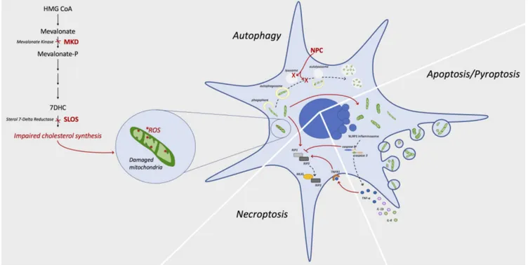
Figure 1 Link between cholesterol deregulation and programmed cell death mechanisms.
Chronic impaired cholesterol synthesis due to mutations in enzymes of the metabolic pathway, as occur in MKD or SLOS, causes a dysfunction of mitochondria. Organelles suffer from alterations of metabolism, mainly due to accumulation of ROS, that induce modifications of the morphology with shrunken shape and condensed cristae. Damaged mitochondria are usually removed by autophagy mechanisms, but accumulation of dam-aged organelles or mutations in such mechanism (as occur in NPC) cause a failed clearance with further accumulation of damdam-aged mitochondria. As effect of this accumulation, the cell is driven toward programmed cell death. Caspase 8 is recognized as a modulator of the different fates of the cell: its activation leads to apoptosis or pyroptosis via NLRP3 inflammosome formation; when inactive or partially activated, it allows the forma-tion of necroptosome consisting in RIP1, RIP3 and MLKL complex. 7DHC: 7-Dehydrocholesterol; HMG CoA: β-hydroxy β-methylglutaryl-co-enzyme A (HMG-CoA); MKD: mevalonate kinase deficiency; MLKL: mixed lineage kinase domain like pseudokinase; NLRP3: NACHT, LRR and PYD domains-containing protein 3; NPC: Niemann-Pick disease type C; RIP: receptor-interacting serine/threonine-protein kinase; ROS: reactive oxygen species; SLOS: Smith-Lemli-Opitz syndrome; TNF-α: tumor necrosis factor α; TNFR1: tumor necrosis factor receptor 1; IL: interleukin.
mechanisms have been identified (Davidovich et al., 2014). Among these, necroptosis has been recently associated to neurodegenerative disorders as a key mechanism worthy of being considered as a potential therapeutic target (Funakoshi et al., 2016; Zhang et al., 2017).
As the term itself suggests, necroptosis shares some fea-tures of the necrosis associated to a highly regulated process seen in apoptosis. When apoptosis failed to be carried for-ward, necroptosis will be engaged. Tumor necrosis factor-α is the main signal for necroptosis and it drives the pathways, shared with apoptosis and nuclear factor-kappa B signalling, which include the trimerization of the tumor necrosis factor receptor and the formation of the intracellular complex-I that involves TRADD (tumor necrosis factor receptor as-sociated death domain protein) and the kinase receptor-in-teracting protein (RIP)1. Complex-I recruits other factors, such as caspase-8, which initiates the apoptotic cascade. When caspase-8 resulted incompletely activated or blocked, the kinase RIP3 is recruited to form the necrosome, leading to the necroptotic cell death (Newton et al., 2014). The re-cruitment of RIP3 causes the engagement and subsequent phosphorylation of the pseudokinase MLKL (mixed lineage kinase domain-like). Although the molecular mechanisms of RIP1, RIP3 and MLKL is not completely depicted, it is clear
that the pathway of necrosome leads to some characteristic features of this programmed cell death: cell and organelles swelling with membrane rupture that results in the release of intracellular content and DAMPs (damage associated mo-lecular patterns) (Moriwaki and Chan, 2016). This implies the activation of the immune system, as occur in necrosis.
Inherited Diseases Related to Cholesterol
Metabolism
Genetic disorders with dysregulation of cholesterol metabo-lism provide valuable models to study therapeutic approaches aimed at preventing neuronal dysfunction. Impaired choles-terol metabolism can be caused either by enzymatic defects of the mevalonate pathway, as in the mevalonate kinase defi-ciency and Smith-Lemli-Opitz syndrome, or by defects in the lysosome trafficking and function, as occurs in Niemann-Pick disease (Jira, 2013). These diseases show a very heterogeneous phenotypic spectrum but share various features of neuronal dysfunction (Figure 1).
Mevalonate kinase deficiency
Mevalonate kinase deficiency is a rare metabolic and au-toinflammatory disorder caused by mutation of the MVK (mevalonate kinase) gene (Muller and Freed, 2017).
Caus-ative mutations result in reduced enzymatic activity of me-valonate kinase, with accumulation of meme-valonate, which can be found in plasma and urine during acute phases, and shortage of downstream compounds, including isoprenoid intermediates and sterols. The onset of the disease usually occurs in the first year of life and presents a continuum spectrum with different levels of severity, ranging from the milder form called hyperimmunoglobulinemia D (OMIM #260920) to the most severe form known as mevalonic ac-iduria (OMIM #610377) (van der Burgh et al., 2013). Com-mon symptoms includ periodic attacks of fever associated with systemic inflammatory symptoms. Patients with hyper-immunoglobulinemia D present headaches, splenomegaly, adenopathy, pharyngitis, abdominal and musculoskeletal pain, while patients with mevalonic aciduria also present a significant psycho-motor and neurological involvement.
The most reliable hypothesis regarding the pathogenesis of mevalonate kinase deficiency is that its typical inflammatory phenotype is caused by the lack of pre-squalene isoprenoid intermediates, with reduced prenylation of the small GT-Pases that would consequently lose their membrane localiza-tion (van der Burgh et al., 2014). The final events lead to the activation of NLRP3 (NACHT, LRR and PYD domains-con-taining protein 3)-inflammasome that triggers the process of pyroptosis with the secretion of the inflammatory cytokines (interleukin-1β, interleukin-6, tumor necrosis factor-α). Furthermore, the incorrect post-translational prenylation of the small GTPase (for example Ras, Rho and Rac), does not allow the formation of autophagosome, and therefore the mitophagy is damaged, with potential consequences on the neurological damage observed in the most severe forms of the disease (van der Burgh et al., 2013). Shortage of choles-terol in immune cells may also play a role in some features of the disease, such as IgA and IgD hypergammaglobulin-emia, due to reduced conversion to 25 hydroxycholesterol, a molecule affecting membrane function and antiviral defense (Simon, 2014).
Smith-Lemli-Opitz syndrome
The Smith-Lemli-Opitz syndrome (OMIM #270400) de-scribed by Smith et al. (1964) is a congenital syndrome characterized by multiple anomaly and intellectual disabil-ity caused by an inborn error of cholesterol metabolism. It is caused by genetic deficiency of 7-dehydrocholesterol (7DHC) reductase, encoded by DHCR7, that leads to toxic effects that can depend both on reduced synthesis of choles-terol and total scholes-terols and on the accumulation of 7DHC-de-rived compounds (Nowaczyk and Irons, 2012; Ramachandra Rao et al., 2018).
The deficiency of cholesterol synthesis can account for a wide spectrum of clinical manifestations involving the ner-vous system, which include prenatal and postnatal growth retardation, microcephaly, intellectual disability (Kelley and Hennekam, 2000).
The potential Smith-Lemli-Opitz syndrome therapy aims to prevent the formation or neutralization of the most toxic 7DHC-derived oxysterols (Korade et al., 2010).
Experimental data show that Smith-Lemli-Opitz syn-drome cells display an elevated autophagy activity, likely in response to the toxic effect of 7DHC accumulation resulting in excessive mitochondrial oxidative stress and activation of mitophagy (Saffari et al., 2017). However, in vitro studies have demonstrated that the accumulation of dysfunctional mitochondria is concomitant with a defective autophagic system which would interfere with the role of mitophagy to clear the defective proteins and organelles (Chang et al., 2014). Thereby, the protective function of the autophagy is altered by the co-existence of dysfunctional mitochondria and impairment in the autophagy process (Chang et al., 2014).
Niemann-Pick disease
Niemann-Pick disease is a very severe rare genetic disorder, which belongs to the family of lysosomal storage diseases, a condition that affects many body systems. Patients with Niemann-Pick disease cannot metabolize cholesterol and other lipids properly, leading to abnormal accumulation of these substances in liver, spleen and other organs (Guo et al., 2016).
Niemann-Pick disease presents a broad clinical spectrum, depending on the degree of defect in lipid trafficking. The onset can be at birth with a fatal disorder, or in children or even adults, with milder phenotypes characterized by pro-gressive psychomotor impairment, in addition to liver and spleen enlargement. The defect of cholesterol trafficking to mitochondria is associated to mitochondrial dysfunction and impairment in antioxidant defense strategies. More-over, besides the neurodegenerative aspect of the disease, Niemann-Pick disease phenotype implies systemic features since non-esterified cholesterol accumulate also in liver and spleen (Vanier, 1999, 2010; Patterson et al., 2012). Different genetic forms of Niemann-Pick disease are known and, in particular, Niemann-Pick disease type C (NPC) is caused by mutations in NPC1 (OMIM #257220, 95% of cases) (Carstea et al., 1993, 1997) and NPC2 genes (OMIM #607625, 5% of cases) (Naureckiene et al., 2000) resulting in functional defects of proteins with lysosomal localization (Torres et al., 2017; Liu and Lieberman, 2018) that trigger an accumulation of non-degraded substrates that interferes with different cel-lular functions (Sarkar et al., 2013). These molecular mech-anisms are not fully elucidated yet, and a deepen knowledge of these processes is of crucial importance because each step of the pathogenetic cascade in Niemann-Pick disease may be a potential target of therapy (Schultz et al., 2018; Wang et al., 2018). Moreover, given the role of autophagy in the clearance of damaged cellular components, the impairment of autophagy itself can contribute in a vicious circle to lipid accumulation and cell injury (Platt et al., 2012; Osellame
and Duchen, 2014). The neuronal manifestations of NPC are related to a selective damage of neurons that have a stronger spontaneous activation of autophagy, if compared to systemic compartments (i.e., fibroblasts), and a block of autophagic progression leads to an exceptionally severe mitochondrial fragmentation. For this reason patients with NPC1 may benefits from the treatment with autophagy in-hibitors (such as 3-methyladenine) or with drugs that mo-bilize cholesterol from the lysosomal compartment (such as cyclodextrin) (Davidson et al., 2016).
Conclusions
Mevalonate kinase deficiency, Smith-Lemli-Opitz syn-drome and Niemann-Pick disease are monogenic disorders, extremely various as regard pathogenesis and molecular mechanisms of onset, but they share some features that can be useful to unravel possible therapeutic targets. First, the onset of these disorders is related with a dysfunctional metabolism of cholesterol; second, recent insights suggest that the dysfunctional cholesterol metabolism, at the basis of disease onset, is related to altered autophagy and other programmed cell death processes; third, all the diseases show an important involvement of the central nervous sys-tem related to altered mechanisms of clearance because of impaired autophagy. Thus, autophagy could be a keystone in the treatment of this rare disorders. Indeed, nowadays, all these pathologies can benefit from only a small repertoire of therapeutics, and none of them are able to completely con-trol the neuronal aspect of the disease. Therefore, it is essen-tial to know the mechanisms that regulate neuronal loss, to evaluate the most suitable pharmacological treatments able to protect from neurodegeneration or prevent the effect of the extended activation of the inflammatory response. Acknowledgments: We thank to the support from the Istituto di Ricov-ero e Cura a Carattere Scientifico.
Author contributions: Manuscript design and writing: EP and AM; manuscript revision and editing: LVB; manuscript revision and con-clusion writing: AT. All authors approved the final version of this man-uscript.
Conflicts of interest: All the authors declare no conflicts of interest. Financial support: This study was supported by a grant from the Institute for Maternal and Child Health–IRCCS “Burlo Garofolo”– Trieste, Italy (RC 24/2017).
Copyright license agreement: The Copyright License Agreement has been signed by all authors before publication.
Plagiarism check: Checked twice by iThenticate. Peer review: Externally peer reviewed.
Open access statement: This is an open access journal, and articles are distributed under the terms of the Creative Commons Attribu-tion-NonCommercial-ShareAlike 4.0 License, which allows others to remix, tweak, and build upon the work non-commercially, as long as appropriate credit is given and the new creations are licensed under the identical terms.
References
Arenas F, Garcia-Ruiz C, Fernandez-Checa JC (2017) Intracellular cholesterol trafficking and impact in neurodegeneration. Front Mol Neurosci 10:382.
Axe EL, Walker SA, Manifava M, Chandra P, Roderick HL, Haber-mann A, Griffiths G, Ktistakis NT (2008) Autophagosome formation from membrane compartments enriched in phosphatidylinositol 3-phosphate and dynamically connected to the endoplasmic reticu-lum. J Cell Biol 182:685-701.
Beaudouin J, Liesche C, Aschenbrenner S, Horner M, Eils R (2013) Caspase-8 cleaves its substrates from the plasma membrane upon CD95-induced apoptosis. Cell Death Differ 20:599-610.
Carstea ED, Polymeropoulos MH, Parker CC, Detera-Wadleigh SD, O’Neill RR, Patterson MC, Goldin E, Xiao H, Straub RE, Vanier MT, et al. (1993) Linkage of Niemann-Pick disease type C to human chromosome 18. Proc Natl Acad Sci U S A 90:2002-2004.
Carstea ED, Morris JA, Coleman KG, Loftus SK, Zhang D, Cummings C, Gu J, Rosenfeld MA, Pavan WJ, Krizman DB, Nagle J, Polymero-poulos MH, Sturley SL, Ioannou YA, Higgins ME, Comly M, Cooney A, Brown A, Kaneski CR, Blanchette-Mackie EJ, et al. (1997) Niemann-Pick C1 disease gene: homology to mediators of cholester-ol homeostasis. Science 277:228-231.
Cerri S, Blandini F (2018) Role of autophagy in Parkinson’s disease. Curr Med Chem doi: 10.2174/0929867325666180226094351. Chan SN, Tang BL (2013) Location and membrane sources for
auto-phagosome formation - from ER-mitochondria contact sites to Gol-gi-endosome-derived carriers. Mol Membr Biol 30:394-402. Chang S, Ren G, Steiner RD, Merkens L, Roullet JB, Korade Z,
DiMuz-io PJ, Tulenko TN (2014) Elevated autophagy and mitochondrial dysfunction in the Smith-Lemli-Opitz syndrome. Mol Genet Metab Rep 1:431-442.
Croft DR, Coleman ML, Li S, Robertson D, Sullivan T, Stewart CL, Olson MF (2005) Actin-myosin-based contraction is responsible for apoptotic nuclear disintegration. J Cell Biol 168:245-255.
Davidovich P, Kearney CJ, Martin SJ (2014) Inflammatory outcomes of apoptosis, necrosis and necroptosis. Biol Chem 395:1163-1171. Davidson CD, Fishman YI, Puskás I, Szemán J, Sohajda T, McCauliff
LA, Sikora J, Storch J, Vanier MT, Szente L, Walkley SU, Dobrenis K (2016) Efficacy and ototoxicity of different cyclodextrins in Nie-mann-Pick C disease. Ann Clin Transl Neurol 3:366-380.
Funakoshi T, Aki T, Tajiri M, Unuma K, Uemura K (2016) Necropto-sis-like Neuronal Cell Death Caused by Cellular Cholesterol Accu-mulation. J Biol Chem 291:25050-25065.
Giampieri F, Afrin S, Forbes-Hernandez TY, Gasparrini M, Cianciosi D, Reboredo-Rodriguez P, Varela-Lopez A, Quiles JL, Battino M (2018) Autophagy in human health and disease: novel therapeutic opportu-nities. Antioxid Redox Signal doi: 10.1089/ars.2017.7234.
Guo H, Zhao M, Qiu X, Deis JA, Huang H, Tang QQ, Chen X (2016) Niemann-Pick type C2 deficiency impairs autophagy-lysosomal activity, mitochondrial function, and TLR signaling in adipocytes. J Lipid Res 57:1644-1658.
Hayashi-Nishino M, Fujita N, Noda T, Yamaguchi A, Yoshimori T, Ya-mamoto A (2009) A subdomain of the endoplasmic reticulum forms a cradle for autophagosome formation. Nat Cell Biol 11:1433-1437. Iwasaki T, Katayama T, Kohama K, Endo Y, Sawasaki T (2013) Myosin
phosphatase is inactivated by caspase-3 cleavage and phosphoryla-tion of myosin phosphatase targeting subunit 1 during apoptosis. Mol Biol Cell 24:748-756.
Jira P (2013) Cholesterol metabolism deficiency. Handb Clin Neurol 113:1845-1850.
Kelley RI, Hennekam RC (2000) The Smith-Lemli-Opitz syndrome. J Med Genet 37:321-335.
Kerr JF, Wyllie AH, Currie AR (1972) Apoptosis: a basic biological phenomenon with wide-ranging implications in tissue kinetics. Br J Cancer 26:239-257.
Kim J, Kundu M, Viollet B, Guan KL (2011) AMPK and mTOR reg-ulate autophagy through direct phosphorylation of Ulk1. Nat Cell Biol 13:132-141.
Klionsky DJ (2008) Autophagy revisited: a conversation with Christian de Duve. Autophagy 4:740-743.
Korade Z, Xu L, Shelton R, Porter NA (2010) Biological activities of 7-dehydrocholesterol-derived oxysterols: implications for Smith-Lemli-Opitz syndrome. J Lipid Res 51:3259-3269.
Lazarus MB, Novotny CJ, Shokat KM (2015) Structure of the human autophagy initiating kinase ULK1 in complex with potent inhibitors. ACS Chem Biol 10:257-261.
Lim Y, Cho H, Kim EK (2016) Brain metabolism as a modulator of au-tophagy in neurodegeneration. Brain Res 1649:158-165.
Liu EA, Lieberman AP (2018) The intersection of lysosomal and en-doplasmic reticulum calcium with autophagy defects in lysosomal diseases. Neurosci Lett doi: 10.1016/j.neulet.2018.04.049.
Maiuri MC, Zalckvar E, Kimchi A, Kroemer G (2007) Self-eating and self-killing: crosstalk between autophagy and apoptosis. Nat Rev Mol Cell Biol 8:741-752.
Marcuzzi A, Piscianz E, Loganes C, Vecchi Brumatti L, Knowles A, Bilel S, Tommasini A, Bortul R, Zweyer M (2015) Innovative tar-get therapies are able to block the inflammation associated with dysfunction of the cholesterol biosynthesis pathway. Int J Mol Sci 17:E47.
Marcuzzi A, Loganes C, Valencic E, Piscianz E, Monasta L, Bilel S, Bortul R, Celeghini C, Zweyer M, Tommasini A (2018) Neuronal dysfunction associated with cholesterol deregulation. Int J Mol Sci 19:E1523.
Meier P, Finch A, Evan G (2000) Apoptosis in development. Nature 407:796-801.
Miettinen TP, Bjorklund M (2016) The mevalonate pathway as a met-abolic requirement for autophagy-implications for growth control, proteostasis, and disease. Mol Cell Oncol 3:e1143546.
Mills JC, Stone NL, Erhardt J, Pittman RN (1998) Apoptotic membrane blebbing is regulated by myosin light chain phosphorylation. J Cell Biol 140:627-636.
Moloudizargari M, Asghari MH, Ghobadi E, Fallah M, Rasouli S, Ab-dollahi M (2017) Autophagy, its mechanisms and regulation: Impli-cations in neurodegenerative diseases. Ageing Res Rev 40:64-74. Moriwaki K, Chan FK (2016) Necroptosis-independent signaling by
the RIP kinases in inflammation. Cell Mol Life Sci 73:2325-2334. Moutinho M, Nunes MJ, Rodrigues E (2017) The mevalonate pathway
in neurons: It’s not just about cholesterol. Exp Cell Res 360:55-60. Muller AL, Freed DH (2017) Basic and clinical observations of
meva-lonate depletion on the mevameva-lonate signaling pathway. Curr Mol Pharmacol 10:6-12.
Naureckiene S, Sleat DE, Lackland H, Fensom A, Vanier MT, Wattiaux R, Jadot M, Lobel P (2000) Identification of HE1 as the second gene of Niemann-Pick C disease. Science 290:2298-2301.
Newton K, Dugger DL, Wickliffe KE, Kapoor N, de Almagro MC, Vucic D, Komuves L, Ferrando RE, French DM, Webster J, Roose-Girma M, Warming S, Dixit VM (2014) Activity of protein kinase RIPK3 determines whether cells die by necroptosis or apop-tosis. Science 343:1357-1360.
Nowaczyk MJ, Irons MB (2012) Smith-Lemli-Opitz syndrome: pheno-type, natural history, and epidemiology. Am J Med Genet C Semin Med Genet 160C:250-262.
Obergasteiger J, Frapporti G, Pramstaller PP, Hicks AA, Volta M (2018) A new hypothesis for Parkinson’s disease pathogenesis: GTPase-p38 MAPK signaling and autophagy as convergence points of etiology and genomics. Mol Neurodegener 13:40.
Osellame LD, Duchen MR (2014) Quality control gone wrong: mito-chondria, lysosomal storage disorders and neurodegeneration. Br J Pharmacol 171:1958-1972.
Park JM, Jung CH, Seo M, Otto NM, Grunwald D, Kim KH, Moriar-ity B, Kim YM, Starker C, Nho RS, Voytas D, Kim DH (2016) The ULK1 complex mediates MTORC1 signaling to the autophagy initi-ation machinery via binding and phosphorylating ATG14. Autopha-gy 12:547-564.
Patterson MC, Hendriksz CJ, Walterfang M, Sedel F, Vanier MT, Wi-jburg F; NP-C Guidelines Working Group (2012) Recommendations for the diagnosis and management of Niemann-Pick disease type C: an update. Mol Genet Metab 106:330-344.
Pfrieger FW, Ungerer N (2011) Cholesterol metabolism in neurons and astrocytes. Prog Lipid Res 50:357-371.
Platt FM, Boland B, van der Spoel AC (2012) The cell biology of dis-ease: lysosomal storage disorders: the cellular impact of lysosomal dysfunction. J Cell Biol 199:723-734.
Ramachandra Rao S, Pfeffer BA, Más Gómez N, Skelton LA, Keiko U, Sparrow JR, Rowsam AM, Mitchell CH, Fliesler SJ (2018) Com-promised phagosome maturation underlies RPE pathology in cell culture and whole animal models of Smith-Lemli-Opitz Syndrome. Autophagy 14:1796-1817.
Riedl SJ, Shi Y (2004) Molecular mechanisms of caspase regulation during apoptosis. Nat Rev Mol Cell Biol 5:897-907.
Rusmini P, Cortese K, Crippa V, Cristofani R, Cicardi ME, Fer-rari V, Vezzoli G, Tedesco B, Meroni M, Messi E, Piccolella M, Galbiati M, Garrè M, Morelli E, Vaccari T, Poletti A (2018) Trehalose induces autophagy via lysosomal-mediated TFEB ac-tivation in models of motoneuron degeneration. Autophagy doi: 10.1080/15548627.2018.1535292.
Saffari A, Kolker S, Hoffmann GF, Ebrahimi-Fakhari D (2017) Linking mitochondrial dysfunction to neurodegeneration in lysosomal stor-age diseases. J Inherit Metab Dis 40:631-640.
Sarkar S, Carroll B, Buganim Y, Maetzel D, Ng AH, Cassady JP, Co-hen MA, Chakraborty S, Wang H, Spooner E, Ploegh H, Gsponer J, Korolchuk VI, Jaenisch R (2013) Impaired autophagy in the lip-id-storage disorder Niemann-Pick type C1 disease. Cell Rep 5:1302-1315.
Schultz ML, Krus KL, Kaushik S, Dang D, Chopra R, Qi L, Shakkot-tai VG, Cuervo AM, Lieberman AP (2018) Coordinate regula-tion of mutant NPC1 degradaregula-tion by selective ER autophagy and MARCH6-dependent ERAD. Nat Commun 9:3671.
Shintani T, Klionsky DJ (2004) Cargo proteins facilitate the formation of transport vesicles in the cytoplasm to vacuole targeting pathway. J Biol Chem 279:29889-29894.
Simon A (2014) Cholesterol metabolism and immunity. N Engl J Med 371:1933-1935.
Singh AK, Kashyap MP, Tripathi VK, Singh S, Garg G, Rizvi SI (2017) Neuroprotection through rapamycin-induced activation of autopha-gy and PI3K/Akt1/mTOR/CREB signaling against amyloid-beta-in-duced oxidative stress, synaptic/neurotransmission dysfunction, and neurodegeneration in adult rats. Mol Neurobiol 54:5815-5828. Smith DW, Lemli L, Opitz JM (1964) A newly recognized syndrome of
multiple congenital anomalies. J Pediatr 64:210-217.
Suárez-Rivero JM, de la Mata M, Pavón AD, Villanueva-Paz M, Pov-ea-Cabello S, Cotán D, Álvarez-Córdoba M, Villalón-García I, Ybot-González P, Salas JJ, Muñiz O, Cordero MD, Sánchez-Alcázar JA (2018) Intracellular cholesterol accumulation and coenzyme Q10 deficiency in Familial Hypercholesterolemia. Biochim Biophys Acta Mol Basis Dis 1864:3697-3713.
Tanida I (2011) Autophagosome formation and molecular mechanism of autophagy. Antioxid Redox Signal 14:2201-2214.
Torres S, Balboa E, Zanlungo S, Enrich C, Garcia-Ruiz C, Fernan-dez-Checa JC (2017) Lysosomal and mitochondrial liaisons in Nie-mann-Pick disease. Front Physiol 8:982.
van der Burgh R, Ter Haar NM, Boes ML, Frenkel J (2013) Mevalonate kinase deficiency, a metabolic autoinflammatory disease. Clin Im-munol 147:197-206.
van der Burgh R, Pervolaraki K, Turkenburg M, Waterham HR, Fren-kel J, Boes M (2014) Unprenylated RhoA contributes to IL-1beta hypersecretion in mevalonate kinase deficiency model through stim-ulation of Rac1 activity. J Biol Chem 289:27757-27765.
Vanier MT (1999) Lipid changes in Niemann-Pick disease type C brain: personal experience and review of the literature. Neurochem Res 24:481-489.
Vanier MT (2010) Niemann-Pick disease type C. Orphanet J Rare Dis 5:16.
Wang YH, Twu YC, Wang CK, Lin FZ, Lee CY, Liao YJ (2018) Nie-mann-Pick type C2 protein regulates free cholesterol accumulation and influences hepatic stellate cell proliferation and mitochondrial respiration function. Int J Mol Sci 19:E1678.
Ward C, Martinez-Lopez N, Otten EG, Carroll B, Maetzel D, Singh R, Sarkar S, Korolchuk VI (2016) Autophagy, lipophagy and lysosomal lipid storage disorders. Biochim Biophys Acta 1861:269-284. Wu Y, Cheng S, Zhao H, Zou W, Yoshina S, Mitani S, Zhang H, Wang
X (2014) PI3P phosphatase activity is required for autophagosome maturation and autolysosome formation. EMBO Rep 15:973-981. Zhang J, Liu Q (2015) Cholesterol metabolism and homeostasis in the
brain. Protein Cell 6:254-264.
Zhang S, Tang MB, Luo HY, Shi CH, Xu YM (2017) Necroptosis in neurodegenerative diseases: a potential therapeutic target. Cell Death Dis 8:e2905.