DEPARTMENT OF BIOMOLECULAR SCIENCES
PhD Course in Life Sciences, Health and Biotechnology
Biochemical and Pharmacological Sciences and Biotechnology
XXXIII cycle
Fine-tuning of intracellular redox state by pro-glutathione molecules
modulates the cellular immune/inflammatory response to infections:
in vivo and in vitro studies
SSD: BIO/13Supervisor: PhD student:
Dr. Alessandra Fraternale Dr. Carolina Zara
1 GENERAL INTRODUCTION ... 4 Redox state ... 5 Oxidative stress ... 5 ROS/RNS production ...5 Oxidative damage ...6
Protective antioxidant systems ... 7
Intracellular redox-sensitive pathways ... 8
Glutathione ... 12
GSH synthesis ...13
GSH degradation ...14
GSH functions ...14
GSH in infections and inflammation ...14
Pro-GSH molecules ...16
I-152 ... 17
Structure of the thesis ... 19
Aim of the study ... 19
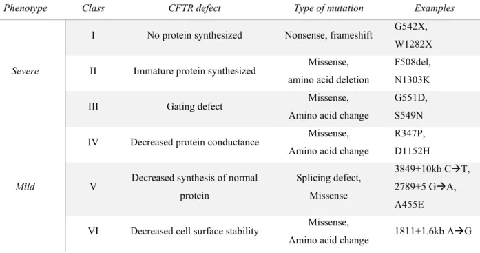
CHAPTER 1... 20 CHAPTER 2... 32 CHAPTER 3... 62 CHAPTER 4... 83 4.1 Introduction ... 84 CFTR mutations...84 Inflammation...85 Redox state in CF ...87
GSH supplementation and GSH precursors in CF ...87
4.2 Aim of the study ... 89
4.3 Materials and methods ... 90
4.3.1 Reagents...90
4.3.2 CF bone marrow-derived macrophages: isolation, culture, and treatments ...90
2
4.3.4 Preparation of samples for the determination of GSH, cysteine, and other thiol species ...91
4.3.5 Spectrophotometric determination of proteins ...91
4.3.6 Determination of GSH and other thiol species by high-performance liquid chromatography (HPLC) ...92
4.3.7 Preparation of standard solutions for HPLC ...92
4.3.8 Cytokine quantification by ELISA ...93
4.3.9 CF: Extraction and quantification of total RNA ...93
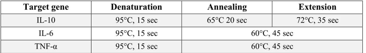
4.3.10 CF: Synthesis of cDNA and Real-Time PCR for the analysis of genes that code for inflammatory cytokines ...94
4.3.11 RAW 264.7: Extraction and quantification of total RNA ...94
4.3.12 RAW 264.7: Synthesis of cDNA and Real-Time PCR for the analysis of genes that code for inflammatory cytokines ...94
4.3.13 Western immunoblotting analysis of the NF-kB pathway ...95
4.3.14 Statistical analysis ...96
4.4 Results ... 97
4.4.1 Results obtained in the CF model ...97
4.4.1.1 Western immunoblotting analysis ...97
4.4.1.2 Determination of GSH levels after stimulation with LPS from P. aeruginosa ...98
4.4.1.3 Determination of GSH and cysteine content after LPS and I-152 treatment ...98
4.4.1.4 Modulation of LPS-induced cytokine production by I-152 ...99
4.4.2 Results obtained in RAW 264.7 ...101
4.4.2.1 Determination of GSH levels after stimulation with LPS from E. coli ...101
4.4.2.2 Determination of GSH and other thiol species content after 30 min I-152 treatment ...102
4.4.2.3 Modulation of LPS-induced cytokine production by I-152 ...103
4.5 Discussion ... 105
4.6 Conclusions ... 106
CHAPTER 5... 107
5.1 Introduction ... 108
5.2 Materials and methods ... 108
3
5.2.2 RAW 264.7 cells: culture and treatments ...108
5.2.3 Preparation of samples for the determination of GSH, cysteine, and other thiol species ...108
5.2.4 Spectrophotometric determination of proteins ...108
5.2.5 Determination of GSH and other thiol species by high-performance liquid chromatography (HPLC) ...108
5.2.6 Preparation of standard solutions for HPLC ...109
5.2.7 Western immunoblotting analysis ...109
5.3.8 Statistical analysis ...109
5.3 Results ... 110
5.3.1 Determination of GSH and other thiol species in I-152-treated cells ...110
5.3.2 Western immunoblotting analysis of Nfr2 and GCLm ...111
5.3.3 Western immunoblotting analysis of ChaC1 ...112
5.4 Discussion ... 114
5.5 Conclusions ... 114
FINAL CONCLUSIONS ... 115
REFERENCES ... 117
4
5 Redox state
The term “redox state” has been utilized historically to indicate the ratio of the oxidized and reduced form of a specific redox couple [1]. However, cells are complex structures and, for this reason, Shafer and Buettner suggested the use of the term “redox environment”. According to their definition, “the redox environment of a linked set of redox couples as found in a biological fluid, organelle, cell, or tissue is the summation of the products of the reduction potential and reducing the capacity of the linked redox couples present” [2]. However, in this work I will refer to “redox state” because it is the most commonly used term.
The functioning of many cellular components may be dependent on the cellular redox state; therefore, it is crucial to maintain correct redox homeostasis [3][4]. This is also important for the proper functioning of the immune system. When an infection occurs (caused by bacteria, or virus), cells respond by activating all of the mechanisms aimed to kill the pathogen, including the production of several reactive oxygen/nitrogen species (ROS/RNS) with the goal to eliminate the non-self. ROS and RNS can affect/modulate the redox-sensitive mechanisms implicated in the immune/inflammatory response, so it is crucial that the redox state is under cellular control; otherwise, a malfunction could lead to many oxidative stress-related diseases [5].
Oxidative stress
The definition of oxidative stress was first formulated by Sies in 1985 [6] as “a disturbance in the prooxidant-antioxidant balance in favor of the former”. This disturbance can be caused by the presence of the so-called reactive oxygen species (ROS), which are derived from oxygen, an obligate component of eukaryotic organisms. These species can be present in free-radical form, like the superoxide anion (O2.-) and the hydroxyl
radical (.OH), or as chemically stable molecules, like hydrogen peroxide (H
2O2). In addition to ROS, other
molecules that have an impact on oxidative stress are reactive nitrogen species (RNS). The RNS system includes different compounds like peroxynitrite (ONOO-) and the free-radical nitric oxide (NO.) [6]. Giles et
al. proposed an additional group of redox-active molecules termed reactive sulfur species (RSS), which “are redox-active sulfur compounds formed under conditions of oxidative stress that may be capable of initiating oxidation reactions” [7]. However, these species are synthesized also under non-oxidative conditions. RSS comprehend different forms of cysteine and methionine, and some low-molecular-mass compounds such as glutathione, thioredoxin, or mycothiol [8]. Although all these molecules have been historically considered harmful agents for cells at high concentrations, recent evidence showed that at moderate/low concentrations they can function as secondary signaling molecules [9], bringing to the updated concept of oxidative stress as “an imbalance between oxidants and antioxidants in favor of the oxidants, leading to a disruption of redox signaling and control and/or molecular damage” [6].
ROS/RNS production
The generation of reactive oxygen and nitrogen species (ROS, and RNS) in the cells is due to both endogenous and exogenous sources. The main ROS source is mitochondria, where adenosine triphosphate (ATP) is generated. Its generation depends on the electron transport chain (ETC), which needs O2; part of this
6 molecular oxygen is incompletely metabolized and converted into O2- by complex I and III of the ETC [10].
The enzyme NADPH oxidase (NOX), which catalyzes the transport of one electron to oxygen from cytosolic NADPH, is another important ROS source. Of the seven isoforms of NOX, NOX4 is the only known form to produce H2O2 instead of O2- [11]. Another important cellular organelle that generates ROS is the endoplasmic
reticulum (ER). One of the ER’s main roles is to promote correct protein folding by forming disulfide bonds, which is ensured by the maintaining of an oxidized environment [12]. Other ROS sources can be microsomes and peroxisomes, which primarily generate H2O2, cytochrome c oxidase, and xanthine oxidase [13]. The
production of these reactive species is one of the major mechanisms to fight and kill pathogens by cells of the immune system, such as neutrophils and macrophages. The RNS NO. is synthesized from L-arginine in the
presence of molecular oxygen through a reaction catalyzed by the nitric oxide synthase (NOS) [14]. There are several known isoforms of this enzyme, such as the neuronal NOS (nNOS), endothelial NOS (eNOS), and the inducible NOS (iNOS) that can be induced by pro-inflammatory cytokines [15]. Nitric oxide can react with O2- to generate ONOO- [16]. Exogenous sources of ROS and RNS are irradiation (e.g. UV irradiation, γ
irradiation), drugs, and atmospheric pollutants [17].
Oxidative damage
ROS and RNS have been historically considered harmful agents for cells because they can react with important cellular structures, like lipids, proteins, and DNA. These modifications could cause damages to these structures, leading to their dysfunction and oxidative stress-related diseases, such as inflammation, neurodegenerative diseases, and cancers [5].
High concentrations of ROS/RNS can lead to lipid oxidation, which causes lipid peroxidation and oxysterol formation. The former is a radical chain reaction, that consists of three major steps: initiation, propagation, and termination [18]. Briefly, a polyunsaturated fatty acid (PUFA) can be attacked by a free radical molecule (initiation) producing an unstable fatty acid radical (L.) that in turn reacts with a molecule of oxygen. The
product of this reaction is a peroxyl-fatty acid radical (LOO.), which can continue the cycle by reacting with
another PUFA (propagation) or stop it by reacting with another radical molecule or with an antioxidant able to break the cycle (e.g., Vitamin E) (termination). This chain reaction mechanism can lead to a loss of membrane components and to the formation of reactive end products, like malondialdehyde (MDA), 4-hydroxy-2-nonenal (HNE), acrolein, and isoprostanes, which can cause further damage to protein and DNA [5]. The reactive end products have been investigated, especially HNE and MDA that were found to easily react with proteins ad DNA and to be highly toxic molecules [19]. Furthermore, MDA is a stable compound used as a biomarker for oxidative stress [20]. In addition to lipid peroxidation, there may be the oxidation of cholesterol caused by specific enzymatic reactions to generate oxysterols. This reaction can be caused either by cytochrome P451 or by non-specific reactions that involve ROS and RNS. Oxysterols are physiologically produced as intermediates in the cholesterol catabolism, but when in excess they can be involved in many diseases (e.g., neurodegenerative diseases and cancers) [21]. It is interesting to note that oxidative stress appears to both induce the formation of oxysterols and to be induced by their presence [22]. All these reactions will eventually
7 lead to a loss of membrane properties where lipids and cholesterol are mainly localized, and their reactive end products can consequently damage other molecules.
Besides lipids, proteins can also be modified by oxidative stress. Protein damages may cause dysfunction in the cellular activity which in turn may lead to the development of diseases, like chronic inflammatory diseases (cancer, atherosclerosis, ischemia), aging, and age-related neurodegenerative disorders (Alzheimer’s disease) [5]. Proteins can be subject to modification on their amino-acid side chains. ROS mainly cause sulfur oxidation of cysteine and methionine, protein hydroxides, and carbonyl derivatives, while RNS induce protein nitration. It is important to note that these protein modifications are mostly irreversible and that damaged proteins, such as carbonylated proteins, are marked for their degradation by proteasomes, and once escaped from degradation they can form high-molecular-weight aggregates that accumulate with age [23]. Carbonylated proteins can be valid biomarkers of oxidative stress because carbonylation is more difficult to be induced compared to the SH oxidation of cysteine and methionine, and they can also be a sign of disease-derived protein dysfunction [24]. Protein oxidative modifications are not always harmful to cells, in fact, this is an important step of protein folding inside the endoplasmic reticulum (ER), in which intra and intermolecular disulfide bonds are introduced [8].
DNA oxidation causes instability and decay of the genome. The most susceptible base to oxidative damage is guanine, which undergoes the major mutagenic lesion is 8-oxo-7,8-dihydroguanine. This mutation is characterized by the pairing of guanine with adenine rather than with cytosine, thus generating transversion mutation after replication [25]. RNA can also be subject to oxidation; for example, the oxidation of microRNA-184 results in the blocking of the translation of anti-apoptotic factors, such as Bcl-xL [26].
Protective antioxidant systems
Under physiological conditions, cells maintain redox homeostasis by the production and elimination of ROS and RNS. To prevent oxidative damages and maintain redox homeostasis, the cells have evolved enzymatic or non-enzymatic antioxidant systems. Enzymatic antioxidants include superoxide dismutase (SOD), glutathione reductase (GR), catalase, and glutathione peroxidase (GPX); while the non-enzymatic antioxidants are thiol molecules, like reduced glutathione (GSH), thioredoxin (Trx), and glutaredoxin (Grx) which take part to thiol-disulfide exchange reactions [3].
SOD catalyzes the dismutation of O2.- to oxygen and H2O2 [4]. This enzyme is present in different cellular
compartments, and in different isoforms. SOD1 (CuZnSOD) is present in the cytoplasm, SOD2 (MnSOD) is found in mitochondria, and SOD4 in the extracellular matrix [27]. The H2O2 produced is then converted into
H2O + O2 by catalase, which is a heme-based enzyme found in the peroxisome [28]. In addition to this pathway,
hydrogen peroxide can be converted into not harmful molecules via the conversion of GSH to oxidized glutathione (GSSG), catalyzed by GPX [29]. GSH, that will be discussed in paragraph “Glutathione”, is the most important low molecular weight antioxidant synthesized in cells. The Trx system consists of thioredoxin reductase (TrxR), and Trx, in which TrxR activity is to reduce the oxidized form of Trx [4]. To date, two Trxs isoforms have been identified, Trx1 (cytosolic), and Trx2 (mitochondrial), and three TrxRs isoforms, TrxR1
8 (cytosolic), TrxR2 (mitochondrial), and thioredoxin glutathione reductase (TGR) [30]. The Grx system also functions to reduce protein disulfides; their oxidized form is reduced by GSH. To date, four Grxs isoforms have been discovered, Grx1, Grx2, Grx3, and Grx5 [31].
Intracellular redox-sensitive pathways
Although ROS are mainly associated with cell damage caused by irreversible oxidation of proteins, lipids, and nucleic acids, low concentrations of ROS play a crucial role in the response and adaptation to local and overall stress conditions. Redox-regulated proteins are the first to respond to changes in the intracellular redox state and induce initial steps to protect against oxidants, repair cellular damage, and restore the redox homeostasis. Those proteins contain highly specific cysteine residues which can undergo reversible oxidative modifications, causing conformational and functional switch of the protein [32]. Oxidative modifications can consist of the oxidation of cysteine residues on proteins, which translate into the formation of reactive sulfenic acid (-SOH). This acid can react with cysteines and form disulfide bonds (-S-S-), or it can be further oxidized and form sulfinic (-SO2H) or sulfonic (-SO3H) acid [33]. These redox oxidations are mostly reversible by
reducing systems, like thioredoxin and peroxiredoxin, and this is because these modifications have important roles in the redox signaling [34]. Overproduction of ROS may contribute to altering these redox-sensitive pathways, which could, in turn, lead to the onset of several diseases, such as cancer, neurodegeneration, atherosclerosis, and diabetes, and also to aging [35]. There are many cellular redox-sensitive pathways, in which several phosphatases are often involved, and the presence of ROS causes their oxidation and subsequent inactivation, leading to an impairment in the phosphatase activity [3][36]. Moreover, each pathway is not isolated from the other, in fact, there is a crosstalk between them; for this reason, their correct activation and inactivation are fundamental for the prevention of diseases [4]. Here are described some of these pathways.
Nrf2 – Nuclear factor erythroid 2-related factor 2 (Nrf2) is a basic leucine zipper transcription factor, that under basal conditions is bound to the dimeric inhibitory protein Kelch-like ECH-associated protein 1 (Keap1) in the cytosol [37]. Keap1 is an adaptor of the Cul3-based E3-ubiquitin ligase complex that polyubiquitinates Nrf2 for proteasomal degradation [38]. Keap1 possesses cysteine residues that under stress conditions (ROS, electrophiles) are oxidized; the result is the dissociation of Nrf2 from Keap1 and its nuclear translocation. Once in the nucleus, Nrf2 forms a complex with Maf proteins (Maf-F, Maf-g, and Maf-K), which binds to the antioxidant response element (ARE) [39]. The effects of Nrf2 can be dual: on the one hand, it can regulate the production of ROS/RNS by controlling the transcription of certain enzymes, such as superoxide dismutase (SOD), and iNOS; on the other hand, it can lead to the transcription of enzymes involved in the antioxidant response, such as catalase (CAT), and enzymes involved in the GSH synthesis, such as GCL [4]. (Figure 1).
9 Figure 1 Nrf2 activation by ROS. ROS cause the oxidation of Keap1 cysteines, leading to its conformational change. Keap1 dissociates from Nrf2, which is now free to translocate into the nucleus. Here it creates a complex with
the Maf protein and binds to the antioxidant response element (ARE) to induce the transcription of several enzymes, such as glutamate-cysteine ligase (GCL), superoxide dismutase (SOD), and catalase (CAT).
NF-kB – Nuclear factor-kappa B (NF-kB) represents a family that consists of five transcription factors (p65, Rel B, c-Rel, p52, and p50). Under non-stress conditions, it is sequestered in the cytosol by the binding of the inhibitor of kappa B (IkB) [40]. In the presence of stress stimuli (e.g. cytokines, toll-like receptor (TLR) ligands), the IkB kinase (IKK) is activated and phosphorylates IkB, which dissociates from NF-kB, and is subsequently ubiquitinated and degraded. NF-kB is now free to translocate to the nucleus, where it binds to specific DNA promoter regions [41]. NF-kB, as a redox-sensitive factor, is a target activated by ROS and is involved in the inflammatory response (e.g. pro-inflammatory cytokines, such as TNF-α, IL-6, and IL-1β). Interestingly, a severe increase of ROS could lead to a different effect, which is the inactivation of NF-kB and subsequently cell death [3]. The binding of activated NF-kB and DNA requires the reduced form of the transcription factor; in fact, the oxidation of some NF-kB cysteines could lead to the binding inability [42] (Figure 2).
10 Figure 2 NF-kB activation by ROS. ROS activates the IkB kinase (IKK), which in turn phosphorylates IkB-α. This phosphorylation causes the dissociation of IkB-α from NF-kB, which is now free to translocate into the nucleus and to
bind to the DNA. NF-kB induces the transcription of factors involved in the inflammation, such as pro-inflammatory cytokines (e.g. TNF-α and IL-6).
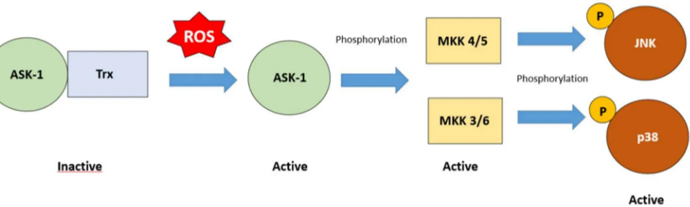
MAPK – The mitogen-activated protein kinase (MAPK) is a family that consists mainly of the extracellular regulated kinases (ERK1/2), the Jun N-terminal kinase (JNK), and the p38 kinase (p38). These factors operate in cascades of sequential phosphorylation that are regulated by different kinases (MAPKKK, and MAPKK), which in turn phosphorylate the MAPK [33]. The outcomes of these pathways can be different: ERK1/2 activation brings to cell survival; while JNK and p38, also known as stress-activated protein kinases (SAPKs), promote cell death (i.e. necrosis, and apoptosis) [43]. It is known that ERK1/2 can be activated by oxidative stress [44]; in fact, its cascade initiates when ROS cause the activation of the growth-factor receptor, which is a tyrosine kinase receptor, that in turn activates Ras. This factor recruits Raf (MAPKKK), which in turn activates MEK1/2 (MAPKK), and ERK1/2 (MAPK) [45]. This pathway is involved in cellular survival by regulating the BCL-2 pro-survival activity [46], and in inflammation by modulating the production of inflammatory cytokines (increased production of TNF-α, 1β, and 10, and decreased production of IL-12) [47]. SAPKs can be activated by multiple stimuli: ligands, such as lipopolysaccharide (LPS), and hormones, cytokines, and many other stresses [48]. Their activation results in pro-inflammatory cytokines production, and apoptosis. The apoptosis-regulating signal kinase 1 (ASK1) is an important redox sensor for the initiation of the SAPKs signaling cascade. ASK1 phosphorylates MKK4/7 and MKK3/6 to induce the activation of JNK and p38, respectively [48]. Under non-stress conditions, ASK1 is inactive because of its binding to the thioredoxin (Trx). When oxidative stress occurs, the cysteines present on both Trx and ASK1
11 are oxidized and the bond can not be formed [49]. The active form of ASK1, in turn, phosphorylates MKK4/7 and MKK3/6 which phosphorylate JNK and p38 (Figure 3).
Figure 3 MAPK activation by ROS. ROS induce the dissociation of Trx from the initiator of the stress-activated protein kinases (SAPKs) signaling cascade, apoptosis-regulating signal kinase 1 (ASK-1). ASK-1 phosphorylates
and activates MKK 4/5 and MKK 3/6, which in turn phosphorylate and activate JNK and p38 respectively. Their activation leads to the production of pro-inflammatory cytokines, such as TNF-α, and IL1β.
PI3K/Akt – The phosphoinositide 3-kinase (PI3K), involved in cell proliferation and survival [50], consists of two subunits, the catalytic (p110) and the regulatory (p85). The activation of this pathway starts when various growth factors, cytokines, and hormones bind to the receptor tyrosine kinase (RTK), causing the bound of PI3K by its p85 subunit, or by the insulin receptor substrate (IRS) protein [51]. Activated PI3K catalyzes the conversion of phosphatidylinositol 4,5-bisphosphate (PIP2) to the second messenger phosphatidylinositol 3,4,5-triphosphate (PIP3), which subsequently recruits to the plasma membrane proteins that contain pleckstrin homology (PH) domain, such as Akt [52]. This pathway is negatively regulated by the activity of the Phosphatase and TENsin homolog (PTEN), which dephosphorylates PIP3 back to PIP2 [53]. Oxidative stress can regulate the PI3K/Akt pathway. In fact, PTEN is normally active when is present in the reduced form, which is promoted by the protective activity of the NADPH/Trx system [54]. The oxidative inactivation of PTEN results in sustained activation of the PI3K/Akt signaling [55]. The outcomes are the synthesis of anti-apoptotic proteins, such as Bcl-2, the inhibition of ASK1 and SAPKs, and the activation of the NF-kB cascade [3][56] (Figure 4).
12 Figure 4 PI3K promotes the activation by ROS. ROS oxidize and inactivate the phosphatase PTEN, which leads to the
incapacity of dephosphorylating PIP3 to PIP2. Therefore, the activity of PI3K, which phosphorylates PIP2 to PIP3, is not counteracted. PIP3 recruits Akt, which in turn inactivates the apoptosis-regulating signal kinase 1 (ASK-1) inhibiting SAPKs action; while it activates both the IkB kinase (IKK) and therefore the NF-kB pathway, and the cyclic
AMP response element-binding protein (CREB) promoting cell survival.
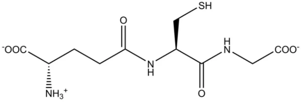
Glutathione
Glutathione (GSH) is a tripeptide, γ-glutamylcysteinglycine (Figure 5), widely distributed in mammalian cells and tissues, in plants, and most microorganisms [57]. It was discovered by De Rey Pailhade in 1888 during his studies in extracts of yeast, and he called it “Philothion” [58]. It is the most abundant non-protein thiol of the cells, with a concentration that ranges between 1-10 mM, with the liver as the main reservoir [59]. Inside the cells, the GSH is mostly present in the cytosol (90%), where its synthesis occurs, followed by mitochondria (10%), and the endoplasmic reticulum (ER) (small percentage) [60]. It is present in the reduced form, GSH, and in the oxidized form, GSSG [61]. The latter consists of two GSH molecules bound by a disulfide bond, and its concentration is less than 1% of GSH [60]. GSH structure and function as an antioxidant were established by Sir Hopkins in 1922 [62].
13 Figure 5 GSH chemical structure.
GSH synthesis
GSH is synthesized from the amino acids glycine, cysteine, and glutamate in the cytosol, and its de novo synthesis involves two ATP-requiring enzymatic steps [63][64].
The first step (Figure 6) is catalyzed by the enzyme glutamate-cysteine ligase (GCL), which produces γ-glutamylcysteine by forming an amide bond between the γ-carboxyl group of glutamate and the amino group of cysteine. This enzyme is a heterodimer, formed by a heavy or catalytic (GCLc), and a light or modifier (GCLm) subunit. The former exhibits the catalytic activity and is regulated by the presence of GSH via negative feedback; the latter is enzymatically inactive but regulates the functioning of the GCLc. In fact, it lowers the Km of GCL for glutamate and raises the Ki for GSH, which means that GCL is more efficient and less subject to inhibition by GSH [65]. Oxidative stress may enhance the activation of the enzyme, even without changing the expression of GCL [66]. Under physiological conditions, GCL is mostly inactive [60]; moreover, its functionality depends on the presence of cysteine which is considered the rate-limiting enzyme [64].
The second step (Figure 6) in GSH synthesis is catalyzed by the glutathione synthase (GS), which is composed of two identical subunits and is not inhibited by GSH [67]. GS is present in higher concentration than GCL, and overexpression of the enzyme does not translate in an increase of GSH concentration [68]; however, the presence of GS has an important role in determining GSH synthesis for example under stressful conditions, such as in response to surgical traumas [60].
14 GSH degradation
The structure of GSH is characterized by the presence of γ-bond between glutamate and cysteine, which makes the tripeptide resistant to hydrolysis from peptidases [69]. For a long time, it has been thought that the degradation of GSH was possible only outside of the cells because the only known enzyme able to break the γ-bond was the γ-glutamyltranspeptidase (GGT or γ-GT) [70]. GGT is located on the plasma membrane and it is implicated in the γ-glutamyl cycle. This enzyme transports GSH outside of the cell in the form of the dipeptide cysteynilglycine and the glutamyl moiety, which is then transferred to an amino acid to form γ-glu-aa [71]. The dipeptide is the substrate of a dipeptidase on the plasma membrane and is broken down into cysteine and glycine. Cysteine is then taken up by the cell and the majority is used to form GSH [58]. γ-glu-aa, on the other hand, is transported back inside the cell and separated from the amino acid; the free glutamate can be used for GSH synthesis [58].
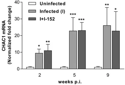
Recently, an intracellular pathway of GSH degradation catalyzed by the ChaC enzyme family was discovered [72]. The mammalian ChaC1 was discovered as a protein upregulated during the Unfolded Protein Response (UPR) caused by ER stress [73], and it was shown to be able to break the γ-bond between glutamate and cysteine because of its γ-glutamylcyclotransferase activity [72]. Chac1 is able to exclusively act on reduced GSH, by forming the dipeptide cysteynilglycine and 5-oxoproline, while GSSG is not affected by its activity [74].
GSH functions
GSH is involved in various cellular functions, which can be categorized into two main reactions: conjugation, and reduction [75]. The first group of reactions is involved in the metabolism of xenobiotics, where GSH is conjugated with the exogenous compounds by the activity of the enzyme glutathione-S-transferase (GST) [58]. The resulted conjugates can be excreted from the cells [75]. The second group of reactions explains its antioxidant activity and includes the GSH itself and several enzymes involved in the intracellular defense against oxidative stress. This mechanism is composed of GSH itself and several enzymes, such as glutathione peroxidase (GPx), GST, and glutathione reductase (GR) [76]. GPx and GST, which not only have conjugating activity, are implicated in the reduction of peroxides, while GR is involved in the conversion of GSSG in two molecules of GSH [77]. Moreover, GSH is involved in the maintenance of the sulfhydryl groups of proteins in the reduced state. It is known that the redox state of proteins regulates different intracellular pathways and that the cysteine residues in the active sites can be easily oxidized [78].
Recent evidence has shown that GSH has a broader activity, in addition to the scavenging one. In fact, GSH deficiency has been described as an important mechanism contributing to the pathogenesis and outcomes of several infections (both viral and bacterial), [79][80].
GSH in infections and inflammation
Alterations of the redox state are common during infections. The consequent modulation of the many intracellular redox-sensitive pathways affects the ability of the cells to cope with pathogens by modifying both
15 the innate and the adaptive immune response [79]. GSH has many important roles in the immune response. Antigen-presenting cells (APCs) need the tripeptide in order to correctly processing the antigen; in fact, the endocytosed proteins must undergo a process of denaturation/unfolding, and the rate-limiting-step seems to be the reduction of disulfide bonds [81]. This antigen processing seems to be correlated with GSH content; indeed, if it is present in APCs at low concentrations the process is defective [82], while high concentrations of GSH promote the capacity to present the antigen [83]. APCs, like macrophages, can influence the differentiation of T helper cells (Th) in Th1 (cellular immunity) and Th2 (humoral immunity) according to the secreted cytokines; thus, the redox state of APCs is important to mount a proper immune response against pathogens [84]. Macrophages with a decreased GSH content were found to produce less IL-12, which in turn inhibit the Th1 differentiation in a murine model [84], while elevated GSH concentrations induced by pro-GSH molecules were found to increase IL-12 secretion associated with a Th1 response [85].
Many viruses are known to alter the intracellular redox state versus a more oxidized, often characterized by GSH depletion, with the aim to make the cellular environment favorable to an efficient virus cycle. For this reason, GSH has been proposed as an effective antiviral molecule, and its capacity to hinder the replication of different viruses has been described. For example, GSH can inhibit NF-kB activation induced by HIV [86], or influenza virus-induced apoptosis resulting in a reduced spread of the virus [87]. Another interesting mechanism could be the ability to inhibit the proper folding and stabilization of viral proteins, like the maturation of the glycoprotein hemagglutinin of the influenza virus in the endoplasmic reticulum (ER) of the host [88]. In this regard, it is known that viruses don’t have the biosynthetic machinery to translate their genome and to produce their structural proteins, thus they take advantage of the host’s protein biosynthetic machinery associated with the ER [89]. The ER is important for the correct folding of proteins, which includes the disulfide bond formation that is important for the protein maturation and stability and to do that the ER lumen is characterized by a low GSH/GSSG ratio [90]. The ER redox state is fundamental for the correct functioning of the protein disulfide isomerase (PDI), which exerts oxidoreductase and redox-regulated chaperone activities. In fact, PDI leads to the formation, isomerization, and reduction of disulfide bonds, and it has also an important role in the degradation of misfolded proteins [91]. As a redox-dependent protein, PDI needs a correct ratio between GSH and GSSG to function properly, otherwise, alterations in the disulfide bond formation could occur, leading to misfolded protein.
The GSH depletion caused by viruses leads to the activation of the NF-kB pathway, which is normally considered as an important factor of the innate immune response against pathogens, promoting viral replication, as said above [92]. GSH has the ability to inhibit NF-kB activation at many levels; as a scavenger it can eliminate oxidants, it can prevent the activation of IKK by inhibiting the kinase, it can interfere with the translocation of activated NF-kB, and it can inhibit the binding of NF-kB to the DNA. However, it is important to remember that this pathway is involved in the immediate early step of immune activation, so in order to promote a correct immune response, the NF-kB pathway can not be completely suppressed [93]. In fact, the outcome is the inflammatory response, which brings to the production of various cytokines and chemokines that have the role to activate the immune response and viral clearance [94].
16 Although an effective anti-viral immune response is necessary for viral elimination, an exaggerated response can be harmful. In particular, pro-inflammatory cascades have a fundamental role in the pathogenesis of lung damage in respiratory virus infections, such as influenza virus, and other diseases, such as Cystic Fibrosis (CF). Redox regulation of inflammatory cytokine production induced by Pseudomonas aeruginosa (P. aeruginosa), which is the most common chronic infection in CF, has been one of the topics of my thesis work [95]. This disease is characterized by a lack/malfunction of the cystic fibrosis transmembrane regulator channel (CFTR) [96], and also by an altered redox state in the CF lung which is translated in a decreased concentration of GSH with respect to healthy lung [97][98]. Furthermore, CF lung has an impaired mucociliary clearance, which means that an eventual P. aeruginosa infection in the lung would not be prevented/resolved properly [99]. Normally, an increase of ROS production leads to the activation of the redox-sensitive inflammatory pathways, such as NF-kB that is considered fundamental for the immune response, and for the production of several inflammatory cytokines, like IL-6, TNF-α [100]; however, CF is characterized by unresolved infection and hyperinflammation, with basal high ROS levels and increased pro-inflammatory cytokines. This alteration leads to increased oxidative stress and impairment of the intracellular pathways involved in the immune response against pathogens [101].
Besides viral infections, bacteria could also invade the cellular host, such as macrophages, and put in place mechanisms aimed to manipulate the cellular immune signaling pathways to survive [102]. It is known that macrophages could be a bacterial reservoir and that the bacterial infection could alter the redox state and in turn influence the antibacterial pathways of the host [103]. One example is represented by infection of Mycobacterium tuberculosis, which is an opportunistic intracellular pathogen able to persist in the macrophage host by inhibiting the killing mechanisms, especially in patients with acquired immune deficiency syndrome (AIDS) [104]. Treatments aimed to increase intracellular GSH levels could influence bacterial survival and replication for three reasons. First, mycobacteria do not produce GSH, but they synthesize mycothiol as a regulator of reduction and oxidation reactions [105]. Therefore, the exposure of this bacterium to high concentrations of GSH could imbalance its redox state and bring to growth inhibition. Second, it is thought that GSH could have evolved in higher eukaryotes as a precursor of antibiotics before the emergence of cellular immunity [106]. Third, the tripeptide is a precursor of GSNO, which may represent one of the most important active forms of NO as an antimicrobial agent [107].
Many diseases, in which GSH alterations have been described, are characterized by impairment in the cellular immune/inflammatory response, and the replenishment/increase of GSH levels, by GSH itself or GSH-boosting drugs, could be considered as a useful approach to modulate the pathways involved in the immune response.
Pro-GSH molecules
Replenishment or augmentation of intracellular GSH concentration can be achieved by treatment with pro-GSH molecules [108]. The use of pro-GSH as it is, in fact, is not ideal because it has a short half-life in plasma (<3 min), and is unable to cross the cell membrane; therefore, high doses of GSH would be required to achieve
17 therapeutic value [109]. For these reasons, several pro-GSH molecules have been designed and used to increase the levels of the tripeptide more easily. For the synthesis of these molecules, different approaches can be followed; it is possible to administer GSH-conjugates to facilitate the passage of the tripeptide across the membrane, such as GSH esters [110]; another potential way is to give GSH precursors, such as the rate-limiting amino acid cysteine, in the form of N-acetylcysteine (NAC) [111] and β-mercaptoethylamine (cysteamine, MEA) [112].
NAC, a precursor of L-cysteine, is widely used in clinical treatments and is recognized by the World Health Organization (WHO). This drug is useful in the treatment of paracetamol overdose [113] and is recognized by the FDA as a mucolytic [114]. NAC can act as an antioxidant via several mechanisms. It can exert an indirect effect by providing cysteine, useful to GSH synthesis; a direct effect against oxidant species; a reducing effect on the sulfhydryl group of protein [115]. For these reasons, NAC has been considered a beneficial drug able to counteract oxidative stress, which is a common characteristic of many diseases [116]. NAC, in fact, is used in several clinical treatments, like neurological disorders [117], substance abuse disorders [118], Alzheimer’s disease [119], asthma [120], influenza [121]; moreover, because of its ability to break bacterial biofilms, it can be used as an adjuvant microbial drug that could improve antibiotic permeability [122]. However, the use of NAC has some limitations since in order to activate the synthesis of GSH the cells must have undamaged enzymatic machinery.
MEA is an aminothiol used in the treatment of the rare disease cystinosis that was approved by the FDA in 1994 [122]. Cystinosis is characterized by a redox alteration that affects GSH content in favor of its oxidized form, GSSG [123]. The effects of MEA had been studied in the cystinosis model, and the results obtained by in vitro studies in proximal tubular epithelial cells showed that this molecule was able to increase GSH levels. The GSH-boosting drug of MEA is presumably linked to its capacity to reduce cystine, the oxidized form of cysteine, in cysteine [124]. Because of its ability to restore GSH and its antioxidant properties, MEA could be used in diseases hallmarked by a damaging oxidative environment. Furthermore, it had been demonstrated that MEA has a synergistic effect when used with the antimalarial artemisinin-derivatives, suggesting a broader effect of this molecule [125].
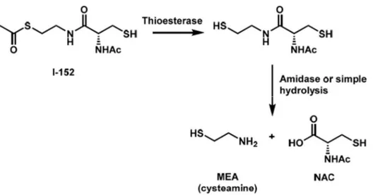
I-152
I-152 is a pro-GSH molecule composed of NAC and MEA linked together by an amide bond; this molecule enters the cells where it is first deacetylated on the MEA moiety and subsequently hydrolyzed to release NAC and MEA [126]. I-152 was synthesized for the first time in 2001 by Oiry et al., and the main goal was to obtain a potent antioxidant molecule able to pass across the cellular plasma membrane and increase GSH intracellular content by taking advantage of the two GSH parent drugs (i.e. NAC and MEA) [127]. The pro-GSH activity of I-152 was reviewed by our research group as reported in Chapter 1 where we collected old and more recent data that can be summarized as follows:
in primary human monocyte-derived macrophages (MDMs) I-152 (i.e. 10 µM – 1 mM) can increase the intracellular content of GSH (old data);
18 in murine macrophage RAW 264.7 cell line and macrophages obtained from the peritoneal cavity of mice low concentrations of I-152 (not higher than 1 mM) can increase GSH content, while high concentrations of I-152 (i.e. 10 and 20 mM), caused a GSH depletion but provided a large amount of thiol species, such as I-152, NAC, MEA, and cysteine (old data);
in GSH-experimentally depleted RAW 264.7 cells I-152, at low concentrations (i.e. 0.1 and 0.5 mM), replenishes GSH (recent data);
in vivo I-152 can deliver NAC and MEA in several mouse organs (recent data);
in vivo I-152 can restore GSH whose content was depleted by the retroviral complex LP-BM5. Moreover, I-152 immunomodulatory activity towards antigens was demonstrated in different animal models. Ova-sensitized and Tat-immunized mice pre-treated with I-152 showed a shift to a Th1 immune response involving splenocyte IFN-gamma production and higher levels of IL-12 in circulation [128][129]. Moreover, in a murine model of AIDS, a correlation between GSH deficiency and a prevalent Th2 immune response was established; in the same animals, I-152 treatment was found to restore a balanced Th1/Th2 response [129][130] (old data).
19 Structure of the thesis
The thesis is divided into 5 chapters. Every chapter is introduced by a brief paragraph explaining what the chapter contains. While the chapters 1-3 report papers which have been already published, the chapters 4 and 5 describe results which have not been yet published.
The References of the section “General introduction” and of the Chapters 4 and 5 can be found in the section “References” at the end of the thesis.
Aim of the study
The study was aimed to modulate intracellular redox-sensitive pathways to hinder directly the pathogen (like in the case of Mycobacterium avium), whose survival depends on the thiol-based redox metabolism which is fundamental to maintain a reducing environment to preserve cellular homeostasis, especially from ROS, and antibiotics, and indirectly by modulating the host’s immune/inflammatory response to the pathogen. Furthermore, it was investigated whether the redox state could play a role in immune/antiviral response by modulating folding and secretion of immunoglobulins.
Alteration of redox state was induced by I-152 whose pro-GSH capacity had already been demonstrated, but the metabolism and the mechanism by which it increases intracellular GSH levels had not been studied in depth. In this thesis, one of the aims was also to deepen this aspect.
20
CHAPTER 1
Chapter 1 – This chapter consists of a review where the pro-GSH activity of I-152 is discussed.
Original article published in Nutrients Volume 11, Issue 6, 2020, 1291
21
nutrients
Review
Boosting GSH Using the Co-Drug Approach: I-152, a
Conjugate of N-acetyl-cysteine and
β-mercaptoethylamine
Rita Crinelli 1, Carolina Zara 1, Michaël Smietana 2 , Michele Retini 1 , Mauro Magnani 1 and
Alessandra Fraternale 1,*
1 Department of Biomolecular Sciences, University of Urbino Carlo Bo, 61029 Urbino, Italy;
[email protected] (R.C.); [email protected] (C.Z.); [email protected] (M.R.); [email protected] (M.M.)
2 Institut des Biomolécules Max Mousseron, Université de Montpellier UMR 5247 CNRS, ENSCM,
34095 Montpellier, France; [email protected]
* Correspondence: [email protected]; Tel.: +39-0722-305243
Received: 12 April 2019; Accepted: 5 June 2019; Published: 7 June 2019 check Eorupdates
Abstract: Glutathione (GSH) has poor pharmacokinetic properties; thus, several derivatives and biosynthetic precursors have been proposed as GSH-boosting drugs. I-152 is a conjugate of N-acetyl-cysteine (NAC) and S-acetyl-β-mercaptoethylamine (SMEA) designed to release the parent drugs (i.e., NAC and β-mercaptoethylamine or cysteamine, MEA). NAC is a precursor of L-cysteine, while MEA is an aminothiol able to increase GSH content; thus, I-152 represents the very first attempt to combine two pro-GSH molecules. In this review, the in-vitro and in-vivo metabolism, pro-pro-GSH activity and antiviral and immunomodulatory properties of I-152 are discussed. Under physiological GSH conditions, low I-152 doses increase cellular GSH content; by contrast, high doses cause GSH depletion but yield a high content of NAC, MEA and I-152, which can be used to resynthesize GSH. Preliminary in-vivo studies suggest that the molecule reaches mouse organs, including the brain, where its metabolites, NAC and MEA, are detected. In cell cultures, I-152 replenishes experimentally depleted GSH levels. Moreover, administration of I-152 to C57BL/6 mice infected with the retroviral complex LP-BM5 is effective in contrasting virus-induced GSH depletion, exerting at the same time antiviral and immunomodulatory functions. I-152 acts as a pro-GSH agent; however, GSH derivatives and NAC cannot completely replicate its effects. The co-delivery of different thiol species may lead to unpredictable outcomes, which warrant further investigation.
Keywords: glutathione; pro-glutathione molecules; co-drug; antiviral activity; immunomodulation; N-acetyl-cysteine; cysteamine
1. Introduction
Glutathione (GSH) is the most powerful antioxidant molecule within cells performing a variety of functions that go beyond its protective activity against reactive oxygen species (ROS). Indeed, in its reduced form, GSH not only acts as a scavenger of ROS and as a substrate for antioxidant enzymes, but also promotes drug detoxification [1]. Moreover, by controlling the redox potential, GSH modulates many redox-sensitive proteins within signaling pathways [2]. Notably, GSH itself can be conjugated to the cysteines of proteins in a process known as S-glutathionylation. It is widely accepted that thiol redox transitions cause changes in protein activity, abundance, localization, and interaction with other macromolecules [3]. These properties of GSH may at least partially explain its ability to stimulate cell proliferation and act as an immunomodulator, although research in this field is still limited.
22 Alterations in GSH levels may be transient in response to an oxidative insult, or they may become chronic under conditions of prolonged oxidation and/or dysfunction/deficiency of the enzymes involved in GSH synthesis/degradation. Inborn errors in GSH metabolism include those arising from defective γ-glutamyl-cysteine ligase and glutathione transferase which are the most frequently occurring disorders [4]. Conversely, examples of acquired GSH deficiency include mitochondrial diseases, cystic fibrosis, and many viral/microbial infections [5,6]. In the last few decades, the number of pathologies found to be associated with low GSH levels has risen rapidly, prompting researchers to consider GSH and its derivatives as possible therapeutic agents. Cellular GSH concentration can be affected by the exogenous administration of GSH or GSH-boosting drugs, such as glutathione esters and GSH biosynthetic precursors, which have been used to overcome the poor pharmacokinetic properties of GSH [7,8]. GSH and N-acetylcysteine (NAC) have also been used in the co-drug approach as bioconjugates of several therapeutics employed in the treatment of neurodegenerative diseases [9]. In this context, the use of I-152 marks the very first attempt to combine two pro-GSH molecules into one to potentiate both the cellular uptake and to improve the biopharmaceutical properties of the parent drugs.
In this review, the design, metabolism and ability of I-152 to affect GSH levels are discussed.
Moreover, the antiviral and immunomodulatory properties of I-152 in light of its GSH-boosting activity are also summarized.
2. I-152 Design andSynthesis
I-152 is a conjugate of NAC and S-acetyl-β-mercaptoethylamine (SMEA) linked together by an amide bond. The molecule was synthesized by Oiry et al., in 2001 using commercially available N-acetyl-S-trityl– L-cysteine and S-acetylcysteamine hydrochloride [10]. Experiments performed in cell-free extracts have shown that this compound is deacetylated to the corresponding dithiol derivative, which may be responsible for the in-situ release of NAC and MEA [10] (Figure 1).
Figure 1. Chemical structure and proposed metabolism of I-152. NAC, N-acetyl-cysteine; MEA, β-mercaptoethylamine or cysteamine.
In an attempt to enhance the lipophilic properties of the molecule, a series of I-152 analogues carrying different S-acyl groups on the MEA moiety and S-acylation of the free thiol group were subsequently synthesized by the same team [11]. In terms of pro-GSH activity, the potency of the molecules increased with the presence of the free thiol group. By contrast, the presence of an R-radical on the MEA moiety had no significant effects.
The initial aim of combining NAC and MEA was to design a new potent antioxidant molecule able to liberate two potential pro-GSH compounds after metabolic conversion. NAC is the N-acetyl derivative of the natural amino acid L-cysteine; thus, it is a direct precursor of glutathione. NAC has long been used therapeutically as a well-tolerated and safe medication for the treatment of various
23 pathologies, including paracetamol intoxication and cystic fibrosis. Its efficacy as an antioxidant has been demonstrated in a wide range of clinical settings where oxidation is tightly linked to GSH deficiency, given that NAC is a poor direct antioxidant [12]. Although NAC was designed to facilitate membrane permeability, it has been suggested that its pharmacological activity might rely on the reduction of plasma cystine to cysteine which then enters the cells and sustains glutathione synthesis [13]. NAC-induced cytoprotection and the NAC signal transduction pathway are not well understood. Recent evidence suggests that NAC may modulate antioxidant pathways by increasing the expression of miR-141 regulating Keap1/Nrf2 signaling, at least under conditions in which the miRNA is downregulated [14]. β-mercaptoethylamine (MEA) or cysteamine is a product of the constitutive degradation of Coenzyme A. It derives from the cleavage of pantetheine to form MEA and pantothenate (vitamin B5). In mammalian cells, MEA can be oxidized to hypotaurine and taurine by aminothiol dioxygenase [15]. The thiol cysteamine can be oxidized into the disulfide cystamine depending on the local redox environment. Both forms have been used in clinical and experimental settings; however, in most cases their specific role in the observed biological effects was not understood. High intracellular levels of GSH are probably sufficient to reduce cystamine to cysteamine; thus, it has been proposed that most of the effects of cystamine may be mediated by its reduced form [16]. On the other hand, there is evidence that cysteamine has the propensity to form disulfides in vivo, suggesting that cysteamine-containing disulfides such as cystamine may normally be present along with cysteamine in mammalian tissue under physiological conditions [17]. Cysteamine can perform a wide range of functions acting as a cystine-depleting agent, pro-GSH molecule, enzyme inhibitor, and gene expression modulator. Interestingly, many of these activities are thought to rely on thiol/disulfide exchange reactions between cystamine/cysteamine and susceptible protein cysteine sulfhydryl groups in a process called cysteaminylation [17]. Cysteamine, in the form of cysteamine bitartrate (Cystagon), has been approved by the Food and Drug Administration (FDA) for the clinical treatment of nephropathic cystinosis acting as a cystine-depleting agent by forming cysteine-cysteamine mixed disulfides [18]. Recent studies suggest that cysteamine may have several other potential therapeutic applications beyond cystinosis, including the treatment of neurodegenerative diseases and cancer. Indeed, cysteamine/cystamine is an efficient inhibitor of transglutaminase and caspase 3 which play a pivotal role in mutant Huntington protein processing [19]. Other proteins whose activity has been reported to be affected by cysteamine/cystamine treatment are protein kinase C and metalloproteinases, important drug targets in cancer progression and metastasis [20,21]. More poorly understood are the mechanisms mediating the pro-GSH activity of cysteamine and its oxidized form. Cysteamine treatment of normal and cystinotic cells has been shown to increase GSH content. However, the authors claimed that the increase of cysteine levels resulting from cystine reduction can only partially explain elevated GSH levels [22]. The ability of cystamine and, less potently, of cysteamine to activate the Nrf2 antioxidant pathway was shown by Calkins et al., in astrocytes [23]. Cystamine-mediated activation of Nrf2 was found to be inversely correlated with the GSH content of the culture and the increase in GSH levels was shown to be dependent on de novo synthesis. Unfortunately, experiments performed with Nrf2 knockout cells revealed that the cystamine-induced GSH increase was Nrf2-independent. Hence, cystamine-induced GSH up-regulation seems to involve other yet-unknown mechanism(s).
3. I-152: Metabolism and Effects on GSH Levels
The ability of I-152 to increase basal intracellular GSH content was evaluated in human and murine cellular models. In primary human monocyte-derived macrophages (MDMs) and other human cell lines, I-152 ranging from 10 µM to 1 mM was found to increase GSH content even at doses which were ineffective when NAC and MEA, alone or in equimolar combinations, were used [10]. In murine monocyte/macrophage-like cell line RAW 264.7 and macrophages obtained from the peritoneal cavity of mice, low concentrations of I-152, i.e., 1 mM, increased GSH level [24]. By contrast, high I-152 concentrations (i.e., 10 and 20 mM) caused GSH depletion but provided large quantities of
24 I-152 and NAC which can be used to resynthesize GSH [24]. The decrease in the GSH content at high I-152 doses has been suggested to be the consequence of a negative feedback of GSH on γ-glutamylcysteinyl synthetase, the first enzyme involved in GSH synthesis [10]. Alternatively, GSH could be depleted because of its conjugation to I-152 in a reaction catalyzed by the detoxification enzyme glutathione-S-tranferase (GST). Preliminary in-vitro experiments indicate that GSH could be indeed conjugated to I-152 by GST. Under the condition of GSH depletion, cysteamine could be converted into cystamine, which has been shown to inhibit γ-glutamylcysteinyl synthetase, further supporting the decrease in GSH levels [25].
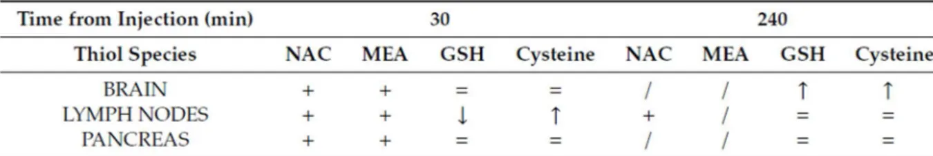
Less information is available on I-152 in-vivo metabolism. Preliminary studies were performed in our laboratory to assess the distribution of I-152 and its metabolites in mouse organs after I-152 intraperitoneal injection (i.p.). GSH and cysteine levels were also measured. The analyses were performed in selected organs, i.e., the lymph nodes, brain and pancreas. The lymph nodes were chosen because it has been reported that lymphoid tissues are significantly more “reduced” than the other tissues, and that the immune response is strongly influenced by variations in the redox state [26]. In particular, some lymphocyte functions, such as DNA synthesis, are favored by high levels of GSH, while other redox-sensitive pathways are favored by low intracellular GSH. However, immunological functions in the diseases characterized by oxidative stress can be restored by cysteine or GSH supplementation [27,28]. The brain generates high levels of ROS due to its high oxygen
consumption, hence it is more susceptible to the damaging effects of ROS than other tissues [29,30]. The GSH concentration has a key role in maintaining redox balance in the central nervous system (CNS) and it is altered in neurodegenerative diseases [5,31,32] such as Alzheimer’s disease (AD), Parkinson’s disease (PD), amyotrophic lateral sclerosis (ALS), multiple sclerosis (MS), HIV-associated neurocognitive disorder (HAND or “NeuroAIDS”), cerebral ischemia/reperfusion injury (I/R), and traumatic brain injury (TBI) [33]. The presence of the tightly regulated blood brain barrier (BBB) represents a serious obstacle to effective treatment of CNS disorders, in fact its selective permeability prevents most bioactive molecules from entering the brain [34]. Several antioxidants, including vitamin E (the important scavenger of lipid peroxidation in the brain), vitamin C (intracellular reducing molecule), coenzyme Q10 (transporter of electrons in the electron transport chain, ETC), and NAC (acting as a precursor of GSH) have been used as therapeutic agents. Although antioxidant therapies have shown benefits in preclinical animal models, negative results have been obtained from clinical trials [30]. Moreover, in such treatments, dosage and additives as well as synergistic interactions with other antioxidants must be considered [35]. Hence, the design and development of antioxidant-based therapies for the brain require a great deal of effort [33,34,36]. Lastly, the pancreas plays a major role, along with several other organs, in GSH metabolism, as shown by the high concentration of the tripeptide, its rapid turnover rate, and the presence of high levels of various enzymes involved in GSH metabolism [37]. Therefore, the pancreas requires a great amount of cystine/cysteine for pancreatic enzymes, which is provided by glutathione (GSH). Moreover, the induction of CYP450 enzymes by xenobiotics in pancreatic acinar cells can cause a decrease in GSH content, which can affect both detoxification and pancreatic enzyme synthesis [38].
Our preliminary data indicate that I-152 can be used to deliver precursors for GSH synthesis, in the form of NAC and MEA, to different organs, including the brain (Table 1).
The presence of NAC within tissues supports the hypothesis that I-152 is able to reach the target site and release in situ the active molecules. In fact, it has been demonstrated that NAC does not pass the cell membrane, but rather reacts with cystine reducing it to cysteine, which then enters the cells and sustains GSH synthesis [13]. Hence, increased cysteine found in the organs can derive from NAC released by I-152 or by the reduction of plasma cystine to cysteine. The presence of NAC in the brain is an interesting aspect since NAC’s ability to cross the BBB is disputed [40]. Hence, I-152 could be a promising pro-drug to release NAC in the brain. Interestingly, increased intracellular thiol availability did not enhance intracellular GSH with the exception of the brain, probably because these cells had normal GSH levels. Indeed, under conditions of GSH depletion, e.g., in cells treated with diethyl
25 maleate (DEM) [41], I-152 even at low concentrations (0.1 and 0.5 mM) exerts a GSH-replenishing effect (Figure 2).
Figure 2. In-vitro replenishment of intracellular GSH by I-152. RAW 264.7 cells were treated with 6 mM diethyl maleate (DEM) for 15 min, then medium not containing I-152 (DEM+medium) or containing I-152 at different concentrations were added for 2h. Reduced glutathione (GSH) content was determined by HPLC [24]. Results represent the mean ± S.D. of two independent experiments.
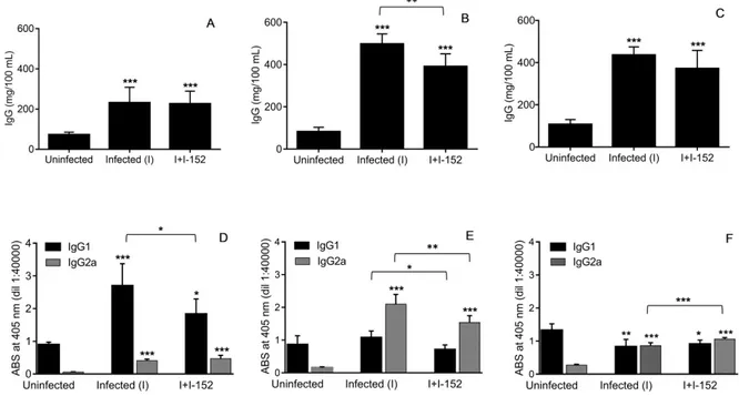
The capacity of I-152 to restore the intracellular GSH content was also studied in vivo in organs of mice experimentally infected with the retroviral complex LP-BM5. This infection causes a disease with many similarities to human Acquired Immuno Deficiency Syndrome (AIDS), including GSH deficiency [42,43]. Glutathione depletion was found in most organs of LP-BM5-infected mice both in the early phases of the disease and later, in particular in the lymphoid organs, e.g., lymph nodes, known to be the site where viral loads are higher [42]. The effect of intraperitoneal administration of I-152 in infected mice is shown in Figure 3, where it can be observed that the treatment was able to re-establish the content of the tripeptide in the lymph nodes of infected mice at all the times of infection. Notably, GSSG content was unaffected by I-152 treatment [42]. Hence, as hypothesized above, I-152 could be used to increase intracellular GSH during aging and various disease states in which antioxidant defense systems can be altered leading to progressive oxidative damage and subsequent cell death and/or significant impairment of several cellular processes.
26 Figure 3. Replenishment of reduced glutathione (GSH) by I-152 in LP-BM5-infected mice. C57BL/6 mice were infected and treated with I-152 (30 µmol/mouse) three times a week, every other day, for a total of 9 weeks. At 2, 5 and 9 weeks after virus inoculation, GSH content in the lymph nodes was determined by HPLC [39,42]. Data are the mean ± S.D. of at least three mice. *p < 0.05; ** p < 0.01; *** p < 0.001 (ANOVA). [42]; (unpublished results).
4. Antiviral and Immunomodulatory Properties of I-152
GSH depletion characterizes several viral infections and associated-disease progression [44]. Numerous studies have demonstrated that the use of GSH is effective in reducing viral production in different experimental models suggesting that the administration of the tripeptide can be considered a useful strategy to hinder viral infection and infection-associated symptoms [2,28,44,45]. Due to the poor pharmacokinetic properties of GSH, high GSH concentrations are necessary to sufficiently increase its content in order to obtain an antiviral effect [7,8]. Hence, taking advantage of the GSH-replenishing capacity of I-152, antiviral effects of the molecule were explored in two retroviral infections associated with systemic and tissue decrease in the GSH content, i.e., HIV and LP-BM5 infections. In HIV-1/BaL-infected MDMs, 150 µM I-152 was able to inhibit viral replication by 90% likely interfering with both early and late steps of the virus life cycle [11]. In LP-BM5-infected mice I-152, when administered at a concentration of about 10 times lower than GSH, significantly reduced murine AIDS (MAIDS) symptoms were observed, i.e., splenomegaly and lymphadenopathy, as well as BM5d proviral DNA in spleen and lymph nodes [46]. Actually, the exact mechanisms through which I-152 can exert antiviral activity are not known. However, since I-152 treatment replenished virus-induced GSH depletion both in human MDMs [10,11] and in mouse lymphoid organs (Figure 3), it could be hypothesized that the antiviral effect observed is dependent on GSH. Indeed, GSH can inhibit the replication of viruses by different modes of action [44,45]. For example, it has been reported that administration of GSH permeable analogue GSH-C4 can interfere with the maturation of influenza virus glycoproteins modifying the activity of the host-cell protein disulphide isomerase (PDI) which is essential for the correct disulphide bond formation of viral proteins [47]. Glutathione can interfere with the entry of rhinovirus by inhibiting rhinovirus induction of intercellular adhesion molecule-1 (ICAM-1) mRNA in respiratory epithelial cells [48]. Furthermore, GSH by counteracting the action of reactive oxygen intermediates (ROI), can prevent the activation of NF-kB and HIV replication [49].
I-152 effects against HIV and LP-BM5 can be due to its direct antiviral action, but also to its immunomodulatory activity. In fact, many studies have correlated altered GSH levels with an impaired immune response, suggesting a combination of the highly active antiviral therapy with the GSH replenishment approach [50,51]. Recently, the main functions of GSH in the immune response have been reviewed [52]. Accordingly, a possible immunomodulatory role of I-152 was investigated. In particular, the role of I-152 in Th1/Th2 polarization was studied. In fact, several studies have underlined the correlation between altered GSH levels and an unbalanced Th1/Th2
27 immune response in favor of Th2 linked to an impaired cytokine production by antigen-presenting cells [42,50,53–56]. The immunomodulatory activity of I-152 was demonstrated in in-vitro systems where the molecule-stimulated IL-27 p28 gene expression and sustained STAT-1-mediated IRF-1 de novo synthesis [24]; moreover, in vivo, it enhanced Th1 response in ovalbumin immunized mice as well as drove Th1 immune responses and CTL activity against HIV antigens [55,56]. Finally, I-152 treatment, by inducing Th1 cytokine production, restored a balanced Th1/Th2 response in mice affected by murine AIDS [42]. Hence, the effect exerted by I-152 on MAIDS can be derived from its dual mechanisms of action: On the one hand, it can directly inhibit viral replication; on the other hand, it can re-establish a correct GSH content favoring the production of cytokines which induce the Th1 immune response (Table 2).
However, the immunomodulatory activity of I-152 could go beyond the regulation of Th1/Th2 response. In fact, I-152 treatment inhibited total IgG secretion in LP-BM5 infected mice [46] and influenced IgG1/IgG2a ratio in favor of the IgG2a subtype (unpublished results). This second effect is likely the consequence of Th1/Th2 response regulation; in fact, the induction of high IgG1 titers is considered indicative of a Th2-type immune response while high IgG2a are typical of a Th1-type response [57,58]. On the contrary, the inhibition of hypergammaglobulinemia could be the consequence of a lower viral load, but it cannot be excluded that I-152 could also directly affect plasma cell maturation and Ig folding/secretion.
One of the features characterizing murine retrovirus LP-BM5-induced MAIDS is decreased T- and B-cell responses. I-152 treatment was demonstrated to partially restore T and B B-cell proliferative capacity to mitogens [46]. In this context, Green et al., who had previously demonstrated that monocytic myeloid-derived suppressor cells (M-MDSCs) suppress both T and B cell responses [59,60], found that I-152 could interfere with the suppressive function of M-MDSCs (Green, personal communication). On the whole, these observations suggest that I-152 can influence different components of the immune system. Future studies aimed to shed light on the molecular mechanisms modulated by I-152 will provide further insights into the immunomodulatory activity of this pro-GSH molecule.
5. Conclusions
The data presented in this review show that I-152 is an excellent co-drug able to generate NAC and MEA which can be used to increase intracellular GSH. I-152 has been shown to regulate the intracellular GSH content differently depending on the concentration used and the pre-existing redox states; consequently, different redox-sensitive pathways could be activated or inhibited. For example, it has been reported that different concentrations of NAC cause opposite effects on the GSH/GSSG ratio and on the production of pro-inflammatory cytokines [61,62]. Moreover, pro-GSH molecules such as NAC and MEA could affect cellular processes independently of their ability to influence intracellular GSH/GSSG balance [63,64]. Furthermore, the exact mechanisms through which NAC and MEA can increase GSH levels are not yet completely understood. Preliminary experiments have shown that I-152 can influence different redox-signaling pathways linked to GSH [24] and that NAC and MEA administered alone or in combination are less efficient in raising intracellular GSH [10]. Hence, it
28 would be interesting to investigate all of these aspects. Moreover, such studies could yield useful information for the design and synthesis of new redox-modulating agents with improved activity.
Author Contributions: A.F. and R.C. literature search and original draft preparation. M.M., M.S., C.Z. and M.R. reviewing and editing. M.S. I-152 synthesis. A.F. and R.C. conception, design, writing, reviewing, and editing of the manuscript. M.M. funding acquisition and supervision.
Funding: This work was partially supported by grants from Urbino University; Ministero dell’Istruzione, dell’Università e della Ricerca (MIUR) (PRIN (Research Projects of National Interest) 2010-2011-prot. 2010PHT9NF_004).
Conflicts of Interest: The authors declare no conflict of interest.
References
1. Forman, H.J.; Zhang, H.; Rinna, A. Glutathione: Overview of its protective roles, measurement, and biosynthesis. Mol. Asp. Med. 2009, 30, 1–12. [CrossRef] [PubMed]
2. Aquilano, K.; Baldelli, S.; Ciriolo, M.R. Glutathione: New roles in redox signaling for an old antioxidant. Front. Pharmacol. 2014, 5, 196. [CrossRef] [PubMed]
3. Wang, Y.; Yang, J.; Yi, J. Redox sensing by proteins: Oxidative modifications on cysteines and the consequent events. Antioxid. Redox Signal. 2012, 16, 649–657. [CrossRef] [PubMed]
4. Ristoff, E.; Larsson, A. Inborn errors in the metabolism of glutathione. Orphanet J. Rare Dis. 2007, 2, 16.
[CrossRef] [PubMed]
5. Ballatori, N.; Krance, S.M.; Notenboom, S.; Shi, S.; Tieu, K.; Hammond, C.L. Glutathione dysregulation and the etiology and progression of human diseases. Biol. Chem. 2009, 390, 191–214. [CrossRef]
6. Morris, D.; Khurasany, M.; Nguyen, T.; Kim, J.; Guilford, F.; Mehta, R.; Gray, D.; Saviola, B.; Venketaraman, V. Glutathione and infection. Biochim. Biophys. Acta 2013, 1830, 3329–3349. [CrossRef] [PubMed]
7. Palamara, A.T.; Brandi, G.; Rossi, L.; Millo, E.; Benatti, U.; Nencioni, L.; Iuvara, A.; Garaci, E.; Magnani, M. New synthetic glutathione derivatives with increased antiviral activities. Antivir. Chem. Chemother. 2004, 15, 83–91. [CrossRef]
8. Cacciatore, I.; Cornacchia, C.; Pinnen, F.; Mollica, A.; Di Stefano, A. Prodrug approach for increasing cellular glutathione levels. Molecules 2010, 15, 1242–1264. [CrossRef]
9. Cacciatore, I.; Baldassarre, L.; Fornasari, E.; Mollica, A.; Pinnen, F. Recent advances in the treatment of neurodegenerative diseases based on GSH delivery systems. Oxid. Med. Cell. Longev. 2012, 2012, 240146. [CrossRef] 10. Oiry, J.; Mialocq, P.; Puy, J.Y.; Fretier, P.; Clayette, P.; Dormont, D.; Imbach, J.L. NAC/MEA conjugate: A new
potent antioxidant which increases the GSH level in various cell lines. Bioorg. Med. Chem. Lett. 2001, 11, 1189–1191. [CrossRef]
11. Oiry, J.; Mialocq, P.; Puy, J.Y.; Fretier, P.; Dereuddre-Bosquet, N.; Dormont, D.; Imbach, J.L.; Clayette, P. Synthesis and biological evaluation in human monocyte-derived macrophages of N-(N-acetyl-L-cysteinyl)-S-acetylcysteamine analogues with potent antioxidant and anti-HIV activities. J. Med. Chem. 2004, 47, 1789–1795.
[CrossRef] [PubMed]
12. Rushworth, G.F.; Megson, I.L. Existing and potential therapeutic uses for N-acetylcysteine: The need for conversion to intracellular glutathione for antioxidant benefits. Pharmacol. Ther. 2014, 141, 150–159. [CrossRef]
[PubMed]
13. Whillier, S.; Raftos, J.E.; Chapman, B.; Kuchel, P.W. Role of N-acetylcysteine and cystine in glutathione synthesis in human erythrocytes. Redox Rep. 2009, 14, 115–124. [CrossRef] [PubMed]
14. Wang, L.L.; Huang, Y.H.; Yan, C.Y.; Wei, X.D.; Hou, J.Q.; Pu, J.X.; Lv, J.X. N-acetylcysteine Ameliorates Prostatitis via miR-141 Regulating Keap1/Nrf2 Signaling. Inflammation 2016, 39, 938–947. [CrossRef] [PubMed]
15. Dominy, J.E., Jr.; Simmons, C.R.; Hirschberger, L.L.; Hwang, J.; Coloso, R.M.; Stipanuk, M.H. Discovery and characterization of a second mammalian thiol dioxygenase, cysteamine dioxygenase. J. Biol. Chem. 2007, 282, 25189– 25198. [CrossRef] [PubMed]
16. Besouw, M.; Masereeuw, R.; van den Heuvel, L.; Levtchenko, E. Cysteamine: An old drug with new potential. Drug Discov. Today 2013, 18, 785–792. [CrossRef] [PubMed]