ARTICLE OPEN ACCESS
Ethnicity-related DMD Genotype Landscapes in
European and Non-European Countries
Rita Selvatici, PhD, Rachele Rossi, PhD, Fernanda Fortunato, MD, Cecilia Trabanelli, MSc, Yamina Sifi, PhD, Alice Margutti, MSc, Marcella Neri, MD, PhD, Francesca Gualandi, MD, PhD, Lena Szab`o, MD, Balint Fekete, MD, Lyudmilla Angelova, MD, Ivan Litvinenko, MD, Ivan Ivanov, MD, Yurtsever Vildan, MD,
Oana Alexandra Iuhas, MD, Mihaela Vintan, MD, Carmen Burloiu, MD, Butnariu Lacramioara, MD, Gabriela Visa, MD, Diana Epure, MD, Cristina Rusu, MD, Daniela Vasile, MD, Magdalena Sandu, MD, Dmitry Vlodavets, MD, Monica Mager, MD, PhD, Theodore Kyriakides, MD, PhD, Sanja Delin, MD, Ivan Lehman, MD, Jadranka Sekelj Fureˇs, MD, Veneta Bojinova, MD, Mariela Militaru, MD,
Velina Guergueltcheva, MD, Birute Burnyte, MD, Maria Judith Molnar, MD, PhD, Niculina Butoianu, MD, Selma Dounia Bensemmane, MD, Samira Makri-Mokrane, MD, Agnes Herczegfalvi, MD, Monica Panzaru, MD, Adela Chirita Emandi, MD, Anna Lusakowska, MD, Anna Potulska-Chromik, MD,
Anna Kostera-Pruszczyk, MD, PhD, Andriy Shatillo, MD, Djawed Bouchenak Khelladi, MD, Oussama Dendane, MD, Mingyan Fang, PhD, Zhiyuan Lu, PhD, and Alessandra Ferlini, MD, PhD Neurol Genet 2021;7:e536. doi:10.1212/NXG.0000000000000536
Correspondence Dr. Ferlini [email protected]
Abstract
Objective
Genetic diagnosis and mutation identification are now compulsory for Duchenne (DMD) and Becker muscular dystrophies (BMD), which are due to dystrophin (DMD) gene mutations, either for disease prevention or personalized therapies. To evaluate the ethnic-related genetic assortments of DMD mutations, which may impact on DMD genetic diagnosis pipelines, we studied 328 patients with DMD and BMD from non-European countries.
Methods
We performed a full DMD mutation detection in 328 patients from 10 Eastern European countries (Poland, Hungary, Lithuania, Romania, Serbia, Croatia, Bosnia, Bulgaria, Ukraine, and Russia) and 2 non-European countries (Cyprus and Algeria). We used both conventional methods (multiplex ligation-dependent probe amplification [MLPA] followed by gene-specific sequencing) and whole-exome sequencing (WES) as a pivotal study ran in 28 patients where DMD mutations were already identified by standard techniques. WES output was also in-terrogated for DMD gene modifiers.
Results
We identified DMD gene mutations in 222 male patients. We identified a remarkable allele heterogeneity among different populations with a mutation landscape often country specific. We also showed that WES is effective for picking up all DMD deletions and small mutations and
From the Medical Genetics Unit (R.S., R.R., F.F., C.T., A.M., M.N., F.G., A.F.), Department of Medical Sciences, University of Ferrara, Italy; Neurologie (Y.S.), CHU de Benbadis, Constantine, Alg´erie; 2nd Department of Paediatrics Clinic (L.S., A.H.), Semmelweis University; Institute of Genomic Medicine and Rare Disorders (B.F., M.J.M.), Semmelweis University, Budapest, Hungary; Department of Medical Genetics (L.A.), Medical University, Varna, Bulgaria; Department of Pediatrics (I. Litvinenko), Medical University Sofia; Department of Child Neurology (I. Litvinenko), University Pediatric Hospital“Prof. Ivan Mitev”, Sofia; Department Pediatrics (I.I.), St. George University Hospital, Medical University Plovdiv, Bulgaria; Pediatric Neurology Department (Y.V.), Pediatric Neurologist County Clinical Emergency Hospital of Constanta; G. Curteanu Municipal Clinical Hospital Oradea (O.A.I.); Department of Neuroscience (M.V., M. Militaru), Iuliu Hatieganu University of Medicine and Pharmacy, Cluj Napoca; Pediatric Neurology Department (C.B., N.B.),“Alexandru Obregia” Clinical Psychiatry Hospital, Bucharest;“Grigore T Popa” University of Medicine and Pharmacy (B.L., C.R., M.P.); “Sfanta Maria” Children’s Hospital (B.L., C.R., M.P.); Pediatric Clinical Hospital Sibiu (G.V.);“Dr. Victor Gomoiu” Children’s Hospital (D.E., D. Vasile, M.S.); “Carol Davila” University of Medicine and Pharmacy (M.S., N.B.), Bucharest, Romania; Russian Children Neuromuscular Center (D. Vlodavets), Veltischev Clinical Pediatric Research Institute of Pirogov Russian National Research Medical University, Moscow, Russia; Department of Neuroscience Neurology and Pediatric Neurology“Iuliu Hatieganu” University of Medicine and Pharmacy (M. Mager), Faculty of Medicine; Pediatric Neurology Department (M. Mager), Emergency Clinical Hospital for Children, Cluj-Napoca, Romania; Department of Basic and Clinical Sciences (T.K.), University of Nicosia, Cyprus; Department of Pediatrics (S.D.), General Hospital Zadar, Zadar, Croatia; Department of Paediatrics (I. Lehman, J.S.F.); University Hospital Centre Zagreb, ; Faculty of Medicine University of Osijek (J.S.F.), Croatia; University Hospital of Neurology and Psychiatry Sveti Naum (V.B.); Clinic of Neurology (V.G.), University Hospital Sofiamed; Sofia University“St. Kliment Ohridski” (V.G.)Bulgaria; Institute of Biomedical Sciences (B.B.), Faculty of Medicine, Vilnius University, Lithuania; Ali Ait Idir Hospital (S.D.B., S.M.-M.), Algiers, Algeria; University of Algiers I. Algeria (S.D.B., S.M.-M.); Center of Genomic Medicine (A.C.E.), University of Medicine and Pharmacy Victor Babes Timisoara; Regional Center of Medical Genetics Timis (A.C.E.), Clinical Emergency Hospital for Children Louis Turcanu Timisoara, Romania; Department of Neurology (A.L., A.P., A.K.-P.), Medical University of Warsaw, Poland; Institute of Neurology (A.S.), Psychiatry and Narkology National Academy of Medical Science of Ukraine; Neurologie (D.B.K., O.D.), CHU Tidjani Damerdji, Tlemcen, Algerie; and BGI-Shenzhen (M.F., Z.L.), Shenzhen, China.
Go to Neurology.org/NG for full disclosures. Funding information is provided at the end of the article. The Article Processing Charge was funded by Consorzio Futuro in Ricerca.
This is an open access article distributed under the terms of the Creative Commons Attribution-NonCommercial-NoDerivatives License 4.0 (CC BY-NC-ND), which permits downloading and sharing the work provided it is properly cited. The work cannot be changed in any way or used commercially without permission from the journal.
its adoption could allow a detection rate close to 90% of all occurring mutations. Gene modifiers haplotypes were identified with some ethnic-specific configurations.
Conclusions
Our data provide unreported mutation landscapes in different countries, suggesting that ethnicity may orient genetic diagnosis flowchart, which can be adjusted depending on the mutation type frequency, with impact in drug eligibility.
Dystrophinopathies are a group of X-linked allelic diseases caused by mutations in the dystrophin gene (DMD gene, Online Mendelian Inheritance in Man [OMIM] *300377) affecting every 1 in 5,000 male births worldwide. Mutations in the DMD gene lead to a spectrum of diseases, from the severe Duchenne (DMD, #310200) and the milder Becker (BMD, #300376) muscle dystrophies to the isolated dilated cardiomyopathy (#302045) and other mild phenotypes or asymptomatic males.1–3 The frame rule assumes that the functional conse-quences of DMD mutations are related to the maintaining of the open reading frame, allowing a shorter and partially functional dystrophin to be translated. Therefore, mutations that maintain the frame result in the milder BMD phenotype, whereas out-of-frame mutations cause multiple premature stop codons down-stream and are associated with the severe DMD phenotype, although some exceptions occur.4Genetic testing is required to confirm a dystrophinopathy diagnosis and allows genetic counseling, prevention, and patients eligibility for already-approved drugs or novel personalized therapeutic treatments.5,6 We approached 328 patients referred to us by 10 Eastern Eu-ropean countries (Poland, Hungary, Lithuania, Romania, Serbia, Croatia, Bosnia, Bulgaria, Ukraine, and Russia) and 2 non-European countries (Algeria and Cyprus) with the aim of identifying DMD mutations. We also validated whole-exome sequencing (WES) as an accurate and highly sensitive method of detecting all DMD deletions and small mutations, although not duplications, and also gene modifiers. Our study allowed to identify different mutation landscapes in the studied populations and primed up considerations for personalized treatments and readjusted diagnosticflowcharts based on the ethnicities.
Methods
Patient Enrollment
Patient enrollment was conducted based on the clinical di-agnosis with a suspicion of a dystrophinopathy. We enrolled 328 male patients (June 2016 and December 2019) from Algeria (68), Cyprus (2), Bosnia (1), Bulgaria (12), Croatia
(5), Hungary (14), Lithuania (6), Poland (66), Romania (62), Russia (1), Serbia (2), and Ukraine (89). A total of 328 affected males and 28 females were tested for DMD gene mutations. Among the 328 males, 229 were clinically di-agnosed as DMD, 27 as BMD, 33 as dystrophinopathy, and 39 as high CK. The 28 at-risk females were from Bosnia (1), Bulgaria (1), Croatia (3), Hungary (4), Poland (1), Romania (13), Ukraine (1), and Algeria (4). The clinical diagnosis was conducted according to established standard clinical outcome measures and scales for DMD mutation.7
Ethical Issues
Ethical consent forms for genetic testing were collected at each clinical center, which referred the patients to our in-stitution as part of the routine diagnostic procedures for DMD genetic diagnosis approved by each local ethical com-mittee. Whole-exome sequencing data were analyzed for DMD mutations and gene modifiers (for scientific purpose) only, as per the ethical consent forms. Study design and data analyses described in this article were performed based on Ethical Approval N. 55/2016 and N. 66/2020/Oss/AOUFe. DMD Deletion and Duplication Identification Genomic DNA was extracted from EDTA-preserved whole blood using QIAsymphony Instrument and Kits following the manufacturer’s instructions. Multiplex ligation-dependent probe amplification (MLPA) assay was performed on all 328 male patients using the P034/P035 DMD Kit (MRC Holland, Amsterdam, the Netherlands) according to the manufacturer’s instructions. The reaction products were analyzed using a DNA analyzer (ABI 3130 XL, Applied Biosystems, Foster City, CA, USA), and data analysis was performed using Coffalyser (MRC Holland, Amsterdam, the Netherlands) and GeneMarker (SoftGenetics, State College, PA, USA) software for MLPA. When the MLPA result suggested a single exon deletion or duplication, the result was always confirmed by an alternative molecular technique, which was PCR and sequencing for single-deleted exons (including the 59 and 39 adjacent exons) and exon-specific real-time PCR for single exon duplications, according to current DMD diagnosis guidelines.8,9
Glossary
ACMG = American College of Medical Genetics and Genomics; BMD = Becker muscular dystrophy; CK = creatine kinase; DMD = Duchenne muscular dystrophy; IGV = Integrative Genomics Viewer; LGMD = limb-girdle muscular dystrophy; NGS = next-generation sequencing; OMIM = Online Mendelian Inheritance in Man; VUS = variant of uncertain/unknown significance; WES = whole-exome sequencing; WGS = whole-genome sequencing.
DMD Small Mutation Identification
The DMD gene was sequenced in all patients resulted negative by MLPA testing using next-generation sequencing (NGS) ap-proach. DMD MASTR™ assay (Multiplicom, Niel, Belgium) was used according to the manufacturer’s instructions. All 79 exons were sequenced with a minimal coverage per allele of 50X and run on MiSeq (Illumina, San Diego, CA, USA). Data analyses were performed using Sophia Genetics pipeline. All detected variants were validated by Sanger sequencing. WES Analysis
WES pilot validation study was performed on 28 male patients already tested using MLPA and NGS sequencing and proven positive for the presence of a DMD mutation. MGIEasy Exome Capture V4 Probe Set (MGI, Shenzhen, China) was performed to enrich the coding regions, and constructed libraries were then sequenced by using Illumina HiSeq2000 platform with 90-bp paired-end reads. Standard analysis of the raw data was performed as previously described,10 including alignment, variant (single nucleotide polymorphism [SNP], insertion/ deletion polymorphism [InDel], and copy number variation [CNV]) calling, and annotation. Integrative Genomics Viewer (IGV) software11was also used to determine the coverage of every exon of the DMD gene and the quality of the reads. Data Analysis and Bioinformatics Tools
All mutations/variations recorded in this study were annotated according to the standards established by the Human Genome Variation Society (HGVS, hgvs.org) and available databases established by the Leiden Muscular Dystrophy pages (dmd.nl), the Leiden Open Variation Database 3.0 (lovd. nl/3.0/home), and UMD-DMD (umd.be/DMD/).12 All variations were checked for their presence in databases (LOVD, ClinVar, and gnomAD), and their possible pathogenic meaning was verified using the MutationTaster, PolyPhen, and Human Splicing Finder in silico prediction tools. All known and novel variations (known pathogenic, likely pathogenic, and variant of uncertain/unknown significance [VUS]) identified in this study were deposited in the LOVD database.
Data were analyzed looking for mutation type (deletions, dupli-cations, and small mutations), frequency, and distribution along the DMD gene and mutation hotspots. We considered mutations occurring in DMD and BMD as main clinical categories. Patients were categorized based on their country of origin to identify mutation type frequency differences related to ethnicity. We also categorized mutations amenable for personalized therapies (exon 44, 45, 51, and 53 skipping by antisense oli-goribonucleotides and stop codon reversion).13,14 We cate-gorized previously unreported variants based on the ACMG classification15as likely pathogenic and variant of uncertain
significance and reported them separately. Data Availability
The data generated and analyzed in the current study will be available and shared by request to the corresponding author (A.F.)
from any qualified investigator. These data include clinical data and raw genomefiles (NGS, MLPA, WES, and Sanger sequences).
Results
Table 1 shows the result of DMD testing in all patients with DMD/BMD. Of the 328 male patients, 222 resulted positive for carrying a hemizygous DMD variation (the detection rate was 68%), whereas 106 resulted negative. Among the 222 patients carrying a DMD mutation/variation, DMD were 188, and BMD were 24. In 10 patients with DMD (1), BMD (1), high CK (7), and dystrophinopathy (1) phenotypes, we identified VUS. Dystrophinopathy definition was adopted by clinicians when a phenotype was not typical or when the boys’ age (younger than 7 years) did not allow unequivocal clinical diagnosis. Among the 106 negative cases, 34 were defined as DMD, 8 as BMD, 39 as high CK, and 25 as dystrophinopathy. Among all identified variants, 26 (all from Eastern European patients) were previously unreported and categorized as likely pathogenic (table e-1, links. lww.com/NXG/A338). Another 9 variations (in 10 patients) were categorized as VUSs (table e-2, links.lww.com/NXG/ A339). Novel, likely pathogenic variants were included in the data analysis, whereas VUSs were not included in the statistics. Frame Rule
In the 72 patients with DMD and 16 patients with BMD carrying DMD deletions, the frame rule applies to 98.5% of DMDs (being out of frame) and 95% of BMDs (being in frame). For duplications, the frame rule applies to 99% of DMDs (18 of 19 patients) and to the only 1 duplicated BMD we have identified, being in frame.
Deletion and Duplication Hotspots
We observed a total of 88 deletions (regardless of the country of origin), 72 in patients with DMD and 16 in patients with BMD with a very low frequency of deletions in Eastern European countries. Deletion distribution shows the absence of the usual hotspot around exons 2–7, with all exons until exon 37 rarely involved in deletion events. The well-known hotspot with breakpoint in intron 44 is present in 21 patients. Also evident is a recurrent breakpoint in intron 45 (18 patients) and intron 50 (25 patients), with therefore 73% of deletions having 1 breakpoint within introns 44, 45, and 50 (figure e-1a, links.lww.com/NXG/ A342). The statistical analysis of duplications is hampered by the very low number of duplicated patients (18 DMDs and only 1 BMD) (figure e-1b, links.lww.com/NXG/A342).
Mutation Frequency and Distribution in Patients With DMD
We considered our patients as belonging to 2 distinct groups: Eastern Europe (Bosnia, Bulgaria, Croatia, Hungary, Serbia, Lithuania, Poland, Romania, Ukraine, and Russia) and Alge-ria. Cyprus was not included in the statistics (only 2 cases). The ethnicity of Eastern Europe is quite heterogeneous, being composed by more than 40 different ethnic groups16
; never-theless, a few studies demonstrated high genetic background
Table 1 Centers and Genotypic Data of the Patients Patients enrolled Negative for DMD mutations Positive for DMD mutations DMD BMD VUS (a) Deletions all Deletions DMD Deletions BMD Duplications all Duplications DMD Duplications BMD Nonsense all Nonsense DMD Nonsense BMD Missense all Missense DMD Missense BMD Frameshifting all Frameshifting DMD Frameshifting BMD Canonical splice sites all Canonical splice sites DMD Canonical splice sites BMD Splicing consensus sites all Splicing consensus sites DMD Splicing consensus sites BMD Algeria 68 16 52 39 9 4 36 30 6 2 2 0 4 2 2 3 1 1 2 2 0 2 2 0 3 0 0 Bosnia 1 0 1 1 0 0 0 0 0 0 0 0 1 1 0 0 0 0 0 0 0 0 0 0 0 0 0 Bulgaria 12 0 12 11 1 0 1 1 0 1 1 0 6 6 0 0 0 0 0 0 0 4 3 1 0 0 0 Croatia 5 0 5 4 1 0 0 0 0 0 0 0 3 3 0 0 0 0 1 0 1 1 1 0 0 0 0 Hungary 14 5 9 9 0 0 1 1 0 0 0 0 4 4 0 0 0 0 3 3 0 1 1 0 0 0 0 Lithuania 6 2 4 4 0 0 0 0 0 1 1 0 2 2 0 0 0 0 1 1 0 0 0 0 0 0 0 Poland 66 30 36 31 1 4 2 2 0 2 2 0 13 12 1 3 0 0 11 11 0 5 5 0 1 0 0 Romania 62 25 37 29 7 1 23 17 6 3 3 0 4 4 0 0 0 0 4 4 0 2 1 1 1 0 0 Russia 1 0 1 1 0 0 0 0 0 0 0 0 1 1 0 0 0 0 0 0 0 0 0 0 0 0 0 Serbia 2 0 2 2 0 0 0 0 0 0 0 0 0 0 0 0 0 0 2 2 0 0 0 0 0 0 0 Ukraine 89 27 62 56 5 1 25 21 4 9 8 1 15 15 0 2 1 0 7 7 0 3 3 0 1 1 0 Cyprus 2 1 1 1 0 0 0 0 0 1 1 0 0 0 0 0 0 0 0 0 0 0 0 0 0 0 0 Total 328 106 222 188 24 10 88 72 16 19 18 1 53 50 3 7 2 1 31 30 1 18 16 2 6 1 0
Abbreviations: BMD = Becker muscular dystrophy; DMD = Duchenne muscular dystrophy; VUS = variant of uncertain/unknown significance.
aSee table e-2 (links.lww.com/NXG/A339).
All the variants identified were submitted to the LOVD database (lovd.nl). The table reports on countries and clinical centers, which have referred the patients to our Unit, and the mutations identified in DMD and BMD phenotypes only. Patients with BMD showing a DMD mutation were only 24. Among these, 16 patients resulted as carrying a DMD deletion and only 1 patient carried a duplication, 3 BMD carried a nonsense, 1 a missense mutation, 1 a small in frame deletion, and 2 patients with BMD showed splicing mutations. Algerian patients showed 6 deletions (5 in frame and 1 out of frame), Romanian patients showed 6 in-frame deletions and 1 canonical splice site mutation; Ukrainian patients showed 4 in-frame deletions and 1 duplication; Polish patients showed 1 nonsense; patients from Bulgaria and Croatia showed 1 canonical splice site and 1 small in-frame deletion, respectively. 4 Neurology: Genetics | Volume 7, Number 1 | February 2021 Neurology. org/NG
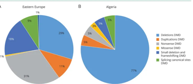
admixture for which we estimated these countries as a single group for our DMD mutation statistical analysis. From the Eastern European countries, a total of 258 patients were gen-otyped, and 169 patients were diagnosed with a DMD muta-tion. Mutations were predominantly nonsense (31%) and deletions (29%), followed by small frameshifting variations (18%), duplications (11%), and canonical site splicing variants (9%). Missense and consensus splicing variations were also identified (1%). Eighty-nine patients did not show a typical DMD mutation (detection rate 65%) (figure 1A).
A DMD mutation was identified in 52 of the 68 samples from Algeria (detection rate 76%) (figure 1B). The vast majority of Algerian patients carried deletions (77%), followed by all other mutations in very similar percentages. In a previous publication, a similar mutation landscape was reported.17By merging these data, 86% of Algerian patients carry deletions and 14% all others.
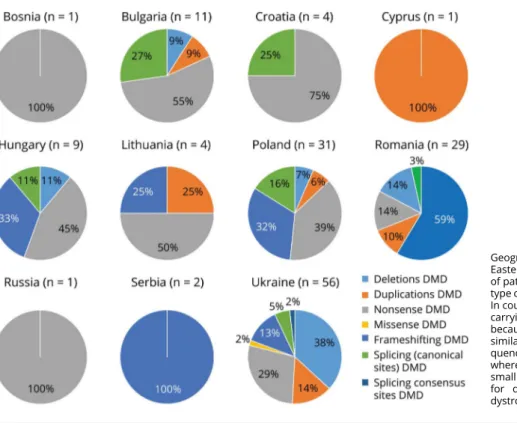
Figure 2 shows the overview of the mutation landscape in each Eastern European country and in Cyprus. The number of patients with BMD (24) is too low to allow statistics. Among the 28 at-risk females, 11 were identified as hetero-zygous carriers.
WES Analysis
Twenty-eight patients with proven DMD mutations (12 de-letions, 15 small mutations, and 1 duplication) were used to validate WES analysis (table e-3, links.lww.com/NXG/ A340).
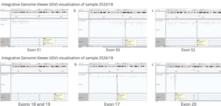
As expected, all DMD small variants previously detected using NGS sequencing were identified by WES analysis. In addition, all MLPA identified deletions were also picked up by WES. Figure 3, A and B shows software visualization of 2 deletions (exon 51 and exons 18–19), where no reads were visible within the deleted interval. In both cases, WES analysis defined the deletion inter-vals within intron borders, thus excluding intraexonic deletions. The duplication was not identified by WES (data not shown). DMD Gene Modifier Haplotyping
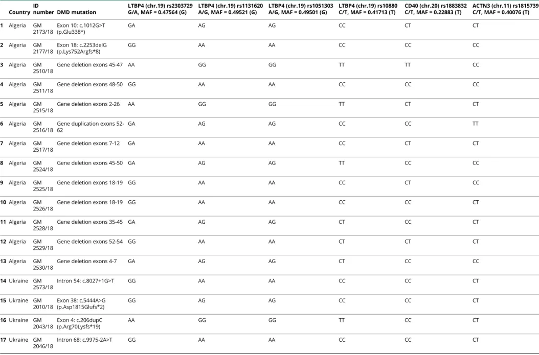
WES analysis performed in the 28 DMDs allowed to call SNPs variations in genes known to be DMD genetic modifiers as SPP1, LTBP4, CD40, THBS1, and ACTN3.18We could not obtain genotypes of SPP1 and THBS1 SNP regions (rs28357094 and rs2725797, respectively) because they were not covered by WES enrichment. LTPB4, CD40, and ACTN3 were fully covered, and we profiled the LFTB4 haplotypes and the CD40 and ACTN3 genotypes. Results are shown in table 2. We also identified in 8 patients other exonic SNPs occurring in 3 gene modifiers, namely 3 SNPs within the LTPB4 gene, 9 SNPs in ACTN3, and 2 SNPs in THBS1 (table e-4, links.lww. com/NXG/A341). The majority are synonymous (6) and missense changes (7), whereas 1 was a nonsense variant in exon 21 of the ACTN3 gene.
Mutations Amenable of New Therapeutic Approaches
Patients carrying deletions amenable of exon-skipping thera-peutic approaches were evaluated. In total, 41% of patients are
Figure 1Distribution of DMD Mutations in Patients With DMD in Countries
Overview of mutation distribution in patients with DMD from Eastern Europe (A) and Algeria (B). Nonsense mutations were the most frequently occurring mutations in Eastern European patients, accounting for 31% of mutation types in the patients with DMD, followed by deletions (29%), frameshifting (18%), duplications (11%), and splicing canonical sites (9%); missense and consensus splicing are the least frequent (1% each). Deletions were the most frequent mutations in Algerian patients (77%), whereas nonsense (5%), frameshifting (5%), splicing canonical sites (5%), duplication (5%), and missense (3%) variations were the least frequent. No splicing consensus sequence mutations were identified in patients from Algeria. The reported numbers include known and novel, likely pathogenic, mutations/variations, but not VUS. Among the 28 females tested for carrier detection, we found 11 carriers, all heterozygous for large deletions (6) and small mutations (5). The remaining females were not carriers. The 2 patients from Cyprus were not included in the statistical analysis as 1 patient showed an exon 2 duplication, whereas the other resulted negative. DMD = Duchenne muscular dystrophy; VUS = variant of uncertain/unknown significance.
eligible for exon 51 skipping (orphan drug being eteplirsen), 30% might be treated by exon 45 skipping, 18% by exon 53 skipping, and 11% by exon 44 skipping. Considering non-sense mutation correction by read-through mechanisms for stop codon reversion, 53 patients overall (50 DMDs or 27%) were found to be carrying a nonsense mutation for which ataluren is an approved orphan drug (figure 1).
Discussion
DMD mutation identification is an integral part of the di-agnosticflowchart for patients and their family, allowing ge-netic diagnosis, family planning, prenatal testing, and eligibility for personalized treatments.19 Because of the enormous size of the dystrophin gene, the mutational spec-trum is tremendously heterogeneous, and the genetic di-agnostic approach must be both accurate and sensitive. Here, we provide DMD gene genetic characterization in a large group of 328 patients with a clinical diagnosis of DMD/ BMD, from Eastern European and non-European patients diagnosed between June 2016 and December 2019. Standard diagnostic approach based on best practice guidelines iden-tified mutations in 222 patients.
Our analysis highlighted a relevant diversity in the muta-tion type across countries. Indeed, the frequency of dele-tions in Algeria (77%) vs Eastern Europe (29%) greatly varies. Conversely, nonsense mutations (31%) and small
mutations (29%) represent the most frequent mutation type in these last countries, while rarely occurring in Algeria (18%). Ethnic origin and genotype assortments may play a role in this evident variable mutation distribution. The high percentage of still undiagnosed negative cases (34% East-ern Europe and 27.4% Algeria) is remarkable. We may firstly consider that some DMD mutations may have es-caped MLPA and sequencing testing, as for atypical mu-tations occurring in regulatory regions or due to deep intronic variants/rearrangements.20 Because these are es-timated to be less than 1% in various patient series,21,22,23,24 their impact on the detection rate should be low, and only a few cases might have been missed. Ethnicity may also play a role in mutation type frequency, as already described23; only the adoption of a single and fully accurate method able to detect all mutation types, such as whole-genome se-quencing (WGS), will allow us to unravel these events and their frequency in the various populations.25Second, a few patients referred as DMD/BMD or generally as possible dystrophinopathies may represent phenocopies of other clinical entities like limb-girdle muscular dystrophies (LGMDs) or other rare hereditary myopathies.26,27 In-deed, many patients with high CK-related phenotypes and resulted as not carrying a DMD mutation showed DMD VUSs which might be difficult to interpret (see below). LGMDs may indeed deserve a more intensive investigation, but this requires a multigene approach, for example, gene panel testing, WES, or, in the near future, WGS.28Finally, in countries with high consanguinity like Algeria,29a lower detection rate due to the occurrence of many recessive
Figure 2Country Distribution of DMD Mutations in Patients With DMD in Eastern European Countries
Geographical distribution of DMD mutations in Eastern European countries and relative number of patients (in brackets). The percentages for each type of DMD mutation are shown for each country. In countries with more than 30 patients with DMD carrying a DMD mutation, differences can be noted because Polish and Ukrainian patients have a similar mutation landscape with a very high fre-quency of small mutations (average of 55%), whereas in Romania, 31% of mutation types are small mutations with deletions, and 69% account for duplications. DMD = Duchenne muscular dystrophy.
LGMDs might be expected. In Algeria, however, the DMD detection rate was higher than in the Eastern European countries. Although the lack of muscle biopsy availability in negative patients did not allow an RNA study to look for DMD mutations, in some negative cases, the new tool of urinary stem cells to profiling the DMD transcript might be considered.30 Because the DNA testing remains the gold standard to achieve a genetic diagnosis, only a genome-based diagnostic approach will allow us to define the full DMD mutation scenario.
We identified 9 previously unreported VUSs (1 shared by 2 brothers) in the DMD gene. The pathogenic effect of these VUSs is often based on an in silico prediction model (table e-2, links.lww.com/NXG/A339), although VUS requires functional validation, possibly by RNA profiling or segrega-tion studies in larger families to assess their pathogenic meaning. Therefore, their meaning remains hypothetical. We fully validated WES analysis in our pilot study. All small mutations and large deletions were correctly identified. Therefore, WES may cover more than 90% of all DMD gene mutations for certain populations, and MLPA might only be performed in negative cases to identify duplications. The adoption of this approach, especially if running many patients in parallel, could reduce costs and time to diagnosis, bringing to light more than 90% of the causative mutations of dystrophinopathies. Furthermore, the use of WES as a first approach would not only allow the study of the
variations of the DMD gene but also all other genes of clinical interest.
WES provides the great advantage to possibly interrogate the data for SNPs in gene modifiers. Indeed, we were able to profile the genotypes of LTBP4, CD40, and ACTN3. Al-though the small numbers (28 patients) do not allow a genotype-phenotype correlation, some differences in geno-type assortment are visible if comparing Algerian and Eastern Europe (Ukraine) patients. Looking at the allele frequency of LTBP4 SNPs (which form a 4-SNP haplotype), Ukrainian patients have higher frequency of the A (19/28), G (19/28), G (19/28), and T (18/28) alleles compared with Algerian patients. This implies that the VAAM (AGGT) protective haplotype frequency in patients may vary, therefore having a less powerful value for predicting milder DMD phenotype (in terms of loss of ambulation). Indeed, ethnic differences in SNPs allele frequency may hamper a meaningful statistical association in population studies. WES also identified pre-viously unreported SNPs in LTBP4, ACTN3, and THBS1. The heterozygous ACTN3 exon 21 nonsense variation is unreported and may have a deleterious effect with a possible role in modulating the DMD phenotype.
The mutation landscape also allows for the evaluation of personalized treatment eligibility.31Eteplirsen (Exondys 51) which induces DMD exon 51 skipping in amenable patients,32 is an approved orphan drug for DMD in the USA. Other molecules that induce exon 45 and 53 skipping are also cur-rently in clinical trials.
Figure 3Integrative Genome Viewer (IGV) Visualization
(A) IGV visualization of sample 2533/18. Visualization of sample 2533/18 with deletion of exon 51: (a) there is no coverage or readings of exon 51. (b and c) Visualization of coverage and reads of exons 50 and 52, which precede and follow the deletion of exon 51, respectively. (B) IGV visualization of sample 2526/ 18. Visualization of sample 2526/18 with deletion of exons 18–19. (a) This figure shows that there is no coverage or readings corresponding to exons 18 and 19. (b and c) Visualization of coverage and reads of the flanking exon 17 preceding the deletion and of exon 20 following the deletion.
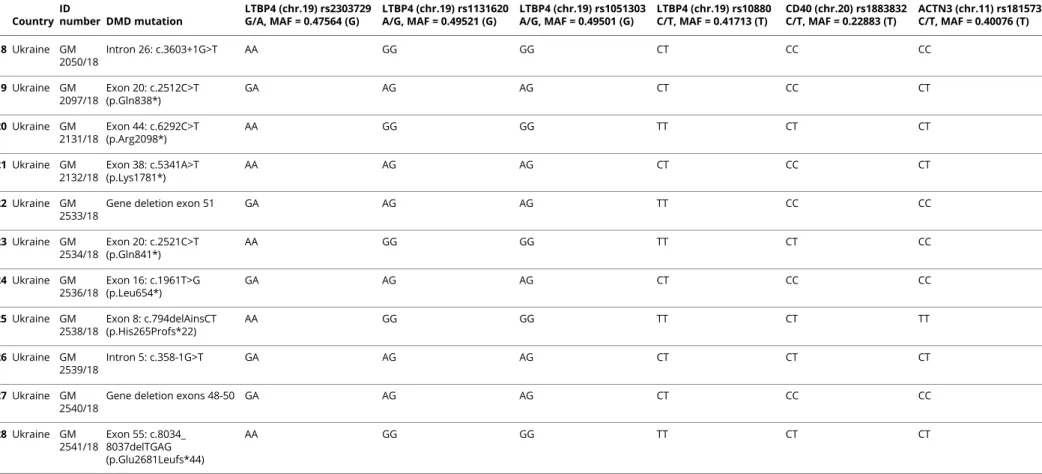
Table 2 Known Gene Modifiers: Genotyping of Patients Studied by WES Country ID number DMD mutation LTBP4 (chr.19) rs2303729 G/A, MAF = 0.47564 (G) LTBP4 (chr.19) rs1131620 A/G, MAF = 0.49521 (G) LTBP4 (chr.19) rs1051303 A/G, MAF = 0.49501 (G) LTBP4 (chr.19) rs10880 C/T, MAF = 0.41713 (T) CD40 (chr.20) rs1883832 C/T, MAF = 0.22883 (T) ACTN3 (chr.11) rs1815739 C/T, MAF = 0.40076 (T) 1 Algeria GM 2173/18 Exon 10: c.1012G>T (p.Glu338*) GA AG AG CC CT CT 2 Algeria GM 2177/18 Exon 18: c.2253delG (p.Lys752Argfs*8) GG AA AA CC CC CC 3 Algeria GM 2510/18
Gene deletion exons 45-47 AA GG GG TT TT CC
4 Algeria GM 2511/18
Gene deletion exons 48-50 GG AA AA CC CC CC
5 Algeria GM 2515/18
Gene deletion exons 2-26 AA GG GG TT CT CT
6 Algeria GM 2516/18
Gene duplication exons 52-62
GA AG AG CC CC TT
7 Algeria GM 2517/18
Gene deletion exons 7-12 GA AA AA CC CT CT
8 Algeria GM 2524/18
Gene deletion exons 45-50 GA AG AG TT CC CC
9 Algeria GM 2525/18
Gene deletion exons 18-19 GG AA AA CC CT CC
10 Algeria GM 2526/18
Gene deletion exons 18-19 GG AA AA CC CC CT
11 Algeria GM 2528/18
Gene deletion exons 35-45 GA AG AG CT CC CT
12 Algeria GM 2529/18
Gene deletion exons 52-54 GG AA AA CT CT CT
13 Algeria GM 2530/18
Gene deletion exons 4-7 GA AG AG CT CC CC
14 Ukraine GM 2573/18 Intron 54: c.8027+1G>T GG AA AA CC CC CT 15 Ukraine GM 2010/18 Exon 38: c.5444A>G (p.Asp1815Glufs*2) GG AG AG CC CC CT 16 Ukraine GM 2043/18 Exon 4: c.206dupC (p.Arg70Lysfs*19) AA GG GG TT CC CT 17 Ukraine GM 2046/18 Intron 68: c.9975-2A>T GG AA AA CC CC CT Continued 8 Neurology: Genetics | Volume 7, Number 1 | February 2021 Neurology. org/NG
Table 2 Known Gene Modifiers: Genotyping of Patients Studied by WES(continued) Country ID number DMD mutation LTBP4 (chr.19) rs2303729 G/A, MAF = 0.47564 (G) LTBP4 (chr.19) rs1131620 A/G, MAF = 0.49521 (G) LTBP4 (chr.19) rs1051303 A/G, MAF = 0.49501 (G) LTBP4 (chr.19) rs10880 C/T, MAF = 0.41713 (T) CD40 (chr.20) rs1883832 C/T, MAF = 0.22883 (T) ACTN3 (chr.11) rs1815739 C/T, MAF = 0.40076 (T) 18 Ukraine GM 2050/18 Intron 26: c.3603+1G>T AA GG GG CT CC CC 19 Ukraine GM 2097/18 Exon 20: c.2512C>T (p.Gln838*) GA AG AG CT CC CT 20 Ukraine GM 2131/18 Exon 44: c.6292C>T (p.Arg2098*) AA GG GG TT CT CT 21 Ukraine GM 2132/18 Exon 38: c.5341A>T (p.Lys1781*) AA AG AG CT CC CT 22 Ukraine GM 2533/18
Gene deletion exon 51 GA AG AG TT CC CC
23 Ukraine GM 2534/18 Exon 20: c.2521C>T (p.Gln841*) AA GG GG TT CT CC 24 Ukraine GM 2536/18 Exon 16: c.1961T>G (p.Leu654*) GA AG AG CT CC CC 25 Ukraine GM 2538/18 Exon 8: c.794delAinsCT (p.His265Profs*22) AA GG GG TT CT TT 26 Ukraine GM 2539/18 Intron 5: c.358-1G>T GA AG AG CT CT CT 27 Ukraine GM 2540/18
Gene deletion exons 48-50 GA AG AG CT CC CC
28 Ukraine GM 2541/18 Exon 55: c.8034_ 8037delTGAG (p.Glu2681Leufs*44) AA GG GG TT CT CT
Abbreviations: DMD = Duchenne muscular dystrophy; SNP = single nucleotide polymorphism; WES = whole-exome sequencing.
The modifier SNPs of genes LTBP4, CD40, and ACTN3, already known to be associated with the DMD phenotypic variability, are reported. Four missense SNPs (rs2303729, rs1131620, rs1051303, and rs10880) along the coding sequence of LTBP4, SNP rs1883832 in the 59-UTR of CD40, and SNP rs1815739 of ACTN3 (R577X) were studied, and the genotypes are reported. For each sample, ID number, DMD mutation, and SNPs genotypes are reported. SNP rs28357094 of the SPP1 gene and SNPs rs2725797 and rs2624259 of the THBS1 gene could not be studied due to their location in the promoter (5 bases upstream of the transcription start site) and in a telomeric region situated about 750 kb flanking a strong enhancer site, respectively.
Neurology. org/NG Neurology: Genetics | Volume 7, Number 1 | February 2021
Indeed, 51 is the most skippable exon (orphan drug ete-plirsen), 30% might be treated by exon 45 skipping, 18% by exon 53 skipping, and 11% by exon 44 skipping. Considering nonsense mutation correction by read-through mechanisms for stop codon reversion, 53 patients overall (23.9%) were found to be carrying a nonsense mutation for which ataluren is an approved orphan drug. There were, however, remarkable differences when considering country distribution, with Eastern European countries showing 31% of patients treatable using a nonsense mutation correction approach and Algeria just 5%.33
Genetic characterization in different and large patient cohorts prompts many reflections on DMD/BMD prevalence, mu-tation type, gene modifiers allele frequency, and personalized therapy, as we have explored in Eastern Europe and in Algeria. We suggest that diagnostic flowcharts should be adjusted depending on ethnic characteristics. In some populations where small mutations are more frequent than deletions and/ or duplications, like in Eastern European countries, WES might be indeed the preferredfirst-step diagnostic tool. These studies will have an impact on diagnostic approaches pipelines with repercussions on care and design of new therapies. Acknowledgment
PTC Therapeutics (United States) is gratefully acknowl-edged for funding the DMD diagnostic project “Interna-tional DMD” (to A. Ferlini, I-DMD; ospfe.it/reparti-e- servizi/reparti-dalla-a-alla-m-1/genetica-medica/genetica- medica-english/medical-genetics-unit/genetica-medica-ser-vices/dmd-international-project). The authors thank Ms. Courtney King (Medical Genetics Unit, Department of Medical Sciences, University of Ferrara, Ferrara, Italy) for the English revision of this article. Thanks are also due to the EU SOLVE-RD project (to A. Ferlini). The authors are grateful to Drs. Orza Diana, MD, Cristea Alexandru, MD, Puia Virgil-Petru, MD (Spitalul Clinic de Urgenta Pentru Copii Louis Turcanu, Timisoara, Romania), Radenka Kuzmanic-Samija, MD (University Clinical Hospital Split, Croatia), and Alexandra Grozavu, MD (Bac˘au County Emergency Hospital, Romania) because they referred to them patients for DMD diagnosis. A. Ferlini and other authors of this article are full members of the European Reference Network (ERN) Euro-NMD.
Study Funding
PTC Therapeutics service grant (to A. Ferlini). All clinicians and authors, listed in authorship, disclose that the patient genetic diagnosis was supported by this PTC service grant. Disclosure
The authors report no disclosures. Go to Neurology.org/NG for full disclosures.
Publication History
Received by Neurology: Genetics May 24, 2020. Accepted infinal form October 21, 2020.
AppendixAuthors
Name Location Contribution Rita Selvatici,
PhD
Medical Genetics Unit, Department of Medical Sciences, University of Ferrara, Italy
Designed and conceptualized the study, wrote the paper, performed and interpreted the DMD molecular testing, interpreted the WES data, and performed the variant calling
Rachele Rossi, PhD
Medical Genetics Unit, Department of Medical Sciences, University of Ferrara, Italy
Performed and interpreted the DMD molecular testing, interpreted the WES data, performed the variant calling, and edited the paper
Fernanda Fortunato, MD
Medical Genetics Unit, Department of Medical Sciences, University of Ferrara, Italy
Performed and interpreted the DMD molecular testing Cecilia
Trabanelli, MSc
Medical Genetics Unit, Department of Medical Sciences, University of Ferrara, Italy
Performed and interpreted the DMD molecular testing Yamina Sifi, PhD Neurologie, CHU de
Benbadis, Constantine, Alg´erie
Collected and referred to us patients for DMD diagnosis
Alice Margutti, MSc
Medical Genetics Unit, Department of Medical Sciences, University of Ferrara, Italy
Performed and interpreted the DMD molecular testing Marcella Neri,
MD, PhD
Medical Genetics Unit, Department of Medical Sciences, University of Ferrara, Italy
Performed and interpreted the DMD molecular testing Francesca
Gualandi, MD, PhD
Medical Genetics Unit, Department of Medical Sciences, University of Ferrara, Italy
Performed and interpreted the DMD molecular testing Lena Szab`o, MD 2nd Department of
Paediatrics, Semmelweis University, Budapest
Collected and referred to us patients for DMD diagnosis
Balint Fekete, MD
Institute of Genomic Medicine and Rare Disorders, Semmelweis University, Budapest, Hungary
Collected and referred to us patients for DMD diagnosis Lyudmilla Angelova, MD Department of Medical Genetics, Medical University, Varna, Bulgaria
Collected and referred to us patients for DMD diagnosis
Ivan Litvinenko, MD
Medical Univesity Sofia, Department of Pediatrics; University Pediatric Hospital“Prof. Ivan Mitev” Sofia, Department of Child Neurology, Bulgaria
Collected and referred to us patients for DMD diagnosis
Ivan Ivanov, MD Department of Pediatrics, St. George University Hospital, Faculty of Medicine, Medical University, Plovdiv, Bulgaria
Collected and referred to us patients for DMD diagnosis
Yurtsever Vildan, MD
Pediatric Neurologist County Clinical Emergency Hospital of Constanta, Pediatric Neurology Department, Romania
Collected and referred to us patients for DMD diagnosis
Appendix (continued)
Name Location Contribution Oana Alexandra
Iuhas, MD
G. Curteanu Municipal Clinical Hospital Oradea, Romania
Collected and referred to us patients for DMD diagnosis
Mihaela Vintan, MD
Iuliu Hatieganu University of Medicine and Pharmacy Cluj-Napoca, Romania, Department of Neuroscience
Collected and referred to us patients for DMD diagnosis
Carmen Burloiu,
MD “Alexandru Obregia”Clinical Psichiatry Hospital, Pediatric Neurology Department, Bucharest, Romania
Collected and referred to us patients for DMD diagnosis
Butnariu Lacramioara, MD
University of Medicine and Pharmacy Grigore T Popa Iasi, Romania Hospital Sf. Maria Iasi
Collected and referred to us patients for DMD diagnosis
Gabriela Visa, MD
Pediatric Clinical Hospital Sibiu, Romania
Collected and referred to us patients for DMD diagnosis
Diana Epure, MD “Dr. Victor Gomoiu” Children’s Hospital, Bucharest, Romania
Collected and referred to us patients for DMD diagnosis
Cristina Rusu, MD
1.“Grigore T Popa” University of Medicine and Pharmacy, Iasi, Romania 2. “Sfanta Maria”Children’s Hospital, Iasi, Romania
Collected and referred to us patients for DMD diagnosis
Daniela Vasile, MD
Victor Gomoiu Children’s Hospital, Bucharest, Romania
Collected and referred to us patients for DMD diagnosis
Magdalena Sandu, MD
Victor Gomoiu Children’s Hospital Carol Davila University of Medicine and Pharmacy, Bucharest, Romania
Collected and referred to us patients for DMD diagnosis Dmitry Vlodavets, MD Russian Children Neuromuscular Center, Veltischev Clinical Pediatric Research Institute of Pirogov Russian National Research Medical University, Moscow, Russia
Collected and referred to us patients for DMD diagnosis
Monica Mager, MD, PhD
Iuliu Hatieganu’ University of Medicine and Pharmacy, Faculty of Medicine, Department of Neuroscience, Neurology and Pediatric Neurology. Emergency Clinical Hospital for Children, Pediatric Neurology Department, Cluj-Napoca, Romania
Collected and referred to us patients for DMD diagnosis
Theodore Kyriakides, MD, PhD
Department of Basic and Clinical Sciences, University of Nicosia, Cyprus
Collected and referred to us patients for DMD diagnosis
Sanja Delin, MD General Hospital Zadar, Department of Pediatrics, Zadar, Croatia
Collected and referred to us patients for DMD diagnosis
Ivan Lehman, MD
Department of Paediatrics; University Hospital Centre Zagreb, Zagreb, Croatia
Collected and referred to us patients for DMD diagnosis
Appendix (continued)
Name Location Contribution Jadranka Sekelj
Fureˇs, MD
Children’s Hospital Zagreb, Faculty of Medicine, University of Osijek, Croatia
Collected and referred to us patients for DMD diagnosis
Veneta Bojinova, MD
University Hospital of Neurology and Psychiatry Sveti Naum, Sofia, Bulgaria
Collected and referred to us patients for DMD diagnosis
Mariela Militaru, MD
University of Medicine and Pharmacy“Iuliu
Hatieganu ’’,Cluj-Napoca,Romania and Genetic Center, Romania
Collected and referred to us patients for DMD diagnosis Velina Guergueltcheva, MD Clinic of Neurology, University Hospital Sofiamed and Sofia University“St. Kliment Ohridski”, Sofia, Bulgaria
Collected and referred to us patients for DMD diagnosis Birute Burnyte, MD Institute of Biomedical Sciences, Faculty of Medicine, Vilnius University, Lithuania
Collected and referred to us patients for DMD diagnosis
Maria Judith Molnar, MD, PhD
Institute of Genomic Medicine and Rare Disorders, Semmelweis University, Budapest, Hungary
Collected and referred to us patients for DMD diagnosis
Niculina Butoianu, MD
Carol Davila Faculty of Medicine and Pharmacy, Bucharest, Pediatric Neurology Clinic“Al. Obregia Hospital”, Romania
Collected and referred to us patients for DMD diagnosis
Selma Dounia Bensemmane, MD
Ali Ait Idir Hospital, Algiers, Algeria. University of Algiers I.
Collected and referred to us patients for DMD diagnosis
Samira Makri-Mokrane, MD
Ali Ait Idir Hospital, Algiers, Algeria. University of Algiers I.
Collected and referred to us patients for DMD diagnosis Agnes Herczegfalvi, MD 2nd Pediatric Clinic of Semmelweis University Budapest, Hungary
Collected and referred to us patients for DMD diagnosis
Monica Panzaru, MD
“Grigore T. Popa” University of Medicine and Pharmacy, Iasi, Romania; “Sfanta Maria” Children’s Hospital Iasi, Romania
Collected and referred to us patients for DMD diagnosis Adela Chirita Emandi, MD 1. Center of Genomic Medicine, University of Medicine and Pharmacy Victor Babes Timisoara, Romania. 2. Regional Center of Medical Genetics Timis, Clinical Emergency Hospital for Children Louis Turcanu Timisoara, Romania
Collected and referred to us patients for DMD diagnosis Anna Lusakowska, MD Department of Neurology, Medical University of Warsaw, Poland
Collected and referred to us patients for DMD diagnosis Anna Potulska-Chromik, MD Department of Neurology, Medical University of Warsaw, Poland
Collected and referred to us patients for DMD diagnosis Anna Kostera-Pruszczyk, MD, PhD Department of Neurology, Medical University of Warsaw, Poland
Collected and referred to us patients for DMD diagnosis
References
1. Muntoni F, Torelli S, Ferlini A. Dystrophin and mutations: one gene, several proteins, multiple phenotypes. Lancet Neurol 2003;2:731–740.
2. Saengpattrachai M, Ray PN, Hawkins CE, Berzen A, Banwell BL. Grandpa and I have dys-trophinopathy?: approach to asymptomatic hyperCKemia. Pediatr Neurol 2006;35:145–149. 3. Zimowski JG, Pilch J, Pawelec M, et al. A rare subclinical or mild type of Becker
muscular dystrophy caused by a single exon 48 deletion of the dystrophin gene. J Appl Genet 2017;58:343.
4. Falzarano MS, Scotton C, Passarelli C, Ferlini A. Duchenne muscular dystrophy: from diagnosis to therapy. Molecules 2015;20:18168–18184.
5. Straub V, Balabanov P, Bushby K, et al. Stakeholder cooperation to overcome chal-lenges in orphan medicine development: the example of Duchenne muscular dys-trophy. Lancet Neurol 2016;15:882–890.
6. Sardone V, Zhou H, Muntoni F, Ferlini A, Falzarano MS. Antisense Oligonucleotide-based therapy for neuromuscular disease. Molecules 2017;22:563.
7. Birnkrant DJ, Bushby K, Bann CM, et al. Diagnosis and management of Duchenne muscular dystrophy, part 1: diagnosis, and neuromuscular, rehabilitation, endocrine, and gastrointestinal and nutritional management. Lancet Neurol 2018;17:251–267. 8. Fratter C, Dalgleish R, Allen SK, et al. Best practice guidelines for genetic testing in
dystrophinopathies. Eur J Hum Genet 2020;28:1141–1159.
9. Abbs S, Tuffery-Giraud S, Bakker E, Ferlini A, Sejersen T, Mueller CR. Best practice guidelines on molecular diagnostics in Duchenne/Becker muscular dystrophies. Neuromuscul Disord 2010;20:422–427.
10. Fang M, Abolhassani H, Lim CK, Zhang J, Hammarstr¨om L. Next generation se-quencing data analysis in primary immunodeficiency disorders—future directions. J Clin Immunol 2016;36(suppl 1):68–75.
11. Robinson JT, Thorvaldsd´ottir H, Wenger AM, Zehir A, Mesirov JP. Variant review with the Integrative Genomics Viewer (IGV). Cancer Research 2017;77:31–34.
12. den Dunnen JT. Sequence variant descriptions: HGVS nomenclature and Mutalyzer. Curr Protoc Hum Genet 2016;90:7.13.1–7.13.19.
13. McDonald CM, Campbell C, Torricelli RE, et al. Ataluren in patients with non-sense mutation Duchenne muscular dystrophy (ACT DMD): a multicentre, randomised, double-blind, placebo-controlled, phase 3 trial. Lancet 2017;390: 1489–1498.
14. Echevarr´ıa L, Aupy P, Goyenvalle A. Exon-skipping advances for Duchenne muscular dystrophy. Hum Mol Genet 2018;27:163–172.
15. Richards S, Aziz N, Bale S, Bick D, Das S, Gastier-Foster J. Standards and guidelines for the interpretation of sequence variants: a joint consensus recommendation of the American College of Medical Genetics and Genomics and the Association for Mo-lecular Pathology. Genet Med 2015;17:405–424.
16. Bhopal R, White DL. European, Western, Caucasian, or what? Inappropriate la-beling in research on race, ethnicity, and health. Am J Public Health 1998;88: 1303–1307.
17. Dalichaouche I, Sifi Y, Roudaut C, et al. γ-sarcoglycan and dystrophin mutation spectrum in an Algerian cohort. Muscle Nerve 2017;56:129–135.
18. Bello L, Pegoraro E. The“usual suspects”: genes for inflammation, fibrosis, re-generation, and muscle strength modify Duchenne muscular dystrophy. J Clin Med 2019;8:649.
19. Shimizu-Motohashi Y, Komaki H, Motohashi N, Takeda S, Yokota T, Aoki Y. Re-storing dystrophin expression in Duchenne muscular dystrophy: current status of therapeutic approaches. J Pers Med 2019;9:1.
20. Aartsma-Rus A, Hegde M, Ben-Omran T, et al. Consensus and systematic review on reducing the time to diagnosis of Duchenne muscular dystrophy. J Pediatr 2019;204: 305–313.
21. Bladen CL, Salgado D, Monges S, et al. The TREAT-NMD DMD Global Database: analysis of more than 7, 000 Duchenne muscular dystrophy mutations. Hum Mutat 2015;36:395–402.
22. Tuffery-Giraud S, B´eroud C, Leturcq F, et al. Genotype-phenotype analysis in 2,405 patients with a dystrophinopathy using the UMD-DMD database: a model of na-tionwide knowledgebase. Hum Mutat 2009;30:934–945.
23. Neri M, Rossi R, Trabanelli C, et al. The genetic landscape of dystrophin mutations in Italy: a nationwide study. Front Genet 2020;11:131.
24. Flanigan KM, Dunn DM, von Niederhausern A, et al. Nonsense mutation-associated Becker muscular dystrophy: interplay between exon definition and splicing regulatory elements within the DMD gene. Hum Mutat 2011;32:299–308.
25. Barseghyan H, Tang W, Wang RT, et al. Next-generation mapping: a novel approach for detection of pathogenic structural variants with a potential utility in clinical di-agnosis. Genome Med 2017;9:90.
26. Barohn RJ, Dimachkie MM, Jackson CE. A pattern recognition approach to patients with a suspected myopathy. Neurol Clin 2014;32:569–593.
27. Neri M, Selvatici R, Scotton C, et al. A patient with limb girdle muscular dys-trophy carries a TRIM32 deletion, detected by a novel CGH array, in compound heterozygosis with a nonsense mutation. Neuromuscul Disord 2013;23: 478–482.
28. Donkervoort S, Dowling JJ, Laporte J, MacArthur D, B¨onnemann CG; 214th ENMC workshop participants. 214th ENMC International Workshop: establishing an In-ternational Consortium for Gene Discovery and Clinical Research for Muscle Disease, Heemskerk, the Netherlands, 6-18 October 2015. Neuromuscul Disord 2019;29: 644–650.
29. Chentouf A, Talhi R, Dahdouh A, et al. Consanguinity and epilepsy in Oran, Algeria: a case-control study. Epilepsy Res 2015;111:10–17.
30. Falzarano MS, Ferlini A. Urinary stem cells as tools to study genetic disease: overview of the literature. A.J Clin Med 2019;8:627.
31. Aartsma-Rus A, Straub V, Hemmings R, et al. Development of exon skipping therapies for Duchenne muscular dystrophy: a critical review and a perspective on the out-standing issues. Nucleic Acid Ther 2017:251–259.
32. Syed YY. Eteplirsen:first Global approval. Drugs 2016;76:1699–1704.
33. Nakamura A. Moving towards successful exon-skipping therapy for Duchenne mus-cular dystrophy. J Hum Genet 2017;62:871–876.
Appendix (continued)
Name Location Contribution Andriy Shatillo,
MD
Institute of Neurology, Psychiatry and Narkology National Academy of Medical Science of Ukraine
Collected and referred to us patients for DMD diagnosis
Djawed Bouchenak Khelladi, MD
Neurologie, CHU Tidjani Damerji, Tlemcen, Algerie
Collected and referred to us patients for DMD diagnosis
Oussama Dendane, MD
Neurologie, CHU Tidjani Damerji, Tlemcen, Algerie
Collected and referred to us patients for DMD diagnosis Mingyan Fang, PhD BGI-Shenzhen, Shenzhen, China
Performed the WES and bioinformatics analysis Zhiyuan Lu, PhD BGI-Shenzhen, Shenzhen,
China
Performed the WES and bioinformatics analysis Alessandra
Ferlini, MD, PhD
Medical Genetics Unit, Department of Medical Sciences, University of Ferrara, Italy
Designed and
conceptualized the study and wrote the paper