S100B Protein in Biological Fluids: A Tool for
Perinatal Medicine
Fabrizio Michetti
1*and Diego Gazzolo
2The diagnosis of perinatal insults currently relies on adequate documentation of general medical and obstet-ric factors and on radiologic and laboratory assessments. The measurement of brain constituents such as S100B protein may offer an alternative and direct indicator of cell damage in the nervous system when clinical and radiologic assessments are still silent and has the addi-tional advantage of providing a quantitative indicator of the extent of brain lesions. S100B protein has been measured by several immunoassays in biological fluids (i.e., cerebrospinal fluid, blood, amniotic fluid, and urine) from fetuses and newborns at high risk of peri-natal brain damage. S100B protein in biological fluids increased at an early stage when standard monitoring procedures were still silent in the study populations that later developed brain damage. S100B concentration was also significantly correlated with the extent of brain lesions. S100B protein appears to satisfy the criteria for a marker for brain injuries in perinatal medicine: (a) simple to perform measurements with good reproduc-ibility; (b) detection in a variety of biological fluids, possibly reducing perinatal stress related to testing; (c) possible use in longitudinal monitoring because of its 1-h half-life; and (d) well-established use as an early and quantitative marker of brain lesions/damage. Finally, because of the neurotrophic role putatively played by S100B, its measurement in biological fluids at pre-/ perinatal ages makes it a candidate for the laboratory evaluation of brain maturation.
© 2002 American Association for Clinical Chemistry
Epidemiology of Perinatal Brain Damage
Our knowledge of the timing of adverse insults is impor-tant in relation to future measures of prevention. How-ever, such knowledge is still incomplete and under de-bate. For example, the reported contribution of asphyxia at birth to cerebral palsy in infants born at term varies from 8% to 28% (1–3 ).
Preterm birth accounts for most cases of perinatal mortality and for ⬃40% of neurologically handicapped children. According to population studies by Hagberg et al. (4 ),⬃60% of neurologic handicaps in preterm infants are attributable to peri-/neonatal events, 10% are of antenatal origin, and 30% are of generally unknown origin. In infants born at term, 50% of cases of cerebral palsy have a prenatal etiology, 36% are of peri-/neonatal origin, and 14% of cases are of unknown etiology. Our understanding of the timing of insults and of contributing factors may be improved by adequate documentation of general medical and obstetric factors, determination of pH and blood gases in cord blood, and neonatal neuro-imaging. Other diagnostic tools that may be of crucial importance are measurements of markers of perinatal brain injury in biological fluids. The key word is “preven-tion”, with the aim of improving our ability to detect fetuses and newborns at risk of brain injury at an earlier stage, when the window for therapeutic action is still open. Both pre- and postnatal interventions show consid-erable promise: antenatal administration of glucocorti-coids, the optimal management of labor and delivery, and the postnatal stabilization of critical newborns seem to dramatically improve the quality of life of these individ-uals. Significant decreases in the rate of cerebral bleeding and of adverse long-term neurologic outcome have been shown (5– 8 ). On the other hand, the same findings support the need for additional markers to optimize the timing of treatment and, at the same time, to monitor their true effectiveness.
The possibility of longitudinal monitoring of the effects of drugs and supportive care is second in importance only to prevention as a means to minimize brain trauma and the numbers of neurologically handicapped children.
1Institute of Anatomy, Catholic University, I-00168 Rome, Italy. 2Department of Pediatrics, Giannina Gaslini Children’s University
Hospi-tal, I-16148 Genoa, Italy
*Address correspondence to this author at: Institute of Anatomy, Catholic University, Largo Francesco Vito, 1, I-00168 Rome, Italy. Fax 39-063054813; e-mail [email protected].
Received October 30, 2001; accepted August 9, 2002.
Standard Diagnostic Procedures for Early Detection of Perinatal Brain Damage
The essential steps for establishing a diagnosis of cerebral bleeding or of hypoxic ischemic events, the main factors involved in the genesis of perinatal brain damage in preterm and term infants, are similar. They are based on clinical examination, continuous electroencephalographic monitoring, cerebral ultrasound and Doppler velocimetry recordings, cerebrospinal fluid (CSF)3 assessment,
cere-bral computerized tomography, magnetic resonance im-aging, and proton magnetic resonance spectroscopy (5, 6 ). These tools can provide useful and crucial information regarding the presence, location, and extent of brain injury and may be useful in establishing a prognosis. Although there is a wide range of diagnostic possibilities, there are several problems associated with the early diagnosis of cases at risk. The limited interval for diagno-sis and therapeutic intervention and the confusing/am-biguous effects of sedative and anticonvulsant drugs are the main factors involved. This particularly applies to neurologic examination (5, 6 ), electroencephalography (9 ), and cerebrovascular recordings (10 –12 ). Other limi-tations are poor reproducibility and the need for complex measurements (CSF measurements), infrequent use of longitudinal monitoring, and high costs (computerized tomography, magnetic resonance imaging, and proton magnetic resonance spectroscopy). For these reasons, ce-rebral ultrasound scanning is the procedure of choice for diagnosis, although the progression and extent of hemor-rhage and brain insults can be defined only at a later stage (i.e., after more than 6 –12 h), which limits the possibility of intervention (5, 6 ). In this respect, therapeutic proce-dures that aim to maintain adequate cerebrovascular perfusion and new approaches, such as selective head cooling with or without moderate hypothermia, have been shown to improve the outcome for these infants (6 ). Nevertheless, the need for additional markers that enable longitudinal monitoring and that assess the effectiveness of these interventions is justified.
S100B Protein: Biochemical, Biological, and Pathophysiologic Features
The term S100B refers to members of a multigenic family of calcium-modulated proteins (S100 proteins), mostly of low molecular mass (⬃10 000 Da), that were first identi-fied (on the basis of methods available at the time) as a protein fraction detectable in brain but not in nonneural extracts and called S100 because of their solubility in a 100%-saturated solution with ammonium sulfate (13 ). At present, at least 20 proteins have been identified as belonging to the S100 protein family, the members of
which are characterized by the presence of a pair of so-called EF-hand (i.e., helix-loop-helix) calcium-binding motifs (14 ), first discovered in the crystal structure of parvalbumin (15 ), that induce conformational changes of the protein after binding to calcium (16, 17 ). This confor-mational change may facilitate the interaction of S100 proteins with a secondary effector: S100 proteins are generally thought to be calcium sensor proteins that modulate biological activity via calcium binding (18 ). In addition, some S100 members have been shown to bind Zn2⫹ and Cu2⫹ (19, 20 ), suggesting the possibility that their biological activity in some cases might be regulated by Zn2⫹and/or Cu2⫹, rather than by Ca2⫹(21 ).
Interestingly, the S100 proteins are highly conserved in amino acid composition among vertebrate species (22 ), and S100-like proteins have also been immunologically detected in planarians and spinach leaves (23, 24 ). Most S100 proteins exist as dimers (frequently homodimers) within cells and are generally expressed and distributed in a cell-specific fashion, indicating a conserved biological role.
In particular, S100B, a homodimer of a subunit ( subunit) that constitutes the bulk of the fraction originally isolated from brain extracts, was regarded for more than a decade as specific to the nervous system. A later study showed that the protein was not restricted to the nervous system (25 ). Since then, the location of S100B has been extensively studied in mammalian tissues, including hu-man tissues (26 –30 ). In the nervous system, the protein appears to be most abundant in glial cells, although its presence in neuronal subpopulations has also been re-ported (31, 32 ). In nonneural tissues, it is distributed widely in definite cell types, including melanocytes, Langerhans cells, chondrocytes, folliculostellate cells of the adenohypophysis, adrenal gland satellite cells, Leydig cells, and interdigitating reticulum cells (27 ), whereas adipose tissue constitutes a site of concentration for the protein comparable to the nervous tissue (33 ).
The biological role of this protein within the cell populations that contain it has not been completely elu-cidated, although many hypotheses have been formu-lated, including the inhibition of protein phosphorylation (34 –38 ), inhibition of cytoskeletal constituent assembly (39 – 42 ), stimulation of enzyme activities (43– 46 ), and interaction with transcription factors (34, 47 ). An extracel-lular biological role has also been hypothesized for S100B, which secreted by astrocytes as a cytokine could have a neurotrophic effect during both development and nerve regeneration at physiologic (nanomolar) concentrations, but at high (micromolar) concentrations could be neuro-toxic, participating in the pathophysiology of neurode-generative disorders (16 ). In this respect, it could be relevant that S100B is coded on the long arm of chromo-some 21 (21q22.3) (48 ), which is also involved in the translocation that causes Down syndrome. An increase in gene expression for the protein has also been related to
3Nonstandard abbreviations: CSF, cerebrospinal fluid; IVH,
intraventric-ular hemorrhage; HIE, hypoxic-ischemic encephalopathy; and IUGR, intra-uterine growth retardation.
the neurodegenerative processes associated with both Down syndrome and Alzheimer disease, on the basis of findings indicating that-amyloid stimulates the synthe-sis of S100B, whereas -amyloid precursor protein in-creases in cultures exposed to S100B (49, 50 ). More gen-erally, S100B could take part in inflammatory diseases that eventually lead to Alzheimer-like disorders, in which chronic gliosis occurs (51 ). Interestingly, most studies of the extracellular function of S100B focus on the nervous system, whereas consistent hypotheses have not yet been formulated for a role of this protein in the extracellular environment in nonneural locations
S100B in Biological Fluids as a Marker of Brain Damage S100B protein has been measured in several biological fluids (CSF, blood, urine, and amniotic fluid) by a series of immunoassays, which were indiscriminately used in var-ious fluids. The earliest studies used immunoassays such as a microcomplement fixation assay, RIA, and a particle-counting immunoassay directly developed by the authors (52–55 ), whereas more recent studies have used very simple, sensitive, and inexpensive commercially available immunoassays such as a two-site IRMA (Sangtec), an immunoluminometric assay (Sangtec), and an ELISA (SynX Pharma). PCR has also been used to analyze S100B in blood and amniotic fluid (56 ).
csf
CSF was the first of various biological fluids in which the role of S100B as a marker of active brain damage was shown (52, 53 ). Since then, several studies have been conducted, first in adults and later in children, and have established that high concentrations of the protein indi-cate the occurrence of brain injury such as neurodegen-erative diseases, cerebral tumors, cerebral trauma, and cerebrovascular diseases (57, 58 ). The finding of a rapid increase in CSF S100B after brain damage, such as trau-matic or focal ischemic insult, has also been confirmed in animal models (59 ). In perinatal medicine, measurements of S100B protein in CSF have been used to monitor infants affected by perinatal asphyxia and posthemorrhagic ven-tricular dilatation brain damage during cardiac surgery (60 – 62 ) (Table 1). In this setting, S100B concentrations correlated with the extent of brain lesions, with long-term prognosis, and with neurologic impairment at 1 year of age or death before that time. The measurement of S100B concentrations in CSF has also been suggested as a tool to screen patients with the most pessimistic prognoses from neuroprotective trials. In this regard, the need to use age-matched reference values when evaluating S100B protein in neurologic diseases has been emphasized (63 ). It is also noteworthy that on the basis of CSF dynamics, a concentration gradient decreasing from ventricular and lumbar CSF (in contrast to blood-derived proteins) has also been shown (64 ).
blood
Since the pioneering observations in CSF, which repre-sents the biological fluid into which brain constituents might most directly be released during active brain dam-age, a series of studies have been performed to investigate the usefulness of measuring S100B in blood as an index of brain damage, given that blood is the most widely used fluid for laboratory tests. The studies were based on the hypothesis that during active brain injury at least some of the S100B released from the damaged tissue could spread into the systemic circulation (54 ), also as a result of hemodynamic rearrangement of the brain– blood barrier. S100B was measured mainly in the blood of patients affected by the above-mentioned diseases (see the section on CSF), to which was added malignant melanoma on the basis of the location of the protein in melanocytes (55, 65– 68 ). Many of the studies focused on trauma, supporting the notion that high blood concentrations of S100B are an index of posttraumatic brain damage (69, 70 ). Similar results were also obtained in rats after controlled cortical impact (71 ). However, caution has recently been sug-gested in this respect because, even in the absence of head injury, trauma has been shown to lead to high serum concentrations of S100B, probably originating from adi-pose tissue, where the protein is highly concentrated (72 ). This also applies when monitoring the occurrence of brain damage attributable to ischemia-reperfusion injury in patients subjected to open-heart surgery, in which thora-cotomy can induce traumatic release of S100B from adi-pose tissue (73 ). The latter studies suggest that nonacute S100B measurements may be of greater prognostic value
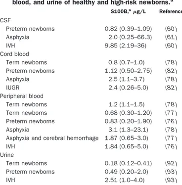
Table 1. S100B (g/L) in CSF, cord blood, peripheral blood, and urine of healthy and high-risk newborns.a
S100B,ag/L Reference CSF Preterm newborns 0.82 (0.39–1.09) (60 ) Asphyxia 2.0 (0.25–66.3) (61 ) IVH 9.85 (2.19–36) (60 ) Cord blood Term newborns 0.8 (0.7–1.0) (78 ) Preterm newborns 1.12 (0.50–2.75) (82 ) Asphyxia 2.5 (1.1–3.7) (78 ) IUGR 2.4 (0.26–5.0) (82 ) Peripheral blood Term newborns 1.2 (1.1–1.5) (78 ) Term newborns 0.68 (0.30–1.20) (77 ) Preterm newborns 0.83 (0.20–1.90) (76 ) Asphyxia 3.1 (1.3–23.1) (78 )
Asphyxia and cerebral hemorrhage 1.87 (0.65–3.0) (77 )
IVH 1.84 (0.65–5.0) (76 )
Urine
Term newborns 0.18 (0.12–0.41) (92 )
Preterm newborns 0.49 (0.20–2.0) (93 )
IVH 2.51 (1.0–4.0) (93 )
aData are expressed as medians. Values in parentheses correspond to the
than acute measurements. As for CSF, the first studies were performed in adult patients (73, 74 ), later in children (75 ), and finally in the perinatal period. At this stage, the usefulness of a marker able to detect the occurrence and the extent of brain lesions is particularly relevant because of the high mortality and morbidity rates in high-risk infants. In addition, the use of a brain constituent as a marker opened the possibility for a direct indication of brain damage when clinical and radiologic signs are still silent. Increased blood concentrations of S100B were in fact detected⬃48 to 72 h before any clinical, laboratory, or ultrasound signs of cerebral bleeding in preterm infants [intraventricular hemorrhage (IVH)] (76 ) or of hypoxic-ischemic encephalopathy (HIE) in full-term infants (Table 1) (77, 78 ). In the latter study, Nagdyman et al. (78 ) reported that S100B protein concentrations in cord blood were already significantly higher (range, 1.1–3.7g/L) in asphyxiated full-term infants suffering from birth as-phyxia and HIE.
The same authors performed longitudinal S100B pro-tein monitoring in peripheral blood and demonstrated a peak concentration of the protein 6 h after birth (range, 2.5–52.3 g/L, according to the severity of HIE) with a progressive decrease in S100B at 24 h (range, 1.2–9.0 g/L). The positive predictive value of S100B for HIE with a protein cutoff of 8.5 g/L at 2 h from birth was 71%, the negative predictive value was 90%, the sensitiv-ity was 71%, and the specificsensitiv-ity was 90% (78 ). S100B blood concentrations also correlated with abnormal cere-bral hemodynamic patterns (increased cerebrovascular resistance) and with the extent of IVH both in preterm and in full-term asphyxiated infants (76, 77 ).
In asphyxiated full-term infants, an early increase in S100B was found to be predictive of HIE and subsequent adverse neurologic outcomes (78 ). Because ischemia-reperfusion injury is known to be one of the major complications in newborn infants undergoing open-heart surgery, increased blood S100B has also been indicated as an index of brain injury attributable to surgical proce-dures (73, 75, 79 ). Similarly, S100B protein has also been used to monitor the occurrence of cerebral complications in preterm and term infants undergoing extracorporeal membrane oxygenation support for treatment of respira-tory distress (80, 81 ). The usefulness of measuring S100B in the cord blood of high-risk pregnancies, such as intra-uterine growth-retarded (IUGR) fetuses, in which in-creased S100B could indicate brain injury attributable to impaired feto-placental perfusion deserves attention (82, 83 ). In particular, S100B concentrations have been shown to be higher (3.6 ⫾ 1.44 g/L) in IUGR fetuses with redistribution of fetal-placental blood flow, the so-called “brain sparing effect”, and correlated with the degree of fetal hemodynamic impairment, as indicated by an altered middle cerebral artery Doppler pattern, whereas IUGR fetuses without “brain sparing effect” showed S100B concentrations similar (1.7⫾ 1.25g/L) to those of non-IUGR fetuses (1.12⫾ 0.74g/L) (82). In this
pathologic condition, disturbances in the organization of quiet or active fetal behavioral states, which are known to constitute echographic signs of brain injury (84, 85 ), were also correlated with high cord blood S100B concentrations (86 ). Cord blood S100B measurements were also used to assess the effects of vasodilation treatment with maternal NO administration in IUGR fetuses (87 ). At present, data correlating S100B protein with clinical prenatal data, other fetal well-being tests, and maternal medical conditions (e.g., hypertension, insulin-dependent diabetes, and co-agulopathies) are lacking: such data could be of use in investigating the role of S100B in fetomaternal diseases.
Finally, because the gene encoding for S100B is located on chromosome 21, higher concentrations of the protein have, not surprisingly, been found in the blood of fetuses with trisomy 21, although attempts to use measurements of S100B in maternal blood in screening for Down syn-drome, which is potentially very interesting, were unsuc-cessful: no significant differences in concentrations be-tween uncomplicated and trisomy 21 pregnancies were found (88, 89 ). These results are probably attributable to the effects of protein dilution or to the inability of the protein to pass into the placenta because of its molecular mass.
urine
Although studies of S100B concentrations in CSF and blood as a pathologic marker have offered consistent and useful results, the potential for developing new areas of investigation in perinatal medicine and improving the care of newborns depends on meeting the requirements of longitudinal monitoring of high-risk patients for the pos-sible occurrence of brain damage. This is particularly important because repeated blood sampling can induce anemia in premature infants (90 ). The usefulness of frequent S100B measurements is supported by data on the kinetic properties of this protein, which has a half-life of ⬃1 h and is eliminated mainly by the kidneys (91). For this reason, urine could constitute an excellent fluid for these studies. An initial report showed the presence of S100B in the urine of healthy preterm and term newborns and its correlation with gestational age at sampling, offering a normality reference curve (Table 1) (92 ). Similar studies were subsequently performed on urine (93 ) on the basis of previous investigations of the blood of brain-damaged preterm and term infants (76 –78 ). The results indicated that urine S100B concentrations at birth were significantly higher in preterm newborns (2.51 ⫾ 0.79 g/L) who later developed cerebral bleeding and/or brain damage at a stage when all routine clinical, labora-tory, and ultrasound investigations were still silent. Lon-gitudinal monitoring of urine S100B concentrations showed a progressive increase in the concentration of the protein with a peak at 72 h from birth (10.51⫾ 3.21g/L). The positive predictive value of S100B for IVH with a protein cutoff of 0.70g/L at 2 h from birth was 80.5%,
the negative predictive value was 100%, the sensitivity was 100%, and the specificity was 100%.
Future research protocols on S100B urinary patterns will probably provide a new and easier means of investi-gating pathophysiologic brain conditions (not only in the perinatal period), which have previously been studied by measuring the protein in CSF and blood, involving pro-cedures that are more stressful for patients.
amniotic fluid
The search for a nervous system constituent, such as S100B, that could be used to detect prenatal brain pathol-ogies in amniotic fluid is not new. In studies using a particle-counting immunoassay, the protein was shown to be present in the amniotic fluid of anencephalic and open spina bifida fetuses (94 ) as well as in cases of fetal death (95 ). Normal amniotic fluid appeared to be devoid of the protein. More recent studies used a more sensitive radio-immunometric assay (limit of detection, 0.2g/L vs 1.5 g/L) to propose a normality reference curve for S100B in amniotic fluid that appeared to be correlated with gesta-tional age, opening intriguing prospects concerning the putative neurotrophic role of the protein (96 ). In this respect, the recent finding of S100B in feto-placental tissues could be relevant (56 ). However, the possibility of a placental contribution to S100B concentrations in fetal fluids, despite a lack of evidence at present, should be taken into consideration (97 ).
The localization of the gene encoding for S100B on the long arm of chromosome 21, which is believed to be responsible for trisomy 21 (98 ), stimulated studies on the use of S100B measurements in amniotic fluid as a diag-nostic aid for Down syndrome. As expected, concentra-tions of the protein were⬃1.5 times higher in trisomy 21 amniotic fluid (0.92⫾ 0.45g/L) than in controls (0.52 ⫾ 0.24g/L) (97–99). However, as the difference in S100B concentrations is not sufficient to validate this test as a clear-cut screening tool and the relevant sampling proce-dures have no advantage over other, more specific meth-ods (reverse transcription-PCR, fluorescence in situ hy-bridization), these results are of scientific rather than clinical diagnostic value.
Future Prospectives
The bulk of studies showing the usefulness of S100B as a marker of brain injury have to date referred to measure-ments in CSF and blood. However, considering the simple procedure involved in urine sampling, it is possible to predict an increase in interest concerning the investigation of S100B in this biological fluid, which at present can be regarded only as a promising field of research. Likewise, the restricted number of studies on amniotic fluid at present offers poor information on fetal brain injury. Nevertheless, because amniotic fluid potentially offers the possibility of monitoring prenatal life, measurement of S100B in amniotic fluid remains a candidate method for the detection of fetal brain injury and/or fetal death.
Another potential use of S100B measurements in bio-logical fluids is to monitor the effectiveness of therapeutic intervention in high-risk term and preterm infants, as has already been reported in IUGR fetuses treated with NO (87 ).
Of the numerous functions attributed to the S100B protein, a neurotrophic role appears to be particularly intriguing in relation to perinatal medicine. After early observations in healthy fetuses by Zuckerman et al. (100 ) showing that the caudo-rostral pattern of accumulation of S100 parallels the biochemical, morphologic, and electro-physiologic maturation of the nervous system, attention has more recently focused on the possibility that informa-tion concerning the role of this protein could be obtained by studying the pattern of S100B concentrations in differ-ent biological fluids in the prenatal and perinatal periods. In the second trimester of pregnancy, when S100B is known to increase progressively in the brain cortex (101 ), a parallel increase at the same gestational age has been shown in the amniotic fluid of uncomplicated pregnan-cies. The amniotic fluid concentration of S100B not only correlated with gestational age, but also with echographic findings suggestive of brain development.
A pattern of S100B concentrations suggestive of a role in brain maturation processes has also been shown in cord blood in the third trimester of pregnancy, when a pro-gressive decrease of the protein is observed, possibly reflecting reduced release of the trophic factor at a later stage of fetal-neonatal brain maturation. The same pattern was observed in urine collected immediately after birth from healthy preterm and term newborns.
Finally, we recently found high concentrations of S100B (80 –100 times higher than in other biological fluids) in human milk (unpublished data), which may hypothet-ically constitute an exogenous source of the trophic factor in early brain developmental stages. On the other hand, to date there are no data on the absorption of S100B in maternal milk by infants and, therefore, on the effect of a potential contribution of exogenous S100B to measure-ments of the protein in the biological fluids of infants. In conclusion, taken together, the above findings could open the way to additional studies aimed at investigating a potential use of S100B measurements in perinatal med-icine and in pediatric patients as a monitoring tool for brain development.
This work was partially supported by grants from the Ministero dell’Universita` and from Universita` Cattolica del S. Cuore (to F.M.) and from the “Let’s Improve Prenatal Life Foundation” (to D.G.).
References
1. Freeman JM, Nelson KB. Intrapartum asphyxia and cerebral palsy. Pediatrics 1988;82:240 –9.
2. Hughes I, Newton R. Genetic aspects of cerebral palsy. Dev Med Child Neurol 1992:34; 80 – 6.
3. Hagberg B, Hagberg G, Olow I, van Wendt L. The changing panorama of cerebral palsy in Sweden. VII. Prevalence and origin in the birth year period 1987–1990. Acta Paediatr 1996;85: 954 – 60.
4. Hagberg B, Hagberg G, Beckung E, Uvebrandt P. Changing panorama of cerebral palsy in Sweden. VIII. Prevalence and origin in the birth year period 1991–1994. Acta Paediatr 2001;90: 271–7.
5. Volpe JJ. Intracranial hemorrhage: germinal matrix-intraventricu-lar hemorrhage of premature infant. In: Volpe JJ, ed. Neurology of the newborn. Philadelphia: WB Saunders, 1995:403– 63. 6. Volpe JJ. Hypoxic-ischemic encephalopathy: clinical aspects. In:
Volpe JJ, ed. Neurology of the newborn. Philadelphia: WB Saun-ders, 1995:314 –70.
7. Leviton A, Pagano M, Kuban KC, Krishnamoorthy KS, Sullivan KF, Allred EN. The epidemiology of germinal matrix hemorrhage during the first half-day of life. Dev Med Child Neurol 1991;33: 138 – 45.
8. Paneth N, Pinto-Martin J, Gardiner J, Wallenstein S, Katsikiotis V, Hegyi T, et al. Incidence and timing of germinal matrix/intraven-tricular hemorrhage in low birth weight infants. Am J Epidemiol 1993;137:1167–76.
9. Pezzani C, Radvanyi MF, Relier JP, Monod N. Neonatal electro-encephalography of the newborn during the first twenty-four hours of life in full-term newborn infants. Neuropediatrics 1986; 17:11– 8.
10. Rennie JM, South M, Morely CJ. Cerebral blood flow velocity variability in infants receiving assisted ventilation. Arch Dis Child 1987;62:1247–51.
11. Ilves P, Talvik R, Talvik T. Changes in Doppler ultrasonography in asphyxiated term infants with hypoxic-ischaemic encephalopa-thy. Acta Paediatr 1998;87:680 – 4.
12. Shortland DB, Gibson NA, Levene MI, Archer LN, Eveans DH, Shaw DE. Patent ductus arteriosus and cerebral circulation in preterm infants. Dev Med Child Neurol 1990;32:386 –93. 13. Moore BW. A soluble protein characteristic of the nervous
system. Biochem Biophys Res Commun 1965;19:739 – 44. 14. Kawasaki H, Nakayama S, Kretsinger RH. Classification and
evolution of EF-hand proteins. Biometals 1998;11:277–95. 15. Kretsinger RH, Nockolds CE. Carp muscle calcium-binding
pro-tein: II Structure determination and general description. J Biol Chem 1973;248:3313–26.
16. Donato R. S100: a multigenic family of calcium-modulated proteins of the EF-hand type with intracellular and extracellular functional roles. Int J Biochem Cell Biol 2001;33:637– 68. 17. Heizmann CW. Calcium-binding proteins in the central nervous
system. Neurochem Res 1999;24:1097–100.
18. Ikura M. Calcium binding and conformational response in EF-hand proteins. Trends Biochem Sci 1996;21:14 –7.
19. Nishikawa T, Lee ISM, Shiraishi T, Ishikawa Y, Ohta M, Nishikimi I. Identification of S100b protein as copper-binding protein and its suppression of copper-induced cell damage. J Biol Chem 1997;272:23037– 41.
20. Schafer BW, Fritschy JM, Murmann P, Troxler H, Durussel I, Heizmann CW, et al. Brain S100A5 is a novel calcium-zinc-, and copper ion-binding protein of the EF-hand superfamily. J Biol Chem 2000;275:30623–30.
21. Heizmann CW, Cox JA. New perspectives on S100 proteins: a multifunctional Ca2⫹, Zn2⫹, and Cu2⫹-binding protein family.
Biometals 1998;11:383–97.
22. Fano` G, Biocca S, Fulle S, Mariggio` MA, Belia S, Calissanpo P. The S100: a protein family in search of a function. Progr Neurobiol 1995;46:71– 82.
23. Michetti F, Cocchia D. S100-like immunoreactivity in a planarian. An immunochemical and immunocytochemical study. Cell Tissue Res 1982;223:575– 82.
24. Michetti F, Grilli Caiola AM, Botti F, Bertini G, Cocchia D. Immunochemical and immunohistochemical detection of S100-like immunoreactivity in spinach tissues. J Histochem Cytochem 1992;40:839 – 43.
25. Cocchia D, Michetti F, Donato R. S100 antigen in normal human skin. Nature 1981;294:85–7.
26. Takahashi K, Isobe T, Ohtsuki Y, Akagi T, Sonobe H, Okuyama T. Immunohistochemical study on the distribution of ␣ and  subunits of S100 protein in human neoplasm and normal tissues. Virchows Arch B 1984;45:385–96.
27. Haimoto H, Hosoda S, Kato K. Differential distribution of immu-noreactive S100␣ and S100 proteins in normal non-nervous human tissues. Lab Invest 1987;57:489 –98.
28. Zimmer DB, Cornwall EH, Landar A, Song W. The S100 protein family: history, function and expression. Brain Res Bull 1995; 37:417–29.
29. Schaefer BW, Heizmann CW. The S100 family of EF-hand calci-um-binding proteins: functions and pathology. Trends Biochem Sci 1996;21:134 – 40.
30. Ilg EC, Schaefer B, Heizmann CW. Expression pattern of S100 calcium-binding proteins in human tumors, Int J Cancer 1996; 68:325–32.
31. Rickmann M, Wolff JR. S100 protein expression in a subpopula-tion of neurons of rat brain. Neuroscience 1995;67:977–91. 32. Yang Q, Hamberger A, Hyden H, Wang S, Stigbrand T, Haglid K.
S100 has a neuronal localization in the rat hindbrain revealed by an antigen retrieval method. Brain Res 1995;696:49 – 61. 33. Michetti F, Dell’Anna E, Tiberio G, Cocchia D. Immunochemical
and immunocytochemical study of S100 protein in rat adipo-cytes. Brain Res 1983;262:352– 6.
34. Baudier J, Cole RD. Interaction between microtubule-associated tau proteins and S100b regulate tau phosphorylation by the Ca2⫹/calmodulin-dependent protein kinase II. J Biol Chem 1988;
263:5876 – 83.
35. Baudier J, Delphin C, Grunwald D, Khochbin S, Lawrence JJ. Char-acterization of the tumor suppressor protein p52 as a protein kinase C substrate and an S100 binding protein. Proc Natl Acad Sci U S A 1992;89:11627–31.
36. Lin LH, Van Eldik LJ, Osheroff N, Nordon JJ. Inhibition of protein kinase C- and casein kinase II-mediated phosphorylation of GAP-43 by S100. Mol Brain Res 1994;25:297–304.
37. Sheu FS, Azmitia EC, Marshak DR, Parker PK, Routtenberg A. Glial-derived S100b protein selectively inhibits recombinant protein kinase C (PKC) phosphorylation of neuron-specific protein F1/GAP43. Mol Brain Res 1994;21:62– 6.
38. Ziegler DR, Innocente CE, Leal RB, Rodnight R, Goncalces CA. The S100B inhibits phosphorylation of GFAP and vimentin in a cytoskeletal fraction from immature rat hippocampus. Neuro-chem Res 1998;23:1259 – 63.
39. Bianchi R, Verzini M, Garbuglia M, Giambanco I, Donato R. Mechanism of S100 protein-dependent inhibition of glial fibrillary acidic protein (GFAP) polymerization. Biochim Biophys Acta 1994;1223:354 – 60.
40. Baudier J, Briving C, Deinum J, Haglid K, Sorskog L, Wallin M. Effect of S100 proteins and calmodulin on Ca2⫹-induced
disas-sembly of brain microtubule proteins in vitro. FEBS Lett 1982; 147:165–7.
41. Endo T, Hidaka H. Effect of S100 protein on microtubule assembly-disassembly. FEBS Lett 1983;161:235– 8.
42. Donato R. Mechanism of action of S100 protein(s) on brain microtubule protein assembly, Biochem Biophys Res Commun 1984;124:850 – 6.
43. Zimmer DB, Van Eldik LJ. Identification of a molecular target for the calcium-modulated protein S100: fructose-1,6-biphosphate aldolase. J Biol Chem 1986;261:11424 – 8.
44. Landar A, Caddel G, Chesser J, Zimmer DB. Identification of an S100A1/S100B target protein: phosphoglucomutase. Cell Cal-cium 1996;20:279 – 85.
45. Heierhorst J, Kobe B, Feil SC, Parker MW, Benians GM, Weiss KR, et al. Ca2⫹/S100 regulation of giant protein kinases. Nature
1996;380:636 –9.
46. Pozdnyakoz N, Goraczniak, Margulis A, Duda T, Sharma RK, Yoshida A, et al. Structural and functional characterization of retinal calcium-dependent guanylate cyclase activator protein (CD-GCAP): identity with S100 protein. Biochemistry 1997;36: 14159 – 66.
47. Scotto C, Deloulme JC, Rousseau D, Chambaz E, Baudier J. Calcium and S100B regulation of p53-dependent cell growth arrest and apoptosis. Mol Cell Biol 1998;18:4272– 81. 48. Allore R, O’Hanlon D, Price R, Neilson K, Willard HF, Cox DR, et
al. Gene encoding the subunit of S100 protein is on chromo-some 21: implications for Down’s syndrome. Science 1988;239: 1311–3.
49. Pena LA, Brecher CW, Marshak DR. Amyloid regulates gene expression of glial trophic substance S100 in C6 glioma and primary astrocyte cultures. Mol Brain Res 1995;34:118 –26. 50. Li YK, Wang JZ, Sheng JG, Liu L, Barger SW, Jones RA, et al.
S100 increases levels of  amyloid precursor protein and its encoding mRNA in rat neuronal cultures. J Neurochem 1998;71: 1421– 8.
51. Griffin SWT, Sheng JG, Royston MC, Gentleman SM, McKenzie JE, Graham DI, et al. Glia-neuronal interactions in Alzheimer’s disease: the potential role of a “cytokine cycle” in disease progression. Brain Pathol 1998;8:65–72.
52. Michetti F, Massaro A, Murazio M. The nervous system-specific S100 antigen in cerebrospinal fluid of multiple sclerosis pa-tients. Neurosci Lett 1979;11:171–5.
53. Michetti F, Massaro A, Russo G, Rigon G. The S-100 antigen in cerebrospinal fluid as a possible index of cell injury in the nervous system. J Neurol Sci 1980;44:259 – 63.
54. Persson L, Hardemark HG, Gustafsson J, Rundstrom G, Mendel-Hartvig I, Esscher T. et al. S-100 protein and neurospecific enolase in cerebrospinal fluid and serum: markers of cell dam-age in human central nervous system. Stroke 1987;18:911– 8. 55. Fagnart OC, Sindic CJ, Laterre C. Particle counting immunoassay S100 protein in serum. Possible relevance in tumors and ischemic disorders of the central nervous system. Clin Chem 1988;34:1387–91.
56. Yang YH, Kim IK, Oh SH, Kim CK, Kim JY. Rapid prenatal diagnosis of trisomy 21 by polymerase chain reaction-associated analysis of small tandem repeats and S100B in chromosome 21. Fetal Diagn Ther 1998;13:361– 6.
57. Beaudeux JL, Dequen L, Foglietti MJ. La prote´ine S100: un nouveau marqueur biologique de pathologie ce´re´brale. Ann Biol Clin 1999;57:261–72.
58. Berger RP, Pierce MC, Wisniewski SR, Adelson PD, Clark RS, Ruppel RA. Neuron-specific enolase and S100B in cerebrospinal fluid after severe traumatic brain injury in infants and children. Pediatrics 2002;109:E31.
59. Hardemark HG, Ericsson N, Kotwica Z, Rundstrom G, Mendel-Hartvig I, Olsson Y, et al. S-100 protein and neuron-specific enolase in CSF after experimental traumatic or focal ischemic brain damage. J Neurosurg 1989;71:727–31.
60. Whitelaw A, Rosengren L, Blennow M. Brain specific proteins in posthaemorrhagic ventricular dilatation. Arch Dis Child (Fetal and Neonatal Ed) 2001;84:F90 –1.
61. Blennow M, Savman K, Ilves P, Thoresen M, Rosengren L.
Brain-specific proteins in the cerebrospinal fluid of severely asphyxiated newborn infants. Acta Paediatr 2001;90:1171–5. 62. Sellman M, Ivert T, Ronquist G, Caesarini K, Persson L, Semb
BKH. Central nervous system damage during cardiac surgery assessed by 3 different biochemical markers in cerebrospinal fluid. Scand J Thor Cardiovasc Surg 1992;26:39 – 45.
63. Van Engelen BG, Lamers KJ, Gabreels FJ, Wevers RA, Gee W, Borm GF. Age-related changes of neuron-specific enolase, S-100 protein and myelin basic protein concentrations in cerebrospinal fluid. Clin Chem 1992;38:813– 6.
64. Reiber H. Dynamics of brain-derived proteins in cerebrospinal fluid. Clin Chim Acta 2001;310:173– 86.
65. Ali MS, Harmer M, Vaughan R. Serum S100 protein as a marker of cerebral damage during cardiac surgery. Br J Anaesth 2000; 85:287–98.
66. Wunderlich MT, Ebert AD, Kratz T, Goertler M, Jost S, Herrmann M. Early neurobehavioural outcome after stroke is related to release of neurobiochemical markers of brain damage. Stroke 1999;30:1190 –5.
67. Cochran AJ, Wen DR. S-100 protein as a marker for melanocytic and other tumours. Pathology 1985;17:340 –5.
68. Brochez L, Naeyaert JM. Serological markers for melanoma. Br J Dermatol 2000;143:256 – 68.
69. de Kruijk JR, Leffers JR, Menheere PP, Meerhoff S, Twijnstra A. S100B and neuron-specific enolase in serum of mild traumatic brain injury patients. A comparison with healthy controls. Acta Neurol Scand 2001;103:175–9.
70. Anderson RE, Hansom LO, Nilsson O, Dijlai-Merzoug R, Setter-gren G. High serum S100B levels from trauma patients without head injuries. Neurosurgery 2001;48:1255– 8.
71. Rothoerl RD, Brawanski A, Woertgen C. S-100B protein serum levels after controlled cortical impact injury in the rat. Acta Neurochir 2001;142:199 –203.
72. Anderson RE, Hansson LO, Liska J, Settergren G, Vaage J. The effect of cardiotomy suction on the brain injury marker S100B after cardiopulmonary by-pass. Ann Thorac Surg 2000;69:847– 50.
73. Westaby S, Johnsson P, Parry AJ, Blomqvist S, Solem JO, Alling C, et al. Serum S100 protein: a potential marker for cerebral events during cardiopulmonary bypass. Ann Thorac Surg 1996; 61:88 –92.
74. Johnsson P, Lundquist C, Lidgren A, Frencz I, Alling C, Stabi C. Cerebral complications after cardiac surgery assessed by S100 and NSE levels in blood. J Cardiothorac Vasc Anesth 1995;9: 694 –9.
75. Gazzolo D, Vinesi P, Geloso MC, Marcelletti CF, Iorio FS, Mari-aneschi SM, et al. S100 blood concentrations in children sub-jected to cardiopulmonary by-pass. Clin Chem 1998;44:1058 – 60.
76. Gazzolo D, Vinesi P, Bartocci M, Geloso MC, Bonacci W, Serra G, et al. Elevated S100 blood level as early indicators of intraven-tricular hemorrhage in preterm infants. Correlation with cerebral Doppler velocimetry. J Neurol Sci 1999;170:32–5.
77. Gazzolo D, Di Iorio R, Marinoni E, Masetti P, Serra G, Giovannini L, et al. S100B Protein is increased in asphyxiated term infants developing intraventricular hemorrhage. Crit Care Med 2002;30: 1356 – 60.
78. Nagdyman N, Komen W, Ko HK, Muller C, Obladen M. Early biochemical indicators of hypoxic-ischemic encephalopathy after birth asphyxia. Pediatr Res 2001;49:502– 6.
79. Gazzolo D, Masetti P, Vinesi P, Meli M, Abella R, Marcelletti C, et al. S100B blood levels correlate with rewarming time and cerebral Doppler in pediatric open heart surgery. J Card Surg 2002;in press.
S100B protein as an early indicator of intracranial haemorrhage in infants subjected to extracorporeal membrane oxygenation. Acta Paediatr 2002;91:218 –21.
81. Golej J, Trittenwein G. Early detection of neurologic injury and issues of rehabilitation after pediatric cardiac extracorporeal membrane oxygenation. Artif Organs 1999;23:1020 –5. 82. Gazzolo D, Marinoni E, Di Iorio R, Lituania M, Bruschettini PL,
Michetti F. Circulating S100B protein is increased in intrauterine growth retarded fetuses. Pediatr Res 2002;51:215–9. 83. Gazzolo D, Vinesi P, Marinoni E, Di Iorio R, Marras M, Lituania M,
et al. S100B protein concentrations in cord blood: correlations with gestational age in term and preterm deliveries. Clin Chem 2000;46:998 –1000.
84. Gazzolo D, Scopesi F, Bruschettini PL, Marasini M, Esposito V, Di Renzo GC, et al. Predictors of perinatal outcome in intrauterine growth retardation: a long term study. J Perinat Med 1994;22: 71– 8.
85. Gazzolo D, Visser GHA, Santi F, Magliano CP, Scopesi F, Russo A, et al. Behavioural development and Doppler velocimetry in relation to perinatal outcome in small for dates fetuses. Early Hum Dev 1995;43:185–95.
86. Gazzolo D, Visser GHA, Lituania M, Sarli R, Bruschettini M, Michetti F, et al. S100B protein cord blood levels and fetal behavioural states of development: a study in normal and small for dates fetuses. J Maternal-Fetal Neonat Med 2002;11:378 – 84.
87. Gazzolo D, Bruschettini M, Di Iorio R, Marinoni E, Lituania M, Marras M, et al. Maternal nitric oxide supplementation de-creases cord blood S100B in intrauterine growth-retarded fe-tuses. Clin Chem 2002;48:647–50.
88. Abraha HD, Noble PL, Nicolaides KH, Sherwood RA. Maternal serum S100 protein in normal and Down syndrome pregnancies. Prenat Diagn 1999;19:334 – 6.
89. Muller F, Ngo S, Rebiffe M, Oury JF, Uzan S, Satge D. Maternal serum S100b protein is ineffective for Down syndrome screen-ing. Prenat Diagn 1999;11:1086.
90. Straus RG. Neonatal anemia: pathophysiology and treatment. Immunol Invest 1995;24:341–5.
91. Jonsson H, Johnsson P, Hoglund P, Alling C, Blomquist S.
Elimination of S100B and renal function after cardiac surgery. J Cardiothorac Vasc Anesth 2000;14:698 –701.
92. Gazzolo D, Bruschettini M, Lituania M, Serra G, Gandullia E, Michetti F. S100B protein in urine are correlated with gestational age in healthy preterm and term newborns. Clin Chem 2001;47: 1132–3.
93. Gazzolo D, Bruschettini M, Lituania M, Serra G, Bonacci W, Michetti F. Increased urinary S100B protein as an early indicator of intraventricular hemorrhage in preterm infants: correlation with the grade of hemorrhage. Clin Chem 2001;47:1836 – 8. 94. Anneren G, Esscher T, Larsson L, Olsen L, Pahlman S. S-100
protein and neuron-specific enolase in amniotic fluid as markers of abdominal wall and neural tube defects in the fetus. Prenat Diagn 1988;8:323– 8.
95. Sindic CJ, Freund M, Van Regemorter N, Verellen-Dumoulin C, Masson PL. S-100 protein in amniotic fluid of anencephalic fetuses. Prenat Diagn 1984;4:297–302.
96. Gazzolo D, Bruschettini M, Corvino V, Sarli R, Lituania M, Bruschettini PL, et al. S100B protein concentrations in amniotic fluid are correlated with gestational age and with cerebral ultrasound scanning parameters results in healthy fetuses. Clin Chem 2001;47:954 – 6.
97. Marinoni E, Di Iorio R, Gazzolo D, Lucchini C, Michetti F, Corvino V, et al. Ontogenic localization and distribution of S-100 protein in human placental tissues. Obstet Gynecol 2002;99:1093–9. 98. Portela LC, Tort AB, Neto EC, Kessler RG, Penchaszadeh V,
Souza DO, et al. High immunocontent of S100  protein in amniotic fluid of pregnancies with Down syndrome. Ultrasound Obstet Gynecol 2000;6:590 –2.
99. Gazzolo D, Bruschettini M, Corvino V, Lituania M, Bruschettini PL, Michetti F. S100B protein amniotic fluid levels in normal and trisomy 21 fetuses. Clin Chim Acta;in press.
100. Zuckerman JE, Herschman HR, Levine L. Appearance of a brain specific antigen (the S-100 protein) during human foetal devel-opment. J Neurochem 1970;17:247–51.
101. Tiu SC, Chan WY, Heizmann CW, Shafer BW, Shu SY, Yew DT. Differential expression of S100B and S100A6(1) in the human fetal cerebral cortex. Brain Res Dev Brain Res 2000;7:159 – 68.