*Correspondence to: Tambella, A. M., Veterinary Teaching Hospital, School of Biosciences and Veterinary Medicine, University of Camerino, Via Circonvallazione, 93/95, 62024 Matelica, MC, Italy. e-mail: [email protected]
©2017 The Japanese Society of Veterinary Science
Bilateral cervical ribs in a mixed breed dog
Andrea CANTALAMESSA
1), Stefano MARTIN
1), Andrea MARCHEGIANI
1),
Alessandro FRUGANTI
1), Fabrizio DINI
1)and Adolfo Maria TAMBELLA
1)*
1)Veterinary Teaching Hospital, School of Biosciences and Veterinary Medicine, University of Camerino,
Via Circonvallazione, 93/95, 62024 Matelica, MC, Italy
ABSTRACT. A 4-year-old intact female, mixed breed dog was presented with a complaint of dyspnea. Clinical examination revealed symptoms related to disease of the upper airways. Radiographic findings were consistent with tracheal collapse associated with anomalies involving the seventh cervical vertebra and the first ribs bilaterally. Radiographs were highly suggestive of cervical ribs; computed tomography and ultrasound examination allowed complete characterization and better localization of the anomalies with relationship to the adjacent muscle and vasculature. Cervical ribs are malformations widely described in human medicine, but only sporadically in dogs. Herein, we discuss etiological, clinical, diagnostic and therapeutic aspects of cervical ribs and possible correlations between the cervical ribs and other anatomical anomalies noted in this dog.
KEY WORDS: cervical ribs, diagnostic imaging, dog, thoracic outlet syndrome, tracheal collapse
A cervical rib is a congenital malformation of the vertebral column. Generally, the term “cervical rib” is used to indicate a supernumerary or accessory rib arising from one or both sides of usually the seventh, rarely the sixth, and very rarely the fifth cervical vertebra [1]. The prevalence of this malformation in the human population is estimated to be 0.05–3% based on radiographic review [7], while it is rarely described in such other mammalian species as dogs [18, 23] and cats [20]. Cervical ribs were first observed by Galen (130–201 AD) and then later by Vesalius (1514–1564 AD), during their dissection of human cadavers [24]. In 1869, Gruber defined four types of cervical ribs ranging from rudimentary ribs barely longer than the C7 transverse process to complete, fully articulated ribs [21]. The majority of patients with cervical ribs are asymptomatic, and these malformations were previously considered an anomaly of no clinical significance [6, 28]. However, cervical ribs are now considered by the medical community to be both a cause of disease and a possible marker for an underlying disease genotype [6, 16]. Considering the paucity of veterinary reports investigating this condition [19, 23], our aim in this study was to describe the clinical findings in a mixed breed dog possessing cervical ribs, focusing on the etiological, clinical, diagnostic and therapeutic aspects. Reports in both human [8] and veterinary medicine [20, 23] claim that cervical ribs are usually found incidentally on routine thoracic radiographs.
A 4-year-old, female, small (10 kg), mixed breed dog was referred to the Veterinary Teaching Hospital at the University of Camerino, because on that day, after physical exercise in a hot environment, the dog experienced an acute respiratory crisis with cyanosis of the mucous membranes. The dyspnea subsided spontaneously with rest. The owner reported a history of a chronic cough and recurrent episodes of dyspnea. A diagnosis of tracheal collapse had been made by the family veterinarian when the patient was one year of age and the patient was managed medically with inhaled steroids, which provided only partial and temporary relief.
On initial physical examination, the patient was bright, alert, responsive and in good body condition. Clinical findings included tachypnea (60 breaths/min), reinforced respiratory sounds and a heart rate of 120 beats/min. A honking cough could be elicited by palpation of the trachea at the thoracic inlet. Hematological and serum biochemical analyses were performed, and the results were within normal limits.
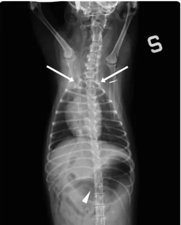
Lateral radiographs (FCR Capsula, Fuji Film Medical System SpA, Milan, MI, Italy) of the neck and thorax (Fig. 1) showed moderate tracheal collapse in the cervical region. The area of tracheal collapse appeared to correspond with an anatomical anomaly involving the first ribs and the seventh cervical vertebra, resembling supernumerary cervical ribs. The lateral radiographic view demonstrated that the bodies of the cervical ribs flared caudally and connected to the cranioventral aspect of the seventh vertebra proximally, and to the cranial aspect of the first rib distally. Spondylosis was detectable at the ventral aspects of the fourth, fifth, sixth and seventh cervical vertebrae. On the ventrodorsal radiographic view (Fig. 2), the absence of the 13th rib on the right was
Received: 31 May 2016 Accepted: 25 April 2017 Published online in J-STAGE:
18 May 2017 J. Vet. Med. Sci.
79(6): 1120–1124, 2017 doi: 10.1292/jvms.16-0281
evident. At the cranial thoracic inlet, two well-defined, rib-like opacities were evident connected on each side to the first rib and to the craniolateral aspect of seventh cervical vertebra (C7). The cervical rib on the right side was bigger than the one on the left, and both appeared completely ossified. A slight increase in the pulmonary vascular pattern was observed radiographically.
To rule out any vascular anomalies, ultrasonographic evaluation of the cranial portion of the thorax was performed using a multi-frequency linear transducer (4–13 MHz) (ESAOTE My Lab Class C, Esaote, Genoa, Italy). The
transducer was positioned between the axilla and the base of neck, just cranial to the first rib, allowing the ultrasound beam to be directed obliquely, from cranial to caudal and from ventral to dorsal. Brightness mode (B-mode or 2D mode), pulsed-wave doppler and color doppler examinations were performed in both hemithoraces. The doppler ultrasonographic evaluation was used to assess the differences in vasculature between the two sides of the cranial thorax. In fact, only a single vessel, the left common carotid artery, was present beneath the dorsal portion of the left first rib, traveling in a cranial to caudal direction. On the opposite side, beneath the dorsal portion of the right first rib, two different vessels were detected. The right common carotid artery traveled in the same direction as its left-sided counterpart and was located parallel to the right internal jugular vein. The inner diameter of all described vessels was approximately 2 mm.
The patient was hospitalized and managed with cage rest, supplemental oxygen, dexamethasone (Dexadreson, Intervet International BV, Boxmeer, Holland) (1 mg/kg, intravenous), ranitidine (Zantadine, Ceva Salute Animale SpA, Agrate Brianza, MB, Italy) (2 mg/kg, orally) and intravenous fluid therapy. After 2 days of hospitalization, the patient showed a clear clinical improvement and was discharged with a therapy consisting of prednisolone (Vetsolone 5 mg, Bayer, Milan, MI, Italy) (1 mg/kg, orally, once daily for 5 days and then every other day for a total of 5 doses), ranitidine (2 mg/kg, orally, twice daily) and stanozolol (Stargate, Acme Srl, Cavriago, RE, Italy) (0.3 mg/kg, orally, once daily for 30 days).
Seven months later, the patient was again referred to the Veterinary Teaching Hospital. The owner reported a full recovery from the respiratory disease with no further episodes of coughing or dyspnea. The main reason for this visit was profuse vaginal bleeding during estrus and the presence of small mammary nodules with discharge. A gynecological examination revealed a 4 cm mass on the dorsal aspect of the vaginal wall. The mammary nodules were clinically and cytologically compatible with mammary neoplasia. Routine preoperative electrocardiogram and blood tests were within normal limits.
Prior to surgery, radiographic and computed tomography (CT) evaluations of the thorax were performed, in order to exclude lung metastasis and to simultaneously clarify the relationship between the cervical ribs and the adjacent tissue. Computed tomography images were acquired using a single-slice multi-detector CT scanner (CT/e, General Electric, Fairfield, CT, U.S.A.) before and after intravenous injection of the iodinated contrast medium, iomeprol (Iomeron, 300 ng/ml, Bracco imaging Italia Srl, Milan, MI, Italy) Fig. 1. Lateral radiographic view of the neck and thorax. Note the
cervical rib (arrow) whose body is caudally flared and connected to the cranioventral aspect of the seventh vertebra proximally, and to the cranial aspect of the first rib distally. Moderate cervical tracheal collapse (arrowheads) can be appreciated mainly in association with the cervical rib. Spondylosis was detectable on the ventral surfaces of the fourth, fifth, sixth and seventh cervical vertebrae.
Fig. 2. Ventrodorsal radiograpic view of neck and thorax. Bilateral
supernumerary cervical ribs (arrows) were connected to the first ribs and to the craniolateral aspect of C7. Both cervical ribs were completely ossified with the right one appearing bigger than the left one. Absence of the right 13th rib was also noted (arrowhead). A slight increase in the pulmonary vascular pattern was observed. S=left.
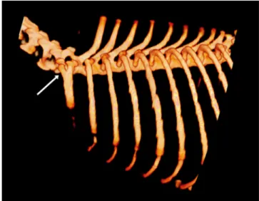
(2 ml/kg). The patient was placed under general anesthesia and was positioned in dorsal recumbency. The technical parameters were as follows: soft tissue acquisition algorithm, scan helical mode, slice thickness 1.0 mm, peak kilovoltage 120 kVp, x-ray tube current 100 mA, rotation time 1.5 sec, beginning at the fourth cervical vertebra and proceeding caudally to the pelvis. Three-dimensional multiplanar reformatted and volume rendered images were obtained using a DICOM (Digital Imaging and Communications in Medicine) workstation (Osirix DICOM-viewer, Pixmeo Sarl, Geneva, Switzerland). Radiographic and CT images confirmed the absence of metastasis and resolution of the tracheal collapse. Moreover, CT confirmed the presence of bilateral cervical ribs, which articulated with the transverse processes of C7 via short bony prominences. The proximal aspect of the left cervical rib was Y-shaped with a linear profile (Fig. 3), while the proximal aspect of the right cervical rib was U-shaped with a flared cranial profile and a linear caudal profile (Fig. 4). Both cervical ribs were completely calcified, and no line of separation was appreciated along the prominence or the body of the ribs; they articulated with the sternum at level of the first sternebra by an isoattenuating line of soft tissue, within a mean value of 100 Hounsfield units. On each side, the insertion of the scalenus muscle was on the cervical rib, and there was no evidence of an abnormal anatomical relationship between the subclavian artery and vein, scalenus muscle and the supernumerary rib. The right internal jugular vein was adhered to the right carotid artery, while the left internal jugular vein was not associated with the ipsilateral carotid artery (Fig. 5).
During the same anesthetic episode, an ovariohysterectomy, bilateral mastectomy and episiotomy were performed with success. A final diagnosis of vaginal leiomyoma and mammary carcinoma was obtained via the histological evaluation of the Fig. 3. Three-dimensional volume rendering of the left hemithorax.
Note the presence of the supernumerary rib at the level of C7 (arrow), articulating with the transverse process via short bony prominences. The proximal aspect of the left cervical rib appears Y-shaped with a linear profile and is completely calcified.
Fig. 4. Three-dimensional volume rendering of the right
hemitho-rax. Note the presence of the supernumerary rib at the level of C7 (arrow), articulating with the transverse process via short bony prominences. The proximal aspect of the right cervical rib appears U-shaped with a flared cranial profile and a linear caudal profile and is completely calcified.
Fig. 5. Three-dimensional volume rendering of the neck of the dog
at the level of C6 in a craniocaudal direction. Note the presence of a single vessel (left common carotid artery) on the left side (arrow), and two vessels (right common carotid artery and right internal jugular vein) on the right side (arrowhead).
masses. After 15 days, the dog was re-evaluated, and no significant abnormalities were noted.
In human beings, cervical ribs are often associated with early childhood cancer [13] and are known to be the cause of thoracic outlet syndrome (TOS) in up to 10% of affected individuals [30]. Early childhood cancers result from aberrant developmental processes and are generally embryonic in origin [2]. Thoracic outlet syndrome consists of upper extremity symptoms caused by compression of the neurovascular bundle in the area of the neck just cranial to the first rib [26]. Affected patients usually have neurogenic or vascular symptoms [25]. Classification of TOS can be based on etiology, symptoms, clinical presentation or anatomy. The neurovascular bundle in question contains 3 structures (brachial plexus, subclavian artery and vein); therefore, there are 3 types of TOS: arterial, venous and neurogenic [26]. Complete cervical ribs behave differently than incomplete cervical ribs. The prevalence of spontaneous onset of neurological symptoms with complete cervical ribs is 50%, compared with 20% for incomplete ribs. In addition, only complete cervical ribs have been noted to produce arterial TOS [25]. Although many reports describing the operative and non-operative management of this syndrome are in existence, rigorous scientific investigation leading to evidence-based management is lacking. In human medicine, the presence of unilateral or bilateral cervical ribs is often correlated with the absence of a radial pulse, reduced sensation, paraesthesia, muscle weakness and hand pain [9, 27]. The most common complaints result from compression of the lower trunk of the brachial plexus as it passes through the narrowed costoscalene triangle [14]. Along with motor and sensory abnormalities, compression of the sympathetic neurons can result in vasomotor disturbances [3]. Less commonly, a cervical rib can cause vascular symptoms related to compression of the subclavian artery. Many therapeutic options have been proposed for the management of TOS. These include physical therapy [17, 26, 29]; administration of medications, such as non-steroidal anti-inflammatory drugs, corticosteroids and muscle relaxants; local injections of botulinum toxin [12, 15]; and surgical treatment including both soft-tissue and bony procedures [22]. Most authors agree that conservative therapy alleviates symptoms in the majority of patients and that surgery should be considered only in those cases that have not responded to conservative therapy within a few months [26].
In veterinary medicine, the diagnosis of cervical ribs is based on imaging techniques and may be associated with clinical findings. A retrospective, epidemiological study evaluating the incidence of congenital vertebral anomalies reported abnormalities in the cervicothoracic spine in the radiographs from two cats [20]. The anomalies consisted of partial thoracicization of C7, with a single extra rib extending ventrally from the cranial aspect of C7. In both cats, the supernumerary rib was smaller than the adjacent thoracic ribs and appeared to taper to a smooth point ventrally, rather than forming a costochondral junction [20]. A recent case report described the incidental radiographic and CT findings as well as the anatomical characterization of bilateral cervical ribs in a dog [23].
In our case, the presence of cervical ribs was not associated with neurovascular symptoms. Advanced imaging allowed a definitive clinical diagnosis to be made, and the characteristics, location and extension of the anatomical anomaly were established. While the plain radiographs were fairly typical for a patient with cervical ribs, CT examination and ultrasound yielded a more accurate description of the anatomical relationship between the two cervical ribs and adjacent structures. The scalenus muscle normally attaches to the first few ribs and to the transverse processes of the cervical vertebrae [10, 11]. In this case, the cranial attachment of the muscle was to the cervical ribs on both sides, without any effect on inspiratory function. The left common carotid artery was located ventral to the esophagus while right common carotid artery was located ventral to the trachea, as is observed in normal dogs [4, 5]. In this case, the proximal aspect of the right internal jugular vein was associated with the internal carotid artery, while the left internal jugular vein was not in contact with the internal carotid artery. In our opinion, these findings are attributable to variation in the normal topographic anatomy of the vessels in the neck. It should be considered that no hemodynamic changes were detected via echocardiography.
In this report, a possible correlation between tracheal collapse and the presence of cervical ribs was suspected based on the first radiographic examination. In fact, the lateral view of the thorax revealed that the collapsed portion of the trachea existed cranial to the cervical ribs. This hypothesis was later dismissed based on the regression of the tracheal collapse with the use of stanozolol. This patient’s complete recovery suggests the tracheal collapse was caused by a temporary abnormality during development rather than by persistent compression by the cervical ribs.
In this report, the histology confirmed that the origin of the tumors was not embryonic, as has been reported in human beings with cervical ribs [2].
Given the limited number of reports on cervical ribs in dogs, the prevalence of this abnormality may be underestimated due to a possibly high rate of stillbirths. Because the clinical significance of cervical ribs in dogs remains unclear, a larger number of patients possessing these anomalies should be evaluated.
The mechanisms involved in the etiopathogenesis of cervical ribs, evidently variable and complex, remain uncertain. The study of familial cases may help to reveal the genetic basis behind cervical ribs.
We aim to raise awareness among the veterinary population regarding this often overlooked anomaly with possible clinical significance. It should especially be considered as a differential diagnosis for dogs presenting with neurological signs, vascular compromise affecting the forelimbs only, neoplasia and obstructive respiratory syndromes.
REFERENCES
1. Adson, A. W. and Coffey, J. R. 1927. Cervical rib: a method of anterior approach for relief of symptoms by division of the scalenus anticus. Ann.
Surg. 85: 839–857. [Medline] [CrossRef]
Pediatr. 26: 138–141 (in Spanish). [Medline]
3. Atasoy, E. 1996. Thoracic outlet compression syndrome. Orthop. Clin. North Am. 27: 265–303. [Medline]
4. Bezuidenhout, A. J. 2013. Veins. pp. 505–534. In: Miller’s Anatomy of the Dog, 4th ed. (Evans, H. E. and De Lahunta, A. eds.), Elsevier Saunders, St. Luois.
5. Bezuidenhout, A. J. 2013. The heart and arteries. pp. 428–504. In: Miller’s Anatomy of the Dog, 4th ed. (Evans, H. E. and De Lahunta, A. eds.) Elsevier Saunders, St. Luois.
6. Bokhari, R. F., Al-Sayyad, M. J. and Baeesa, S. S. 2012. Prevalence of cervical ribs and elongated transverse processes in Saudi Arabia. Saudi Med.
J. 33: 66–69. [Medline]
7. Brewin, J., Hill, M. and Ellis, H. 2009. The prevalence of cervical ribs in a London population. Clin. Anat. 22: 331–336. [Medline] [CrossRef] 8. Chang, K. Z., Likes, K., Davis, K., Demos, J. and Freischlag, J. A. 2013. The significance of cervical ribs in thoracic outlet syndrome. J. Vasc. Surg.
57: 771–775. [Medline] [CrossRef]
9. Cooke, R. A. 2003. Thoracic outlet syndrome--aspects of diagnosis in the differential diagnosis of hand-arm vibration syndrome. Occup. Med.
(Lond.) 53: 331–336. [Medline] [CrossRef]
10. Evans, H. E. and De Lahunta, A. 2010. The neck, thorax and thoracic limb. pp. 93–136. In: Guide to the Dissection of the Dog, 7th ed. (Evans, H. E. and De Lahunta, A. eds.), Elsevier Saunders, St. Louis.
11. Evans, H. E. and De Lahunta, A. 2010. The skeletal and muscular system. pp. 6–92. In: Guide to the Dissection of the Dog, 7th ed. (Evans, H. E. and De Lahunta, A. eds.), Elsevier Saunders, St. Louis.
12. Finlayson, H. C., O’Connor, R. J., Brasher, P. M. and Travlos, A. 2011. Botulinum toxin injection for management of thoracic outlet syndrome: a double-blind, randomized, controlled trial. Pain 152: 2023–2028. [Medline] [CrossRef]
13. Galis, F. 1999. Why do almost all mammals have seven cervical vertebrae? Developmental constraints, Hox genes, and cancer. J. Exp. Zool. 285: 19–26. [Medline] [CrossRef]
14. Hempel, G. K., Shutze, W. P., Anderson, J. F. and Bukhari, H. I. 1996. 770 consecutive supraclavicular first rib resections for thoracic outlet syndrome. Ann. Vasc. Surg. 10: 456–463. [Medline] [CrossRef]
15. Jordan, S. E., Ahn, S. S., Freischlag, J. A., Gelabert, H. A. and Machleder, H. I. 2000. Selective botulinum chemodenervation of the scalene muscles for treatment of neurogenic thoracic outlet syndrome. Ann. Vasc. Surg. 14: 365–369. [Medline] [CrossRef]
16. Keeling, J. W. and Kjaer, I. 1999. Cervical ribs: useful marker of monosomy X in fetal hydrops. Pediatr. Dev. Pathol. 2: 119–123. [Medline] [CrossRef]
17. Lindgren, K. A. 1997. Conservative treatment of thoracic outlet syndrome: a 2-year follow-up. Arch. Phys. Med. Rehabil. 78: 373–378. [Medline] [CrossRef]
18. Morgan, J. P. 1968. Congenital Anomalies of the Vertebral Column of the dog: A study of the Incidence and Significance Based on a Radiographic and Morphologic Study. Vet. Radiol. Ultrasound 9: 21–29. [CrossRef]
19. Morgan, J. P. 1999. Transitional lumbosacral vertebral anomaly in the dog: a radiographic study. J. Small Anim. Pract. 40: 167–172. [Medline] [CrossRef]
20. Newitt, A., German, A. J. and Barr, F. J. 2008. Congenital abnormalities of the feline vertebral column. Vet. Radiol. Ultrasound 49: 35–41. [Medline] [CrossRef]
21. Pollack, E. W. 1980. Surgical anatomy of the thoracic outlet syndrome. Surg. Gynecol. Obstet. 150: 97–103. [Medline]
22. Povlsen, B., Hansson, T. and Povlsen, S. D. 2014. Treatment for thoracic outlet syndrome. Cochrane Database Syst. Rev. 11: CD007218. [Medline] 23. Ricciardi, M., De Simone, A., Gernone, F. and Giannuzzi, P. 2015. Bilateral cervical ribs in a Dobermann Pinscher. Vet. Comp. Orthop. Traumatol.
28: 145–150. [Medline] [CrossRef]
24. Roos, D. B., Annest, S. J. and Brantigan, C. O. 1999. Historical and anatomic perspectives on thoracic outlet syndrome. Chest. Surg. Clin. 9: 713–723.
25. Sanders, R. J. and Hammond, S. L. 2002. Management of cervical ribs and anomalous first ribs causing neurogenic thoracic outlet syndrome. J.
Vasc. Surg. 36: 51–56. [Medline] [CrossRef]
26. Sanders, R. J., Hammond, S. L. and Rao, N. M. 2008. Thoracic outlet syndrome: a review. Neurologist 14: 365–373. [Medline] [CrossRef] 27. Sharma, P., Rasheed, I., Ansari, M. A., Gurung, G. S. and Chataut, S. P. 2010. Cervical rib causing thrombosis of subclavian artery. JNMA J. Nepal
Med. Assoc. 49: 161–163. [Medline]
28. Solecki, R., Bürgin, H., Buschmann, J., Clark, R., Duverger, M., Fialkowski, O., Guittin, P., Hazelden, K. P., Hellwig, J., Hoffmann, E., Hofmann, T., Hübel, U., Khalil, S., Lingk, W., Mantovani, A., Moxon, M., Müller, S., Parkinson, M., Paul, M., Paumgartten, F., Pfeil, R., Platzek, T., Rauch-Ernst, M., Scheevelenbos, A., Seed, J., Talsness, C. E., Yasuda, M., Younes, M. and Chahoud, I. 2001. Harmonisation of rat fetal skeletal terminology and classification. Report of the Third Workshop on the Terminology in Developmental Toxicology. Berlin, 14-16 September 2000.
Reprod. Toxicol. 15: 713–721. [Medline] [CrossRef]
29. Taskaynatan, M. A., Balaban, B., Yasar, E., Ozgul, A. and Kalyon, T. A. 2007. Cervical traction in conservative management of thoracic outlet syndrome. J. Musculoskeletal Pain 15: 89–94. [CrossRef]
30. Viertel, V. G., Intrapiromkul, J., Maluf, F., Patel, N. V., Zheng, W., Alluwaimi, F., Walden, M. J., Belzberg, A. and Yousem, D. M. 2012. Cervical ribs: a common variant overlooked in CT imaging. AJNR Am. J. Neuroradiol. 33: 2191–2194. [Medline] [CrossRef]