Microbiota modulation as preventative and therapeutic
approach in Alzheimer’s disease
Laura Bonfili, Valentina Cecarini, Olee Gogoi, Chunmei Gong, Massimiliano Cuccioloni, Mauro Angeletti, Giacomo Rossi and Anna Maria Eleuteri
School of Biosciences and Veterinary Medicine, University of Camerino, Camerino, Italy
Keywords
Alzheimer’s disease; dysbiosis; gut–brain axis; inflammation; microbiota modulation; oxidative stress; prebiotics; probiotics; proteolysis; therapeutic approaches
Correspondence
A. M. Eleuteri, School of Biosciences and Veterinary Medicine, University of Camerino, Via Gentile III da Varano, 62032 Camerino (MC), Italy
Tel:+390737403267
E-mail: [email protected]
(Received 8 June 2020, revised 27 August 2020, accepted 17 September 2020)
doi:10.1111/febs.15571
The gut microbiota coevolves with its host, and numerous factors like diet, lifestyle, drug intake and geographical location continuously modify its composition, deeply influencing host health. Recent studies demonstrated that gut dysbiosis can alter normal brain function through the so-called gut–brain axis, a bidirectional communication network between the central nervous system and the gastrointestinal tract, thus playing a key role in the pathogenesis of neurodegenerative disorders, such as Alzheimer’s disease (AD). In this perspective, in the constant search for novel treatments in AD, the rational modulation of gut microbiota composition could repre-sent a promising approach to prevent or delay AD onset or to counteract its progression. Preclinical and human studies on microbiota modulation through oral bacteriotherapy and faecal transplantation showed anti-inflammatory and antioxidant effects, upregulation of plasma concentra-tion of neuroprotective hormones, restoraconcentra-tion of impaired proteolytic pathways, amelioration of energy homeostasis with consequent decrease of AD molecular hallmarks and improvement of behavioural and cognitive performances. In this review, we dissect the role of gut microbiota in AD and highlight recent advances in the development of new multitarget strate-gies for microbiota modulation to be used as possible preventative and therapeutic approaches in AD.
Introduction
Microbiota is a community of symbiotic microorgan-isms that can be neutral, beneficial or detrimental to the host, with important regulatory functions in health and disease. The human body hosts trillions of microorganisms (bacteria, archaea, fungi and viruses) that colonize the skin surface, the respiratory tract, genitourinary organs and, most importantly, the gas-trointestinal tract. Approximately 95% of the symbi-otic organisms of the human microbiome can be found in the gut (gut microbiota) [1]. Gut microbial
ecosystem consists mainly of bacteria, mostly obligate anaerobes, fungi and viruses [2]. These diverse groups of microorganisms play multiple roles in humans, such as the fermentation of undigested carbohydrates, the production of short-chain fatty acids (SCFAs) and other metabolites, the synthesis of vitamins B and K, the metabolism of important substances (bile acids, sterols and drugs), and the protection against exoge-nous pathogens [3]. Bacteroidetes (~ 48%), Firmicutes (~ 51%), Proteobacteria and Actinobacteria (1%) are
Abbreviations
AD, Alzheimer’s disease; APP, amyloid precursor protein; Ab, amyloid beta; BBB, blood–brain barrier; BDNF, brain-derived neurotrophic factor; CNS, central nervous system; FMT, faecal microbiota transplantation; GABA,c-aminobutyric acid; GIP, glucose-dependent insulinotropic polypeptide; GLP1, glucagon-like peptide-1; LPS, lipopolysaccharide; MCI, mild cognitive impairment; NFT, neurofibrillary tangles; SCFAs, short-chain fatty acids; TLRs, Toll-like receptors; UPS, ubiquitin–proteasome system.
the main four bacterial phyla present in adults[4]. The gut microbiota coevolves with humans and regulates host pathophysiology and health status via symbiotic interactions[5–8]. An example of this tight relationship is the crosstalk between microbiota and mitochondria [9–11], and these subcellular organelles (which evolved from ancestral bacteria) mediate the transduction of stress and metabolic signals, and are particularly sensi-tive to metabolites produced by other microbes associ-ated with the gut and other mucosa[12].
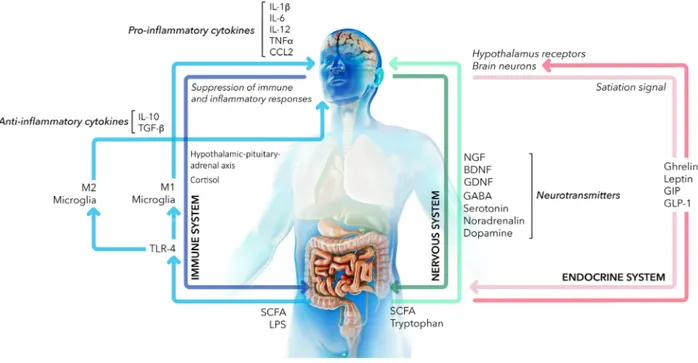
The gut microbiota communicates with the central nervous system (CNS) through the bidirectional gut– brain axis involving different pathways: the neuroim-mune system, sympathetic and parasympathetic branches of the autonomic nervous system, and neu-roendocrine system[13] (Fig.1). The gut–brain axis is responsible not only for the proper function of the digestive tract, but it also represents a biochemical sig-nalling system linked to the functionality of sympa-thetic nervous system, endocrine glands and specific brain regions, such as the hypothalamus and the fron-tal cortex. Moreover, the gut–brain axis influences
CNS development and behavioural performances in both normal and pathological conditions.
Although most bacterial metabolites are required for anabolic and catabolic functions, many of these compounds have other physiological roles. The gut microbiota synthetizes neurotransmitters such as c-aminobutyric acid (GABA), noradrenaline and dopa-mine, modulates systemic immune cells, produces metabolites like SCFAs, metabolizes essential amino acids like tryptophan and activates the secretion of the nerve growth factor (NGF), the glial-derived rotrophic factor (GDNF) and the brain-derived neu-rotrophic factor (BDNF) with consequent implications in neurodegenerative disorders [14–16]. In particular, BNDF plays a crucial role in the normal function and survival of neurons in mature peripheral and central nervous system [17]. Interestingly, germ-free mice showed a decreased expression of BDNF in the hip-pocampus, at both protein and mRNA levels, associ-ated with impaired cognition[18,19].
Bacterial fermentation of indigestible carbohydrates in the colon produces SCFAs, metabolites implicated
Fig. 1. Gut–brain axis. Schematic representation of the bidirectional communication network between the gut microbiota and the brain. Principal molecular mediators in the nervous system (green arrows), immune system (blue arrows) and endocrine system (pink arrows) are reported. In detail, gut bacteria produce metabolites like SCFAs and process essential amino acids like tryptophan triggering the secretion of the nerve growth factor (NGF), the brain-derived neurotrophic factor (BDNF) and the glial-derived neurotrophic factor (GDNF) and the synthesis of neurotransmitters such as c-aminobutyric acid (GABA), noradrenaline and dopamine. Bacterial LPS stimulates TLR4 thus modulating systemic immune cells. Microglia cells are activated and polarized to the pro-inflammatory (M1) phenotype, resulting in the production of cytokines and chemokines like IL-1b, IL-6, IL-12, TNF-a and CCL2. Differently, SCFAs producing bacteria favour the anti-inflammatory M2 phenotype, with the secretion of IL-10 and TGF-b. Moreover, SCFAs stimulate endocrine cells of the gastrointestinal tract to synthetize neuroactive compounds like ghrelin, leptin GIP and GLP-1 that exert neuroprotective effects and regulate important metabolic functions.
also in neurotransmission, since they modulate the syn-thesis of several neurotransmitters regulating behaviour and cognition [20]. Acetate, propionate and butyrate are the most abundant gut bacterial metabolites that can act either as substrates for host metabolism or as signalling molecules. In particular, butyrate and its pro-tonated form, butyric acid (pKa= 4.82), exist in differ-ent parts of the gastrointestinal tract (stomach: 1.5< pH < 3.5; intestine: 5.5 < pH < 7.4), where both forms exert beneficial health effects, improving food digestion and nutrient absorption, downregulating the proliferation of pathogenic microflora and favouring the colonization of anti-inflammatory bacteria[21].
Specifically, systemic administration of butyrate determines an antidepressant-like behavioural response [22]and butyric acid, as well as valeric acid and propi-onic acid, was shown to successfully counteract the conversion of Ab peptides to neurotoxic aggregates in vitro[23].
The gut microbiota exert a fundamental role in the digestion and absorption of amino acids that are important not only in protein synthesis, but also in the production of bioactive molecules that regulate key signalling pathways and metabolic pathways of the host [24]. Dysregulation of amino acid homeostasis can contribute to AD pathogenesis. In fact, changes in glutamate metabolism in AD brain consequently alter GABA concentrations thus affecting neural function-ing. GABA is produced from glutamate metabolism by different bacterial species, mainly lactic acid bacte-ria [25,26]. It is the major inhibitory neurotransmitter in the brain that regulates adult brain function, synap-tic plassynap-ticity, and corsynap-tical adaptation and reorganiza-tion thus representing an important mediator through which bacteria can modulate brain chemistry [27]. Altered concentrations of this neurotransmitter were detected in several conditions, including epilepsy [28] and schizophrenia[29], and dysfunctions of the GABA system were implicated in the pathophysiology of sev-eral chronic neurological diseases[30].
Changes in methionine, tryptophan, tyrosine and purine metabolism pathways were observed in both mild cognitive impairment (MCI) and AD subjects [31]. Interestingly, tryptophan is an essential amino acid largely found in meats, dairy products, fruits and seeds. It is not only absorbed through the intestinal epithelium to enter the blood circulation, but also directly and indirectly metabolized by the gut micro-biota into several compounds with an active role in gut–brain axis [32]. It is the precursor of several metabolites, most notably kynurenine and serotonin [33]. Kynurenine can cross the blood–brain barrier (BBB) [34] and, once in the brain, it is the precursor
of neuroactive glutamatergic compounds, including the neuroprotective kynurenic acid. Serotonin, a neuro-transmitter active both in the central nervous system and in the gut, plays an important role in maintaining mood and cognition [35]. Alterations in the levels of serotonin can be associated with the onset of gastroin-testinal and mood disorders, and tryptophan dysregu-lation is linked to detrimental conditions both in the brain and in the gastrointestinal tract[36].
Microbiota and related metabolites regulate BBB permeability. Among them, SCFAs can access the BBB via the bloodstream and can regulate its integrity through the upregulation of tight junction proteins [37]. Interestingly, the lack of gut microbiota is associ-ated with increased BBB permeability and altered expression of tight junction proteins. Braniste et al. demonstrated the tight communication between gut microbiota and BBB that initiates during gestation and propagates throughout life. They showed that fae-cal transfer from mice with pathogen-free gut flora into germ-free mice or treatment of germ-free mice with bacteria that produce SCFAs decreased the per-meability of the BBB and upregulated the expression of tight junction proteins[38].
The composition of the microbiota loses diversity and the abundance of beneficial bacteria decreases with ageing. This alteration directly and indirectly influences mitochondria functionality and energy production in intestinal cells, reducing the integrity of the intercellular junctional apparatus and increasing the translocation of bacterial products (principally lipopolysaccharide, LPS). This condition favours the so-called ‘inflammag-ing’, which is strongly associated with numerous age-re-lated diseases, whereas a healthy microbiota represents a key condition for longevity[25,39].
Moreover, dietary changes, antibiotic exposure and infections impair intestinal homeostasis promoting a condition known as dysbiosis [15], during which altered gut microbiota composition can damage the normal function of the intestinal barrier and increase intestinal permeability. Consequently, neuroactive compounds and gut microbial metabolites can reach areas of the central nervous system that regulate cogni-tion[25]. Dysbiosis is observed in neurodevelopmental diseases such as autism and in neurodegenerations such as Huntington’s disease, Parkinson’s disease and AD [40]. Current research is trying to gather new knowledge on the underlying mechanisms.
In the following sections, the impact of dysbiosis on gut–brain axis in AD is described and recent advances in the identification of multitarget strategies for micro-biota modulation to counteract the onset and progres-sion of AD are summarized.
Alzheimer’s disease
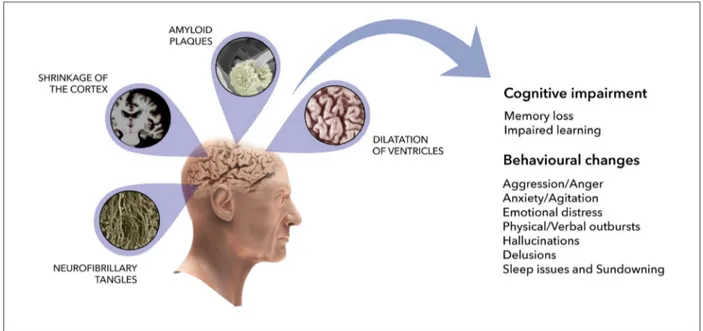
Alzheimer’s disease (AD) is the most common neu-rodegenerative disorder in the elderly and is a major challenge for the healthcare system given the impact it has on both economy and society. It is the main cause of dementia, and it is characterized by loss of neurons in the hippocampus and cerebral cortex, shrinkage of the cortex, enlargement of ventricles, resulting in the progressive decline in cognitive function. Memory impairment accompanied by visual space dysfunction and sleep deprivation was also linked to AD progres-sion [22]. Key molecular hallmarks of the disease are the extracellular amyloid beta (Ab) plaques and the intraneuronal neurofibrillary tangles (NFT) composed of hyperphosphorylated tau protein (Fig.2).
Plaques are extracellular deposits of amyloid pep-tides deriving from the amyloid precursor protein (APP), a type I membrane protein. APP was consid-ered a good target for therapeutic interventions in the early treatment of AD. APP can undergo nonamy-loidogenic cleavage process mediated by a-secretase andc-secretase that generates a soluble APP fragment (sAPPa) and a membrane-bound C-terminal fragment of APP (aCTF). Alternatively, APP can be cleaved by b-secretase and c-secretase releasing two major
fragments, sAPPb and a C-terminal fragment located in the membrane (bCTF). Further cleavage of bCTF results in the production of Ab peptides ranging from 37 to 43 amino acid in length, with Ab(1-40) and Ab (1-42) being the most dominant and neurotoxic [41,42]. Ab monomers form oligomeric structures that can further aggregate into regular fibrils. According to the Ab oligomer hypothesis, small soluble Ab oligo-mers are considered more neurotoxic than insoluble fibres or amyloid plaques[43].
Defective proteolysis is another important contribu-tor to AD pathogenesis, because it favours the accu-mulation of detrimental aggregates. In addition, it may also be secondary to accumulation of aggregates that can act as inhibitors for cellular proteolytic machiner-ies, mainly the ubiquitin–proteasome system (UPS) and autophagy. The UPS is the major intracellular degradation system, responsible for the removal of short-lived, misfolded and defective proteins [44], and proteasome inhibition impairs both APP processing and Ab production[45]. Autophagy includes degrada-tion pathways that finally transport their targets to lysosomes, acidic membrane-surrounded compartments containing hydrolytic enzymes involved in the intracel-lular breakdown of long-lived proteins, organelles and substrates with limited access to catalytic chamber of
Fig. 2. Schematic representation of cerebral modifications in Alzheimer’s disease. AD is characterized by loss of neurons in the hippocampus and cerebral cortex, shrinkage of the cortex, enlargement of ventricles, resulting in the progressive decline in cognitive function. The principal AD molecular hallmarks are the extracellular amyloid beta plaques and the intraneuronal neurofibrillary tangles composed of hyperphosphorylated tau protein. SEM micrographs for neuronal deposition of amyloid plaques and neurofibrillary tangles are from Meyeret al.[194]and Itohet al.[195], respectively. M.R.I. of the brain is from Lediget al.[196].
the proteasome, such as larger aggregates[46]. Lysoso-mal enzymes, particularly cathepsin B and cathepsin L, can interfere with APP processing, thus altering Ab formation [47]. Ab deposition and removal are finely regulated by the UPS and autophagy [48–51]. Impair-ment of proteolysis, which is typical of AD neurons, favours the accumulation of harmful Ab structures, which in turn alter both proteasome and autophagy functionality [52]. Failure of autophagy –lysosome-me-diated proteolysis in AD brain leads to a massive accumulation of autophagic vacuoles and lysosomes in dystrophic neurites. Vacuoles contain incompletely digested proteins, including toxic autophagic sub-strates, ubiquitinated proteins and Ab, indicating the importance of targeting autophagy to ameliorate neu-ropathology and cognitive deficits in AD[53]. Regard-ing UPS, some authors considered the proteasome a potential target in AD therapy because this multicat-alytic protease complex regulates the intracellular con-centration of both presenilins 1 and 2, and/or their presenilinase-derived C-terminal maturation fragments, thereby modulating both a- and b/c-secretase-derived products APPa and Ab(1-40) and Ab(1-42). Based on the amyloidogenic hypothesis, proteasome activators would enhance presenilin degradation and lower Ab peptide secretion [54]. Later, deficiencies of the amy-loidogenic hypothesis were identified and a major role of tau in the development and progression of AD emerged[55]. Failure of proteasomal- and autophagy-mediated clearance of tau and its aggregates leads to a progressive neurofibrillary degeneration in AD [56,57]. Nontoxic approaches to restore both UPS and autop-hagy represent an important challenge to achieve Ab and tau proteins correct balance.
The proteasome system is also responsible for the clearance of oxidized proteins[58,59]. Oxidative stress is one of the mechanisms through which Ab neuro-toxic peptides and tau protein cause impaired synaptic plasticity, neuroinflammation, neuronal and synaptic loss, and neurotransmitter imbalance in AD [60], con-tributing to the observed behavioural disturbances [61]. The key role of oxidative stress in the onset and progression of AD is largely documented: inadequate antioxidant defence systems, high O2consumption, the presence of excitotoxic amino acids and high iron con-tent promote the production of unstable reactive oxy-gen and nitrooxy-gen species (ROS and RNS) in the brain [62,63]. ROS and RNS easily react with proteins, lipids, carbohydrates and nucleic acids, causing oxida-tive modifications that finally result in dysfunctions of cellular processes [64,65], among them impaired pro-teasome-mediated proteolysis [66]. Numerous evi-dences have described a crosstalk between proteasome
and autophagy, with the overexpression of APP corre-lating with a reorganization of the cellular proteolytic machineries and with an increased oxidative status [48]. Also tau and tau aggregates are degraded by both the proteasome[67]and lysosomes[68].
Alzheimer’s disease subjects are characterized by a compromised blood–brain barrier (BBB) that is perme-able to neurotoxic components as well as pathogens and favours neuroinflammatory and neurodegenerative processes[69]. Both genetic and environmental factors can be involved. Apolipoprotein E variant 4 (APOE4) was found to be responsible for the increased risk of AD due to a leaky BBB. Individuals carrying APOE4 have a breakdown of the BBB in the hippocampus and parahippocampal gyrus, the regions of the brain responsible for memory and cognition. This condition is already detectable in individuals with MCI[70]. The increased permeability of BBB is also favoured by a dysregulated microbiota and causes the penetration of microbiota-derived products from the blood into the brain. In particular, gut microbes can excrete complex and immunogenic factors like LPSs and amyloids which may leak from the gastrointestinal tract and accumulate at the systemic and brain level contributing to the production and release of pro-inflammatory cytokines and reactive oxygen species, both responsible for AD neuroinflammation[71].
Neuroinflammation plays a major role in the patho-genesis of AD. A cascade of molecular events involves the activation of microglia, primary immune cells of CNS. Dysbiosis and intestinal infection trigger sys-temic immune response and cerebral inflammatory processes in AD[72]. Toll-like receptors (TLRs), which are present throughout the intestinal mucosa and in CNS, are fundamental in transducing molecular patho-genic patterns from the gut to the brain by modulating microglia response. TLRs are involved in commensal colonization, homeostasis maintenance and intestinal barrier integrity[73]. Ab binding to TLRs was demon-strated to trigger an inflammatory process in the gut and the brain, contributing to the development of AD and other neurodegenerative diseases. Although all TLRs are highly expressed in intestinal epithelial cells, they have been also detected in CNS: in particular, TLR1-9 encoding mRNA is present in microglia; TLR2 and TLR3 are expressed in astrocytes and oligodendrocytes, in association with TLR1 and TLR4 [74]. Besides, certain TLRs have been also found in neurons[75].
Under physiological conditions, the CNS-resident macrophages constituting microglia ensure neuronal health by secreting trophic factors such as BDNF[76], by playing a role in synaptic pruning [77,78] and by
exerting phagocytic activity towards senescent cells[79] and toxic forms of Ab[80]. Aberrant microglia activity or age-related decline of phagocytosis can cause inflammatory processes in CNS leading to cognitive impairment [81]. It was demonstrated that microglia activation by Ab requires CD14 (lipopolysaccharide receptor), TLR2 and TLR4 [82]. These receptors are overexpressed in 3xTgAD mice [83] as well as in the brain of AD patients[84]. Contrasting studies indicate that microglia can exert a protective role in AD [85,86], or can induce inflammation [87]and accelerate the disease [88]. Since microglia and blood-derived macrophages share many membrane determinants and possess phagocytic activity [89], peripheral monocytes can enter the BBB and, upon differentiation, can exert an essential role for reparative and regenerative func-tions in AD[90].
TLR4, localized on the surface of microglia, con-tributes to induce amyloid phagocytosis [91]. Ab is highly hydrophobic and can induce innate immune responses like those triggered by LPS. Furthermore, it has been shown that acute or chronic administration of LPS into the brain ventricles of rats and mice results in gliosis, cytokine production, increased Ab concentrations and occasional cognitive deficits [92]. Glial activation due to LPS administration was thought to mimic some features of AD [93]. In this perspective, several works were conducted to directly stimulate TLR4 or to administer TLR4 agonists to enhance the activation of monocyte-derived macro-phages and microglia, and to increase the natural clearance of amyloid deposits. Active phagocytosis of Ab(1–42) deposits in the brain of AD patients can be obtained through the natural stimulation of the innate immune response by stimulating TLR4-mediated phagocytosis, or the active immunization through the administration of adjuvanted Ab(1-42).
Alteration of gut microbiota in
Alzheimer’s disease
Gut microbiota can influence brain activity [19], and changes in relative abundance of taxa are documented in cognitively impaired subjects. In detail, a compara-tive analysis of the microbiome between healthy sub-jects and AD patients showed a lower prevalence of bacteria synthesizing the anti-inflammatory and neuro-protective SCFA butyrate and higher levels of pro-in-flammatory taxa [94]. Older adults showed increased abundance of the pro-inflammatory bacteria Escheri-chia/Shigella, whereas individuals with evidence of amyloid deposition on PET imaging exhibited decreased abundance of the anti-inflammatory bacteria
Eubacterium rectale [95]. Bacterial taxa correlate also with cerebrospinal fluid (CSF) biomarkers of AD pathology. Brandscheid et al. [96] observed an increased abundance of Firmicutes with a significant decrease in Bacteroidetes in 5xFAD mice, a model with severe amyloid pathology. Additionally, an increase in Clostridium leptum group was observed in AD mice [96]. Consistently, a study from Vogt et al. revealed reduced richness and abundance in alpha diversity and beta diversity in AD patients compared to healthy sub-jects. They evidenced differences in bacterial abun-dance such as decreased Firmicutes, increased Bacteroidetes and decreased Bifidobacteria in the microbiome of AD participants [97]. Colonization of Bacteroides fragilis was linked to exacerbation of AD along with increasing Ab plaques in AD mice [98]. Interestingly, Helicobacter pylori was largely studied not only for its pro-inflammatory properties, but also for its ability to stimulate Ab deposition[99]as well as tau hyperphosphorylation [100]. A recent comparative microbiome study confirmed a decreased microbial diversity in human AD patients with respect to mild cognitive impairment (MCI) patients and healthy con-trols. The study highlighted a progressive growth in the abundance of Gammaproteobacteria, Enterobacteri-ales and Enterobacteriaceae from healthy controls to AD subjects, increased glycan biosynthesis and meta-bolism in AD and MCI patients and decreased immune system-related pathways in AD patients. Among Firmicutes, Lachnospiraceae, Clostridiaceae and Ruminococcaceae were major SCFAs producers found to be lacking in AD patients[101]. Zhang et al. observed in APPSwe/PS1E9 transgenic mice a signifi-cant alteration of gut microbiome on the level of phy-lum, genus and species with age. Changes in the diversity and the composition of the faecal microbiota and SCFAs in these mice correlated with alterations of important metabolic pathways, which are associated with amyloid deposition and ultrastructural abnormali-ties in AD mouse intestine[102].
Microbiota modulation influences
multiple molecular mechanisms
through the gut
–brain axis
The modulation of intestinal homeostasis triggers mul-tiple mechanisms, among them the increase of anti-inflammatory microbial metabolism. As abovemen-tioned, the gut microbiota is an important source of LPS and amyloid peptides. LPS stimulates TLR4 via CD14. Bacterial amyloid proteins help bacterial cells to aggregate in biofilms and to resist destruction by physical or immune factors [103,104]and induce CNS
inflammation in different ways. Despite the differences in the sequence, bacterial amyloid peptides share simi-larities in their tertiary structure with CNS amyloids [105,106]. The exposure to bacterial amyloid proteins in the gut can cause priming of the monocytes/macro-phages system, leading to endogenous production of neuronal amyloid in the brain[104]. Microglia can rec-ognize and phagocyte Ab by receptor-mediated endo-cytosis that activates signalling pathways and cytokine production in a ligand-dependent manner [107]. In AD, microglia are activated and polarized to the pro-inflammatory (M1) phenotype. M1 microglia produce cytokines and chemokines (IL-1b, IL-6, IL-12, TNF-a, CCL2) and express NADPH oxidase generating reac-tive oxygen and nitrogen species. M1 microglia also express MHC-II, integrins (CD11b, CD11c), costimu-latory molecules (CD36, CD45, CD47) and Fc recep-tors. Differently, M2 microglia produce anti-inflammatory cytokines (IL-10, TGF-b), growth fac-tors (IGF-1, FGF, CSF1) and neurotrophic facfac-tors (nerve-derived growth factor (NGF), BDNF, neu-rotrophins, glial cell-derived neurotrophic factor (GDNF))[108].
Scientists totally agree on the fundamental neuro-modulatory role of SCFAs. These well-recognized anti-inflammatory bioactive bacterial metabolites pro-vide cells (including nervous cells) with energy; they are involved in cell signalling systems by influencing the intracellular levels of potassium, and directly affect brain neurochemistry by regulating the expres-sion of the gene coding for tryptophan hydroxylase, the key enzyme in the serotonin biosynthesis pathway [109]. SCFAs interfere with the activity of DNA repair enzymes by decreasing the activity of chromo-some histone deacetylases (HDACs); for example, the HDAC inhibitor 4-phenylbutyrate was investigated for its ability to restore dendritic spine density in the hippocampus of Tg2576 mice, coupled with decreased Ab load and tau phosphorylation, producing positive effects on cellular protein homeostasis [110], with the main disadvantage of a high therapeutic dosage required (up to 15 g/day) [111]. Undoubtedly, micro-biota modulation strategies that favour SCFAs pro-ducing bacteria represent a feasible and sustainable approach to ameliorate intestinal ad neuronal home-ostasis.
Interestingly, SCFAs are able to stimulate endocrine cells of the gastrointestinal tract to synthetize neuroac-tive compounds like histamine, serotonin, c-aminobu-tyric acid,b-alanine, peptide YY, leptin and glucagon-like peptide-1 (GLP-1) [112-114]. Gut peptide hor-mones have a documented role in AD as they regulate energy homeostasis and food intake and modulate
nervous functions like learning and memory[115-117]. For example, ghrelin and leptin are neurotrophic hor-mones [115,118]. Specifically, ghrelin affects both glu-cose and lipid metabolism, and it influences mitochondrial respiration and exerts neuroprotective effects and therefore is involved in the aetiopathogene-sis of neurodegenerative disorders, representing a link between metabolism and neurodegeneration [119]. In AD, memory and learning impairment is closely asso-ciated with the age-related decline of plasma ghrelin concentration[120]. Conversely, plasma leptin concen-tration is negatively correlated with Ab levels due to its direct regulatory effect on c-secretase [121]. Inter-estingly, treatment with leptin reduced both Ab and p-tau levels in AD animal models[122,123]. Also, GLP-1 and glucose-dependent insulinotropic polypeptide (GIP) act as neuroprotective hormones. Several studies described GLP-1 ability to protect cultured neurons from oxidative damage and Ab plaque formation, and the capability to improve synaptic plasticity in mice [124,125]. Additionally, increased plasma levels of gut hormones such as GLP-1, leptin, ghrelin and GIP par-tially restore hippocampus functions in AD subjects [119,126]. In this framework, the use of GIP analogues has emerged as promising strategy in AD treatment [127,128].
Since in many cases of early-onset AD, Ab peptides originate from mutations in genes encoding APP and presenilins (1 and 2), past and present therapies focused on the improvement of a-secretase activity to promote the nonamyloidogenic pathway, and on the modulation of proteolytic systems in charge of the clearance of amyloid deposits. The UPS and autop-hagy are the main cellular proteolytic systems[48–51], and there is a mutual effect of harmful Ab structures on both proteasome and autophagy functionality [52]. Both autophagy [53] and UPS have been identified as attractive therapeutic targets in AD, not only to degrade presenilins and lower Ab peptides production [54], but also for the clearance of tau and its aggre-gates[56,57]. Drug-based pharmacological modulators of proteolytic machineries are detrimental because of their toxicity, whereas natural product-derived approaches represent nontoxic options to restore both UPS and autophagy in several pathologic conditions including AD. A partial but significant recovery of both proteolytic pathways was obtained through microbiota modulation in AD transgenic mice, evi-dencing another important molecular target to achieve Ab and tau proteins correct balance. Moreover, autop-hagy induction can mediate the inhibition of Wingless-related integration site (Wnt) signalling, which affects differentiation of the CNS [129] and causes the
reduced expression of fibroblast growth factor 9 (FGF9) in the CNS, thus explaining the improvement of behavioural performances in probiotic treated ani-mals [130]. The crucial role of the proteasome in the clearance of oxidized proteins has been described [58,59]. A proper modulation of gut microbiota can directly and/or indirectly influence the oxidative bal-ance in the CNS, increasing the degradation of oxi-dized proteins and positively interfering with antioxidant systems[131]. Interestingly, probiotics oral administration ameliorated oxidative status in trans-genic AD mice by improving the functionality of antioxidant enzymes and decreasing oxidized proteins, lipids and DNA also through the activation of sirtuin-1 (SIRT-sirtuin-1) [132], a deacetylase enzyme with estab-lished neuroprotective action, able to lower ROS levels and promote cell survival[133].
Gut microbiota modulation inevitably impacts key metabolic functions such as glucose homeostasis which is impaired in AD. Defective insulin signalling strongly contributes to brain metabolic impairments in AD. For example, deficient cerebral amount of insulin growth factor I (IGF-I) is associated with compro-mised Ab clearance [134] and reduced proteolysis of oxidized proteins by the proteasome [135]. The levels of both IGF-I and its receptor significantly decrease in the hippocampus and somatosensory cortex of aged mice, causing age-related changes in the brain [136]. Moreover, tau hyperphosphorylation occurs in mice having abnormal brain insulin levels[137]. Specific gut microbial alterations can indicate a signature of the pathology. In this perspective, personalized interven-tion, using, for example, probiotics and prebiotics, rep-resents a successful revolutionary approach to restore the optimal concentrations of healthy promoting
bacteria that contribute to normalize host glycaemic response and insulin sensitivity.
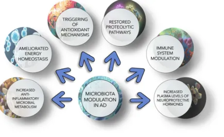
Figure3 summarizes the multiple molecular and metabolic mechanisms that are influenced by gut–brain axis modulation. Studies on animal models and human subjects show that modifications of gut microbiota reflect changes in genes involved in inflammatory and neuronal plasticity processes, with a positive impact on neuronal function[138,139].
Novel therapeutic strategies:
diet-based approaches and faecal
transplantation
As previously described, ageing, infections, unhealthy dietary habits and lifestyle behaviours can alter gut microbiota composition and diversity favouring the onset and progression of neurodegenerative disorders including AD. Since dysbiosis is strictly correlated with alterations of intestinal permeability, dysfunctions of BBB and neuroinflammatory processes [140,141], strongly participating in the development of AD, gut microbiota can also represent a key to tackle AD. Specifically, in the absence of a definitive treatment for AD, with most therapies simply delaying the loss of cognition and memory, recent studies have focussed on the role of the human microbiome in regulating multiple neurochemical pathways through the gut– brain axis and on looking for new therapeutic approaches for microbiota modulation [142–144]. A diet rich in saturated fat, carbohydrates and highly processed foods may have detrimental effects on health contributing to the reduction of microbiota diversity, neuroinflammation and cognitive impairment. On the other hand, healthy dietary patterns show
Fig. 3. Schematic representation of metabolisms and pathways affected by gut microbiota modulation. Microbiota modulation targets multiple molecular mechanisms to ameliorate AD condition, among them inflammatory and oxidative processes, proteolytic pathways, gut–brain axis, immune system components and energy metabolism.
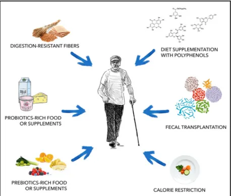
neuroprotective properties and can be beneficial to host cognitive health. On this regard, the Mediter-ranean diet is rich in many components considered helpful for AD subjects, among them vegetables, legumes, fruits, cereals and a high intake of unsatu-rated fatty acids and polyphenols [145]. In addition, oral bacteriotherapy has been recently identified as an accepted practice for the prevention and treatment of gastrointestinal infections[146] and inflammatory con-ditions[147,148]. Beneficial effects of lactic acid bacte-ria and bifidobactebacte-ria in CNS-related diseases have been reported [149–154]. Diet-based therapeutic inter-ventions include probiotics administration through specific supplements or probiotics enriched foods, foods rich in prebiotics, supplementation with polyphenols, calorie restriction and consumption of digestion-resistant fibres.
Besides dietary interventions, faecal microbiota transplantation (FMT) is another promising therapeu-tic option against AD that targets intestinal microbes, which involves the transfer of stool from a healthy donor into the gastrointestinal tract of a patient in order to restore diversity and function of the microbial population. This approach is currently considered a valid treatment for recurrent Clostridioides difficile infections, and it was successfully tested in intestinal conditions including inflammatory bowel disease (IBD), diarrhoea, irritable bowel syndrome (IBS) and constipation, and is now being investigated for its
possible application in extraintestinal conditions such as metabolic and neuropathological conditions (Fig.4) [155]. FTM is considered a safe therapeutic procedure with minor and transient side effects due to the intro-duction of live microorganisms and associated metabo-lites. However, it is of extreme importance to properly screen the donor and the faecal material in order to avoid contamination of the patient with pathogenic microorganisms that could lead to serious infections.
In the following sections, we report recent findings on the use of these new microbiota-modulating strate-gies.
Preclinical studies in animal models
Acting on the microbiota through specific strategies such as intervention with beneficial microbes or diet modifications could be considered a promising preven-tative and therapeutic approach in AD. Particularly, single- or multistrain probiotic preparations turned out to be successful examples of oral treatments. These formulations are usually made of Lactobacillus and Bifidobacterium species since members of both groups have been used extensively in promoting human health and are classified as GRAS (generally regarded as safe) for human consumption [156]. We recently reported on the beneficial properties of SLAB51, a formulation of lactic acid bacteria and bifidobacteria able to modu-late microbiota in 3xTg-AD mice increasing the
Fig. 4. Strategies used to modulate gut microbiota composition. Diet-based strategies and faecal transplantation are considered promising approaches to regulate function and composition of gut microbial population, favouring the abundance of beneficial bacterial groups.
relative abundance of Bifidobacterium spp. and decreasing Campylobacterales, bacterial groups differ-ently involved in the regulation of inflammatory path-ways. These changes in microflora composition together with enriched gut concentration of SCFAs and increased plasma levels of neuroprotective gut peptide hormones contributed to counteract cognitive decline through a reduction of Ab aggregates and brain damages, and a partial restoration of impaired neuronal proteolytic pathways [130]. SLAB51-medi-ated microbiota modulation also mitigSLAB51-medi-ated oxidative stress by activating SIRT1-dependent mechanisms and restored glucose homeostasis in 3xTg-AD mouse brain [132,157]. ProBiotic-4, another formulation containing B. lactis, L. casei, B. bifidum and L. acidophilus, signif-icantly improved cognitive functions and attenuated intestinal and BBB injury in aged SAMP8 mice through inhibition of both TLR4- and RIG-I-mediated NF-jB signalling pathways and inflammatory responses [158]. The mixture exerted a modulation of SAMP8 mouse microbiota with a marked decrease of Proteobacteria (phylum), Pseudomonas (genus) and Lachnospiraceae_NK4A136_group(genus) and a signifi-cant lower Firmicutes/Bacteroidetes ratio than vehicle-treated SAMP8 mice [158]. A probiotic mixture con-taining Lactobacillus acidophilus, L. fermentum, Bifi-dobacterium lactis and B. longum administered for 8 weeks to Ab(1-42)-injected rats improved spatial memory and learning deficits and decreased oxidative stress by modifying microbiota [159]. Manipulation of the gut microbiota with a combination of Lactobacillus helveticus R0052 and Bifidobacterium longum R0175 significantly decreased serum and hippocampus levels of pro-inflammatory cytokines, alleviated hippocampal apoptosis and attenuated the detrimental effect of LPS on memory through BDNF protein expression in LPS-induced rats [160,161]. Short-term administration of Bifidobacterium breve strain A1 prevented cognitive decline in AD mice, with a reduction in the immune response and neuronal inflammation. However, the authors did not detect a marked effect on intestinal microbiota composition, indicating the involvement of other mechanisms in the probiotic final effect, such as the gut–brain communication via stimulation of the vagus nerve[162]. The effects of bacteria on cognition can also depend upon the type of strain that is admin-istered. On this regard, Savignac H.M. et al. demon-strated that treatment with B. longum 1714 improved the ability of learning and memory in an anxious mouse model, whereas B. breve 1205 had little or no positive impact on memory [163,164]. A recent article introduced the possibility for probiotics to be used also in combination with traditional AD drugs to
potentiate their beneficial effects. In details, L. plan-tarum augmented the therapeutic efficacy of meman-tine in APP/PS1 mice by remodelling the intestinal microbiota, inhibiting the synthesis of trimethylamine-N-oxide (TMAO), a gut microbial metabolite able to promote AD progression, and reducing clusterin levels. Moreover, a 12-week treatment with memantine in combination with L. plantarum ameliorated cognitive deterioration, decreased hippocampus Αb levels, and protected neuronal integrity and plasticity[165].
Several studies on microbiota and probiotic interac-tions involve Drosophila melanogaster, which is consid-ered an excellent model for microbiota research in view of its easily manipulated microbiome, high-throughput screening capabilities, low costs, and fast reproduction and is currently used to investigate the mechanisms at the basis of AD[166,167]. The adminis-tration of Lactobacillus plantarum DR7 to an AD-in-duced Drosophila melanogaster model rescued the rough eye phenotype and restored the gut microbiota diversity with a significant reduction in Wolbachia’s relative abundance, positively correlated with neurode-generative disorders, accompanied by an increase of Stenotrophomonas and Acetobacter [168]. A symbiotic preparation containing three metabolically active pro-biotics and a polyphenol-rich prebiotic increased sur-vivability and motility, rescuing Ab deposition and acetylcholinesterase activity in a transgenic humanized Drosophila melanogaster model of AD through the effect on gut–brain axis components and on PPARc activity[169].
Numerous studies also highlighted that dietary inter-ventions with specific nutrients or combination of nutrients may act on gut microbes and their metabo-lites to ameliorate AD neuropathology. Diet supple-mentation with prebiotics was demonstrated to be a possible strategy to attenuate AD symptoms by modu-lating the microbiota. Prebiotics are dietary supple-ments used as food source by the microflora that offer a health benefit to the host regulating gut microbiota composition. Inulin, a well-studied prebiotic com-pound, enhanced systemic metabolism and decreased hippocampus inflammatory gene expression modulat-ing gut microbiome composition in E4FAD mice even before the development of Ab [170]. Treatment of APP/PS1 transgenic mice with fructooligosaccharides (FOS), commonly found in fruits and vegetables, chan-ged microbiota composition and activated the GLP-1 pathway with consequent amelioration of cognitive deficits and pathological changes. In details, FOS reduced the groups of Proteobacteria, associated with dementia and immunological reactions and inflamma-tion, of Helicobacteraceae and Desulfovibrionaceae and
reversed the decrease of Lactobacilli observed in untreated transgenic animals [171]. Oligosaccharides from Morinda officinalis administered to APP/PS1 transgenic mice significantly improved memory, brain tissue swelling and neuronal apoptosis and downregu-lated the expression of Ab(1-42). These molecules were able to regulate the composition and metabolism of the gut microbiota in treated transgenic mice [172]. Syeda et al. demonstrated that the ingestion of bioac-tive food composed of nopal, soy protein, chia seed and turmeric reduced the amount of pro-inflammatory bacteria simultaneously increasing the anti-inflamma-tory ones, including A. muciniphila and F. prausnitzii, in 3xTg-AD mice [173]. This variation in microflora exerted numerous beneficial effects on the main patho-logical markers of AD such as a better cognitive outcome, decreased amyloid aggregates and hyper-phosphorylation of tau, diminished oxidative damage, neuroinflammation, synaptic and metabolic alterations [173]. Sesamol, an antioxidant lignan from sesame oil, was able to reshape gut microbiome and improve the generation of SCFAs in high-fat diet rats. The effect on the gut was then accompanied by an ApoE-depen-dent improvement of cognitive deficits, anxiety, and synapse ultrastructure and inhibition of Ab accumula-tion [174]. Treatment with jatrorrhizine, a main com-ponent of Coptidis rhizome, a traditional Chinese herbal, alleviated learning and memory deficits, reduced the levels of Ab plaques in the cortex and hip-pocampus of APP/PS1 mice. Jatrorrhizine administra-tion clearly affected mouse microbiota modulating the relative abundance of the most predominant phylum Firmicutes and Bacteroidetes in transgenic mice [175]. Also calorie restriction (CR) was effective in promot-ing neuroprotective effects and health benefits in dif-ferent animal models, being also able to counteract AD symptoms [176-178]. Cox et al. recently reported on the effects of CR on the microbiome of Tg2576 mice during ageing, showing that CR can rescue age-and APP-related microbiome alterations age-and prevent the enrichment of microbes associated with AD age-re-lated cognitive decline[98].
Besides oral administration of bacterial strains or nutrients, faecal microbiota transplantation (FMT) from a healthy donor to a patient or diseased animal is an alternative microbiota-targeted intervention, which represents a potentially attractive therapeutic approach against AD. A recent article by Kim et al. focused on this strategy, further contributing to eluci-date the intricate relationship among the gut, blood and brain axis and AD. They demonstrated that fresh faecal matters oral transfer of healthy wild-type mice in ADLPAPTmice for 16 weeks and faecal microbiota
transplantation in antibiotics-pretreated ADLPAPT mice for 4 weeks alleviated Ab deposition, tau pathol-ogy, reactive gliosis and memory impairment in these transgenic AD mice. Interestingly, FMT successfully reversed abnormalities in intestinal macrophage activ-ity and circulating blood inflammatory monocytes in the ADLPAPT recipient mice [179]. 16S ribosomal RNA sequencing analyses revealed that FMT reversed the alterations observed in microbial composition of APP/PS1 transgenic mice such as the abnormal enrich-ment in Proteobacteria, Verrucomicrobia, Akkermansia and Desulfovibrio, and the downregulation of Bac-teroidetes and Alloprevotella. Modulating transgenic mouse microbiota and the associated SCFAs produc-tion, FMT improved cognitive deficits, decreased phos-phorylation of tau protein and the levels of amyloid peptides, and ameliorated synaptic plasticity [180]. Cognitive dysfunctions and a- and b-diversity indices of pseudo germ-free mice (in detail, C57BL/6 mice receiving broad-spectrum antibiotics dissolved in drinking water for 14 consecutive days) were deeply ameliorated upon FMT from senescence-accelerated mouse resistant 1 (SAMR1) mice, but not from SAMP8 mice, further confirming that improving unhealthy gut microbiota may provide valid treatment for AD[181]. Interestingly, FMT associated with calo-rie restriction improved glucose tolerance, insulin sen-sitivity and lipid metabolism and regulated immune system in mice, indicating positive implications in metabolic disorders [182] and suggesting larger thera-peutic potential, also in AD, considering that glucose intolerance and impairment of insulin metabolism are strictly connected with a higher risk of developing AD [183].
Human clinical studies on microbiota
modulation
An increasing number of nutritional interventions to modify gut microbiota are documented in humans, principally involving old adults with memory com-plaints and healthy volunteers, also considering that AD, insulin resistance, diabetes, obesity and cardiovas-cular disease are strongly interconnected[152]and that there is an urgent need to establish preventative strate-gies. Specifically, it was reported that chronic supple-mentation with Bifidobacterium breve A1 restored cognitive functions in old people with impaired mem-ory [184]. Conversely, in some cases, experimental groups receiving placebo, instead of probiotics, obtained significantly better memory scores[185]. Nag-pal et al. identified significant differences between the gut microbiome of MCI patients and cognitively
normal subjects, highlighting specific gut microbiome signatures that are associated with MCI and that cor-relate with cerebrospinal fluid levels of Ab, total tau and phosphorylated tau [186]. In the same study, a modified Mediterranean ketogenic diet influenced the abundance of specific bacterial taxa (increasing the abundance of Enterobacteriaceae, Akkermansia, Slack-ia, Christensenellaceae and Erysipelotriaceae and reduc-ing Bifidobacterium and Lachnobacterium) and SCFAs (increasing propionate and butyrate) consequently improving AD biomarkers in the cerebrospinal fluid of treated MCI patients[186].
Interestingly, a randomized, double-blind and con-trolled trial conducted by Akbari and collaborators evidenced that chronic supplementation with milk enriched with L. acidophilus, L. casei, B. bifidum and L. fermentum improved learning and memory in AD patients. Probiotics positively influenced the levels of malondialdehyde and high-sensitivity C-reactive pro-tein, improved insulin resistance, pancreatic beta cell secretion, and metabolic status with respect to controls [187].
Another study reported that AD patients supple-mented with a multispecies probiotic formulation influ-enced gut bacteria composition decreasing faecal zonulin concentrations and increasing Faecalibacterium prausnitzii. In these patients, enhanced kynurenine serum concentrations influenced tryptophan metabo-lism and stimulated the immune system[188].
No data on FMT in human AD patients were found but results from ongoing clinical trials are expected in the near future. Although animal models represent great opportunities to reveal new insights into micro-biota–host interactions, they cannot fully nor accu-rately reproduce the human phenotype and there is an urgent need for additional human clinical studies with well-defined targets, and standardized protocols and outcome measures.
Conclusions
Alzheimer’s disease is the most diffuse incurable dementia, and the identification of a definitive thera-peutic intervention is a major challenge of our time. Dysbiosis was demonstrated to be a relevant risk fac-tor for AD [101], with lifestyle, geographical location, drug assumption and dietary habits continuously being capable of modifying the gut microbiota composition. Diet rich in saturated fat and simple carbohydrates increases the risk of dementia and a suboptimal diet is associated with a more severe impaired cognition in AD [189]. Differently, a high quality diet like the Mediterranean diet correlates with better cognitive
status in healthy people with reduced risk of develop-ing MCI and AD [190]. In this context, the possibility to modulate the composition of gut microbiota using probiotics, prebiotics and other dietary intervention represents a promising and sustainable approach. Diet-ary interventions are generally safe and more advanta-geous than drug-based therapies since probiotics, prebiotics and synbiotics are cheap and easy to handle, thus reducing the burden also for AD patient care-givers. Similarly, FMT was described as a promising procedure, although some adverse effects were docu-mented in infections from Clostridium difficile [191] or ulcerative colitis [192], indicating that intersubject vari-ability must be considered and that a long-term fol-low-up is necessary to assess the risks and benefits. Moreover, standardization of methods used for micro-biota analysis (sampling, preservation and storage of samples, and analytic procedures) will facilitate com-parison between studies, enhancing the reproducibility [193]. The rapid advances of metabolomics and infor-matics will help in managing the vast databases deriv-ing from ongoing and short-coming microbiota studies.
Successful results depend upon the optimization of different factors including proper combinations of bac-terial strains and nutrients, time of treatment, disease stage therefore the presence of specific procedures and guidelines are necessary to enhance effectiveness of gut microbiota modulation.
The identification of AD-specific signatures in gut microbiota together with a better knowledge of the molecular mechanisms triggered upon microbiota modification will contribute to identify multiple per-sonalized interventions for decreasing AD risk, delay-ing the onset of the pathology, and counteractdelay-ing the appearance or improving the clearance of AD hall-marks.
Acknowledgments
This study was financially supported by ‘Noemi Avi-colli’ private donation to the University of Camerino.
Author contributions
LB organized the contents, and drafted and wrote the manuscript. VC wrote, edited and revised the article. OG and CG drafted and wrote the manuscript. MC designed the figures and revised the article. GR and MA wrote and critically revised the article. AME con-ceived and designed the work, critically revised the manuscript, and edited and approved the final version to be published.
Conflict of interest
The authors declare no conflict of interest.
Peer Review
The peer review history for this article is available at https://publons.com/publon/10.1111/febs.15571.
References
1 De-Paula VJR, Forlenza AS & Forlenza OV (2018) Relevance of gutmicrobiota in cognition, behaviour and Alzheimer’s disease. Pharmacol Res136, 29–34. 2 Giau VV, Wu SY, Jamerlan A, An SSA, Kim SY &
Hulme J (2018) Gut Microbiota and their
neuroinflammatory implications in Alzheimer’s disease. Nutrients10, 1765.
3 Quigley EM (2013) Gut bacteria in health and disease. Gastroenterol Hepatol9, 560–569.
4 Villaran RF, Espinosa-Oliva AM, Sarmiento M, De Pablos RM, Arguelles S, Delgado-Cortes MJ, Sobrino V, Van Rooijen N, Venero JL, Herrera AJ et al. (2010) Ulcerative colitis exacerbates lipopolysaccharide-induced damage to the nigral dopaminergic system: potential risk factor in Parkinson‘s disease. J Neurochem114, 1687–1700.
5 Nishida A, Inoue R, Inatomi O, Bamba S, Naito Y & Andoh A (2018) Gut microbiota in the pathogenesis of inflammatory bowel disease. Clin J Gastroenterol11, 1– 10.
6 Chow J, Lee SM, Shen Y, Khosravi A & Mazmanian SK (2010) Host-bacterial symbiosis in health and disease. Adv Immunol107, 243–274.
7 Greiner T & Backhed F (2011) Effects of the gut microbiota on obesity and glucose homeostasis. Trends Endocrinol Metab22, 117–123.
8 Erny D, Hrabe de Angelis AL, Jaitin D, Wieghofer P, Staszewski O, David E, Keren-Shaul H, Mahlakoiv T, Jakobshagen K, Buch T et al. (2015) Host microbiota constantly control maturation and function of microglia in the CNS. Nat Neurosci18, 965–977. 9 d’Ettorre G, Rossi G, Scagnolari C, Andreotti M,
Giustini N, Serafino S, Schietroma I, Scheri GC, Fard SN, Trinchieri V et al. (2017) Probiotic
supplementation promotes a reduction in T-cell activation, an increase in Th17 frequencies, and a recovery of intestinal epithelium integrity and mitochondrial morphology in ART-treated HIV-1-positive patients. Immun Inflamm Dis5, 244–260. 10 Mottawea W, Chiang CK, Muhlbauer M, Starr AE,
Butcher J, Abujamel T, Deeke SA, Brandel A, Zhou H, Shokralla S et al. (2016) Altered intestinal microbiota-host mitochondria crosstalk in new onset Crohn’s disease. Nat Commun7, 13419.
11 Saint-Georges-Chaumet Y & Edeas M (2016) Microbiota-mitochondria inter-talk: consequence for microbiota-host interaction. Pathog Dis74, ftv096. 12 Galluzzi L, Kepp O & Kroemer G (2012)
Mitochondria: master regulators of danger signalling. Nat Rev Mol Cell Biol13, 780–788.
13 Carabotti M, Scirocco A, Maselli MA & Severi C (2015) The gut-brain axis: interactions between enteric microbiota, central and enteric nervous systems. Ann Gastroenterol28, 203–209.
14 O’Mahony SM, Clarke G, Borre YE, Dinan TG & Cryan JF (2015) Serotonin, tryptophan metabolism and the brain-gut-microbiome axis. Behav Brain Res 277, 32–48.
15 Sherwin E, Dinan TG & Cryan JF (2018) Recent developments in understanding the role of the gut microbiota in brain health and disease. Ann N Y Acad Sci1420, 5–25.
16 Silva YP, Bernardi A & Frozza RL (2020) The role of short-chain fatty acids from gut microbiota in gut-brain communication. Front Endocrinol (Lausanne)11, 25. 17 Casoli T, Di Stefano G, Fattoretti P, Giorgetti B,
Balietti M, Lattanzio F, Aicardi G & Platano D (2012) Dynamin binding protein gene expression and memory performance in aged rats. Neurobiol Aging33, 618 e15–618 e19.
18 Gareau MG, Wine E, Rodrigues DM, Cho JH, Whary MT, Philpott DJ, Macqueen G & Sherman PM (2011) Bacterial infection causes stress-induced memory dysfunction in mice. Gut60, 307–317.
19 Wang T, Hu X, Liang S, Li W, Wu X, Wang L & Jin F (2015) Lactobacillus fermentum NS9 restores the antibiotic induced physiological and psychological abnormalities in rats. Benef Microbes6, 707–717. 20 Luca M, Di Mauro M, Di Mauro M & Luca A (2019)
Gut microbiota in Alzheimer’s disease, depression, and type 2 diabetes mellitus: the role of oxidative stress. Oxid Med Cell Longev2019, 4730539.
21 Ahsan U, Cengiz €O, Raza I, Kuter E, Chacher MFA, Iqbal Z, Umar S & Cß akir S (2019) Sodium butyrate in chicken nutrition: the dynamics of performance, gut microbiota, gut morphology, and immunity. Worlds Poult Sci J72, 265–275.
22 Jiang HY, Zhang X, Yu ZH, Zhang Z, Deng M, Zhao JH & Ruan B (2018) Altered gut microbiota profile in patients with generalized anxiety disorder. J Psychiatr Res104, 130–136.
23 Ho L, Ono K, Tsuji M, Mazzola P, Singh R & Pasinetti GM (2018) Protective roles of intestinal microbiota derived short chain fatty acids in Alzheimer’s disease-type beta-amyloid
neuropathological mechanisms. Expert Rev Neurother 18, 83–90.
24 Wu G (2013) Functional amino acids in nutrition and health. Amino Acids45, 407–411.
25 Filosa S, Di Meo F & Crispi S (2018) Polyphenols-gut microbiota interplay and brain neuromodulation. Neural Regen Res13, 2055–2059.
26 Jamwal S & Kumar P (2019) Insight into the emerging role of striatal neurotransmitters in the
pathophysiology of Parkinson’s disease and
Huntington’s disease: a review. Curr Neuropharmacol 17, 165–175.
27 Stagg CJ (2014) Magnetic Resonance Spectroscopy as a tool to study the role of GABA in motor-cortical plasticity. NeuroImage86, 19–27.
28 Macdonald RL, Kang JQ & Gallagher MJ (2010) Mutations in GABAA receptor subunits associated with genetic epilepsies. J Physiol588, 1861–1869. 29 Reynolds GP, Beasley CL & Zhang ZJ (2002)
Understanding the neurotransmitter pathology of schizophrenia: selective deficits of subtypes of cortical GABAergic neurons. J Neural Transm (Vienna)109, 881–889.
30 Kim YS & Yoon BE (2017) Altered GABAergic signaling in brain disease at various stages of life. Exp Neurobiol26, 122–131.
31 Kaddurah-Daouk R, Zhu H, Sharma S, Bogdanov M, Rozen SG, Matson W, Oki NO, Motsinger-Reif AA, Churchill E, Lei Z et al. (2013) Alterations in metabolic pathways and networks in Alzheimer’s disease. Transl Psychiatry3, e244.
32 Agus A, Planchais J & Sokol H (2018) Gut microbiota regulation of tryptophan metabolism in health and disease. Cell Host Microbe23, 716–724.
33 Jenkins TA, Nguyen JC, Polglaze KE & Bertrand PP (2016) Influence of tryptophan and serotonin on mood and cognition with a possible role of the gut-brain axis. Nutrients8, 56.
34 Fukui S, Schwarcz R, Rapoport SI, Takada Y & Smith QR (1991) Blood-brain barrier transport of kynurenines: implications for brain synthesis and metabolism. J Neurochem56, 2007–2017.
35 Szapacs ME, Mathews TA, Tessarollo L, Ernest Lyons W, Mamounas LA & Andrews AM (2004) Exploring the relationship between serotonin and brain-derived neurotrophic factor: analysis of BDNF protein and extraneuronal 5-HT in mice with reduced serotonin transporter or BDNF expression. J Neurosci Methods 140, 81–92.
36 Clarke G, Grenham S, Scully P, Fitzgerald P, Moloney RD, Shanahan F, Dinan TG & Cryan JF (2013) The microbiome-gut-brain axis during early life regulates the hippocampal serotonergic system in a sex-dependent manner. Mol Psychiatry 18, 666– 673.
37 Parker A, Fonseca S & Carding SR (2020) Gut microbes and metabolites as modulators of blood-brain barrier integrity and blood-brain health. Gut Microbes 11, 135–157.
38 Braniste V, Al-Asmakh M, Kowal C, Anuar F, Abbaspour A, Toth M, Korecka A, Bakocevic N, Ng LG, Kundu P et al. (2014) The gut microbiota influences blood-brain barrier permeability in mice. Sci Transl Med6, 263ra158.
39 Biagi E, Franceschi C, Rampelli S, Severgnini M, Ostan R, Turroni S, Consolandi C, Quercia S, Scurti M, Monti D et al. (2016) Gut microbiota and extreme longevity. Curr Biol26, 1480–1485.
40 Di Meo F, Donato S, Di Pardo A, Maglione V, Filosa S & Crispi S (2018) New therapeutic drugs from bioactive natural molecules: the role of gut microbiota metabolism in neurodegenerative diseases. Curr Drug Metab19, 478–489.
41 Haass C, Kaether C, Thinakaran G & Sisodia S (2012) Trafficking and proteolytic processing of APP. Cold Spring Harb Perspect Med2, a006270.
42 Kummer MP & Heneka MT (2014) Truncated and modified amyloid-beta species. Alzheimers Res Ther6, 28.
43 Chen GF, Xu TH, Yan Y, Zhou YR, Jiang Y, Melcher K & Xu HE (2017) Amyloid beta: structure, biology and structure-based therapeutic development. Acta Pharmacol Sin38, 1205–1235.
44 Wilkinson KD, Urban MK & Haas AL (1980) Ubiquitin is the ATP-dependent proteolysis factor I of rabbit reticulocytes. J Biol Chem255, 7529–7532. 45 Flood F, Murphy S, Cowburn RF, Lannfelt L, Walker
B & Johnston JA (2005) Proteasome-mediated effects on amyloid precursor protein processing at the gamma-secretase site. Biochem J385, 545–550. 46 Hara T, Nakamura K, Matsui M, Yamamoto A,
Nakahara Y, Suzuki-Migishima R, Yokoyama M, Mishima K, Saito I, Okano H et al. (2006)
Suppression of basal autophagy in neural cells causes neurodegenerative disease in mice. Nature441, 885– 889.
47 Klein DM, Felsenstein KM & Brenneman DE (2009) Cathepsins B and L differentially regulate amyloid precursor protein processing. J Pharmacol Exp Ther 328, 813–821.
48 Cecarini V, Bonfili L, Cuccioloni M, Mozzicafreddo M, Rossi G, Buizza L, Uberti D, Angeletti M & Eleuteri AM (2012) Crosstalk between the ubiquitin-proteasome system and autophagy in a human cellular model of Alzheimer’s disease. Biochim Biophys Acta 1822, 1741–1751.
49 Zheng Q, Li J & Wang X (2009) Interplay between the ubiquitin-proteasome system and autophagy in proteinopathies. Int J Physiol Pathophysiol Pharmacol 1, 127–142.
50 Nilsson P, Loganathan K, Sekiguchi M, Matsuba Y, Hui K, Tsubuki S, Tanaka M, Iwata N, Saito T & Saido TC (2013) Abeta secretion and plaque formation depend on autophagy. Cell Rep5, 61–69.
51 van Tijn P, Dennissen FJ, Gentier RJ, Hobo B, Hermes D, Steinbusch HW, Van Leeuwen FW & Fischer DF (2012) Mutant ubiquitin decreases amyloid beta plaque formation in a transgenic mouse model of Alzheimer’s disease. Neurochem Int61, 739–748. 52 Keller JN, Hanni KB & Markesbery WR (2000)
Impaired proteasome function in Alzheimer’s disease. J Neurochem75, 436–439.
53 Yang DS, Stavrides P, Mohan PS, Kaushik S, Kumar A, Ohno M, Schmidt SD, Wesson DW,
Bandyopadhyay U, Jiang Y et al. (2011) Therapeutic effects of remediating autophagy failure in a mouse model of Alzheimer disease by enhancing lysosomal proteolysis. Autophagy7, 788–789.
54 Checler F, da Costa CA, Ancolio K, Chevallier N, Lopez-Perez E & Marambaud P (2000) Role of the proteasome in Alzheimer’s disease. Biochim Biophys Acta1502, 133–138.
55 Kametani F & Hasegawa M (2018) Reconsideration of amyloid hypothesis and Tau hypothesis in Alzheimer’s disease. Front Neurosci12, 25.
56 Yen SS (2011) Proteasome degradation of brain cytosolic tau in Alzheimer’s disease. Int J Clin Exp Pathol4, 385–402.
57 Wang Y & Mandelkow E (2012) Degradation of tau protein by autophagy and proteasomal pathways. Biochem Soc Trans40, 644–652.
58 Jung T & Grune T (2008) The proteasome and its role in the degradation of oxidized proteins. IUBMB Life 60, 743–752.
59 Davies KJ (2001) Degradation of oxidized proteins by the 20S proteasome. Biochimie83, 301–310.
60 Varadarajan S, Yatin S, Aksenova M & Butterfield DA (2000) Review: Alzheimer’s amyloid beta-peptide-associated free radical oxidative stress and
neurotoxicity. J Struct Biol130, 184–208.
61 Bloom GS (2014) Amyloid-beta and tau: the trigger and bullet in Alzheimer disease pathogenesis. JAMA Neurol71, 505–508.
62 Halliwell B (2006) Oxidative stress and neurodegeneration: where are we now? J Neurochem97, 1634–1658.
63 Kim GH, Kim JE, Rhie SJ & Yoon S (2015) The role of oxidative stress in neurodegenerative diseases. Exp Neurobiol24, 325–340.
64 Sultana R, Perluigi M & Butterfield DA (2009) Oxidatively modified proteins in Alzheimer’s disease (AD), mild cognitive impairment and animal models of AD: role of Abeta in pathogenesis. Acta Neuropathol 118, 131–150.
65 Dawnay AB & Millar DJ (1997) Glycation and advanced glycation end-product formation with icodextrin and dextrose. Perit Dial Int17, 52–58. 66 Breusing N & Grune T (2008) Regulation of
proteasome-mediated protein degradation during oxidative stress and aging. Biol Chem389, 203–239.
67 David DC, Layfield R, Serpell L, Narain Y, Goedert M & Spillantini MG (2002) Proteasomal degradation of tau protein. J Neurochem83, 176–185.
68 Kenessey A, Nacharaju P, Ko LW & Yen SH (1997) Degradation of tau by lysosomal enzyme cathepsin D: implication for Alzheimer neurofibrillary degeneration. J Neurochem69, 2026–2038.
69 Sweeney MD, Sagare AP & Zlokovic BV (2018) Blood-brain barrier breakdown in Alzheimer disease and other neurodegenerative disorders. Nat Rev Neurol 14, 133–150.
70 Montagne A, Nation DA, Sagare AP, Barisano G, Sweeney MD, Chakhoyan A, Pachicano M, Joe E, Nelson AR, D’Orazio LM et al. (2020) APOE4 leads to blood-brain barrier dysfunction predicting cognitive decline. Nature581, 71–76.
71 Pistollato F, Sumalla Cano S, Elio I, Masias Vergara M, Giampieri F & Battino M (2016) Role of gut microbiota and nutrients in amyloid formation and pathogenesis of Alzheimer disease. Nutr Rev74, 624– 634.
72 Montacute R, Foley K, Forman R, Else KJ, Cruickshank SM & Allan SM (2017) Enhanced susceptibility of triple transgenic Alzheimer’s disease (3xTg-AD) mice to acute infection. J
Neuroinflammation14, 50.
73 Rakoff-Nahoum S, Paglino J, Eslami-Varzaneh F, Edberg S & Medzhitov R (2004) Recognition of commensal microflora by toll-like receptors is required for intestinal homeostasis. Cell118, 229–241.
74 Bsibsi M, Ravid R, Gveric D & van Noort JM (2002) Broad expression of Toll-like receptors in the human central nervous system. J Neuropathol Exp Neurol61, 1013–1021.
75 Prehaud C, Megret F, Lafage M & Lafon M (2005) Virus infection switches TLR-3-positive human neurons to become strong producers of beta interferon. J Virol79, 12893–12904.
76 Parkhurst CN, Yang G, Ninan I, Savas JN, Yates JR 3rd, Lafaille JJ, Hempstead BL, Littman DR & Gan WB (2013) Microglia promote learning-dependent synapse formation through brain-derived neurotrophic factor. Cell155, 1596–1609.
77 Paolicelli RC, Bolasco G, Pagani F, Maggi L, Scianni M, Panzanelli P, Giustetto M, Ferreira TA, Guiducci E, Dumas L et al. (2011) Synaptic pruning by microglia is necessary for normal brain development. Science333, 1456–1458.
78 Stevens B, Allen NJ, Vazquez LE, Howell GR, Christopherson KS, Nouri N, Micheva KD, Mehalow AK, Huberman AD, Stafford B et al. (2007) The classical complement cascade mediates CNS synapse elimination. Cell131, 1164–1178.
79 Takahashi K, Rochford CD & Neumann H (2005) Clearance of apoptotic neurons without inflammation
by microglial triggering receptor expressed on myeloid cells-2. J Exp Med201, 647–657.
80 Farfara D, Trudler D, Segev-Amzaleg N, Galron R, Stein R & Frenkel D (2011) gamma-Secretase component presenilin is important for microglia beta-amyloid clearance. Ann Neurol69, 170–180.
81 Hickman SE, Allison EK & El Khoury J (2008) Microglial dysfunction and defective beta-amyloid clearance pathways in aging Alzheimer’s disease mice. J Neurosci28, 8354–8360.
82 Reed-Geaghan EG, Savage JC, Hise AG & Landreth GE (2009) CD14 and toll-like receptors 2 and 4 are required for fibrillar A{beta}-stimulated microglial activation. J Neurosci29, 11982–11992.
83 Bilkei-Gorzo A (2014) Genetic mouse models of brain ageing and Alzheimer’s disease. Pharmacol Ther142, 244–257.
84 Letiembre M, Liu Y, Walter S, Hao W, Pfander T, Wrede A, Schulz-Schaeffer W & Fassbender K (2009) Screening of innate immune receptors in
neurodegenerative diseases: a similar pattern. Neurobiol Aging30, 759–768.
85 Jay TR, Miller CM, Cheng PJ, Graham LC, Bemiller S, Broihier ML, Xu G, Margevicius D, Karlo JC, Sousa GL et al. (2015) TREM2 deficiency eliminates TREM2+ inflammatory macrophages and ameliorates pathology in Alzheimer’s disease mouse models. J Exp Med212, 287–295.
86 Wang Y, Ulland TK, Ulrich JD, Song W, Tzaferis JA, Hole JT, Yuan P, Mahan TE, Shi Y, Gilfillan S et al. (2016) TREM2-mediated early microglial response limits diffusion and toxicity of amyloid plaques. J Exp Med213, 667–675.
87 Heppner FL, Ransohoff RM & Becher B (2015) Immune attack: the role of inflammation in Alzheimer disease. Nat Rev Neurosci16, 358–372.
88 Yamasaki R, Lu H, Butovsky O, Ohno N, Rietsch AM, Cialic R, Wu PM, Doykan CE, Lin J, Cotleur AC et al. (2014) Differential roles of microglia and monocytes in the inflamed central nervous system. J Exp Med211, 1533–1549.
89 Gundra UM, Girgis NM, Ruckerl D, Jenkins S, Ward LN, Kurtz ZD, Wiens KE, Tang MS, Basu-Roy U, Mansukhani A et al. (2014) Alternatively activated macrophages derived from monocytes and tissue macrophages are phenotypically and functionally distinct. Blood123, e110–e122.
90 Corraliza I (2014) Recruiting specialized macrophages across the borders to restore brain functions. Front Cell Neurosci8, 262.
91 Lehnardt S, Massillon L, Follett P, Jensen FE, Ratan R, Rosenberg PA, Volpe JJ & Vartanian T (2003) Activation of innate immunity in the CNS triggers neurodegeneration through a Toll-like receptor 4-dependent pathway. Proc Natl Acad Sci USA100, 8514–8519.
92 Stalder AK, Pagenstecher A, Yu NC, Kincaid C, Chiang CS, Hobbs MV, Bloom FE & Campbell IL (1997) Lipopolysaccharide-induced IL-12 expression in the central nervous system and cultured astrocytes and microglia. J Immunol159, 1344–1351.
93 Thomas CJ, Kapoor M, Sharma S, Bausinger H, Zyilan U, Lipsker D, Hanau D & Surolia A (2002) Evidence of a trimolecular complex involving LPS, LPS binding protein and soluble CD14 as an effector of LPS response. FEBS Lett531, 184–188.
94 Haran JP, Bhattarai SK, Foley SE, Dutta P, Ward DV, Bucci V & McCormick BA (2019) Alzheimer’s disease microbiome is associated with dysregulation of the anti-inflammatory P-glycoprotein pathway. MBio 10, e00632-19.
95 Cattaneo A, Cattane N, Galluzzi S, Provasi S, Lopizzo N, Festari C, Ferrari C, Guerra UP, Paghera B, Muscio C et al. (2017) Association of brain
amyloidosis with pro-inflammatory gut bacterial taxa and peripheral inflammation markers in cognitively impaired elderly. Neurobiol Aging49, 60–68. 96 Brandscheid C, Schuck F, Reinhardt S, Schafer KH,
Pietrzik CU, Grimm M, Hartmann T, Schwiertz A & Endres K (2017) Altered gut microbiome composition and tryptic activity of the 5xFAD Alzheimer’s mouse model. J Alzheimers Dis56, 775–788.
97 Vogt NM, Kerby RL, Dill-McFarland KA, Harding SJ, Merluzzi AP, Johnson SC, Carlsson CM, Asthana S, Zetterberg H, Blennow K et al. (2017) Gut
microbiome alterations in Alzheimer’s disease. Sci Rep 7, 13537.
98 Cox LM, Schafer MJ, Sohn J, Vincentini J, Weiner HL, Ginsberg SD & Blaser MJ (2019) Calorie restriction slows age-related microbiota changes in an Alzheimer’s disease model in female mice. Sci Rep9, 17904.
99 Roubaud-Baudron C, Krolak-Salmon P, Quadrio I, Megraud F & Salles N (2012) Impact of chronic Helicobacter pyloriinfection on Alzheimer’s disease: preliminary results. Neurobiol Aging33, 1009.e11– 1009.e19.
100 Zhu S, Jiang Y, Xu K, Cui M, Ye W, Zhao G, Jin L & Chen X (2020) The progress of gut microbiome research related to brain disorders. J
Neuroinflammation17, 25.
101 Liu P, Wu L, Peng G, Han Y, Tang R, Ge J, Zhang L, Jia L, Yue S, Zhou K et al. (2019) Altered microbiomes distinguish Alzheimer’s disease from amnestic mild cognitive impairment and health in a Chinese cohort. Brain Behav Immun80, 633–643. 102 Zhang L, Wang Y, Xiayu X, Shi C, Chen W, Song N,
Fu X, Zhou R, Xu YF, Huang L et al. (2017) Altered gut microbiota in a mouse model of Alzheimer’s disease. J Alzheimers Dis60, 1241–1257.
103 Cherny I, Rockah L, Levy-Nissenbaum O, Gophna U, Ron EZ & Gazit E (2005) The formation of