Ionizing Radiation Induces Apoptotic Signal Through
Protein Kinase C
␦ (delta) and Survival Signal
Through Akt and Cyclic-Nucleotide Response
Element-Binding Protein (CREB) in Jurkat T Cells
AMELIA CATALDI1,2,*, VIVIANA DI GIACOMO1,2, MONICA RAPINO3, SUSI ZARA1,2, AND ROSA ALBA RANA1
1
Dipartimento di Biomorfologia,2Cattedra di Anatomia Umana, Facolta` di Farmacia, Universita` G. d’Annunzio, Chieti-Pescara;3Istituto di Genetica Molecolare del CNR, Unita` di Chieti, Italy
Abstract. Although ionizing radiation induces a loss of proliferative capacity as well as cell death by apoptosis and necrosis, cells can oppose the damaging effects by activat-ing survival signal pathways. Here we report the effect of 1.5- and 6-Gy doses of ionizing radiation on apoptotic protein kinase C␦ (PKC␦) and survival cyclic-nucleotide response element-binding protein (CREB) signal in Jurkat T cells. Cell cycle analysis, performed by flow cytometry, showed a significant G2M arrest 24 h after exposure to 6 Gy. This arrest was accompanied by dead cells, which increased in number up to 7 days, when cell viability was further reduced. The response was apparently promoted by caspase-3-mediated PKC␦ activation, and thus apoptosis. Moreover, the presence of viable cells up to 7 days in samples exposed to 6 Gy is explained by Akt activation, which may influence the nuclear transcription factor CREB, leading to resistance to ionizing radiation. Thus, the knowl-edge of apoptotic and survival pathways activated in tumor cells may help in establishing specific therapies by combin-ing selective inhibitors or stimulators of key signalcombin-ing pro-teins with conventional chemotherapy, hormone therapy, and radiotherapy.
Introduction
Ionizing radiation induces a loss of proliferative capacity as well as cell death by apoptosis and necrosis. Apoptosis
can occur during interphase before division after the G2 block induced by radiation (fast apoptosis) or after one or more divisions (late apoptosis). Some radiosensitive cells, such as thymocytes, lymphocytes, and intestinal crypt cells, undergo fast apoptosis without cell division after ionizing radiation, while irradiated mouse leukemia cells undergo a block at G2 and the apoptotic fraction begins to increase on the release from this G2 block (Radford et al., 1994; Tauchi and Sawada, 1994; Merritt et al. 1997).
Moreover, the cell can respond by trying to counteract radiation-induced damage to the membrane and to repair single- and double-stranded breaks in DNA by activating survival-signal pathways. Whether apoptosis ensues or the cell survives, genes controlling complex pathways are acti-vated (Cohen-Jonathan et al., 1999; Miura, 2004).
Among the proteins involved in cell responses to ionizing radiation, a role has been assigned to members of the protein kinase C (PKC) family (Haimovitz-Friedman, 1998; Cataldi et al., 2002; Dent et al., 2003) and to the cyclic-nucleotide response element-binding (CREB) nuclear transcription fac-tor (Shaywitz and Greenberg, 1999; Shi et al., 2004; Dod-son and Tibbetts, 2006; Cataldi et al., 2006). PKCs belong to a class of serine-threonine kinases comprising at least 12 closely related isozymes that have distinct, and in some cases opposing, roles in cell growth, differentiation, apo-ptosis, and survival (Nishizuka,1995; Dempsey et al., 2000; Musashi et al., 2000). Among these 12 isozymes, PKC␦ (delta) is known to be activated in response to a variety of genotoxic stresses (Kronfeld et al., 2000; Kikkawa et al., 2002; Brodie and Blumberg, 2003; Jackson and Foster, 2004; Nakajima, 2008).
Received 25 March 2009; accepted 14 June 2009.
* To whom correspondence should be addressed at Dipartimento di Biomorfologia, Via dei Vestini 6, 66100 Chieti, Italy. E-mail: [email protected]
© 2009 Marine Biological Laboratory
PKC␦ is regulated not only by binding diacylglycerol or phorbol esther, but also by molecular mechanisms such as phosphorylation and proteolysis. In particular, it shows an activation loop, a turn, and a hydrophobic motif site at Thr 505, Ser-643, and Ser-662, respectively, and these sites are substantially phosphorylated in vivo (Konishi et al., 2001). It can be further phosphorylated on tyrosine residues such as Tyr-52, Tyr-155, Tyr-187, Tyr-311,Tyr-332, and Tyr-565, depending on the cell stimulus (Li et al., 1994). In addition, a catalytically active 40-kDa fragment of PKC␦ is generated by proteolysis in cells exposed to ionizing radiation, DNA-damaging drugs, or anti-FAS antibody (Emoto et al., 1995; Ghayur et al., 1996; Mizuno et al., 1997; Takahashi et al., 1998; Lewis et al., 2005).
CREB protein is a 43-kDa basic leucine zipper (bzip) transcription factor composed of a C-terminal basic DNA-binding domain, an adjacent leucine zipper dimerization domain, and a kinase-inducible transcriptional activation domain. It is implicated in numerous cell functions includ-ing proliferation, apoptosis, survival, differentiation, and adaptive response (Wilson et al., 1996; De Cesare et al., 1999; Saeki et al., 1999; Andreatta et al., 2004; Di Pietro et al., 2007; Caravatta et al., 2008). CREB activation depends not only on the cell type but also on the stimulus adminis-tered (Wilson et al., 1996; Du and Montminy, 1998; Walton et al., 1999; Choi et al., 2003; Dodson and Tibbetts, 2006; Di Giulio et al., 2007), and it results from post-translational modifications such as phosphorylation of serine-133 (Ser-133) by several factors (Muthusamy and Leiden, 1998; Shi et al., 2004). The signaling molecules responsible for CREB Ser -133 phosphorylation, which differ among cell types, include Akt/protein kinase B (PKB) (Du and Montminy, 1998; Pugazenthi et al., 2000; Das et al., 2005; Yano et al., 2005). Akt stimulates target gene expression via CREB in a phospho-(ser-133)– dependent manner, promoting cell sur-vival in response to growth factor stimulation (Uchiyama et al., 2004).
In the light of this evidence, the present study addressed the effect of ionizing radiation on apoptotic PKC␦ signal and survival Akt/CREB signal activation in Jurkat T human lymphoblastoid cells, which are sensitive to ionizing radia-tion (Dahlberg et al., 1999; Di Pietro et al., 2004). A 1.5-Gy dose was chosen as suboptimal, and 6 Gy is the threshold above which cells lose clonogenicity and the apoptosis rate increases (Cataldi et al., 2006). In addition, 6 Gy is the suboptimal dose delivered in radiotherapeutic protocols for human tumors and induces evident damage in many cancer cell lines (Dahlberg et al., 1999).
Materials and Methods
Cell culture and ionizing radiation exposure
Jurkat T leukemic cells, grown in suspension in RPMI 1640 medium, supplemented with 10% FCS (fetal calf
serum), glutamine, HEPES, and penicillin/streptomycin in a controlled atmosphere, were irradiated at room temperature by a Siemens Mevatron 74 linear accelerator (Munich, Germany) (photonic energy: 10 MV) with doses of 1.5 and 6 Gy (dose rate 3 Gy/min). To ensure that the effects of ionizing radiation observed were specifically attributable to the activation of PKC␦, 3 mol l⫺1rottlerin (Sigma Co., St. Louis, MO), a highly selective inhibitor of such enzymes (Gschwendt et al., 1994), was added to some cultures 1 h before irradiation. The cells were diluted and then reseeded in fresh RPMI, and viability and death were assessed using a Trypan blue dye exclusion test until day 7. For fluores-cence and electron microscopy and western blotting, cells were recovered within 1 h after exposure to the radiation, a reasonable time interval to study molecular signaling acti-vation; TUNEL analysis and cell cycle were evaluated 24 h later.
Evaluation of cell cycle and apoptosis
Samples containing 2–5 ⫻ 105 cells were harvested by
centrifugation at 200⫻ g for 10 min at 4 °C, fixed with 70% cold ethanol for at least 1 h at 4 °C, and resuspended in 20 g/ml propidium iodide (PI) and 100 g/ml RNAse (final concentration). Cell cycle profiles (10,000 cells) were ana-lyzed by an EPICS Coulter flow cytometer (Fullerton, CA) with an FL2 detector in a linear mode using the Expo 32 analysis software (Beckman Coulter, Miami, FL). For each sample, 10,000 –20,000 events were collected. Multicycle software (Phoenix Flow Systems, San Diego, CA) was used for analysis of cell cycle phase. For quantitative evaluation of apoptosis, subdiploid (less than 2n) DNA content was calculated and expressed as the percentage of apoptotic versus non-apoptotic cells, regardless of the specific phase of the cell cycle.
Electron microscopy analysis
Cells were fixed in a mixture of 4% paraformaldehyde and 0.1% glutaraldehyde in 0.1 mol l⫺1cacodylate buffer, pH 7.4, for 60 min at 4 °C. Samples were dehydrated using an alcohol series and embedded in hydrophilic Bioacryl resin, followed by UV polymerization. Ultrathin sections, cut with a Reichert ultramicrotome and mounted on 300-mesh nickel grids, were counterstained in uranyl acetate and lead citrate to preserve cell morphology, and then photo-graphed using a Zeiss electron microscope 109.
TUNEL analysis
After centrifugation in a cytospin, cells were fixed in paraformaldehyde (4% v/v in phosphate buffered saline [PBS], pH 7.4) for 30 min at room temperature and perme-abilized with 0.1% Triton, 0.1% sodium citrate for 2 min on ice. DNA strand breaks, characteristic of apoptotic cells,
were identified by labeling the free 3⬘-OH nucleotide ter-mini with fluorescein– dUTP with an In Situ Cell Death Detection kit (Roche Diagnostics GmbH, Roche Applied Science, Mannheim, Germany). Slides were mounted in glycerol and examined under fluorescence microscope (Leica Microscopy System, Heidelberg, Germany). Nega-tive and posiNega-tive controls were performed according to the manufacturer’s protocol (not shown).
Western blotting analysis
Samples, 20g each, of total cellular protein lysates were subjected to electrophoresis on a 10% sodium dodecyl sul-phate(SDS)-polyacrylamide gel. The gel was then electro-blotted onto nitrocellulose. Blots were blocked with 5% nonfat milk, 10 mmol l⫺1 Tris (pH 7.5), 100 mmol l⫺1 NaCl, 0.1% Tween-20; probed with rabbit polyclonal anti-bodies against PKC␦, Akt-1, p-Akt-1 (Ser473), CREB, and p-CREB (Ser133) or with mouse monoclonal antibodies against cleaved caspase-3 (Santa Cruz Biotechnology, Santa Cruz, CA), PARP (Poly(ADP-Ribose)Polymerase) (Onco-gene Research Products, La Jolla, CA); and developed with specific enzyme-conjugated horseradish-peroxidase. Bands were detected by an ECL detection system (Amersham Intl., UK). When required, the blots were stripped of bound antibodies by incubating the membranes in wash buffer containing 2% SDS at 50 °C for 30 min, blocked, and re-probed with other primary and secondary antibodies. Fluorescence microscopy
After centrifugation in a cytospin, cells were fixed in 4% paraformaldehyde for 10 min, washed in PBS, and treated
with 5% donkey serum in PBS for 20 min at room temper-ature. Immunolabeling was performed for 1 h at 37 °C in the presence of 5g/ml rabbit polyclonal antibodies (diluted in PBS, 5% Tween-20, and 2% bovine serum albumin) against p-PKC (Thr 507) and p-Akt (Ser 473) (Santa Cruz Biotech-nology, Santa Cruz, CA). The p-PKC, which represents the activation loop of PKC, identifies the catalytic domain of the protein (Cheng et al., 2007). The slides were washed in PBS and treated for 45 min at 37 °C with FITC (fluorescein-isothiocyanate)-conjugated anti-rabbit IgG (immunoglobu-lin) antibody or TRITC (tri-rhodamine-isothiocyanate)-con-jugated anti-goat IgG antibody (Boheringer Mannheim, Germany) diluted 1:50 in PBS, 5% Tween-20, 2% BSA. After several washes in PBS, the slides were mounted in glycerol-DABCO (1-4-diazabicyclo[2-2-2]octane) contain-ing 5g/ml DAPI (4-6,diamidino-2-phenyl-indol) to coun-terstain nuclei. Internal controls, performed by omitting the primary antibody, showed no FITC staining. The labeled slides were examined under a Leica light microscope (Hei-delberg, Germany) equipped with a Coolsnap videocamera (RS Photometrics, Tucson, AZ) to acquire computerized images.
Densitometry analysis and statistics
Densitometric values of protein bands, expressed as in-tegrated optical intensity, were estimated in a CHEMIDOC XRS System by the QuantiOne 1-D analysis software, ver. 22 (BIORAD, Richmond, CA). Values obtained were nor-malized with reference to densitometric values of internal beta tubulin. Data were analyzed using the tailed,
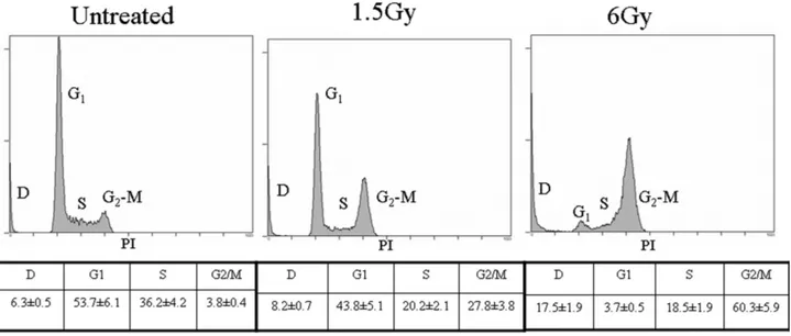
two-Figure 1. Flow cytometry analysis of cell cycle performed 24 h after exposure of Jurkat T cells to ionizing radiation. Letters indicate the different phases of the cell cycle. D: dead cells. The most representative of three independent experiments whose results were consistent is shown. Tables show cell percentages quantified by the mean (⫾SD) of three different peak analyses.
sample Student’s t test. Results were expressed as mean⫾ SD. Values of P⬍ 0.05 were considered significant.
Results
Cell cycle analysis, performed 24 h after the exposure to ionizing radiation, showed that 27.8% of the cells congre-gated in G2-M phase after 1.5 Gy, and 60.3% after 6 Gy; the corresponding percentages of dead and early apoptotic cells were 8.2% and 17.5%, respectively (Fig. 1). Decreased cell viability was also apparent for up to 7 days after 6-Gy
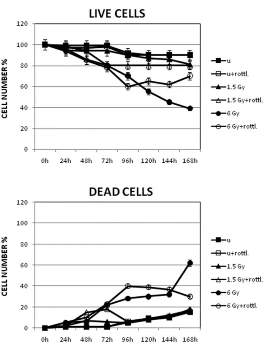
exposure (Fig. 2), even though compensatory cellular pro-liferation occurred during the course of the assay, which may affect the results of such assessment. The percentage of proliferation in unexposed and 1.5-Gy-exposed cells de-creased by 10%–20%, while the rate after exposure to 6 Gy decreased to 60% instead of 38% as reported in Figure 2 (live cells). Lastly, the percentage of TUNEL-positive ap-optotic cells, the characteristic features of which are evi-denced by electron microscopy analysis (Fig. 3), increased up to 40% in the 6-Gy-exposed sample (Table 1).
Figure 2. Cell viability and death assessed by Trypan blue dye exclusion test on Jurkat T cells up to 7 days after exposure to 1.5- and 6-Gy ionizing radiation. Data, presented as percentages, are the mean (⫾SD) of three independent experiments whose results were consistent.
In view of these preliminary results, we next investigated key signal transduction pathways involved in the apoptotic and survival responses of Jurkat T cells to ionizing radia-tion. Because the percentage of dead cells was significant
after exposure to 1.5 Gy and especially after 6 Gy, we investigated the signaling pathway likely to be involved in this death response. Since our group, among the PKC iso-forms checked (␦, , ), previously assigned a role to PKC␦
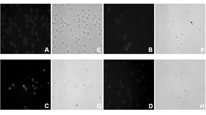
Figure 3. (a) TUNEL detection of apoptosis and phase contrast light microscopy (E–H) of Jurkat T cells exposed to 6-Gy ionizing radiation (A-D). When required, 3mol l⫺1rottlerin, a highly selective PKC␦ inhibitor, was added to the culture 1 h prior to irradiation. The extent of DNA fragmentation was quantified by direct visual counting of fluorescent-labeled nuclei by light microscopy, as shown in Table 1. Phase contrast light microscopy shows cells with apoptotic features. Magnification: 40⫻. A, E: unexposed; B, F: unexposed ⫹ rottlerin; C, G: 6 Gy; D, H: 6 Gy⫹ rottlerin. (b) Morphological features of apoptosis revealed by electron microscopy in Jurkat T cells exposed to ionizing radiation. Note the presence of membrane blebbing and micronuclei (mi) with highly compact, electron-dense chromatin, which are typical features of the apoptotic state. Magnification: 4400⫻. A: unexposed; B: 6 Gy.
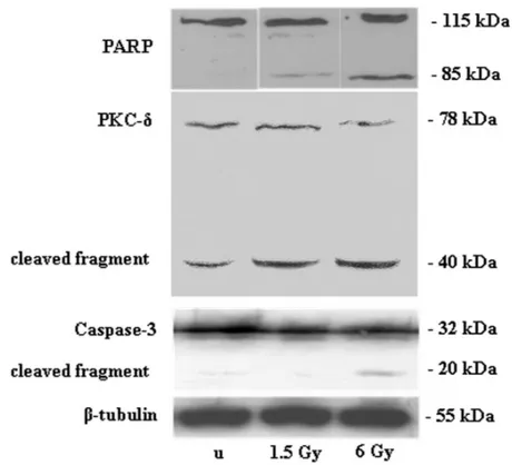
in the apoptotic response of Jurkat cells to ionizing radiation (Cataldi et al., 2002) and since cleavage by caspase-3 is reasonable evidence of activation of this protein (Emoto et al., 1995; Lewis et al., 2005), we checked the effects of 1.5-and 6-Gy ionizing radiation on caspase-3 mediated PKC␦ activation. When PKC␦ expression was checked after ex-posure to ionizing radiation, western blotting revealed a cleaved fragment of 40 kDa, coinciding with the 20-kDa
caspase-3 cleaved fragment and the 85-kDa PARP cleaved fragment (Fig. 4), suggesting a new role for proteolytic activation of PKC␦ by caspase-3. This biochemical re-sponse was corroborated by the effects observed in this cell line when the PKC␦ highly selective inhibitor, rottlerin, was added to the cultures. In the presence of rottlerin, the num-ber of dead cells decreased, as shown by the Trypan blue dye exclusion test and TUNEL analysis (Figs. 2–3 and Table 1), even though cell viability also decreased.
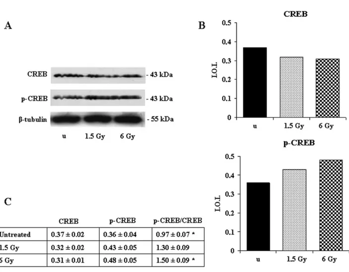
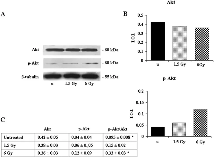
Since the CREB nuclear transcription factor is an impor-tant element in the cAMP-dependent survival signaling (Shaywitz and Greenberg, 1999), we then checked its phos-phorylation state by western blotting analysis. CREB ex-pression was unchanged, but its phosphorylation on Ser 133 increased after irradiation with 1.5 Gy and particularly after 6 Gy (Fig. 5). In addition, given that CREB is considered a regulatory target for the protein kinase Akt/PKB (Du and Montminy, 1998), we investigated the response of Akt to the ionizing radiation. Akt expression was unchanged, but its phosphorylation on Ser 473 increased after exposure to 6 Gy (Fig. 6), suggesting that its activation could determine CREB phosphorylation.
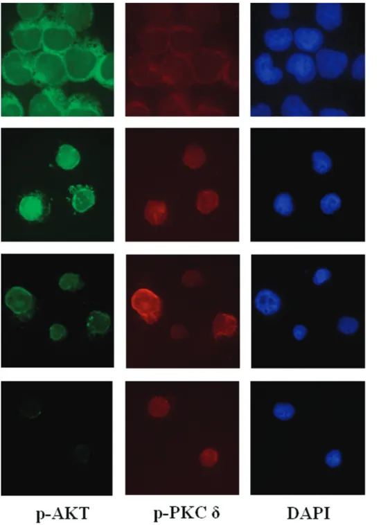
Lastly, we performed a double immunolabeling of p-PKC␦ (Thr 507) and p-Akt (Ser 473). 1.5-Gy-exposed
sam-Table 1
TUNEL detection of apoptosis in Jurkat cells exposed to ionizing radiation
Experimental points Apoptotic cells
Unexposed cells 4.0⫾ 0.09
Unexposed cells⫹ rottlerin 2.0⫾ 0.07
6 Gy 40.0⫾ 3.08*
6 Gy⫹ rottlerin 20.0⫾ 1.12*
Five slides were examined per sample within 5 h after exposure to ionizing radiation. The presence of DNA fragmentation was quantified by direct visual counting of fluorescent-labeled nuclei at 40⫻ magnification (light microscopy). Apoptotic cells were counted out of a total of 100 cells. Values (expressed as %) are means⫾ SD; n ⫽ 3 for all groups.
* Values of P⬍ 0.05 are considered statistically significant.
Figure 4. Effect of 1.5- and 6-Gy ionizing radiation on PARP, PKC␦, and caspase-3 expression and
cleavage in Jurkat T cells. The 85-kDa PARP cleaved subunit, 40-kDa PKC␦ fragment, and 20-kDa caspase-3 fragment are apparent after exposure to ionizing radiation. Immunostaining with a monoclonal antibody against -tubulin confirms equal loading (20 g). The blots shown are the most representative of three independent experiments whose results were consistent. u⫽ unexposed.
ples showed an increased level and nuclear translocation of p-Akt, while 6-Gy-exposed samples showed more p-PKC ␦-positive than p-Akt-␦-positive nuclei (Fig. 7), thus explaining the presence of a large number of dead cells, evidenced by TUNEL analysis (Fig. 3a). Interestingly, the number of apoptotic cells decreased in the presence of rottlerin (Table 1), while phosphorylation of the Akt/CREB signaling ma-chinery increased after exposure to a 6-Gy dose of ionizing radiation (Table 2).
Discussion
Ionizing radiation is known to cause cell cycle arrest, allowing cells to repair some of the damages induced to DNA by this oxidative stress. Such damage occurs in G1 prior to DNA replication, in S phase, and also in G2 before mitosis.
Moreover, the cells are activated against ionizing
radia-tion mechanisms, allowing them to repair membrane dam-age and single- and double-stranded breaks in DNA. In this study we wanted to investigate the effect of ionizing radi-ation on apoptotic and survival molecular response in Jurkat T lymphoblastoid cells in order to identify molecular targets for rational therapeutic intervention strategies. The thresh-old over which ionizing radiation induces considerable damage at the cellular level seems to be 6 Gy, even though a proportion of the cells activate repair mechanisms and still survive. In fact, significant G2 arrest occurs 24 h after exposure, along with generation of dead cells whose number increases in parallel to the decrease in cell proliferation for up to 7 days. These responses seem to be promoted by caspase-3-mediated PKC␦ activation, as indicated by the appearance of a 40-kDa PKC␦ fragment along with a phos-phorylated active form of the enzyme translocated into the nucleus, shown by immunofluorescence microscopy,
espe-Figure 5. Effect of 1.5- and 6-Gy ionizing radiation on CREB and p-CREB expression in Jurkat T cells. (A) Western blotting analysis of CREB and p-CREB (Ser 133) expression. Immunostaining with a monoclonal antibody against-tubulin confirms equal loading (20 g). Blots are the most representative of three independent experiments whose results were consistent. (B) Densitometric analysis of CREB and p-CREB (Ser 133) expression of the blot shown. (C) In the table, the ratio between the densitometric values of p-CREB (Ser133) and CREB is given⫾ S.D. u ⫽ unexposed. *6 Gy pCREB/CREB vs. unexposed pCREB/CREB: P ⬍ 0.05
cially after exposure to 6 Gy. This result is consistent with publications reporting that phosphorylation at Tyr 311 pro-motes degradation of PKC␦, presumably after ubiquination (Lu et al., 1998; Cataldi et al., 2002). Death-activated PKC␦ transiently accumulates in the nucleus where it is cleaved by caspase-3 to generate the constitutively activated PKC␦ catalytic fragment. Nuclear accumulation of pPKC␦ thus commits a cell to undergo apoptosis (De Vries-Selmon et al., 2007; Sun et al., 2008). If caspase-3 is not active it cannot cleave PKC␦ and thus induce apoptosis. In fact, the sequential regulation of PKC␦ and caspase-3 may assure that sustained nuclear accumulation of p-PKC␦ occurs only when caspase-3 is activated (Reyland, 2007). Nevertheless, a smaller relative amount of PKC␦ fragment is also present in untreated cells, as elsewhere reported in other experimen-tal models (Lewis et al., 2005). The absence of detectable proteolytic cleavage of other PKC isoforms (not shown)
provides further support for the selectivity of PKC␦ cleav-age induced by ionizing radiation.
Moreover, the cells seem to activate a survival signal system driven by Akt soon after exposure to ionizing radi-ation, and this, in turn, could activate the nuclear transcrip-tion factor CREB. The transient activatranscrip-tion of this signaling system is further confirmed by the presence of viable cells in the samples exposed to 6 Gy up to 3 days after treatment, accompanied by a number of dead cells similar to that present in the 1.5-Gy-exposed sample. It is evident that the cells try to counteract the effect of 6 Gy, but they are less viable after 7 days. In fact, when the dead cell population was evaluated, more apoptotic cells were apparent in the 6-Gy-exposed samples.
Cell death was reduced when the PKC␦ highly selective inhibitor rottlerin was added to the culture 1 h before exposure to the ionizing radiation. Rottlerin does not
di-Figure 6. Effect of 1.5- and 6-Gy ionizing radiation on Akt and p-Akt (Ser 473) expression in Jurkat T cells. (A) Western blotting analysis of Akt and p-Akt (Ser 473) expression. Immunostaining with a monoclonal antibody against-tubulin confirms equal loading (20 g). The blots are the most representative of three independent experiments whose results were consistent. (B) Densitometric analysis of Akt and p-Akt(Ser 473) expression of the blot shown. (C) In the table, the ratio between the densitometric values of p-Akt and Akt is given⫾SD. u ⫽ unexposed. *6 Gy pAkt/Akt vs. unexposed pAkt/Akt: P ⬍ 0.05.
rectly block PKC␦ activity but—by uncoupling mitochon-dria, depolarizing their membrane potential, reducing cel-lular ATP levels, activating 5⬘-AMP-activated protein kinase(AMPK), and affecting mitochondrial production of reactive oxygen species (ROS)—it ultimately precludes PKC␦ tyrosine phosphorylation and activation (Soltoff,
2007). Lastly, since it has been reported that BCR-induced CREB phosphorylation on Ser 133 in B lymphocytes re-quires the novel PKC␦ isoform (Blake et al., 1999; Blois et al., 2004), we checked the phosphorylation state of Akt and CREB in the presence of rottlerin. The phosphorylation levels of both Akt and CREB increased in the presence of
Figure 7. Light microscopy immunofluorescence analysis of p-PKC␦ (Thr 507) and p-Akt (Ser 473) expression in Jurkat T cells exposed to 1.5- and 6-Gy ionizing radiation. Green fluorescence shows p-Akt (left column), red fluorescence shows p-PKC␦, blue fluorescence represents DAPI (4-6-diamino-phenyl-indol) counterstaining nuclei (right column). Negative controls were performed by omitting the primary antibody (bottom row). Magnification: 40⫻
rottlerin, and the number of dead cells decreased concomi-tantly (Tables 1–2), suggesting that Akt/CREB phosphory-lation follows a PKC-independent route and constitutes part of the survival pathway.
Overall, our results are consistent with the response showed by other types of cells “in vivo” (Chong et al., 2005) and confirm the importance of Akt as a vital and broad cytoprotectant against cellular stresses in parallel to pro-apoptotic PKC␦ activator (Nakajima et al., 2006).
Thus, knowledge about some of the molecular mecha-nisms that drive cellular survival or death responses to ionizing radiation can help to establish therapies for several types of cancer by combining selective inhibitors or stimu-lators of key signaling proteins with conventional options for chemotherapy, hormone therapy, and radiotherapy.
Acknowledgments
This work has been supported by the FIRB 2001 Grant Project, cod. RBAU01EN5W-001: Interazioni tra radiazioni ionizzanti e fattori di trascrizione della famiglia CREB/ CREM, and a MIUR grant 60% 2008 to Prof. Cataldi. Authors thank Prof. Ausili and Dr. D. Genovesi, who set up the ionizing radiation protocol and performed radiation ex-posure of the cells
Literature Cited
Andreatta, C. P., P. Nahreini, A. J. Hanson, and K. N. Prasad. 2004.
Regulated expression of VP16CREB in neuroblastoma cells: analysis of differentiation and apoptosis. J. Neurosci. Res. 78: 570 –579.
Blake, R. A., P. Garcia-Paramio, P. J. Parker, and S. A. Courtneidge. 1999. Src promotes PKC␦ degradation. Cell Growth Differ. 10: 231–
241.
Blois, J. T., J. M. Mataraza, I. Mecklenbrauker, A. Tarakhovsky, and T. Chiles. 2004. B cell receptor-induced cAMP-response element-binding proactivation in B lymphocytes requires novel protein kinase C␦. J. Biol. Chem. 279: 30123–30132.
Brodie, C., and P. M. Blumberg. 2003. Regulation of cell apoptosis by PKC␦. Apoptosis 8: 19–27.
Caravatta, L., S. Sancilio, V. di Giacomo, R. A. Rana, A. Cataldi, and
R. Di Pietro. 2008. PI3-K/Akt-dependent activation of cAMP-re-sponse element-binding (CREB) protein in Jurkat T leukemia cells treated with TRAIL. J. Cell. Physiol. 214: 192–200.
Cataldi, A., S. Miscia, L. Centurione, M. Rapino, D. Bosco, G. Grifone, V. Di Valerio, F. Garaci, and R. A. Rana. 2002. Role of nuclear PKC␦ in mediating caspase-3-upregulation in Jurkat T leukemic cells exposed to ionizing radiation. J. Cell. Biochem. 86: 553–560.
Cataldi, A., V. di Giacomo, M. Rapino, D. Genovesi, and R. A. Rana. 2006. Cyclic nucleotide response element binding protein (CREB) activation promotes survival signal in human K562 erythroleukemia cells exposed to ionising radiation/Etoposide combined treatment. J.
Radiat. Res. 47: 113–120.
Cheng N., R. He, J. Tian, M. C. Dinauer, and R. D. Ye. 2007. A critical role of protein kinase C␦ activation loop phosphorylation in formyl-methionyl-leucyl-phenylalanine-induced phosphorylation of p47phox and rapid activation of nicotinamide adenine dinucleotide
phosphate oxidase. J. Immunol. 179: 7720 –7728.
Choi, S. C., B. S. Kim, M. Y. Song, E. Choi, H. M. Oh, J. H. Lyou, W. C. Han, H. B. Moon, T. H. Kim, J. M. Oh, et al. 2003.
Downregulation of p38 kinase pathway by cAMP response element binding protein protects HL-60 cells from iron chelator induced apo-ptosis. Free Radic. Biol. Med. 35: 1171–1184.
Chong, Z. Z., F. Li, and K. Malese. 2005. Activating AKT and the brain’s resources to drive cellular survival and prevent inflammatory injury. Histol. Histopathol. 20: 299 –315.
Cohen-Jonathan, E., E. J. Bernhard, and W. Gillies McKenna. 1999.
How does radiation kill cells? Curr. Opin. Chem. Biol. 3: 77– 83.
Dahlberg, W. K., E. I. Azzam, Y. Yu, and J. B. Little. 1999. Response of human tumor cells of varying radiosensitivity and radiocurability to fractionated irradiation. Cancer Res. 59: 5365–5369.
Das, S., A. Tosaki, D. Bagchi, N. Maulik, and D. K. Das. 2005.
Resveratrol-mediated activation of cAMP response element-binding protein through adenosine A3 receptor by Akt-dependent and -inde-pendent pathways. J. Pharmacol. Exp. Ther. 314: 762–769.
De Cesare, D., G. M. Fimia, and P. Sassone Corsi. 1999. Signaling routes to CREM and CREB: plasticity in transcriptional activation.
Trends Biochem. Sci. 24: 281–285.
Dempsey, E. C., A. C. Newton, D. Mochly-Rosen, A. P. Fields, M. E. Reyland, P. A. Insel, and R. O. Messing. 2000. Protein kinase C isozymes and the regulation of diverse cell responses. Am. J. Physiol.
279: L429 – 438.
Dent, P., A. Yacoub, J. Contessa, R. Caron, G. Amorino, K. Valerie, M. P. Hagan, S. Grant, and R. Schmidt-Ulrich 2003. Stress and radiation-induced activation of multiple intracellular signaling path-ways. Radiat. Res. 159: 283–300.
De Vries-Selmon, T. A., A. M. Ohm, M. J. Humpries, and M. Reyland. 2007. Induction of apoptosis is driven by nuclear retention of protein kinase C␦. J. Biol. Chem. 282: 22307–22314.
Di Giulio, C., M. Rapino, M. Zingariello, A. Antonucci, and A. Cataldi. 2007. PKC alpha-mediated CREB activation is oxygen and age-dependent in rat myocardial tissue. Histochem. Cell Biol. 127: 327– 333.
Di Pietro, R., L. Centurione, N. Sabatini, D. Bosco, S. Sancilio, F. Garaci, R. A. Rana, and A. Cataldi. 2004. Caspase-3 is dually regulated by apoptogenic factors mitochondrial release and by SAPK/ JNK metabolic pathway in leukemic cells exposed to etoposide-ioniz-ing radiation combined treatment. Int. J. Immunopathol. Pharmacol.
17: 181–190.
Di Pietro, R., V. di Giacomo, L. Caravatta, S. Sancilio, R. A. Rana, and A. Cataldi. 2007. Cyclic nucleotide response element binding (CREB) protein activation is involved in K562 erythroleukemia cells differentiation. J. Cell. Biochem. 100: 1070 –1079.
Dodson, G. E., and R. S. Tibbetts. 2006. DNA replication
stress-Table 2
Quantitative analysis by western blotting of CREB and Akt phosphorylation in Jurkat T cells exposed to ionizing radiation
Experimental points p-CREB p-Akt-1 Unexposed cells 0.14⫾ 0.03 0.04⫾ 0.006 Unexposed cells⫹ rottlerin 0.28⫾ 0.06 0.13⫾ 0.010
1.5 Gy 0.18⫾ 0.06 0.08⫾ 0.002
1.5 Gy⫹ rottlerin 0.30⫾ 0.09 0.14⫾ 0.020
6 Gy 0.35⫾ 0.12* 0.12⫾ 0.010*
6 Gy⫹ rottlerin 0.46⫾ 0.16* 0.28⫾ 0.030* Data are the mean⫾SD of three different experiments whose results were consistent.
induced phosphorylation of cyclic AMP response element-binding pro-tein mediated by ATM. J. Biol. Chem. 281: 1692–1697.
Du, K., and M. Montminy. 1998. CREB is a regulatory target for the protein kinase Akt/ PKB. J. Biol. Chem. 273: 32377–32379.
Emoto, Y., Y. Manome, G. Meinhardt, H. Kisaki, S. Kharbanda, M. Robertson, T. Ghayur, W. W. Wong, R. Kamen, R. Weichselbaum, and D. Kufe. 1995. Proteolytic activation of protein kinase C␦ by an ICE-like protease in apoptotic cells. EMBO J. 14: 6148 – 6156.
Ghayur, T., M. Hugunin, R. V. Talanian, S. Ratnofsky, C. Quinlan, Y. Emoto, P. Pandey, R. Datta, Y. Huang, S. Kharbanda, et al. 1996.
Proteolytic activation of protein kinase C␦ by an ICE-CED 3-like protease induces characteristics of apoptosis. J. Exp. Med. 184: 2399 – 2404.
Gschwendt, M., H. J. Mu¨ller, K. Kielbassa, R. Zang, W. Kittstein, G. Rincke, and F. Marks. 1994. Rottlerin, a novel protein kinase in-hibitor. Biochem. Biophys. Res. Commun. 199: 93–98.
Haimovitz-Friedman, A. 1998. Radiation-induced signal transduction and stress response. Radiat. Res. 150: S102–S108.
Jackson, D. N., and D. A. Foster. 2004. The enigmatic protein kinase C␦: complex roles in cell proliferation and survival. FASEB J. 18: 627– 636.
Kikkawa, U., H. Matsuzaki, and T. Yamamoto. 2002. Protein kinase C␦: activation, mechanisms and functions. J. Biochem. 132: 831–839.
Konishi, H., E. Yamauchi, H. Taniguchi, T. Yamamoto, H. Matsuzaki, Y. Takemura, K. Ohmae, U. Kikkawa, and Y. Nishizuka. 2001.
Phosphorylation sites of protein kinase C␦ in H2O2-treated cells and its
activation by tyrosine kinase in vitro. Proc. Natl. Acad. Sci. USA 98: 6587– 6592.
Kronfeld, I., G. Kazimirsky, P. S. Lorenzo, S. H. Garfield, P. M. Blumberg, and C. Brodie. 2000. Phosphorylation of PKC␦ on dis-tinct tyrosine residues regulates specific cellular functions. J. Biol.
Chem. 275: 35491–35498.
Lewis, A. E., R. Susarla, B. C. Wong, M. J. Langman, and M. C. Eggo. 2005. Protein kinase C delta is not activated by caspase-3 and its inhibition is sufficient to induce apoptosis in the colon cancer line, COLO 205. Cell. Signal. 17: 253–262.
Li, W., H. Mischak, J. C. Yu, L. M. Wang, J. F. Mushinski, M. A. Heidaran, and J. H. Pierce. 1994. Tyrosine phosphorylation of protein kinase C␦ in response to its activation. J. Biol. Chem. 269: 2349 –2352.
Lu, Z., D. Liu, A. Hornia, W. Devonish, M. Pagano, and D. A. Foster. 1998. Activation of protein kinase C triggers its ubiquitination and degradation. Mol. Cell. Biol. 18: 839 – 845.
Merritt, A., T. D. Allen, C. S. Potten, and J. A. Hickman. 1997.
Apoptosis in small intestinal epithelia from p-53-null mice: evidence for a delayed p53-independent G2/M associated cell death after ␥-ir-radiation. Oncogene 14: 2759 –2766.
Miura, Y. 2004. Oxidative stress, radiation-adaptive responses, and ag-ing. J. Radiat. Res. 45: 357–372.
Mizuno, K., K. Noda, T. Araki, T. Imaoka, Y. Kobayashi, Y. Akita, M. Shimonaka, S. Kishi, and S. Ohno. 1997. The proteolytic cleavage of protein kinase C isotypes, which generates kinase and regulatory fragments, correlates with Fas-mediated and 12-O-tetradecanoyl-phor-bol-13-acetate-induced apoptosis. Eur. J. Biochem. 250: 7–18.
Musashi, M., S. Ota, and N. Shiroshita. 2000. The role of protein kinase C isoforms in cell proliferation and apoptosis. Int. J. Hematol.
72: 12–19.
Muthusamy, N., and J. M. Leiden. 1998. A protein kinase C-, Ras-, and
RSK2 dependent signal transduction pathway activates the cAMP-responsive element binding protein transcription factor following T cell receptor engagement. J. Biol. Chem. 273: 22841–22847.
Nakajima, T. 2008. Positive and negative regulation of radiation-in-duced apoptosis by protein kinase C. J. Radiat. Res. 49: 1– 8.
Nakajima, T., T. Wakasa, Y. Okuma, O. Inanami, Y. Nomura, M. Kuwabara, and K. Kawahara. 2006. Dual inhibition of protein phosphatase-1/2A and calpain rescues nerve growth factor-differenti-ated PC12 cells from oxygen glucose-deprivation-induced cell death.
J. Neurosci. Res. 83: 459 – 468.
Nishizuka, Y. 1995. Protein kinase C and lipid signalling for sustained cellular responses. FASEB J. 9: 484 – 496.
Pugazenthi, S., A. Nesterova, K. A. Heidenreich, L. M. Boxer, L. E Heasley, and J. E. Reusch. 2000. Akt/protein kinase B up-regulates Bcl-2 expression through cAMP-response element-binding protein
J. Biol. Chem. 275: 10761–10766.
Radford, I. R., T. K. Murphy, J. M. Radley, and S. L. Ellis. 1994.
Radiation response of mouse lymphoid and myeloid cell lines. Part II. Apoptotic death is shown by all lines examined. Int. J. Radiat. Biol. 65: 217–227.
Reyland, M. E. 2007. Protein kinase C␦ and apoptosis. Biochem. Soc.
Trans. 35: 1001–1004.
Saeki, K., A. You, E. Suzuki, Y. Yazaki, and F. Takaku. 1999. Ab-errant expression of c-AMP-response-element-binding protein (CREB) induces apoptosis. Biochem. J. 343: 249 –255.
Shaywitz, A. J., and M. E. Greenberg. 1999. CREB: a stimulus-induced transcription factor activated by a diverse array of extracellular signals. Annu. Rev. Biochem. 68: 821– 861.
Shi, Y., S. L. Venkataraman, G. E. Dodson, A. M. Mabb, S. LeBlanc, and R. S. Tibbetts. 2004. Direct regulation of CREB transcriptional activity by ATM in response to genotoxic stress. Proc. Natl. Acad. Sci.
USA 101: 5898 –5903.
Soltoff, S. P. 2007. Rottlerin: an inappropriate and ineffective inhibitor of PKC delta. Trends Pharmacol. Sci. 28: 453– 458.
Sun, F., A. Kanthasamy, C. Song, Y. Yang, V. Anantharam, and A. G. Kanthasamy. 2008. Proteasome inhibitor-induced apoptosis is me-diated by positive feedback amplification of PKC␦ proteolytic activa-tion and mitochondrial translocaactiva-tion J. Cell Mol. Med. 12: 2467–2472.
Takahashi, M., H. Mukai, M. Toshimori, M. Miyamoto, and Y. Ono. 1998. Proteolytic activation of PKN by caspase-3 or related protease during apoptosis. Proc. Natl. Acad. Sci. USA 95: 11566 –11571.
Tauchi, H., and S. Sawada. 1994. Analysis of mitotic cell death caused by radiation in mouse leukaemia L5178Y cells: apoptosis is the ulti-mate form of cell death following mitotic failure. Int. J. Radiat. Biol.
65: 449 – 455.
Uchiyama, T., R. M. Engelman, N. Maulik, and D. K. Das. 2004. Role of Akt signaling in mitochondrial survival pathway triggered by hy-poxic preconditioning. Circulation 109: 3042–3049.
Walton, M., A. M. Woodgate, A. Muravlev, R. Xu, M. J. During, and M. Dragunow. 1999. CREB phosphorylation promotes nerve cell survival. J. Neurochem. 73: 1836 –1842.
Wilson, B. E., E. Mochon, and L. Boxer. 1996. Induction of bcl-2 expression by phosphorylated CREB proteins during B-cell activation and rescue from apoptosis. Mol. Cell. Biol. 16: 5546 –5556.
Yano, T., Y. Itoh, T. Kubota, T. Sendo, T. Koyama, T. Fujita, K. Saeki, A. Yuo, and R. Oishi. 2005. A prostacyclin analog prevents radio-contrast nephropathy via phosphorylation of cyclic AMP response element binding protein. Am. J. Pathol. 166: 1333–1342.