1
Research Doctorate School in Biological and Molecular Sciences
Ph.D. in BIOMATERIALS – XXVI Cycle (2011-2013)
Epidermis-targeted drug-loaded nanoparticles
for the treatment of psoriasis
Giulia Ciampi
Supervisor: Dr. Federica Chiellini
Tutors: Dr. Anna Maria Piras
Dr. Marcella Ferri
Laboratory of Bioactive Polymeric Materials for Biomedical and
Environmental Applications (BIOlab) – Department of Chemistry and
3
Table of contents1 Chapter I : Introduction ... 6
1.1Polymeric Biomaterials ... 6
1.1.1 Natural Polymeric Biomaterials ... 7
1.1.1.1 Chitosan ... 8
1.1.1.2 Hyaluronic Acid ... 10
1.1.1.3 Poly(γ-glutamic acid) ... 12
1.1.1.4 Poly(3-hydroxybutyrate-co-3-hydroxyhexanoate) ... 13
1.1.2 Synthetic Polymeric Biomaterials ... 15
1.1.2.1 Poly(ε-caprolactone) ... 18
1.1.2.2 Polymethacrylamide ... 19
1.1.3 Polymeric Drug Delivery Systems ... 20
1.1.3.1 Amphiphilic Copolymers as Polymeric Drug Delivery Systems ... 23
1.1.3.2 Living radical Polymerization by Reversible Addition- Fragmentation Chain Transfer (RAFT) Process ... 25
1.1.3.3 Micro-Nanoparticles for Biomedical and Pharmaceutical Applications ... 27
1.1.3.4 Techniques for Micro-Nanoparticles Preparation ... 31
1.2Nanomedicine in Dermatology ... 33
1.2.1 The Skin Structure and Function ... 34
1.2.2 Dermatological Diseases ... 36
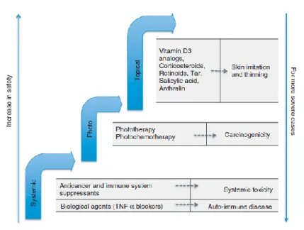
1.2.2.1 Psoriasis ... 38
1.2.2.2 Disease Management and Therapeutic Target in Psoriasis ... 41
1.2.2.3 Treatment of Psoriasis by Methotrexate ... 42
1.2.3 Topical and Transdermal Drug Delivery ... 44
1.2.3.1 Polymeric Nanoparticles Based Topical Delivery Systems ... 46
1.3 Tissue Engineering for Cartilage Repair... 49
1.3.2 Strategies for Cartilage Repair ... 50
1.3.3 Hydrogels for Tissue Engineering ... 52
1.3.3.1 In Situ Forming Biomimetic Hydrogels for Tissue Regeneration .... 54
1.3.3.2 Strategies to Design in Situ Forming Hydrogel ... 56
1.4References: ... 59
I. AIM OF THE WORK ... 75
II. GLOSSARY ... 78
III. LIST OF ABBREVIATION ... 83
2 Chapter II : Synthesis and Characterization of Amphiphilic Copolymer based on Poly (ε-caprolactone) and Poly(methacryloylglycylglycine) ... 85
2.1Introduction ... 85
2.2Materials ... 86
2.3 Methods ... 87
2.4Results and Discussions... 92
2.4.1 Synthesis and Characterization of the Block Copolymer PCL-b-p(MAGG) ... 92
4
2.4.1.1 Preparation and characterization of MAGG ... 92
2.4.1.2 Preparation and characterization of xanthate-terminated PCL ... 94
2.4.1.3 Preparation and characterization of Amphiphilic PCL and p(MAGG) Block Copolymer ... 98
2.4.1.4 Thermal characterization of PCL-b-p(MAGG) copolymer ... 101
2.4.1.5 Preparation and characterization of PCL-b-p(MAGG) micelles ... 101
2.4.1.6 In Vitro Biological Evaluation of the new copolymer ... 103
2.4.2 Preparation and characterization of the copolymer Poly[(polycaprolactonyl)methacrylate-co-glycylglycinemethacrylamide] PCL-co-p(MAGG) ... 104
2.4.2.1 Preparation and characterization of PCL macromonomer ... 104
2.4.2.2 Preparation and characterization of Poly[(polycaprolactonyl)methacrylate-co-glycylglycinemethacrylamide] PCL-co-p(MAGG) ... 106
2.4.2.3 Thermal characterization of PCL-co-p(MAGG) copolymer ... 108
2.5Conclusions ... 108
2.6References ... 109
3 Chapter III : Chitosan and Poly( γ-glutamic acid) based Nanoparticles Loaded with Methotrexate ... 111
3.1Introduction ... 111
3.2 Materials ... 113
3.3Methods ... 113
3.4Results and Discussion ... 115
3.5Conclusions ... 121
3.6References ... 122
4 Injectable and Biodegradable Thermogels Based on Hyaluronic Acid and Chitosan Loaded with Poly[(R)-3-hydroxybutyrate-co-(R)-3-hydroxyhexanoate] Polymeric Nanoparticles for the Sustained and Controlled Release of Dexamethasone ... 124
4.1Introduction ... 124
4.2Materials: ... 126
4.3Methods: ... 126
4.4 Results and Discussions... 132
4.4.1 DEXA-loaded PHBHHx NPs ... 132
4.4.2 HA-AHP-CS thermogel ... 137
4.5Conclusions ... 141
4.6References: ... 142
5 Chapter V : Overall Conclusive Remarks ... 145
5.1Synthesis and Characterization of Amphiphilic Copolymers based on Poly(ε-caprolactone) and Poly(methacryloylglycylglycine) ... 145
5.2
Chitosan and Poly(γ-glutamic acid) based Nanoparticles Loaded with
Methotrexate... 1465
5.3Injectable and Biodegradable Thermogels Based on Hyaluronic acid and Chitosan Loaded with
Poly[(R)-3-hydroxybutyrate-co-hydroxyhexanoate] Polymeric Nanoparticles for the Sustained
Controlled Release of Dexamethasone ... 146 ACKNOWLEDGMENTS ... 147
6
1 Chapter I : Introduction
1.1 Polymeric Biomaterials
Over the past few decades polymeric biomaterials have revolutionized the medical field and have been used in a wide variety of applications such as controlled drug delivery systems, cell culture supports, scaffolds for tissue engineering, biosensor, actuators, bioseparation devices, reactive coatings, medical imaging and devices.
Biomaterials science is highly interdisciplinary in nature, involving materials science, physical, engineering, biological and clinical sciences principles.
The definition for the term “biomaterial” has been widely accepted as the following:
A biomaterial is any material, natural or synthetic, that comprises a whole or part of a living structure or biomedical device that performs , augments, or replaces a natural function.
A biomaterial is a nonviable material used in medical device intended to interact with a biological system without harming it.
The most important requirements for biomaterials are biocompatibility and controlled degradability. Biocompatibility prevents toxic effects of materials on the physiological system. Synthetic biomaterials are not typically rejected by the body, although solid scaffolds or devices may be encapsulated in a fibrous capsule after implantation, and soluble polymers or small particles undergo various routes of elimination, sometimes including chemical breakdown. While the biocompatibility is an intrinsic property of the polymer (related to chemical structure), degradation can be directly engineered, chemically tuned, and controlled by various stimuli. Indeed, degradation of polymeric biomaterials can be initiated by an environmental, internal or external stimulus. For example, hydrolysis is a commonly used controlled degradation mechanism, and the stimulus for degradation is environmental (water). Enzymolysis is another popular method that relies on an internal stimulus (enzyme production) to control degradation [Deshayes 2013].
Stimulus-responsive polymers, as ‘intelligent’, ‘smart’ or ‘environmentally sensitive’ polymers, are biomaterials systems that exhibit large, sharp changes in response to physical stimuli (such as temperature, solvents, or light) or to chemical stimuli (such as reactants, pH, ions in solution, or chemical recognition). Responses differ depending on the stimulus applied and may include changes in shape, volume, mechanical properties, or permeation rates, among other things. These systems possess a variety of interesting applications for encapsulation, controlled delivery, or as intelligent switches to mention just a few [York, 2008]. Engineering polymeric nanostructures such as hyperbranched polymers, dendrimers and polymeric nanoparticles (NPs) [Xu 2012; Gong 2012] are a growing area of contemporary biomaterials science, due to their unique properties and large potential in drug delivery [Kim 2012; Lim 2012; Bielawski et al., 2011]. For using
7
polymers in drug delivery, a polymer must be biocompatible. Biocompatibility was defined as the ability of a material to act with an appropriate host response in a specific application [Williams 1999]. Moreover, biocompatible polymers used in drug delivery are often biodegradable with the formation of non-harmful byproducts, such as non-toxic alcohols, acids and other easily eliminated low molecular weight products. They can indeed contribute to the drug release as a result of their erosion/degradation, in addition to drug diffusion through the polymeric material. Biodegradable polymer, in the development of drug delivery systems, must meet very specific requirements such as: a) Biocompatibility of the polymer backbone and its degradation products; b) Mechanical strength sufficient to meet the needs of specific applications, c) Degradability with degradation kinetics matching a biological process such as wound healing, d) Processibility using available equipment. e) Solubility in various solvents. f) Chemical, structural and application versatility d)Economically acceptable shelf life and obviously, the agreement of the European Medicine Evaluation Agency (EMEA) or Food and Drug Administration (FDA), USA.
Polymers remain the most versatile class of biomaterials, being extensively applied in medicine and biotechnology, as well as in the food and cosmetics industries. Applications include surgical devices, implants and supporting materials (e.g. artificial organs, prostheses and sutures), drug-delivery systems with different routes of administration and design, carriers of immobilized enzymes and cells, biosensors, components of diagnostic assays, bioadhesives, ocular devices, and materials for orthopaedic applications. Polymers used as biomaterials can be synthesized to have appropriate chemical, physical, interfacial and biomimetic characteristics, which permit various specific applications [Chiellini 2008; Williams 2009; Kohane 2010]. Compared with other types of biomaterials, such as metals and ceramics, polymers offer the advantage that they can be prepared in different compositions with a wide variety of structures and properties.
Polymers are mainly divided into two groups, natural polymers, such as proteins, cellulose, silk and synthetic polymers, such as full carbon backbone polymers: polystyrene, polyethylene, polypropylene, poly(vinyl chloride) and heteropolymers: polyamides, polyesters, polyurethanes.
1.1.1
Natural Polymeric Biomaterials
Natural polymers are produced by several living organisms. Their synthesis is due to the presence of enzymatic catalysts through reactions of chain growth polymerization of activated monomers, formed within cells as a result of complex metabolic processes. Use of biopolymers for diversified applications in life sciences has several advantages, such as availability from replenishable agricultural or marine food resources, biocompatibility, biodegradability, therefore leading to ecological safety and the possibility of preparing a variety of chemically or enzymatically modified derivatives for specific end uses.
8
Human serum albumin (HSA), a protein polymer, is one of the most frequently used biopolymers in medicine. HSA is known to be used for treating shock, burns, hypoalbuminemia, after surgery trauma, cardiopulmonary bypass, acute respiratory distress and hemodialysis. Albumin-conjugates are also used in treatment of arthritic diseases, for liver targeting, and others. Albumin is known to be accumulated in malignant and inflamed tissues and nanoparticles made of HSA were found to be non-toxic and well-tolerated by human organism. Owing to its capability to transport low molecular weight compounds it is a unique carrier for drugs. Binding drugs with albumin provided prolonged effect of such proteins and peptides as Albuferon and Levemir. Functional groups (carboxylic and amine groups) which are present in the structure of albumin allow to modify the surface of the particles by attaching the molecules for targeting purposes [Langer 2007; Elzoghby 2012]. Collagen is the most abundant protein of connective tissues in all animals, it is a nonantigenic, biocompatible, biodegradable and bioreabsorbable biopolymer and it can be easly purified from living organism. Gelatin is a natural polymer is obtained by controlling the hydrolysis of collagen, and it is used widely for pharmaceutical (as a plasma expander, wound dressing, adhesive, and absorbent pad for surgical use) and cosmetics applications.
Silks have been investigated as biomaterials due to the successful use of silk fibers from B. mori as suture material for centuries. The silk bio-polymer is used in tissue regeneration for treating burn victims and as matrix of wound healing. The silk fibroin peptides are used in cosmetics due to their glossy, flexible, elastic coating power, easy spreading and adhesion characters. The silk is used to fight edema, adenosine augmentation therapy and cancer [Vepari 2007].
Alginate is a polysaccharide that offers an attractive alternative for sustained-release systems. It offers advantages over synthetic polymers as it forms hydrogels under relatively mild pH and temperature and is generally regarded as non-toxic, biocompatible, biodegradable, less expensive and abundantly available in nature; in addition, alginate meets the important requirement of being amenable to sterilization and storage. All these advantages make alginates very useful materials for biomedical applications, especially for controlled delivery of drugs and other biologically active compounds and for the encapsulation of cells [Chiellini 2008; Ha 2013].
Dextran is a polysaccharide composed of branched glucan and synthesized by bacteria and yeast. Due to dextran’s biocompatibility, dextran-based hydrogels have been widely used as a blood plasma substitute, as delivery systems of various drugs and for tissue engineering scaffolds [Sun 2012].
1.1.1.1 Chitosan
Chitosan (CS) is a linear, randomcopolymer of β-(1–4)-linked D-glucosamine and N-acetyl-D-glucosamine whos emolecular structure comprises a linear backbone linked through glycosidic bonds (Figure 1).
9
Figure 1: Chemical structure of chitosan.
The basic amine groups of this polysaccharide are protonated and, thus positively charged in most physiological fluids. Chitosan is a product derived from alkaline N-deacetylation of chitin. Chitin, being the second most abundant natural biopolymer mainly derived from exoskeletons of crustaceans and also from cell walls of fungi and insects, is a linear cationic heteropolymer of randomly distributed GlcNAc and GlcN residues with β-1, 4 linkage. In general, the degree of deacetylation (DD) of chitosan ranges from 40 to 98% and the molecular weight ranges in between 50 kD and 2000 kD. CS can be considered mostly hydrophilic, but the percentage of acetylated units and their distribution in the chains has a critical effect on its solubility and conformation in aqueous media. Because of these molecular features, CS exhibits in solution a pH-dependent behavior and interesting biopharmaceutical properties such as muco-adhesiveness and ability to open epithelial tight junctions. These characteristics have attracted the attention of many scientists working in biomedical fields, and particularly in drug delivery. CS is biodegradaded by a number of enzymes, such as lysozyme, di-N-acetylchitobiase, N-acetyl-beta-D-glucosaminidase and chitiotriosidase, which are present in human mucosas and other physiological fluids [Garcia-Fuentes 2012].
This renewable polysaccharide is currently being explored extensively for its applications in various fields such as pharmaceutical, cosmetics, biomedical, biotechnological, agriculture, and both food and non-food industries. It has emerged as a new class of biomaterial with highly sophisticated functions due to their versatile biological activity, excellent biocompatibility, and complete biodegradability, in addition to low toxicity [Jain 2013].
The degree of deacetylation and the degree of polymerization (DP) controls the
molecular weight of a polymer and these two are important parameters deciding the use of chitosan and its modifications for various applications. Chitosan possesses reactive hydroxyl and amino groups but is usually less crystalline than chitin, which presumably makes chitosan more accessible to reagents. After heating, it decomposes prior to melting; thus, it has no melting point. The solubility profile of chitosan well depicts that it is soluble in dilute aqueous organic or mineral acids below pH 6.5, p-Toulene sulfonic acid, DMSO, and 10-Camphorsulfonic acid. Almost all aqueous acids dissolve chitosan, out of which the most commonly used are formic acid and acetic acid. The increase in its solubility by increasing both the polarity and the degree of electrostatic repulsion is due to generation of an enormous number of cationic sites formed due to protonation of
10
aminogroups by acids along the chitosan backbone. Water-soluble salts of chitosan include formate, acetate, lactate, maleate, citrate, tartarate, glyoxylate, pyruvate, glycolate, malonate, and ascorbate.
Chitosan is widely regarded as being a nontoxic, biologically compatible polymer. It has been approved for dietary applications in Japan, Italy, and Finland, and additionally, it has been approved by the FDA for wound dressings [Jain 2013].
This interesting biopharmaceutical and toxicological profile of CS has encouraged its use as a biomaterial for drug delivery applications, it has been presented in several pharmaceutical forms including polymer solutions, micro/nanoparticles, and hydrogels [ Dash 2011, Nagpa 2010].
The potential of CS nanocarrier technology has been reported for a variety of applications that include immunization, topical ocular/dermal drug delivery, transmucosal oral/nasal peptide absorption, anti-cancer drug delivery, brain delivery and gene delivery [Garcia Fuentes 2012].
Many preparation methods for chitosan-based nanocarriers have already been developed. Among them, ionic cross-linking methods based on the electrostatic interactions between positively charged chitosan and negatively charged polyanions, demonstrated advantages for bioactive molecular and hydrophilic drug encapsulation owing to their simple and mild processes. In the ionic gelation method, chitosan nanoparticles are constructed by dropping CS droplets into a solution of a polyanion such as tripolyphosphate (TPP)[Lopez-leon 2005] . The coacervation/precipitation method is based on the insolubility of CS in an alkaline solution, therefore when it is put into a solution with an alkaline pH such as sodium hydroxide or ethane diamine, precipitate/coacervate particles are produced [Amidi 2010]. In the emulsion cross-linking method, due to the reactive amine group, CS can easily be cross-linked with the aldehyde groups of cross-linking agents such as glutaraldehyde [Karnchanajindanun 2011]. Recently, amphiphilic chitosan-based polymers were synthesized for self-assembled nanocarriers, which exhibit significant potential for the delivery of both hydrophobic and hydrophilic drugs. Due to the fairly abundant primary amine groups of chitosan, they are easily conjugated to hydrophobic polymers such as polylactide, polycaprolactone and polymers containing acid moieties [ Huo 2010; Jain 2013; Hu 2013].
1.1.1.2 Hyaluronic Acid
Hyaluronic acid (HA), also named hyaluronan, is a high molecular weight (105–107 Da), naturally occurring biodegradable polymer. HA is an unbranched non-sulfated glycosaminoglycan (GAG) composed of repeating disaccharides (β-1,4-D-glucuronic acid (known as uronic acid) and β-1,3-N-acetyl-D-glucosamide) (Figure 2).
11
Figure 2: Chemical structure of hyaluronic acid.
HA is produced and secreted by cells including fibroblasts, keratinocytes or chondrocytes; it is naturally synthesized by hyaluronan synthases (HAS1, HAS2 and HAS3), a class of integral membrane proteins [Hunt 2013].The mechanism of HA synthesis involves chain extension by addition of a monosaccharide (alternating addition of glucuronic acid and N-acetyl glucosamine) to the reducing end of the chain. The number of repeat disaccharides units in a completed HA molecule can reach a value as high as 10,000 or more, and a molecular weight of 4 million Da.
HA is a polyanion that can self-associate and can also bind to water molecules (when not bound to other molecules), giving it a stiff, viscous quality similar to gelatin. HA is one of the major elements in the extracellular matrix (ECM) of vertebrate tissues. It is available in almost all body fluids and tissues, such as the synovial fluid, the vitreous humor of the eye, and hyaline cartilage. This biopolymer functions as a scaffold, binding other matrix molecules, including aggrecan [Garg 2004]. HA is also involved in several important biological functions, such as regulation of cell adhesion and cell motility, manipulation of cell differentiation and proliferation, and providing mechanical properties to tissues. Several cell surface receptors, such as CD44, RHAMM and ICAM-1, have been shown to interact with HA, influencing cellular processes, including morphogenesis, wound repair, inflammation and metastasis [Eng 2010]. Moreover, HA is responsible for providing the viscoelasticity of some biological fluids (synovial fluid and vitreous humor of the eye) and controlling tissue hydration and water transport. In addition, HA has been found during embryonic development in the umbilical cord, suggesting that materials composed of HA may persuade favorable conditions for tissue regeneration and growth. HA’s characteristics, including its consistency, biocompatibility and hydrophilicity have made it an excellent moisturizing component in cosmetic dermatology and skin-care products [Falcone 2006]. Its unique viscoelasticity and limited immunogenicity have led to its use in several biomedical applications, such as viscosupplementation in osteoarthritis treatment, as an aid in eye surgery and for wound regeneration. Hyaluronan has been widely studied for drug delivery, for dermal, nasal, pulmonary, topical, parenteral, liposome-modified, implantable delivery devices and for gene delivery [Liao 2005, Fakhari 2013, Burdick 2011, Yadav 2008]. Hyaluronan for tissue engineering has been focused on cartilage, bone and osteochondral applications, most likely due to the fact that it is a major macromolecular component of the extracellular matrix [Malafaya 2007] .
Commercially available hyaluronan is obtained from different sources, mainly by extraction from umbilical cord, rooster comb, synovial fluid, or vitreous humour. In
12
addition, hyaluronic acid can be easily and controllably produced in large scales through microbial fermentation, from strains of bacteria such as Streptococci, enabling the scale-up of derived products and avoiding the risk of animal-derived pathogens. Hyaluronan is available for several applications, for lubrication and mechanical support for the joints in osteoarthritis (Hyalgan® and Hyalubrix® from Fidia in Italy; Artz® from Seikagaku Corporation in Japan) as a viscoelastic gel for surgery and wound healing (Bionect® from CSC Pharmaceutical in USA; Jossalind® from Hexal in Germany), for implantation of artificial intraocular lens (Healon® from OVD from Advanced Medical Optics in USA, Opegan R® from Seikagaku in Japan, Opelead® from Shiseido in Japan, Orthovisc® from Anika in USA) and as culture media for use in in vitro fertilization (EmbryoGlue® from Vitrolife, USA)[Liao 2005, Malafaya 2007]. Hyaff® commercialized by Fidia in Italy has been widely used as a biomaterial for biomedical applications.
1.1.1.3 Poly(γ-glutamic acid)
Poly-γ-glutamic acid (γ-PGA) in an anionic naturally occurring homo-polyamide that is made of D- and L-glutamic acid units coonected by amide linkages between α-amino and γ-carboxylic acid groups (Figure 3).
Figure 3: Chemical structure of γ-PGA.
γ-PGA is produced predominantly by bacteria belonging to Bacillus sp., such as B. licheniformis, B. subtilis, B. megaterium, B. pumilis, B. mojavensis and B. amyloliquefaciens. γ-PGA has diverse physiological functions that vary according to the species synthesizing it and its environment. Soil bacteria (mostly from the genus Bacillus, but excluding B. anthracis) use the released γ-PGA for sequestration of toxic metal ions, thereby increasing their resistance to adverse environments. γ-PGA may also be a source of glutamate for bacteria during starvation in the late stationary phase. Two highly pathogenic bacteria viz., B. anthracis and Staphylococcus epidermidis synthesizes surface-associated γ-PGA, which enables them to escape phagocytosis, and enables them to act as virulence factor. Furthermore, B. anthracis capsule is composed exclusively of the D-enantiomer, making it particularly non-immunogenic. This capsule also prevents antibodies from gaining access to the bacterium. It protects B. anthracis against phage infections and S. epidermidis against antimicrobial peptides, probably by acting as a passive barrier. Thus, anchored γ-PGA is a bacterial virulence factor that protects the
13
pathogen from the immune system, whereas released PGA may be a persistence factor that protects the bacterium from its environment.
Multienzyme system mostly operates on the basis of the thiotemplate mechanism which generally offers high sterioselectivity for amino acid precursors and productivity of comparatively small peptides in which amino acid arrangements are strictly regulated. Synthesis of highly elongated γ-PGA involves extremely unique mechanism [Ashiuchi 2005]. Biosynthesis of γ-PGA in bacteria is carried out in two steps: in the first step, synthesis of and D-glutamic acid takes place; where as in second step, these D and L-glutamic acid units are joined together to form γ-PGA.
γ- PGA is water soluble, biodegradable, edible and non toxic towards human and the environment. Therefore potential applications of γ-PGA and its derivatives have been of interest in a broad range of industrial field such as food, water treatment and biomedical field as cosmetics and medicine being used as thickener, humectants, cryoprotectant, sustained release materials, drug carrier, adhesive, fibers, hydrogels, biopolymer flocculants.
Its multifunctionality, biodegradability, nontoxicity, compatibility, and edibility have made it a promising biopolymer for use as a health food, a thickener, an osteoporosis-preventing factor, and a stabilizer in the food industry; as a moisturizing agent in cosmetics; as a chelating agent in waste-water treatment; as a hydrogel (especially super absorbent polymer, SAP) for environmental, agricultural, and biomedical product applications; as a biodegradable packing material, and in many other possible applications including Liquid Crystal Displays (LCDs) and conductive display material, drug deliverer, gene vector, curative biological adhesive, dispersant, and enzyme-immobilizing material [Bajaj 2011].
Carboxyl groups of the side chains of γ-PGA offer attachment points for the conjugation of chemotherapeutic agents, thereby rendering the drug more soluble and easier to administer. Its breakdown product, glutamic acid, can enter normal cellular metabolism and is not excreted by the kidney [Singer 2005]. A wide variety of anticancer agents have been conjugated to γ-PGA and the resultant conjugates have been tested. Chemical crosslinking of gelatin and γ-PGA has been shown to be promising as a surgical adhesive and hemostatic agent that may be a potential replacement for the blood-originated fibrin glue [Bajaj 2011]. Self-assembled particulate carriers γ-PGA based materials have been produced by self-assembly of oppositely charged biopolymers. Utility of γ-PGA NPs as an efficient antigen delivery carriers in dendritic cell-based cancer immunotherapy has been demonstrated by Matsuo et al, 2010. Lin et al (2007) showed chitosan/poly-γ-glutamic acid-based self-assembling nanoparticulate system as a drug delivery platform for insulin oral delivery.
Moreover biodegradable nanoparticles, composed of chitosan (CS) and poly-g-glutamic acid (γ-PGA), were prepared by an ionic-gelation method for transdermal DNA delivery
[Lee 2008].
14
Polyhydroxyalkanoates (PHAs) are a group of naturally occurring biodegradable and biocompatible polymers that belong to the aliphatic polyester family (Figure 4). PHAs act as storage compounds of carbon and energy stored in inclusion bodies within the cytoplasm that accumulate during imbalanced growth by various microorganisms in the presence of an excess of a carbon source and nutrient (Figure 4) limiting conditions (e.g., nitrogen, phosphorus). PHAs occur naturally in a variety of organisms, but microorganisms can be employed to tailor their production in cells1.The simplest PHAs, polyhydroxybutyrate (PHB), was discovered in 1926 by Maurice Lemoigne as a constituent of the bacterium Bacillus megaterium. Since that time, many discoveries have generated excitement over the potential of utilizing PHA in household, industrial, medical, and other applications [Misra 2006].
Figure 4: Schematic structures of representative poly(hydroxyalkanoate)s. Members of the PHA family can exist as homopolymers or copolymers of two or more hydroxyalkanoic acids. The monomer composition of PHAs is variable and can be manipulated by means of the carbon source used and by changing the growth conditions. Due to their natural origin and enhanced biocompatibility, PHAs are widely used as biomedical implant materials including wound management (sutures, skin substitutes, nerve cuffs, surgical meshes, staples, swabs), vascular system devices (heart valves, cardiovascular fabrics, pericardial patches, vascular grafts), orthopedics (scaffolds for cartilage engineering, spinal cages, bone graft substitutes, meniscus regeneration,
15
internal fixation devices), and drug delivery systems [Chen 2009, Park 2012, Babu 2013, Puppi 2010].
Poly(R-3-hydroxybutyrate-co-R-3-hydroxyhexanoate) (PHBHHx) is a newly developed member of the PHA family, with improved physical and chemical properties compared with other PHAs as PHB and PHBV.
PHBHHx had improved hemocompatibility and cytocompatibility; it holds promise as a blood-contact material with less platelet adhesion, reduced erythrocyte contact and hemolysis reactivity compared with PHB and PHBV films [Qu 2006]. PHBHHx had better performances on attachment, proliferation, and differentiation of osteoblasts [Li 2005, Wang 2004 ] compared with PHB. In vitro chondrocytes proliferated better and preserved their phenotype on the PHBHHx/PHB scaffolds than on PHB ones. Scaffolds made of PHBHHx/PHB consisting of 60 wt% PHBHHx showed strong growth and proliferation of chondrocytes on the blending materials . PHBHHx had a positive effect on ECM production of chondrocytes in the composite PHB/PHBHHx scaffolds.
Recently, PHBHHx was successfully used as an osteosynthetic material for stimulating bone growth owing to its piezoelectric properties ,and for effectively repairing damaged nerves [Wang 2009]. PHBHHx was found to be totally biodegradable, and its degraded products are nontoxic and nonimmunogenic. The main degraded product, 3-hydroxybutyrate, is a kind of ketone body, which can even been metabolized by extrahepatic tissues to supply energy.
PHBHHx/collagen scaffolds have been successfully used to culture hMSC and SDhESC over an extended period supporting the potential of this scaffold combination in future tissue engineering applications [Lomas 2013].
PHBHHx scaffolds with or without seeded chondrocytes were evaluated in a rabbit articular cartilage defect model and the defects were found to be filled with white cartilaginous tissue throughout 16 weeks of implantation in vivo [Wang 2008].
PHBHHx based 3D scaffold has a great potential for sustained delivery of proteins, especially growth factors. When growth factors are incorporated, it can serve as a bifunctional system that provides a porous skeleton for cells attachment and proliferation, as well as a matrix for long term release of the loaded growth factors [Peng 2013]. PHBHHx based NPs, loaded with Etoposide and attached folic acid as a ligand on the nanoparticles, were produced for targeted cancer therapy [Kilicai 2011]
PHBHHx is a promising kind of biomaterial and is expected to have special applications, such as scaffolding for tissue engineering and drug-delivery carriers. Because of its ester backbone and alkane side chain, PHBHHx is a promising carrier candidate, specifically the delivery of hydrophobic therapeutic agents.
1.1.2
Synthetic Polymeric Biomaterials
Synthetic polymers are available in a wide variety of compositions with readily adjusted properties. Processing, copolymerization and blending provide simultaneous means of optimizing a polymer’s mechanical characteristics and its diffusive and biological
16
properties. The types of synthetic polymers used in biomaterials include hydrophilic and nonhydrolytically degradable materials such as poly(ethylene glycol) (PEG), poly(vinyl alcohol) (PVA), and poly(acrylamide) (PAAm). Hydrophobic polymers, such as poly(n-butyl acrylate), as well as hydrophobic and hydrolytically susceptible materials such as poly-(α-esters),PGA, PLA, PLGA, are also widely employed. Amphiphilic block polymers such as (PEG-b-PPO-b-PEG) and thermally sensitive polymers such as poly(N-isopropylacrylamide) (pNIPAAM) have also been widely employed owing to their lower critical solution temperatures, which afford thermal sensitivity to control microstructure formation, drug delivery, and cell adhesion. The nonhydrolytically degradable polymers are often employed at lower molecular weights (<30-50 kDa) to allow for renal clearance from the body, or are engineered via conjugation techniques to impart points of local hydrolytic or enzymatic degradation [Duncan 2011]. Of these polymers, PEG in particular has enjoyed tremendous use as a biomaterial because of its solubility in a range of organic solvents, ease of end-functionalization, low PDI, and reasonable cost. Consequently, functionalization with PEG of small molecule drugs, peptides, and proteins has been widely employed, with such benefits as increased circulation lifespan, reduced elimination pathways, and improved efficacy [Hamidi 2006].
Table 1: Overview of synthetic polymer and relative pharmaceutical applications
Polymer Main applications
Poly (glycolic acid); Poly (lactic acid) and their copolymers
Used in sutures, drug delivery systems and tissue engineering. Copolymerized to regulate degradation time. Poly(hydroxybutyrate)s Biodegradable, used as matrix for drug. Poly (caprolactone) and
copolymers Delivery systems, cell microencapsulation. Poly(alkylene succinate) Properties can be tuned by chemical modification,
copolymerization and blending.
Polyanhydrides Biodegradable, useful in tissue engineering and for the release of bioactive molecules.
Poly(ortho esters) Surface–eroding polymers. Application in sustained drug delivery, ophthalmology.
Poly(cyanoacrylate)s Biodegradable, depending on the length of the alkyl chain. Used as surgical adhesives, glues, and in drug delivery. Poly(acrylonitrile) Dialysis membranes.
17
Polyphosphazenes Can be tailored with versatile side–chain functionality. Applications in drug delivery.
Polyamides Used for sutures and hemofiltration membranes. Inhibitors of DNA transcription.
Polyurethanes Used for prostheses, vascular grafts, catheters tissue adhesives, artificial cardiac structures, coatings.
Polypropylene Membrane plasmapheresis, sutures, external medical devices. Polyethylene Used for orthopaedic implants and catheters.
Poly(methyl methacrylate)
This and its copolymers are used as dental implants, in bone replacement and as intraocular lenses.
Poly(hydroxyethyl methacrylate)
Used for contact lenses, ocular prostheses, skin coatings, catheters.
Poly(tetrafluoroethylene) (Teflon®)
Used for vascular grafts, clips and sutures facial prosthesis, coatings.
Polydimethylsiloxanes Used for implants in plastic surgery, orthopedics, blood bags, pacemakers, drug delivery devices, membrane oxygenators. Poly(N-vinyl
pyrrolidone)
Hydrogels for controlled release of drugs from metal stents, or in blood substitutes.
Polysulfone Heart valves, penile prostheses. Poly(vinyl chloride) Used for tubing, blood bags.
Polystyrene Used as substrates for cell cultures.
Poly(ethylene oxide) Different polymer derivatives and copolymers used in a variety of biomedical applications.
Poly (ethylene glycol) Several applications as spacer, in bioconjugations, coatings, excipient in many pharmaceutical products. Polylysine Applied in gene delivery, formation of conjugate vectors. Poly(ethylenimine) Applied in gene delivery, formation of conjugate vectors.
18
1.1.2.1 Poly(ε-caprolactone)
Polycaprolactone (PCL) is an aliphatic polyester composed of hexanoate repeat units. PCL is a hydrophobic, semi-crystalline polymer having a glass transition temperature (Tg) of −60◦ C and melting point ranging between 59 and 64°C, dictated by the crystalline nature of PCL which enables easy formability at relatively low temperatures. Its crystallinity tends to decrease with increasing molecular weight ; the number average molecular weight of PCL samples may generally vary from 3,000 to 80,000 g/mol and can be graded according to the molecular weight. The good solubility of PCL, its low melting point (59-64°C) and exceptional blend-compatibility has stimulated extensive research into its potential application in the biomedical field [Labet 2009 ].
The monomer ε-CL can be produced by a number of microorganisms that oxidise cyclohexanol into adipic acid [Thomas 2002]. In this process, both ε-CL and 6-hydroxyhexanoic acid are intermediary products. Industrially, ε-CL is produced from the oxidation of cyclohexanone by peracetic acid [Rocca 2003].
Two main pathways to produce PCL have been described in the literature: the polycondensation of a hydroxycarboxylic acid: 6-hydroxyhexanoic acid, and the ring-opening polymerisation (ROP) of a lactone: ε-caprolactone (ε-CL) [Labet 2009]. The ROP of lactones is an attractive method to synthesize aliphatic polyesters such as PCL because it enables living polymerizations to be conducted and therefore providesa route to tightly control the polymers’ physical properties and polydispersity indices. The thermodynamic driving force for the polymerization is the relief of ring strain, which enables the entropy, unfavourable in all polymerizations, to be overcome [Williams 2007]. A mechanism for ROP using lipases has been proposed by several authors : first, the lipase reacts with the monomer to form a lipase-activated CL complex, then the alcohol reacts with the complex (Figure 5).
Figure 5 : Mechanism of the eROP
Modelling studies on the eROP of ε-CL have revealed that polymerisation, degradation and enzyme deactivation all occur simultaneously [Sivalingham 2004]. There are various mechanisms which affect the polymerisation of PCL and these are anionic, cationic, co-ordination and radical. Each method affects the resulting molecular weight, molecular
19
weight distribution, end group composition and chemical structure of the copolymers [Okada 2002].
PCL has uses in different fields such as scaffolds in tissue engineering [Hutmacher 2001], in long-term drug delivery systems [Sinha 2004] in microelectronics, as adhesives, and in packaging. Its wide applicability and interesting properties (controlled degradability, miscibility with other polymers, biocompatibility and potential to be made from monomers derived from renewable sources) makes PCL a very useful polymer if its properties can be controlled and it can be made inexpensively [Labet 2009].
PCL is suitable for controlled drug delivery due to a high permeability to many drugs excellent biocompatibility and its ability to be fully excreted from the body once bioresorbed. However, the rather high crystallinity of PCL decreases its compatibility with soft tissues and lowers its biodegradability. These drawbacks may obstruct its application in drug-controlled release systems. This problem might be overcome by blending PCL with various polymersor preferably by copolymerization of e-caprolactone with other monomers as demonstrated for PCL-based block copolymers.
Mainly two synthetic pathways are available for the preparation of block copolymers comprising a PCL block. Each block can be prepared separately and connected by coupling of functional end groups, by the so-called ‘‘click’’ chemistry [Binder 2008]. An alternative consists in preparing the first sequence of the copolymer with a chain end functionality suitable to initiate the polymerization of the second monomer.
Up to now, a variety of well-defined copolymers comprising PCL blocks have been synthesized by a combination of controlled/‘‘living’’ polymerizatio methods, such as anionic or coordinated anionic for the PCL synthesis [Jacquier 1996], atom transfer radical polymerization (ATRP) [Nasser-Eddine 2005], nitroxide-mediated polymerization (NMP) [Yoshida 1998], and ROP-Aldol-GTP for the synthesis of the second block [Zhou 2004].‘‘Click’’ reactions, have been applied in polymer synthesis because of their quantitative yields, mild reaction conditions, and tolerance with a wide range of functional groups.
1.1.2.2 Polymethacrylamide
Polymethacrylamide and poly(amido-amine)s are a unique family of synthetic functional polymers that have been widely developed for use both as biomedical materials and polymer therapeutics [Ferruti 2002].
Poly[N-(2-hydroxypropyl)methacrylamide] [p(HPMA)] is a hydrophilic, biocompatible polymers that has been widely used in biomedical field. Water soluble HPMA copolymers have developed into a group of mostly frequently studied hydrophilic polymers utilizable for synthesis of polymer drug conjugates in order to reduce non specific toxicity, and increase the therapeutic index of low molecular weight anticancer drugs, to deliver tumor-specific of antisense oligonucleotides to their target mRNAs [Ulbrich 2010].
The first example of HPMA copolymer conjugate was represented by HPMA copolymer–drug (N-(4-aminobenzensulfonyl)-N′-butylurea) conjugate in the 1977. At
20
the same time studies in the modification of proteins were initiated – HPMA copolymer– insulin and HPMA copolymer–chymotrypsin conjugates [Kopeček 2010].
Methacryloilglycilglicine (MAGG), is an anionic methacrylamide monomer that has been widely used as a co-monomer for the delivery of anticancer drugs, as a spacer in HPMA copolymer and as polymer drug-conjugate [Journo-Gershfeld G. 2012 , Ulbrich K., 2010].
Bioeliminable co-polymers based on poly(methacryloylglycylglycine-OHx
-co-hydroxypropylmethacrylamidey) (p(MAGG-OH-co-HPMA) were successfully converted
into nanoparticles and loaded with human serum albumin as model protein drug [Chiellini 2007].
1.1.3
Polymeric Drug Delivery Systems
The most common problems and challenges concerning bioactive molecules are their absorption, biodistribution, metabolism and excretion, named ADME profile. Excipients, an essential part of the drug formulation have been traditionally used in order to ameliorate the ADME profile of the bioactive substance. During the last decade, excipients – named as drug delivery system- have evolved to the extent that they represent a very important component of the drug compound playing a key role in the final formulation almost as important as the bioactive ingredient a role that has begun to take new dimension with the arrival of nanotechnology. Drug delivery is one of the scientific fields that benefits to a large extent from this new technology breakthrough by the formation of Drug Delivery Nanosystems. The new nano-scale drug carriers offer the possibility of increasing the therapeutic index of drug molecules by increasing their effectiveness, diminishing their toxicity against physiological tissues and achieving controlled and sustained therapeutic levels of the drug for prolonged time. Controlled release systems can be effective if the release of the drug can be modulated by the biomaterials (natural or synthetic) from which the system has been produced.
Polymer DDS can be used to transport a large number of different types of therapeutic agents (including small-molecule drugs and biopharmaceuticals) to the site of action precisely and enable controlled release of payload over a prolonged period of time. The polymers DDS can be designed and formulated in different forms, such as polymer-drug conjugates [Fante 2013], polymeric micelles [Breitenkamp 2013; Gong 2012], nanoparticles [Chiellini 2008; Parven 2012], polymer-modified liposomes [Watarai 2013; Park 2013], multi-component polyplexes (complexes of polymers and genes or proteins) [Shim 2012; Palivan 2012], and polymersomes [Lee 2012 ; Zhao 2014], where drug payloads can be covalently bound, complexed or encapsulated.
Polymeric DDS technologies may improve the therapeutic index of drugs by enhancing their efficacy and/or increasing their tolerability in the body. For example, polymeric NPs can improve the bioavailability of water-insoluble drugs, carry large payloads, protect the therapeutic agents from physiological barriers, as well as enable the development of novel classes of bioactive macromolecules (e.g., DNA and siRNA).
21
Additionally, the incorporation of imaging contrast agents within nanoparticles can allow us to visualize the site of drug delivery or monitor the in vivo efficacy of the therapeutic agent [ Wagner 2006; Cai 2007] . Thus far, over two-dozen polymeric DDS products have been approved by the US Food and Drug Administration (FDA) and European Medicine Evaluation Agency (EMEA) for clinical use, and many are under clinic and preclinic development [ Zhang 2007; Davis 2008 ].
In recent years both natural and synthetic polymers have been investigated as carriers for controlling drug release. The first generation DDS were based on non degradable polymers matrix, although the disadvantage of this systems was that the release kinetics was incontrollable and totally dependent of the diffusive behaviour of the drug through the polymer matrix. The second generation of DDS, based on natural or synthetic biodegradable polymers, allowed the controlled release of the encapsulated drug and the possibility to address the active agents to the target site [Hoffman 2008].
The majority of these clinically approved, first-generation nanotechnology products are comprised of liposomal drugs and polymer–drug conjugates, which are relatively simple and generally lack active targeting or controlled drug release components. To develop safer and more effective therapeutic nanoparticles, researchers have designed novel multifunctional nanoparticle platforms for cell/tissue-specific targeting, sustained or triggered drug delivery, co-delivery of synergistic drug combinations, etc. Among these functions the spatial and temporal controls in drug delivery is critical for the successful development of next-generation nanotechnology products [Shi 2010]
Controlled release polymer technology, resulting in the temporal control of drug
exposure, has benefited virtually every branch of medicine over the past 4 decades. Many products utilizing this technology are now in clinical use, including Atridox®, Lupron Depot®, Gliadel®, Zoladex®, Trelstar® Depot, and Sandostatin® LAR [Farokhzad 2006] and more are listen on Table 2.
22
Interestingly, various polymeric DDS have been described to treat different diseases. For representative examples: polymeric micelle formulated paclitaxel (Genexol-PM®) with free of Cremophor EL reducing Cremophor EL-related toxicities and increasing therapeutic efficacy (Phase II studies, breast and lung cancers), Adagen® which is a conjugate of monomethoxypolyethylene glycol (PEG) covalently attached to the adenosine deaminase (ADA) enzyme for the treatment of severe combined immunodeficiency disease (SCID) associated with a deficiency of ADA) had received FDA approval in 1990 (intramuscular injection), and Oncaspar® which is a Pegylated formulation of L-asparaginase, the enzyme that depletes the amino acid asparagine. In 2006, the FAD approved Oncaspar®for the first-line treatment of patients with acute lymphoblastic leukemia. Commercialized Neulasta® which is a PEGylated recombinant methionyl human granulocyte colony stimulating factor (G-CSF) stimulating the bone marrow and promoting the growth of white blood cells, prevents neutropenia. Neulasta® is given intravenously once for each cycle of high-dose chemotherapy [Green M.R. 2006; Harris J.M. 2003, Tardi P.G. 1996].
In general, drug release can be regulated by diffusion of the drug molecules through the polymer matrix or by differential surface and bulk erosion of the polymer [Wang 2008] . Alternatively, drug release can be triggered by specific body (e.g., changes in pH, temperature, and enzymatic activities) or manipulated by external events (e.g., electric field, magnetic field, and ultrasound) [Svenson 2012] . By further functionalization with targeting ligands, controlled release polymeric nanoparticles could deliver therapeutic agents in a spatiotemporally regulated fashion, which may be essential to many medical applications.
Spatially controlled drug delivery can be obtained by conjugating drug-encapsulated nanoparticles with targeting ligands, which could facilitate the preferential delivery of nanotherapeutics to the sites of interest while reducing undesired side effects elsewhere. Since the first description of cell-specific targeted liposomes in 1980 [Leserman 1980] , targeted nanoparticles have shown some promising clinical and preclinical results in the treatment of different diseases. For tumor cell targeting, the presence of targeting ligands could enhance cellular uptake and retention of drugs via receptor-mediated endocytosis, although tumor accumulation through the enhanced permeability and retention (EPR) effect [Maeda 2001] is largely determined by the physicochemical properties of nanoparticles and long circulation half-life. Active nanoparticle targeting is particularly essential for the delivery of biomacromolecules (e.g., DNA and siRNA) that require intracellular delivery for bioactivity [Farokhzad 2009]. In the case of vascular endothelial targeting for oncology or cardiovascular indications, ligand-mediated targeting may be critically important as nanoparticle localization is not a function of EPR [Dhar 2011; Rothenfluh 2008 ]. In addition, efforts have been made to transport drugs across tight epithelial and endothelial barriers with nanotherapeutics (e.g., the blood– brain barrier) via ligand-mediated transcytosis [Georgieva 2011] . More recently, targeted nanoparticles have been employed in solving the complex problems of multidrug resistance [Davis 2008].
23
1.1.3.1 Amphiphilic Copolymers as Polymeric Drug Delivery Systems
The use of nano-delivery systems formed through assembly of synthetic amphiphilic block copolymers (ABCs) in experimental medicine and pharmaceutical sciences is experiencing rapid development. This rapid development is driven by a crucial need in improving the performance of existing therapeutic agents, as well as the necessity for the development of advanced delivery systems for complex new entities such as genes, proteins and other cellular components [Xiong 2011].
The utility of amphiphilic copolymers for delivery of therapeutic agents results from their unique chemical composition, which is characterized by a hydrophilic block that is chemically tethered to a hydrophobic block. In aqueous solution, polymeric micelles are formed via the association of ABCs into nanoscopic core/shell structures at or above the critical micelle concentration. ABCs consist of at least two regions of distinct chemical nature that undergo phase separation as a result of chain association in solvents that selectively dissolve one of the blocks. These materials, when intended for use in drug delivery, are generally composed of biocompatible, biodegradable hydrophobic polymer blocks such as polyesters or poly(amino acids) covalently bonded to a biocompatible hydrophilic block such as PEG, poly(N-vinyl-2-pyrrolidone), poly(2-ethyl-2-oxazoline), and poly(acrylic acid) [Letchford 2007, Gaucher 2005] .To date, numerous block copolymers have been synthesized, not only with a variety of block combinations, but also varying hydrophilic and hydrophobic block lengths. The literature abounds with studies using amphiphilic block copolymers of different compositions and various methods of preparation that produce nanoparticles referred to as micelles, nanospheres, core-shell nanoparticles, micelle-like nanoparticles, crew cut micelles, nanocapsules and polymersomes.
In addition to the benefit of being able to custom synthesize these copolymers for their intended use, some amphiphilic copolymers have been shown to modulate the activity of the efflux pump, P-glycoprotein [Zastre 2002]. These materials may prove to be useful in the delivery of drugs across membranes expressing this protein such as the intestinal epithelia, drug resistant tumours and the blood–brain barrier [Letchford 2007].
Micelles typically have diameters ranging from 10 to 100 nm and are characterized by a core-shell architecture in which the inner core is composed of the hydrophobic regions of the amphiphiles creating a cargo space for the solubilization of lipophilic drugs. These conformations sterically suppress opsonization by blood components, thus resisting phagocytosis by macrophages and decreasing clearance by the reticuloendothelial system (RES), resulting in prolonged circulation times [Kataoka K. 2001; Tirrel 2010].
24
Figure 7: Micellization model for an amphiphilic diblock copolymer.
Amphiphilic molecules orientate themselves so that the hydrophobic blocks are removed from the aqueous environment in order to achieve a state of minimum free energy. As the concentration of amphiphile in solution is increased, the free energy of the system begins to rise due to unfavourable interactions between water molecules and the hydrophobic region of the amphiphile resulting in structuring of the surrounding water and a subsequent decrease in entropy. At a specific and narrow concentration range of amphiphile in solution, termed the critical micelle concentration (CMC), several amphiphiles will self-assemble into colloidal-sized particles termed micelles. If the amphiphile concentration in solution remains above the CMC, micelles are thermodynamically stabilized against disassembly. Upon dilution below the CMC, micelles will disassemble with the rate of disassembly being largely dependent on the structure of the amphiphiles and interactions between the chains [Rösler 2012 ].
A polymer approach to vesicle formation can broaden the range of the properties achievable due to plethora of possibilities to generate such amphiphiles by controlling the polymer’s molecular weight, polydispersity and relative hydrophilic to hydrophobic block ratio. The use of such amphiphilic block copolymers for generation of polymer vesicles, referred to as “polymersomes”, has now become an attractive strategy to create stable and biocompatible structures for drug delivery applications. Polymersomes are hollow, lamellar and spherical structures whose dimensions and morphologies can be controlled by varying chemical constitution and size of copolymer, preparation methods, solution properties such as initial copolymer concentration, pH, temperature, solvent type, [Jain 2011]. The representative structures of copolymers used, types of encapsulants, model structure of polymersomes, and types of stimuli conditions responsible for degradation of polymersomes are demonstrated in Figure 8.
Polymersomes are one of the most interesting and versatile architectures among various self assembled systems for drug delivery. The stability and ability to load both hydrophilic and hydrophobic molecules make them excellent candidates to use as drug delivery systems.
25
Figure 8 : A systematic demonstration of various types of amphiphilic block copolymers which can self assemble into polymersomes for encapsulation and controlled release of
bioactive moieties in passive way or in response to stimuli. [ Jain J.P. 2011 ] Concluding, the most important feature that makes amphiphilic block copolymers attractive for drug delivery applications is the fact that their chemical composition, total molecular weight, and block length ratios can be easily changed, which allows control of the size and morphology of the micelles [Rösler 2012].
1.1.3.2 Living radical Polymerization by Reversible Addition- Fragmentation
Chain Transfer (RAFT) Process
RAFT polymerization has emerged as one of the most important methods for living radical polymerization providing easy access to a large variety of complex polymer architectures such as block co-polymers and star polymers with excellent control over molecular mass, a narrow molecular mass distribution and appropriate functionality for conjugation to and targeted delivery of diagnostic and therapeutic agents. It can be performed by simply adding a chosen quantity of an appropriate RAFT agent, a reversible chain transfers agent (CTA), to a conventional free-radical polymerization [Smith 2010].
RAFT has been used to polymerize a wide range of monomers, such as styrenic, (meth)acrylic, (meth)acrylamido,isoprene, vinylic (e.g., vinyl acetate, vinyl pyridine, vinyl carbazole, vinyl pyrrolidone), and diallyl monomers, such as diallyldimethylammonium chloride. As RAFT polymerization is adaptable to use in both organic and aqueous media, charged polymers can be successfully polymerized at room or high temperatures [Boyer 2011].
26
The first step of polymerization is the initiation step, where a radical is created (Scheme 1) Many different sources of initiation have been reported for a RAFT polymerization, such as the thermal autoinitiation of monomers such as styrene, direct photochemical stimulation of the CTA by ultraviolet light, γ radiation,and pulsed laser irradiation. The thermal decomposition of radical initiators is, however, the most widely adopted method of initiation, due to the commercial availability of such compounds. The oligomeric radicals produced in the initiation step react with the RAFT agent (1) in a step of initialization The radical intermediate (2) can fragment back to the original RAFT agent (1) and an oligomeric radical or fragment to yield an oligomeric RAFT agent (3) and a reinitiating R radical. Following initialization, polymer chains grow by adding monomer, and they rapidly exchange between existing growing radicals (as in the propagation step) and the thiocarbonylthio group capped species (4). The rapid interchange in the chain transfer step ensures that the concentration of growing radical chains is kept lower than that of the stabilized radical intermediates (4), therefore limiting termination reactions.
Scheme 1 : Mechanism of RAFT polymerization
The key to the RAFT process and subsequently control over molecular weight is the thiocarbonylthio moiety of the CTA. Various thiocarbonylthio groups have been reported in the literature and can be classified in one of the following categories: trithiocarbonates, dithioesters, xanthates , and dithiocarbamates [Moad 2008]. The CTA consists of a stabilizing or destabilizing Z group and the previously mentioned R group that must efficiently reinitiate the polymerization [York 2008].
Reports of RAFT polymers for biomedical and pharmaceutical applications have increased dramatically over the last few years, likely due to facile synthesis of biologically relevant architectures with water solubility under mild conditions[Li 2013;
27
Boyer 2009]. The RAFT techniques offer unprecedented opportunities for size selection since polymers with predetermined molecular weights can be easily synthesized. Furthermore, it is possible to generate complicated molecular architectures, such as block, star and comb copolymers [Barner 2004]. Selecting monomers with appropriate biocompatibility and CTAs with reactive functionality also has allowed facile bioconjugation to drugs, peptides, proteins, targeting moieties, fluorescent dyes [Stenzel 2008; Duong 2013,Boyer 2011; Harvison 2011].
1.1.3.3 Micro-Nanoparticles for Biomedical and Pharmaceutical Applications
Nanoparticle delivery to the skin is being increasingly used to facilitate local therapies. The nanoparticle definition designated by the National Nanotechnology Initiative has been adopted by the American National Standards Institute as particles with all dimensions between 1 nm and 100 nm.There are several advantages to using nanoparticles for therapeutics delivery. The use of materials on the nanoscale level provides unprecedented freedom to modify some of the most fundamental properties of therapeutic carriers, such as solubility, diffusivity, biodistribution, release characteristics and immunogenicity. Precise nanoparticle engineering has yielded longer circulation half-lives, superior bioavailability and lower toxicity [Gronemberg 2006; Brannon 2012 ]. As therapeutic delivery systems, nanoparticles allow targeted delivery and controlled release. For diagnostic applications, nanoparticles allow detection on the molecular scale: they help identify abnormalities such as fragments of viruses, precancerous cells, and disease markers that cannot be detected with traditional diagnostics. Nanoparticle-based imaging contrast agents have also been shown to improve the sensitivity and specificity of magnetic resonance imaging [Zhang 2008].
Over the last several decades, numerous nanoparticle platforms have been studied for their use in therapeutic applications. These nanoparticle platforms include liposomes, polymertherapeutic conjugates, polymeric micelles, dendrimers, nanoshells and nucleic acid-based nanoparticles.
Liposomes have been used widely as pharmaceutical carriers in the past decade, with 11 formulations approved for clinical use and many more in clinical development. Some of the commonly used therapeutics include liposomal amphotericin, liposomal doxorubicin and liposomal daunorubicin [Wang 2008].
Doxil (Table 1) was the first liposomal drug formulation approved by the Food and Drug Administration, USA (FDA) for the treatment of AIDS associated with Kaposi’s sarcoma in 1995 [Northfelt 1998]. By encapsulating doxorubicin (a widely used anticancer chemotherapeutic drug) into stealth liposome carriers comprised of hydrogenated soy phosphatidylcholine, cholesterol, and PEGylated phosphoethanolamine, Doxil has dramatically prolonged doxorubicin circulation half-life and enhanced drug deposition in the tumor tissue [Kaminskas 2012]. Liposomes are spherical vesicles that contain a bilayered membrane structure composed of natural or synthetic amphiphilic lipid molecules [Torchilin 2013] . Their biocompatible and
28
biodegradable composition, as well as their unique ability to encapsulate both hydrophilic and hydrophobic therapeutic agents, make liposomes excellent therapeutic carriers. Liposomes can also be coated with biocompatible and antibiofouling polymers, such as polyethylene glycol (PEG), to prolong their circulation half-life. Another nanoparticle drug delivery platform, polymer-drug conjugates, has been studied extensively [Fante 2013] . Small molecule therapeutic agents and proteins usually have two unfavourable properties: short circulation half-life, leading to the need for frequent administration, and non-site-specific targeting, resulting in undesired systemic side effects. The conjugation of drugs to polymeric nanocarriers can reduce these undesirable adverse effects. Polymer-drug conjugates not only prolong the in vivo circulation time from several minutes to several hours but also reduce cellular uptake along the endocytic route. This characteristic enhances the passive delivery of drugs to tissues with leaky blood vessels, such as tumors and atherosclerotic plaques [Tanaka 2004; Deguchi 2006]. PEG was the first polymer-drug conjugates introduced into clinical use in the early 1990s, enhances plasma stability and drug solubility while reducing drug immunogenicity [Davis 2002]. Today, there are six examples of PEGylated drugs in clinical practice (Table 3). For example, Adagen (PEG–adenosine deaminase) is used to treat immunodeficiency disease; Macugen (PEG–anti-vascular endothelial growth factor aptamer) is used to treat agerelated macular degeneration; Pegasys (PEG-α-interferon 2a) is used to treat hepatitis B and hepatitis C; and Oncaspar (PEG–L-asparaginase) is used to treat acute lymphoblastic leukemia. Besides PEG, other linear polymers such as polyglutamic acid, polysaccharide, and poly(allylamine hydrochloride) have also been harnessed as polymeric drug delivery carriers.
In addition to PEG, other linear polymers such as poly(glutamic acid), poly [N -(2-hydroxypropyl) methacrylamide p(HPMA), polysaccharide and poly(allylamine hydrochloride), have been harnessed as polymeric drug delivery carriers. The polymers in the polymer-drug conjugates can be used for conjugation to a targeting ligand, thus in turn creating biologically targeted therapeutics.
Another example is Abraxane, a 130-nm albumin-bound paclitaxel drug, that was approved by the FDA in 2005 as a second-line treatment for patients with breast cancer. Abraxane concentrates in the tumor partly through the passive enhanced permeability and retention effect and partly through the transendothelial transport mechanisms via the albumin-binding protein gp60. Clinical studies have shown that Abraxane almost doubles the therapeutic response rate and also increases time to disease progression and overall survival in patients with breast cancer.
Besides liposomes and polymeric conjugates, the most common nanoparticle platforms today include polymeric nanoparticles, micelles, nanoshells, dendrimers, engineered viral nanoparticles, albumin-based nanoparticles, polysaccharide-based nanoparticles, metallic nanoparticles, and ceramic nanoparticles. These nanoparticles have shown therapeutic potential for almost every branch of medicine such as oncology, cardiology, immunology, neurology, endocrinology, ophthalmology, pulmonary, orthopedics, and dentistry (Table 4). [Farokhzad 2006; Peer 2007; Kamali 2012]


![Table 2: Marketed and Scientifically Explored Nanosystems [Patel 2014]](https://thumb-eu.123doks.com/thumbv2/123dokorg/8001187.121174/21.774.119.657.662.968/table-marketed-scientifically-explored-nanosystems-patel.webp)