AperTO - Archivio Istituzionale Open Access dell'Università di Torino
Original Citation:
1,2,5-Oxadiazole analogues of leflunomide and related compounds
Published version:
DOI:10.1016/j.ejmech.2010.10.029 Terms of use:
Open Access
(Article begins on next page)
Anyone can freely access the full text of works made available as "Open Access". Works made available under a Creative Commons license can be used according to the terms and conditions of said license. Use of all other works requires consent of the right holder (author or publisher) if not exempted from copyright protection by the applicable law. Availability:
This is the author's manuscript
This Accepted Author Manuscript (AAM) is copyrighted and published by Elsevier. It is posted here by agreement between Elsevier and the University of Turin. Changes resulting from the publishing process - such as editing, corrections, structural formatting, and other quality control mechanisms - may not be reflected in this version of the text. The definitive version of the text was subsequently published in [European Journal of Medicinal Chemistry, 46, (2011),383-392,
doi:10.1016/j.ejmech.2010.10.029].
You may download, copy and otherwise use the AAM for non-commercial purposes provided that your license is limited by the following restrictions:
(1) You may use this AAM for non-commercial purposes only under the terms of the CC-BY-NC-ND license.
(2) The integrity of the work and identification of the author, copyright owner, and publisher must be preserved in any copy.
(3) You must attribute this AAM in the following format: Creative Commons BY-NC-ND license (http://creativecommons.org/licenses/by-nc-nd/4.0/deed.en),[doi:10.1016/j.ejmech.2010.10.029to
1,2,5-Oxadiazole analogues of leflunomide and related
compounds
Marta Giorgisa, Marco Lucio Lollia, Barbara Rolandoa, Angela Raoa, Paolo Toscoa, Shilpi Chaurasiaa, Domenica Marabellob, Roberta Frutteroa, Alberto Gascoa,*
a Dipartimento di Scienza e Tecnologia del Farmaco, Università di Torino, Via Pietro Giuria
9, I-10125 Torino, Italy
b Dipartimento di Chimica I.F.M. e Centro Interdipartimentale di Cristallografia
Diffrattometrica CrisDi, Università di Torino, Via Pietro Giuria 7, I-10125 Torino, Italy * Corresponding author. Dipartimento di Scienza e Tecnologia del Farmaco, Università di Torino, Via Pietro Giuria 9, I-10125 Torino, Italy; Tel.: +390116707670, Fax:
Keywords: cyano-oximes, dihydroorotate dehydrogenase inhibitors, furazans, leflunomide Abstract
A new series of compounds, structurally related to leflunomide, based on the 1,2,5-oxadiazole ring system (furazan) has been synthesised, and their ability to undergo ring scission at physiological pH to afford the corresponding cyano-oximes has been analysed. The latter, together with the respective nitro derivatives obtained by oxidation, have been characterised as weak inhibitors of rat dihydroorotate dehydrogenase (DHODH).
1. Introduction
Leflunomide 1 is a drug used in the treatment of rheumatoid arthritis [1]. It behaves as a prodrug, since its isoxazole heterocyclic system is rapidly transformed, upon absorption, into the active metabolite A771726, which was assigned the Z-configuration 1a (Figure 1) [2]. This transformation has been principally regarded as a non-enzymatic reaction, amenable to the capacity of 3-unsubstituted isoxazoles to undergo ring opening to nitrile enolates under mild basic conditions [3]. More recently, the possibility of heteroring scission catalysed by cytochrome P-450 has also been taken into account [4]. This drug seems to exert its anti-inflammatory action through a number of mechanisms. In particular, it is able to inhibit de
novo pyrimidine biosynthesis through blockade of dihydroorotate dehydrogenase (DHODH)
[5] and to reduce the release of histamine from mast cells [6]. In addition, A771726 acts as inhibitor of tyrosine kinase [7]. Also 3-monosubstituted 1,2,5-oxadiazoles (furazans) (1b, Figure 1) are quite sensitive to bases, which induce ring opening yielding
-oximinoacetonitrile derivatives (cyano-oximes) (1c, Figure 1) [8-10]. In order to explore the potential bioisosterism between isoxazoles and furazans, we designed a series of
compounds structurally related to leflunomide, containing the furazan ring (3a-14a, Scheme 1). In this paper, we report the synthesis of these compounds, their ability to undergo ring
scission under different pH conditions to afford the corresponding cyano-oximes 3b-14b, and the oxidation of selected cyano-oximes to the nitro derivatives 3c-10c, 12c, 14c (Scheme 1). The ionization behaviour of the newly synthesised products as well as the results of their preliminary pharmacological characterisation as inhibitors of rat DHODH are also discussed. A molecular modelling investigation was carried out to shed light on the biological activity profile obtained from in vitro assays.
2. Chemistry
The synthetic pathway used to prepare the products described in the present work is reported in Scheme 1. The N-substituted furazancarboxamides 3a-14a were obtained by direct
coupling in tetrahydrofuran (THF) solution of furazancarboxylic acid 2 with the appropriate anilines 3-14 in the presence of dichloro(triphenyl)phosphorane (Ph3PCl2). These compounds were converted into the corresponding oximes 3b-14b by treatment with NaOH in methanol solution. KMnO4 oxidation in acetone/water of a number of oxime derivatives afforded the nitro compounds 3c-10c, 12c, 14c. 1H and 13C-NMR spectra are in keeping with the proposed structures. In particular, we assigned the E configuration to the oxime compounds on the basis of their 13C-NMR spectra; for all these products the 13CN chemical shifts fell in the very narrow range 108.6-109.6 ppm (DMSO-d6). A similar chemical shift (109.66 ppm) was
measured, in the same solvent, for the 13CN group of the E-2-cyano-2-isonitroso-N-morpholinylacetamide (A, Figure 2) [11]. When A was heated at 100 °C in DMSO-d6, it
underwent partial conversion into its Z-stereoisomer (B, Figure 2), which showed the 13CN resonance at 113.66 ppm. This is in keeping with the known shielding effect exerted by the oxime OH that induces an upfield shift on the adjacent carbon (steric compression shift) [12]. All the attempts to obtain thermal isomerization under the same conditions of cyano-oximes described in the present work failed. A similar behaviour had already been observed for some
carbamoyl-substituted cyano-oximes [13]. Also the preparation of the oxime derivatives
3b-14b by action of isopentylnitrite on the corresponding sodium salts of
2-cyano-N-phenylacetamides afforded only the E forms. A theoretical DFT study was carried out to compute the relative thermodynamic stabilities of E and Z isomers for compounds 3b, 10b,
14b, which confirmed that the E isomer is consistently about 3.7 kcal mol-1 more stable (Table 1). For compound 3b the minimum energy reaction path for the E-Z isomerisation was computed as previously described [14], finding an activation energy of 52.7 kcal mol-1
(Figure 3), which is in excellent accordance with the one reported for
(hydroxyimino)propanedinitrile (54.9 kcal mol-1) [14]. Both the greater thermodynamic stability of the E isomer and the high energy barrier make isomerisation not viable under the experimental conditions described above.
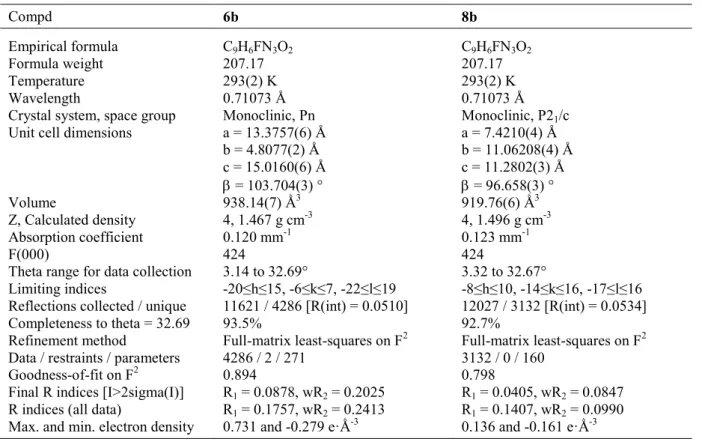
2.1. X-ray diffraction
An X-ray diffraction study confirmed the assigned E configuration for compounds 6b, 8b (Figure 4). Both molecules deviate from planarity and the phenyl ring is rotated around C(1)-N(1) by 17° in 6b and by 23° in 8b with respect to the nearly planar part of the molecule. The figure shows the E configuration with respect to the C(8)-N(2) double bond (1.277(6) and 1.279(1) Å for 6b and 8b respectively). All the C-N bond distances are in agreement with the values reported in literature for analogous bonds [15]. Also the C-C and C-O bond distances agree with literature values. The packing diagram of both structures shows two types of intermolecular hydrogen bonds: H(1)···N(3) (2.41-2.57 Å) and H(2)···O(1) (1.83 Å average).
2.2. Kinetics of furazan ring opening
Kinetics of ring scission (Scheme 2) of furazancarboxamides 3a-14a were assessed in buffered solution at physiological pH (7.4) at 37 °C by monitoring the disappearance of compounds by reversed-phase high-performance liquid chromatography (RP-HPLC). The ring opening strictly followed first order kinetics. The measured half-lives (t1/2) fall in the
range 69.0-123.4 min; all values are reported in Table 2, together with the data found for leflunomide 1, taken as reference. After 24-h incubation under the same conditions, only 20% of 1a was formed from 1. The most stable product was the phenyl substituted term 11a, and the less stable the 4-nitrophenyl derivative 10a. The behaviour of leflunomide and its strict analogue 3a was also studied at pH 1 and 10. After 6 h both compounds were unaltered in acidic medium, while in basic conditions ring opening was strongly accelerated (t1/2 < 1 min and t1/2 = 70 min for 3a and leflunomide 1, respectively). This picture is in keeping with the knowledge that the furazan system undergoes base-induced ring scission much faster than the isoxazole ring [9].
2.3. Dissociation constants
Potentiometric titrations of the oxime compounds 3b-14b were carried out with a Sirius GLpKa automated potentiometric system. The titrations were performed in water using methanol in different ratios as co-solvent. The aqueous pKas (Table 3) were determined by extrapolation to 0% methanol, according to the Yasuda-Shedlovsky procedure [16]. All the oximes behave as quite strong acids in accordance with the presence of two electron withdrawing groups on the hydroxyimino moiety. Their pKa values fall in the range 4.61-5.12, the most acidic product being the 4-nitrophenyl substituted compound 10b, the least acidic the phenyl substituted term 11b. Consequently, as in the case of 1a, the ionised forms are largely predominant for all the products at physiological pH. The determination of dissociation constants of all nitro derivatives by potentiometric titration was prevented by their marked acidity.
3. Results and discussion
DHODH (EC 1.3.99.11) is a mitochondrial protein involved in de novo pyrimidine biosynthesis. It catalyses the ubiquinone-mediated oxidation of dihydroorotate (DHO) to
orotate [17]. DHODH is considered as an important target for the development of immunomodulating agents. A771726 1a is a potent inhibitor of this enzyme. The Z
configuration of 1a bears the enolic hydroxy group in cis position with respect to the amide moiety. The experimental binding mode of this configuration within the active site of human DHODH has been reported [18]. In particular, the 4-trifluoromethylphenyl moiety establishes several hydrophobic contacts with residues of the tunnel leading to active site. The enolic OH group interacts via hydrogen bonding with Tyr356, while the amide carbonyl is hydrogen bonded to Arg136 through a water molecule. SAR studies carried out on analogues of
A771726 show that the most potent inhibitors contain a cyclopropyl residue at the 3-position of the 3-hydroxy propenamide scaffold, while larger substituents are detrimental to the activity. In addition, they bear at the 4-position of the phenyl ring small lipophilic groups, as well as weak hydrogen bond acceptors [19]. In view of the quick conversion at physiological pH of furazans 3a-14a into the related oxime derivatives 3b-14b, only these latter compounds and some of their nitro counterparts were assayed for their DHODH inhibitory potency on rat liver mithocondrial/microsomal membranes. A procedure adapted from literature was
employed, in which oxidation of DHO to orotate is monitored following the concomitant reduction of the chromophore 2,6-dichlorophenolindophenol (DCIP) [20]; the results are collected in Table 3. Analysis of the data shows that most products are inactive when tested at 100 M. 4-Fluorophenyl and 3,5-difluorophenyl substituted compounds 6b and 9b at this concentration were able to trigger feeble inhibition of the enzyme, 27% and 23%
respectively. Only the strict oxime analogue of A771726 3b was able to inhibit the enzyme in a concentration-dependent manner (IC50 = 60 M), although much less effectively than the lead. The general very low inhibitory activity of these products could be ascribed to the E configuration of the oxime group, which prevents them from assuming the same binding mode of the lead in the active cleft of the enzyme. Also the nitro substituted compounds
3c-10c, 12c, 14c are not potent inhibitors, but overall more effective than the corresponding
oximes. The most potent product was 3c, the strict analogue of A771726, followed by the 4-phenyl and 4-nitro substituted products 14c and 10c, respectively. All the other terms of the series inhibited the activity of the enzyme only to the extent of 20-29%, when tested at 100 M. Docking of the most active compounds into the active site of DHODH confirmed the
inability of oximes to hydrogen bond to Tyr356 due to their inappropriate stereochemistry (Figure 5a). While nitro compounds have the correct geometry to interact productively with Tyr356, on one side the charge-enhanced hydrogen bond is not as strong as for the
leflunomide metabolite 1a since the negative charge is spread on two oxygen atoms instead of being mostly localized on the enolic oxygen. On the other side, the other negatively charged oxygen does not interact as effectively as leflunomide’s methyl group with the lipophilic pocket delimited by Val134, Val143 and the two methyl groups of flavin
mononucleotide (FMN) (Figure 5b). These considerations provide a rationale for the lower activity of the newly synthesised analogues compared to the lead. Both the human DHODH in complex with leflunomide [18] and rat DHODH in complex with brequinar [21] were used as docking targets obtaining the same results, proving that the small differences in terms of primary and tertiary structure between the two isozymes do not influence the binding mode of the compounds object of this work.
4. Conclusions
A series of furazan analogues of leflunomide have been realised (3a-14a). These compounds are readily converted at physiological pH into their oxime counterparts (3b-14b). The latter have been assayed as DHODH inhibitors; their low potency is probably due to the
unfavourable stereochemistry of the oxime substructure, which cannot effectively mimic the enolate moiety of A771726. Oxidation of the oxime group to nitro improved the activity on
DHODH, putting into evidence the importance of a properly oriented charge-enhanced hydrogen bond acceptor. Further efforts are ongoing to obtain more potent leflunomide bioisosters.
5. Experimental protocols
5.1. Chemistry
Melting points were determined on a Büchi 530 apparatus. The compounds were routinely checked by IR spectroscopy (Shimadzu, FT-IR 8101 M) and mass spectrometry (Finnigan-Mat TSQ-700 spectrometer, 70 eV, direct inlet). 1H and proton decoupled 13C-NMR spectra were recorded on a Bruker AC-300 spectrometer. The following abbreviations are used: s, singlet; d, doublet; q, quartet; m, multiplet; Fz, furazan; PE, petroleum ether (40-60). Flash column chromatography was performed on silica gel (Merck Kieselgel 60, 230-400 mesh ASTM) using the indicated eluents. Thin layer chromatography (TLC) was carried out on 5-20 cm plates with a layer thickness of 0.25 mm; when necessary they were developed with iodine and KMnO4. Anhydrous magnesium sulphate was used as drying agent of the organic extracts. THF was distilled immediately before use from sodium and benzophenone under N2. Compounds 1 [22], 1a [22] and 2 [23] were synthesized according to literature methods. Elemental analyses of the new compounds are within 0.4% of the theoretical values and are reported in the Supplementary Material. In case of compounds 3c, 5c and 9c, that showed elemental analyses slightly out of the commonly accepted range, the purity was also assessed by RP-HPLC (> 99 %). Analyses were performed on a HP1100 chromatograph system (Agilent Technologies, Palo Alto, CA, USA) equipped with an injector (Rheodyne, Cotati, CA, USA), a quaternary pump (model G1311A), a membrane degasser (model G1379A), and a diode-array detector (DAD, model G1315B) integrated in the HP1100 system. Data
analytical column was a Nucleosil 100-5C18 Nautilus (2504.6 mm, 5 mm particle size) (Macherey-Nagel). Compounds were dissolved in CH3CN and injected through a 20 L loop. The mobile phase consisted of CH3CN/water with 0.1% trifluoroacetic acid (ranging from 60/40 to 80/20 ratio, according to the retention factor of the compounds). HPLC retention times (tR) were obtained at flow rates of 1.2 mL min-1, and the column effluent was monitored at 210 and 270 nm referenced against 360 nm.
5.1.1 Furazancarboxamide derivatives: general synthetic procedure
To a stirred solution of furazancarboxylic acid 2 (2.6 mmol) and the appropriate aniline 3-14 (2.6 mmol) in dry THF (25 mL) Ph3PCl2 (5.46 mmol, 2.1 eq) was added portionwise under an inert atmosphere. The resulting suspension was stirred for 2 h, then poured into chilled 2 M HCl (50 mL). The mixture was extracted exhaustively with Et2O, then the collected organic layers were dried and concentrated under reduced pressure. The residue was purified by flash chromatography obtaining the title compound.
5.1.1.1. N-[4-(Trifluoromethyl)phenyl]furazancarboxamide (3a)
White solid, flash chromatography eluent: PE/EtOAc 9:1 v/v; yield 40%; m.p. 145 °C (triturated with hexane); 1H-NMR (DMSO-d6): 7.77-8.03 (m, 4H, aromatic protons), 9.46
(s, 1H, FzCH), 11.43 (s, 1H, NH); 13C-NMR (DMSO-d6): 119.8, 124.1 (q, CF3, 1JCF 271.7 Hz), 124.7 (q, C(4), 2JCF 32.1 Hz), 126.1 (q, C(3), 3JCF 3.9 Hz), 141.3 (q, C(2), 4JCF 1.3 Hz), 142.6, 151.4, 155.2; MS (CI) m/z 258 (M+1)+. Anal. (C10H6F3N3O2) C, H, N.
5.1.1.2. N-[3-(Trifluoromethyl)phenyl]furazancarboxamide (4a)
White solid; yield 57%; m.p. 46-47 °C (twice purified by flash chromatography; eluents: PE/EtOAc 7:3 v/v, then PE/EtOAc/CH2Cl2 95:5:50 v/v/v); 1H-NMR (DMSO-d6): 7.54-8.24
(m, 4H, aromatic protons), 9.45 (s, 1H, FzCH), 11.40 (s, 1H, NH); 13C-NMR (DMSO-d6):
124.1 (m, C(5)), 129.5 (q, C(3), 2JCF 31.7 Hz), 130.15, 138.5, 151.4, 155.1; MS (CI) m/z 258 (M+1)+. Anal. (C10H6F3N3O2) C, H, N.
5.1.1.3. N-[3,5-Bis(trifluoromethyl)phenyl]furazancarboxamide (5a)
White solid; flash chromatography eluent: PE/EtOAc 7:3 v/v; yield 37%; m.p. 117-118 °C (from hexane/diethyl ether 1:1 v/v); 1H-NMR (DMSO-d6): 7.91 (s, 1H, C(4)H), 8.45 (s, 2H,
C(2)H), 9.48 (s, 1H, NH), 11.70 (s, 1H, FzCH); 13C-NMR (DMSO-d6): 117.5-117.8 (m,
C(4)), 120.2-120.5 (m, C(2)), 123.8 (q, CF3, 1JCF 272.7 Hz), 130.8 (q, C(3), 2JCF 33.0 Hz), 139.7, 142.6, 151.2, 155.5; MS (CI) m/z 326 (M+1)+. Anal. (C11H5F6N3O2) C, H, N.
5.1.1.4. N-(4-Fluorophenyl)furazancarboxamide (6a)
White solid; flash chromatography eluent: PE/EtOAc 7:3 v/v; yield 64%; m.p. 101 °C dec.(triturated with hexane/iPr2O); 1H-NMR (DMSO-d6): 7.22-7.82 (m, 4H, aromatic
protons), 9.43 (s, 1H, FzCH), 11.16 (br s, 1H, NH); 13C-NMR (DMSO-d6): 116.5 (d, C(3),
2J
CF 22.8 Hz), 124.1 (d, C(2), 3JCF 8.05 Hz), 135.0 (d, C(1), 4JCF 2.9 Hz), 142.8, 152.8, 156.7, 161.4 (d, C(4), 1JCF 243.3 Hz); MS (CI) m/z 208 (M+1)+. Anal. (C9H6FN3O2) C, H, N.
5.1.1.5. N-(3-Fluorophenyl)furazancarboxamide (7a)
White solid; flash chromatography eluent: PE/EtOAc 7:3 v/v; yield 57%; m.p. 109 °C (from hexane/diethylether); 1H-NMR (DMSO-d6): 7.00-7.74 (m, 4H, aromatic protons), 9.43 (s,
1H, FzCH), 11.29 (br s, 1H, NH); 13C-NMR (DMSO-d6): 109.1 (d, C(4), 2JCF 26.7 Hz), 112.7 (d, C(2), 2JCF 21.5 Hz), 117.4 (d, C(6), 4JCF 3.0 Hz), 131.4 (d, C(5), 3JCF 9.3 Hz), 140.6 (d, C(1), 3JCF 10.8 Hz), 142.8, 152.8, 156.8, 164.3 (d, C(3), 1JCF 243.2 Hz); MS (CI) m/z 208 (M+1)+. Anal. (C9H6FN3O2) C, H, N.
5.1.1.6. N-(2-Fluorophenyl)furazancarboxamide (8a)
White solid; flash chromatography eluent: PE/EtOAc 7:3 v/v; yield 37%; m.p. 81 °C (from hexane/isopropyl ether 1:1 v/v); 1H-NMR (DMSO-d6): 7.24-7.76 (m, 4H, aromatic
protons), 9.45 (s, 1H, FzCH), 10.96 (br s, 1H, NH), 13C-NMR (DMSO-d6): 115.9 (d, C(3), 2J CF 19.6 Hz), 123.8 (d, C(1), 2JCF 12.3 Hz), 124.4 (d, C(4), 3JCF 3.6 Hz), 126.7 (d, C(5), 4JCF 1.40 Hz), 127.8 (d, C(6), 3JCF 7.9 Hz), 142.3, 150.9, 154.9, 155.4 (d, C(2), 1JCF 248.1 Hz); MS (CI) m/z 208 (M+1)+. Anal. (C9H6FN3O2) C, H, N. 5.1.1.7. N-(3,5-Difluorophenyl)furazancarboxamide (9a)
White solid; flash chromatography eluent: PE/EtOAc 7:3 v/v; yield 53%; m.p. 112-113 °C (from hexane/diethylether 1:1 v/v); 1H-NMR (DMSO-d6): 7.04-7.60 (m, 3H, aromatic
protons), 9.45 (s, 1H, NH), 11.45 (s, 1H, FzCH); 13C-NMR (DMSO-d6): 100.7 (t, C(4), 2JCF 26.1 Hz), 104.7 (m, C(2)),141.4 (t, C(1), 3JCF 13.6 Hz), 142.8, 152.6, 156.9, 164.5 (dd, C(3), 1J
CF 244.9 Hz, 3JCF 14.6 Hz); MS (CI) m/z 226 (M+1)+. Anal. (C9H5F2N3O2) C, H, N.
5.1.1.8. N-(4-Nitrophenyl)furazancarboxamide (10a)
White solid; flash chromatography eluent: PE/EtOAc 8:2 v/v; yield 25%; m.p. 266.9-267.1 °C dec. (from hexane/isopropyl ether 1:1 v/v); 1H-NMR (DMSO-d6): 8.05-8.32 (m, 4H,
aromatic protons), 9.47 (s, 1H, NH), 11.62 (s, 1H, FzCH); 13C-NMR (DMSO-d6): 124.8,
142.6, 143.3, 143.8, 151.3, 155.4, 157.6; MS (CI) m/z 235 (M+1)+. Anal. (C9H6N4O4) C, H, N.
5.1.1.9. N-Phenylfurazancarboxamide (11a)
Pale yellow solid; flash chromatography eluent: PE/EtOAc 6:4 v/v; yield 34%; m.p. 138 °C (from hexane/isopropyl ether 9:1 v/v); 1H-NMR (DMSO-d6): 7.16-7.79 (m, 5H, aromatic
protons), 9.43 (s, 1H, FzCH), 11.08 (br s, 1H, NH); 13C-NMR (DMSO-d6): 121.4, 125.7,
129.7, 138.6, 143.4, 152.5, 155.6; MS (CI) m/z 190 (M+1)+. Anal. (C9H7N3O20.1H2O) C, H, N.
5.1.1.10. N-(4-Methylphenyl)furazancarboxamide (12a)
White solid; flash chromatography eluent: PE/EtOAc 7:3 v/v; yield 46%; m.p. 138 °C (from hexane/isopropyl ether 1:1 v/v); 1H-NMR (DMSO-d ): 3.33 (s, 3H, CH ), 7.19-7.66 (m, 4H,
aromatic protons), 9.40 (s, 1H, NH), 10.99 (s, 1H, FzCH); 13C-NMR (DMSO-d6): 21.1,
122.2, 130.5, 136.2, 136.3, 142.8, 153.0, 156.6; MS (CI) m/z 204 (M+1)+. Anal. (C10H9N3O2) C, H, N.
5.1.1.11. N-(4-Methoxyphenyl)furazancarboxamide (13a)
Pale gray solid; flash chromatography eluent: PE/EtOAc 7:3 v/v; yield 62%; m.p. 131 °C (from hexane/isopropyl ether 1:1 v/v); 1H-NMR (DMSO-d6): 3.76 (s, 3H, OCH3), 6.96-7.71 (m, 4H, aromatic protons), 9.40 (s, 1H, NH), 10.96 (s, 1H, FzCH); 13C-NMR (DMSO-d6):
55.1, 113.9, 122.1, 130.6, 142.4, 151.6, 154.2, 156.2; MS (CI) m/z 220 (M+1)+. Anal. (C10H9N3O3) C, H, N.
5.1.1.12. N-(Biphenyl-4-yl)furazancarboxamide (14a)
Yellow solid; flash chromatography eluent: PE/EtOAc 7:3 v/v; yield 39%; m.p. 199 °C (from hexane/isopropyl ether 1:1 v/v); 1H-NMR (DMSO-d6): 9.79 (s, 1H, FzCH), 7.33-7.90 (m,
9H, aromatic protons), 11.20 (br s, 1H, NH); 13C-NMR (DMSO-d6): 122.0, 127.4, 128.0,
128.1, 129.7, 138.0, 138.4, 141.0, 142.8, 152.6, 155.9; MS (CI) m/z 266 (M+1)+. Anal. (C15H11N3O2) C, H, N.
5.1.2. 2-Hydroxyiminopropionitrile derivatives: general synthetic procedure
1 M NaOH (4.2 mmol, 4.2 mL) was added to an ice cooled solution of the amide 3a-13a (1 mmol) in MeOH (11 mL). The mixture was stirred at room temperature for 2 h, then diluted with water (5 mL). The pH of the mixture was adjusted to 1 by addition of 6 M HCl. The resulting suspension was exhaustively extracted with Et2O. The organic layers were
collected, dried and concentrated under reduced pressure to afford the pure title compound.
5.1.2.1. 2-Hydroxyimino-3-oxo-3-[4-(trifluoromethyl)phenyl]aminopropionitrile (3b)
White solid; yield 89%; m.p. 210-211 °C (from toluene); 1H-NMR (DMSO-d6): 7.72-7.96
(m, 4H, aromatic protons), 10.78 (s, 1H, NH), 14.76 (br s, 1H, OH); 13C-NMR (CD3OD): 108.7, 120.6, 124.1 (q, CF3, 1JCF 271.7 Hz), 124.3 (q, C(4), 2JCF 32.0 Hz), 125.9 (q, C(3), 3JCF
3.8 Hz), 141.2 (q, C(2), 4JCF 1.4 Hz), 157.3; MS (CI) m/z 258 (M+1)+. Anal. (C10H6F3N3O2) C, H, N.
5.1.2.2. 2-Hydroxyimino-3-oxo-3-[3-(trifluoromethyl)phenyl]aminopropionitrile (4b)
White solid; yield 74%; m.p. 181 °C (from toluene); 1H-NMR (DMSO-d6): 7.49-8.16 (m,
4H, aromatic protons), 10.75 (br s, 1H, NH), 14.78 (br s, 1H, OH); 13C-NMR (DMSO-d6):
109.6, 117.8 (q, C(4), 3JCF 4.2 Hz), 121.7 (q, C(2), 3JCF 3.8 Hz), 124.9 (q, CF3, 1JCF 272.3 Hz), 125.2-125.3 (m), 129.3, 130.3 (q, C(3), 2JCF 31.8 Hz), 130.8, 139.3, 158.3; MS (CI) m/z 258 (M+1)+. Anal. (C10H6F3N3O2) C, H, N.
5.1.2.3. 3-[3,5-Bis(trifluoromethyl)phenyl]amino-2-hydroxyimino-3-oxopropionitrile (5b)
White solid; yield 98%; m.p. 191 °C dec. (from toluene); 1H-NMR (DMSO-d6): 7.85 (s,
2H, C(2)H), 8.45 (s, 2H, C(2)H), 11.03 (s, 1H, NH), 14.89 (s, 1H, OH); 13C-NMR
(DMSO-d6): 108.6, 117.2-117.3 (m, C(4)), 120.4-120.6 (m, C(2)), 123.8 (q, CF3, 1JCF 272.7 Hz), 128.3, 130.8 (q, C(3), 2JCF 32.9 Hz), 139.7, 157.8; MS (CI) m/z 326 (M+1)+. Anal. (C11H5F6N3O2) C, H, N.
5.1.2.4. 3-(4-Fluorophenyl)amino-2-hydroxyimino-3-oxopropionitrile (6b)
Pale yellow; yield 78%; m.p. 184 °C dec. (triturated with toluene); 1H-NMR (DMSO-d6):
7.17-7.63 (m, 4H, aromatic protons), 14.65 (br s, 1H, OH), 10.49 (br s, 1H, NH); 13C-NMR (DMSO-d6): 109.1, 116.4 (d, C(3), 2JCF 22.8 Hz), 124.1 (d, C(2), 3JCF 8.0 Hz), 129.8, 134.8 (d, C(1), 4JCF 2.9 Hz), 158.8, 161.2 (d, C(4), 1JCF 243.0 Hz); MS (CI) m/z 208 (M+1)+. Anal. (C9H6FN3O2) C, H, N.
5.1.2.5. 3-(3-Fluorophenyl)amino-2-hydroxyimino-3-oxopropionitrile (7b)
White solid; yield 86%; m.p. 190 °C (from toluene); 1H-NMR (DMSO-d6): 6.94-7.65 (m,
4H, aromatic protons), 14.71 (s, 1H, OH), 10.61 (s, 1H, NH); 13C-NMR (DMSO-d6): 108.9,
131.3 (d, C(5), 3JCF 9.3 Hz), 140.9 (d, C(1), 3JCF 10.9 Hz), 129.8, 158.7, 164.2 (d, C(3), 1JCF 243.1 Hz); MS (EI) m/z 207 (M)+. Anal. (C9H6FN3O2) C, H, N.
5.1.2.6. 3-(2-Fluorophenyl)amino-2-hydroxyimino-3-oxopropionitrile (8b)
White solid, yield 99%; m.p. 198 °C dec. (from toluene); 1H-NMR (DMSO-d6): 7.20-7.64
(m, 4H, aromatic protons), 10.11 (s, 1H, NH), 14.82 (s, 1H, OH); 13C-NMR (DMSO-d6):
108.6, 115.8 (d, C(3), 2JCF 19.6 Hz), 124.1 (d, C(1), 2JCF 12.0 Hz), 124.4 (d, C(4), 3JCF 3.7 Hz), 126.5 (d, C(5), 4JCF 1.3 Hz), 127.5 (d, C(6), 3JCF 7.9 Hz), 127.8, 155.4 (d, C(2), 1JCF 247.3 Hz), 156.9; MS (CI) m/z 208 (M+1)+. Anal. (C9H6FN3O2) C, H, N.
5.1.2.7. 3-(3,5-Difluorophenyl)amino-2-hydroxyimino-3-oxopropionitrile (9b)
White solid; yield 89%; m.p. 202 °C dec. (from toluene); 1H-NMR (DMSO-d6): 6.98-7.53
(m, 4H, aromatic protons), 10.76 (s, 1H, NH), 14.78 (s, 1H, OH); 13C-NMR (DMSO-d6):
100.7 (t, C(4), 2JCF 26.1 Hz), 104.4-104.8 (m, C(2)), 108.8, 129.7, 141.4 (t, C(1), 3JCF 13.5 Hz), 158.9, 164.5 (dd, C(3), 1JCF 244.9 Hz, 3JCF 14.7 Hz); MS (CI) m/z 226 (M+1)+. Anal. (C9H5F2N3O2) C, H, N.
5.1.2.8. 2-Hydroxyimino-3-(4-nitrophenyl)amino-3-oxopropionitrile (10b)
Pale yellow solid; yield 83%; m.p. 273.6-274.5 °C dec. (from toluene); 1H-NMR (DMSO-d6):
7.96-8.29 (m 4H, aromatic protons), 10.99 (br s, 1H, NH), 14.84 (br s, 1H, OH); 13C-NMR (DMSO-d6): 108.6, 120.4, 124.7, 128.4, 143.0, 143.8, 157.6; MS (CI) m/z 235 (M+1)+.
Anal. (C9H6N4O40.1 H2O) C, H, N.
5.1.2.9. 2-Hydroxyimino-3-oxo-3-phenylaminopropionitrile (11b)
Pale yellow solid; yield 91%; m.p. 202.2-202.7 °C dec. (triturated with toluene; lit 229-230 °C [24]); 1H-NMR (DMSO-d6): 7.12-7.71 (m, 5H, aromatic protons), 10.43 (s, 1H, NH),
14.66 (br s, 1H, OH); 13C-NMR (DMSO-d6): 108.8, 120.7, 124.5, 128.5, 128.6, 137.5,
156.7; MS (CI) m/z 190 (M+1)+. Anal. (C9H7N3O2) C, H, N.
Yellow solid; yield 94%; m.p. 238 °C (from toluene; lit242-244 °C [24]);1H-NMR (DMSO-d6) 2.27 (s, 3H, CH3), 7.14-7.58 (m, 4H, aromatic protons), 10.35 (s, 1H, NH),
14.61 (br s, 1H, OH); 13C-NMR (DMSO-d6): 20.4, 108.8, 120.6, 128.5, 129.0, 133.3, 134.9,
156.5; MS (CI) m/z 204 (M+1)+. Anal. (C10H9N3O2) C, H, N.
5.1.2.11. 2-Hydroxyimino-3-(4-methoxyphenyl)amino-3-oxopropionitrile (13b)
Yellow solid; yield 81%; m.p. 221 °C (from toluene; lit216-218 °C [24]);1H-NMR
(DMSO-d6): 3.74 (s, 3H, OCH3), 6.91-7.61 (m, 4H, aromatic protons), 10.32 (s, 1H, NH),
14.58 (br s, 1H, OH); 13C-NMR (DMSO-d6): 55.1, 108.8, 113.7, 122.3, 128.5, 130.4, 156.0,
156.4; MS (CI) m/z 220 (M+1)+. Anal. (C10H9N3O3) C, H, N.
5.1.2.12. 2-Hydroxyimino-3-oxo-3-(biphenyl-4-yl)aminopropionitrile (14b)
Yellow solid; yield 80%; m.p. 250-251 °C (from toluene); 1H-NMR (DMSO-d6) 7.33-7.82
(m, 9H, aromatic protons), 10.55 (s, 1H, NH), 14.68 (br s, 1H, OH); 13C-NMR (DMSO-d6):
108.8, 121.0, 126.3, 126.8, 127.2, 128.6, 128.8, 136.1, 137.0, 139.4, 156.8; MS (CI) m/z 266 (M+1)+. Anal. (C15H11N3O20.15H2O) C, H, N.
5.1.3. 2-Nitropropionitrile derivatives: general procedure
KMnO4 (1.5 mmol, 237 mg) was added portionwise to an ice-cooled solution of the oxime (1.0 mmol) in acetone/water (1:2 v/v; 20 mL). The mixture was stirred at room temperature for 24 h, then filtered under Celite. The filtrate was acidified to pH 3 by adding glacial CH3COOH, then washed with Et2O (220 mL), and concentrated under reduced pressure. The resulting solid was triturated with isopropyl ether obtaining the title compound as potassium salt.
5.1.3.1. 2-Nitro-3-oxo-3-[(4-trifluoromethyl)phenyl]aminopropionitrile potassium salt (3c)
White solid; yield 80%; m.p. 256.0-256.3 °C dec. (triturated with iPr2O); 1H-NMR (DMSO-d6): 7.65-7.82 (m, 4H, aromatic protons), 11.43 (s, 1H, NH); 13C-NMR
C(3), 3JCF 3.1 Hz), 129.8, 142.2 (m, C(2)), 160.3 (br s, NC-C(=NO2)CONH). Anal. (C10H5F3N3O3K) calc.: C, 38.59; H, 1.62; N, 13.50; found: C, 38.92; H,1.72; N, 12.68.
PHPLC = 99% (mobile phase: CH3CN/water with 0.1% trifluoroacetic acid 60/40 v/v, tR = 16.2 min).
5.1.3.2. 2-Nitro-3-oxo-3-[3-(trifluoromethyl)phenyl]aminopropionitrile potassium salt (4c) White solid; yield 71%; m.p. 255.5-265.5 °C dec. (from H2O); 1H-NMR (DMSO-d6):
7.34-8.20 (m, 4H, aromatic protons), 11.35 (br s, 1H, NH), 13C-NMR (DMSO-d6): 98.3, 115.3
(q, C(4), 3JCF 4.1 Hz), 116.8, 119.1 (q, C(2), 3JCF 3.8 Hz), 124.1 (q, CF3, 1JCF 272.3 Hz), 122.8-122.9 (m), 129.4 (q, C(3), 2JCF 31.5 Hz), 129.8, 139.4, 160.4. Anal.
(C10H5F3N3O3K·0.5 H2O)C, H, N.
5.1.3.3. 3-[3,5-Bis(trifluoromethyl)phenyl]amino-2-nitro-3-oxopropionitrile potassium salt (5c)
Pale yellow solid; yield 82%; m.p. 275.7 °C dec. (triturated with iPr2O); 1H-NMR (DMSO-d6): 7.68 (s, 1H, C(4)H), 8.40 (s, 2H, C(2)H), 11.47 (s, 1H, NH);13C-NMR
(DMSO-d6): 98.1, 115.3-115.4 (t, C(4) 3JCF 3.9 Hz,), 116.6, 119.3-119.4 (q, C(2), 3JCF 3.9 Hz), 123.4 (q, C(3), 1JCF 272.7 Hz), 130.5 (q, C(3), 2JCF 32.7 Hz), 140.6, 160.7. Anal. (C11H4F6N3O3K·1.65 H2O) calc.: C, 32.38; H, 1.56; N, 10.30; found: C, 32.79; H, 2.09; N,
10.34. PHPLC = 99% (mobile phase: CH3CN/water with 0.1% trifluoroacetic acid 80/20 v/v, tR = 10.6 min).
5.1.3.4. 3-(4-Fluorophenyl)amino-2-nitro-3-oxopropionitrile potassium salt (6c)
Pale yellow solid; yield 89%; m.p. 265.0-266.9 °C dec. (from EtOH/iPr2O 9:1 v/v); 1H-NMR (DMSO-d6): 7.08-7.63 (m, 4H, aromatic protons), 11.15 (br s, 1H, NH); 13C-NMR
(DMSO-d6): 98.2, 115.2 (d, C(3), 2JCF 22.1 Hz), 117.0, 120.9 (d, C(2), 3JCF 7.9 Hz), 135.0 (d, C(1), 4JCF 2.5 Hz), 157.8 (d, C(4), 1JCF 239.3 Hz), 159.9. Anal. (C9H5FN3O3K·0.4H2O) C, H, N.
5.1.3.5. 3-(3-Fluorophenyl)amino-2-nitro-3-oxopropionitrile potassium salt (7c)
White solid; yield 76%; m.p. 266.3-266.7 °C dec. (from H2O); 1H-NMR (DMSO-d6):
6.84-7.71 (m, 4H, aromatic protons), 11.29 (s, 1H, NH);13C-NMR (DMSO-d6): 98.3, 106.0 (d,
C(4), 2JCF 26.4 Hz), 109.2 (d, C(2), 2JCF 21.2 Hz), 115.0 (d, C(6), 4JCF 2.6 Hz), 116.8, 130.2 (d, C(5), 3JCF 9.7 Hz), 140.3 (d, C(1), 3JCF 11.3 Hz), 160.1, 162.1 (d, C(3), 1JCF 241.0 Hz). Anal. (C9H5FN3O3K) C, H, N.
5.1.3.6. 3-(2-Fluorophenyl)amino-2-nitro-3-oxopropionitrile potassium salt (8c)
Pale yellow solid, yield 80%; m.p. 262.7-263.2 °C dec. (triturated with iPr2O); 1H-NMR (DMSO-d6): 7.05-7.40 (m, 3H, aromatic protons), 8.35-8.63 (m, 1H, C(3)H), 11.65 (s, 1H,
NH); 13C-NMR (DMSO-d6): 98.5, 114.8 (d, C(3), 2JCF 19.0 Hz), 116.7, 120.6 (d, C(5), 4JCF 1.6 Hz), 123.0 (d, C(4), 3JCF 7.6 Hz), 124.5 (d, C(6), 3JCF 3.5 Hz), 150.3, 151.6 (d, C(2), 1JCF 241.7 Hz), 159.8. Anal. (C9H5FN3O3K·0.1H2O) C, H, N.
5.1.3.7. 3-(3,5-Difluorophenyl)amino-2-nitro-3-oxopropionitrile potassium salt (9c)
White solid; yield 87%; m.p. 267.4-268.9 °C dec. (from H2O); 1H-NMR (DMSO-d6):
6.81-7.46 (m, 4H, aromatic protons), 11.31 (s, 1H, NH); 13C-NMR (DMSO-d6) 98.4, 98.7 (t,
C(4), 2JCF 26.3 Hz), 102.9-103.3 (m, C(2)), 117.6, 142.1 (t, C(1), 3JCF 14.1 Hz), 161.3, 163.3 (dd, C(3), 1JCF 242.5 Hz, 3JCF 15.5 Hz). Anal. (C9H4F2N3O3K) calc.: C, 38.71; H, 1.44; N,
15.05; found: C, 39.19; H, 2.11; N, 14.23. PHPLC = 98% (mobile phase: CH3CN/water with 0.1% trifluoroacetic acid 60/40 v/v, tR = 13.7 min).
5.1.3.8. 2-Nitro-3-(4-nitrophenyl)amino-3-oxopropionitrile potassium salt (10c)
Yellow solid; yield 68%; m.p. 252.8-253.6 °C dec. (triturated with isopropylether); 1H-NMR (DMSO-d6): 7.86-8.20 (m, 4H, aromatic protons), 11.59 (s, 1H, NH); 13C-NMR
(DMSO-d6): 98.5, 116.5, 118.8, 124.9, 141.8, 144.8, 160.3. Anal. (C9H5N4O5K) C, H, N.
5.1.3.9. 3-(4-Methylphenyl)amino-2-nitro-3-oxo-propionitrile potassium salt (12c)
(DMSO-d6): 2.27 (s, 3H, CH3), 7.07-7.46 (m, 4H, aromatic protons), 11.10 (s, 1H, NH);
13C-NMR (DMSO-d
6): 21.3, 106.1, 118.0, 120.0, 130.1, 132.7, 137.0, 160.7. Anal.
(C10H8N3O3K) C, H, N.
5.1.3.10. 2-Nitro-3-oxo-3-(biphenyl-4-yl)aminopropionitrile potassium salt (14c)
Yellow solid; yield 20%; m.p. 269.1-271.7 °C dec. (triturated with iPr2O); 1H-NMR (DMSO-d6): 7.29-7.70 (m, 9H, aromatic protons), 11.27 (s, 1H, NH);13C-NMR
(DMSO-d6): 110.0, 117.9, 120.4, 127.1, 127.8, 127.9, 129.8, 135.4, 139.0, 140.8, 160.9.
Anal. (C15H10N3O3K) C, H, N.
5.2. X-ray structure determination of 6b and 8b
Crystal and refinement data are reported in Table 4. The reflection data, using a crystal obtained from toluene solution, have been collected on a Gemini R Ultra diffractometer [25]. The hydrogen atom coordinates have been calculated (6b) or found (8b) and refined with Uiso set at 1.5 times Ueq of the corresponding atom (6b) or with Uiso free (8b). All other atoms have been anisotropically refined. Programs used were CrysAlisPro [25] for data collection and correction and SHELXTL [26] for structure solution, refinement and molecular graphics. No absorption correction has been made. CIF files have been deposited at the Cambridge Crystallographic Data Centre as CCDC 784463 for 6b and CCDC 784464 for 8b; bond length and angles are reported in the Supplementary Material.
5.3. Evaluation of stability in aqueous buffer solutions
A solution of each compound (10 mM) in methanol was added to phosphate buffer (pH 7.4, 50 mM) preheated at 37 °C. To carry out the assay at pH 1 and pH 10, 0.1 M HCl and 0.1 M phosphate buffer were used, respectively. The final concentration of the compound was 100 M. The resulting solution was maintained at 37.0 ± 0.5 °C, and at 30-min time intervals the
reaction mixture was analyzed by RP-HPLC. Incubations were conducted in triplicate. The RP-HPLC procedure allowed the separation and quantitation of the unchanged compound
and newly formed oxime derivatives. HPLC analyses were performed with the HP1100 chromatograph system previously described. The analytical column was a Zorbax Eclipse XDB-C8 (Agilent, 1504.6 mm, 5 m particle size). The mobile phase consisted of
methanol/water with 0.1% trifluoroacetic acid (ranging from 60/40 to 75/25 ratio, according to the retention factor of the compounds); flow rate and injection volume were 1.2 mL min-1 and 20 L, respectively. The column effluent was monitored at 230 and 270 nm referenced against a 360 nm wavelength. Quantitation was performed by comparison of peak areas with standards chromatographed under the same conditions. The percentage of unchanged
compound versus time was fitted with a one-phase exponential decay equation using GraphPad Prism version 5 to obtain kobs; t1/2 was calculated from equation 1:
t1/2 = 0.693 / kobs (1)
5.4. Dissociation constant determination
Potentiometric titrations were performed using the GLpKa apparatus (Sirius Analytical Instruments Ltd., Forest Row, East Sussex, UK). Because of their low aqueous solubility all the compounds required titrations in the presence of methanol as cosolvent: at least five different hydro-organic solutions (ionic strength adjusted to 0.15 M with KCl) of the compounds (20 mL, about 1 mM in 24-58 wt% methanol) were initially acidified to pH 1.8 with 0.5 N HCl; the solutions were then titrated with standardized 0.5 N KOH to pH 10.5. The initial estimates of the psKa values (the apparent ionization constants in the
water-methanol mixtures) were obtained and aqueous pKa values were determined by extrapolation to zero content of cosolvent according to the Yasuda-Shedlovsky procedure [16]. All
titrations were performed under N2 at 25.0 ± 0.1 °C.
5.5. Biological evaluation
5.5.1. Preparation of rat liver membranes
were prepared by homogenisation and differential centrifugation [20, 27]. Homogenisation buffer was 25 mM sodium phosphate, 250 mM sucrose and 0.3% v/v Protease Inhibitor Cocktail for use with mammalian cell and tissue extracts (SIGMA Catalog Number: P8340) at pH 7.4. The homogenate (9 mL homogenisation buffer/g wet tissue) was centrifuged at 470g for 10 min, the supernatant retained, and the pellets re-homogenised in buffer (4.5 mL/g wet tissue). This homogenate was centrifuged (470g, 10 min) and the supernatant was
combined with the one obtained earlier and centrifuged (50000g, 60 min). The membranes were washed by resuspension in homogenisation buffer plus 150 mM NaCl, 1 mM EDTA and 1 mM EGTA before final centrifugation (50000g, 60 min) and re-suspension in the homogenisation buffer (2 mL/g wet tissue). All of the above steps were performed at 4 °C and aliquots of the final membrane preparation were stored at -80 °C.
5.5.2. Dihydroorotate dehydrogenase assay
Inhibition of rat liver DHODH activity by tested compounds was assessed using a DCIP-linked assay. Membranes (0.8 mg protein mL-1) were incubated at 37 °C in 50 mM Tris-HCl, 0.1% Triton X-100, 1 mM KCN, pH 8.0 with coenzyme Q10 (100 M) and the tested
compounds at different concentrations (final DMSO 0.1% v/v). The reaction was initiated by addition of DHO (500 M), and the reduction of DCIP (50 M) was monitored by the
decrease in absorbance at 650 nm. The initial rate of the enzyme reaction, in the presence and in the absence of potential inhibitor, was measured in the first five minutes ( = 10400 M-1 cm-1 in our experimental conditions) and a preliminary IC50 value was calculated, when possible, according to Equation 2 [28]:
v = V / (1 + [I] / IC50) (2) where [I] is the inhibitor concentration.
Michaelis-Menten constant (Km)for DHO, determined by the usual procedure, was found to be 14.6 M, in keeping with previous work [28].
5.6. Molecular modelling
The molecular models of compounds 3b, 3c, 10b, 10c, 14b, 14c and orotate were built using standard bond lengths and angles with the MOE software suite [29]. In accordance with their pKa values (Table 3), all compounds were modelled as anions; their geometries were
optimized with the Newton-Raphson method (MMFF94s force field, GB/SA implicit solvent model) until the gradient was lower than 0.001 kcal mol-1 Å-1. A Monte Carlo conformational search was carried out with the ConfSearch module implemented in MOE. The most stable conformers were then subjected to further optimization by an ab initio HF/6-31G(d) method using GAMESS-US [30]. The experimental crystallographic structures of human DHODH in complex with 1a (PDB ID 1D3H) as well as of rat DHODH in complex with brequinar (PDB ID 1UUO) were retrieved from the Protein Data Bank [31]. The coordinates of a few second-shell residues surrounding the active site (70-72 in 1D3H, 70-73 in 1UUO) were missing and were modelled with the Homology module implemented in MOE. Missing hydrogen atoms were added to both enzymes in standard positions, then their coordinates were optimized with the SANDER module of the AMBER 10 software package [32], while harmonically
restraining heavy atoms to the initial crystallographic coordinates with a force constant of 1000 kcal mol-1 Å-2. AMBER FF99 parameters were assigned to protein atoms and GAFF parameters to the co-crystallized inhibitor and orotate, while atom-centred point charges were fitted to the quantum-mechanical electrostatic potential according to the RESP method [33]. Equilibrium internal coordinates, force-field parameters and electrostatic charges for the FMN cofactor were taken from literature [34]. After removing the co-crystallized inhibitor, docking of compounds 3b, 3c, 10b, 10c, 14b, 14c was carried out using AutoDock 4.0.1 [35-37]. A grid with a 0.375 Å step size was centred on the inhibitor binding site and energy maps were pre-computed with AutoGrid, then flexible docking was carried out with AutoDock. The target proteins were kept rigid, while ligands were left free to explore the
conformational space inside the enzyme cavity; 200 separate docking simulations were run on each target using the Lamarckian genetic algorithm with default parameters.
The geometries of the most stable conformer of both E and Z isomers of compounds 3b, 10b,
14b were optimized with a DFT method at the RB3LYP/6-31G(d) level of theory. Single
point energy calculations at the RB3LYP/6-311G(2d,2p) level were then carried out to compute the relative stabilities of the two isomers. For compound 3b the minimum energy reaction path for the E-Z isomerisation was computed as previously described [14] by constraining the C-N-O angle at 33 intermediate values with the same
RB3LYP/6-31G(d)//RB3LYP/6-311G(2d,2p) DFT method used for ground state geometries. Frequency calculations were performed at the same level of theory as the geometry
optimizations to obtain zero-point energies (no scaling) and to characterize the stationary points as local minima, or as a first-order saddle point for the transition state structure in the isomerisation of 3b.
Acknowledgements
This work was supported by a grant from Regione Piemonte Progetto Ricerca Sanitaria Finalizzata 2009.
REFERENCES
[1] A.M. Krensky, F. Vincenti, Immunosuppressants, tolerogens and immunostimulants, in: L.L. Brunton, J.S. Lazo, K.L. Parker (Eds.), Goodman & Gilman’s The Pharmacological Basis of Therapeutics, Mc Graw-Hill, New York, 2006, pp. 1405-1432.
[2] a) H.T. Silva Jr., R.E. Morris, Leflunomide and malononitrilamides, Am. J. Med. Sci. 313 (1997) 289-301; C. Papageorgiou, M. Zurini, H.P. Weber, X. Borer, Leflunomide's bioactive metabolite has the minimal structural requirements for the efficient inhibition of human dihydroorotate dehydrogenase, Bioorg. Chem. 25 (1997) 233-238; b) C. Papageorgiou, K. Akyel, X. Borer, L. Oberer, G. Rihs, 3-Hydroxy-2-cyanoalk-2-enamides, and 2-cyano-2-(tetrahydrofuran-2-ylidene)- and 2-cyano-2-(tetrahydropyran-2-ylidene)acetamides: Synthesis, structure, and solvent-dependent (Z)/(E)-isomerism Helv. Chim. Acta 81 (1998) 1319-1328; c) G. Bertolini, M. Aquino, M. Biffi, G. d’Atri, F. Di Pierro, F. Ferrario, P. Mascagni, F. Somenzi, A. Zaliani, F. Leoni, A new rational hypothesis for the pharmacophore of the active metabolite of leflunomide, a potent immunosuppressive drug, J. Med. Chem. 40 (1997) 2011-2016; d) R.I. Fox, M.L. Herrmann, C.G. Frangou, G.M. Wahl, R.E. Morris, V. Strand, B.J. Kirschbaum,
Mechanism of action for leflunomide in rheumatoid arthritis, Clin. Immunol. 93 (1999) 198-208.
[3] a) P. Pino, A. Scartabelli, E. Lombardi, Rend. Istit. Lombardo Sci. Lettere. 87 (1954), 229-246; b) T.L. Gilchrist, Ring-opening of five-membered heteroaromatic anions, Adv. Heterocycl. Chem. 41 (1987) 41-74.
[4] A.S. Kalgutkar, H.T. Nguyen, A.D.N. Vaz, A. Doan, D.K. Dalvie, D.G. McLeod, J.C. Murray, In vitro metabolism studies on the isoxazole ring scission in the
anti-inflammatory agent leflunomide to its active -cyanoenol metabolite A771726:
mechanistic similarities with the cytochrome P450-catalyzed dehydration of aldoximes, Drug Metab. Dispos. 31 (2003) 1240-1250.
[5] J.P. Davis, G.A. Cain, W.J. Pitts, R.L. Magolda, R.A. Copeland, The
immunosuppressive metabolite of leflunomide is a potent inhibitor of human dihydroorotate dehydrogenase, Biochemistry 35 (1996) 1270-1273.
[6] R.R. Bartlett, H. Anagnostopulos, T. Zielinski, T. Mattar, R. Schleyerbach, Effects of leflunomide on immune responses and models of inflammation, Semin. Immunopathol. 14 (1993) 381-394.
[7] T. Mattar, K. Kochhar, R. Bartlett, E.G. Bremer, A. Finnegan, Inhibition of the epidermal growth factor receptor tyrosine kinase activity by leflunomide, FEBS Lett. 334 (1993) 161-164.
[8] A. De Munno, V. Bertini, A. Menconi, G. Denti, Su alcuni derivati del 3-fenil-1,2,5-oxadiazolo, Atti Soc. Tosc. Sci. Nat. Mem. (1975) 334-342.
[9] A. Olofson, J.S. Michelman, Furazan, J. Org. Chem. 30 (1965) 1854-1859.
[10] A.B. Sheremetev, N.N. Makhova, W. Friedrichsen, Monocyclic furazans and furoxans, Adv. Heterocycl. Chem. 78 (2001) 65-188.
[11] D. Eddings, C. Barnes, N. Gerasimchuk, P. Durham, K. Domasevich, First bivalent palladium and platinum cyanoximates: synthesis, characterization, and biological activity, Inorg. Chem. 43 (2004) 3894-3909.
[12] G.C. Levy, G.L. Nelson, Carbon-13 NMR study of aliphatic amides and oximes. Spin-lattice relaxation times and fast internal motions, J. Am. Chem. Soc. 94 (1972) 4897-4901.
[13] N. Gerasimchuk, T. Maher, P. Durham, K.V. Domasevitch, J. Wilking, A. Mokhir, Tin(IV) cyanoximates: synthesis, characterization, and cytotoxicity, Inorg. Chem. 46 (2007) 7268-7284.
[14] J. Gálvez, A. Guirado, A theoretical study of topomerization of imine systems: inversion, rotation or mixed mechanisms?, J. Comput. Chem. 31 (2010) 520-531. [15] F.H. Allen, O. Kennard, D.G. Watson, L. Brammer, A.G. Orpen, R. Taylor, Tables of
bond lengths determined by x-ray and neutron diffraction Part 1. Bond lengths in organic compounds, J. Chem. Soc., Perkin Trans. 2 (1987) S1-S19.
[16] A. Avdeef, J.E.A. Comer, S.J. Thomson, pH-Metric log P. 3. Glass electrode calibration in methanol-water, applied to pKa determination of water-insoluble substances, Anal. Chem. 65 (1993) 42-49.
[17] M.E. Jones, Pyrimidine nucleotide biosynthesis in animals: genes, enzymes, and regulation of UMP biosynthesis, Annu. Rev. Biochem. 49 (1980) 253-279. [18] S. Liu, E.A. Neidhardt, T.H. Grossman, T. Ocain, J. Clardy, Structures of human
dihydroorotate dehydrogenase in complex with antiproliferative agents, Structure 8 (2000) 25-33.
[19] E.A. Kuo, P.T. Hambleton, D.P. Kay, P.L. Evans, S.S. Matharu, E. Little,
N. McDowall, C.B. Jones, C.J.R. Hedgecock, C.M. Yea, A.W.E. Chan, P.W. Hairsine, I.R. Ager, W.R. Tully, R.A. Williamson, R. Westwood, Synthesis, structure-activity relationships, and pharmacokinetic properties of dihydroorotate dehydrogenase inhibitors:
2-cyano-3-cyclopropyl-3-hydroxy-N-[3’-methyl-4’-(trifluoromethyl)phenyl]propenamide and related compounds, J. Med. Chem. 39 (1996) 4608-4621.
[20] R.A. Williamson, C.M. Yea, P.A. Robson, A.P. Curnock, S. Gadher, A.B. Hambleton, K. Woodward, J.M. Bruneau, P. Hambleton, D. Moss, T.A. Thomson, S.
Spinella-Jaegle, P. Morand, O. Courtin, C. Sautes, R. Westwood, T. Hercend, E.A. Kuo, E. Ruuth, Dihydroorotate dehydrogenase is a high affinity binding protein for A771726 and mediator of a range of biological effects of the immunomodulatory compound, J. Biol. Chem. 270 (1995) 22467-22472.
[21] M. Hansen, J. Le Nours, E. Johansson, T. Antal, A. Ullrich, M. Loffler, S. Larsen, Inhibitor binding in a class 2 dihydroorotate dehydrogenase causes variations in the membrane-associated N-terminal domain, Protein Sci. 13 (2004) 1031-1042.
[22] A. Ramakrishnan, K. Gobind, K. Neeraj, S. Dnyaneshwar, An improved process for preparation of Leflunomide, Intl. Appl. No.: PCT/IN2006/000359.
[23] Y.A. Strelenko, A.B. Sheremetev, L.I. Khmel’nitskii, Monosubstituted furazans. I. NMR investigation, Chem. Heterocycl. Comp. 28 (1992) 927-930.
[24] Y. Yu. Morzherin, M. Yu. Kolobov, V. S. Mokrushin, M. Brauer, E. Anders, V. A. Bakulev heterocyclization of compounds containing diazo and cyano groups. Part 6. Theoretical and experimental investigations of cyclization of 2-cyano-2-diazoacetamides to 5-hydroxy-1,2,3-triazole-4-carbonitriles, Chem. Heterocycl. Comp. 36 (2000) 22-36. [25] CrysAlisPro, Oxford Diffraction Ltd., Yarnton, UK.
[26] SHELXTL Version 5.1, Bruker AXS Inc., Madison, WI, USA.
[27] R.A. Williamson, P.G. Strange, Evidence for the importance of a carboxyl group in the binding of ligands to the D2 dopamine receptor, J. Neurochem. 55 (1990) 1357-1365. [28] A. Ullrich, W. Knecht, M. Fries, M. Loffler, Recombinant expression of N-terminal
truncated mutants of the membrane bound mouse, rat and human flavoenzyme
dihydroorotate dehydrogenase: a versatile tool to rate inhibitor effects?, Eur. J. Biochem. 268 (2001) 1861-1868.
[30] M.W. Schmidt, K.K. Baldridge, J.A. Boatz, S.T. Elbert, M.S. Gordon, J.H. Jensen, S. Koseki, N. Matsunaga, K.A. Nguyen, S.J. Su, T.L. Windus, M. Dupuis, J.A.
Montgomery, General atomic and molecular electronic structure system, J. Comput. Chem. 14 (1993) 1347-1363.
[31] The RCSB Protein Data Bank, http://www.rcsb.org/ (Last accessed 25 October 2010). [32] AMBER 10, D.A. Case, T.A. Darden, T.E. Cheatham III, C.L. Simmerling, J. Wang,
R.E. Duke, R. Luo, M. Crowley, R.C. Walker, W. Zhang, K.M. Merz, B. Wang, S. Hayik, A. Roitberg, G. Seabra, I. Kolossvary, K.F. Wong, F. Paesani, J. Vanicek, X. Wu, S.R. Brozell, T. Steinbrecher, H. Gohlke, L. Yang, C. Tan, J. Mongan, V. Hornak, G. Cui, D.H. Mathews, M.G. Seetin, C. Sagui, V. Babin, P.A. Kollman, University of California, San Francisco (USA), 2008.
[33] J. Wang, P. Cieplak, P.A. Kollman, How well does a restrained electrostatic potential (RESP) model perform in calculating conformational energies of organic and biological molecules?, J. Comput. Chem. 21 (2000) 1049-1074.
[34] C. Schneider, J. Sühnel, A molecular dynamics simulation of the flavin mononucleotide-RNA aptamer complex, Biopolymers 50 (1999) 287-302.
[35] D.S. Goodsell, A.J. Olson, Automated docking of substrates to proteins by simulated annealing, Proteins: Struct., Funct., Bioinf. 8 (1990) 195-202.
[36] G.M. Morris, D.S. Goodsell, R. Huey, A.J. Olson, Distributed automated docking of flexible ligands to proteins: parallel applications of AutoDock 2.4, J. Comput.-Aided Mol. Des. 10 (1996) 293-304.
[37] G.M. Morris, D.S. Goodsell, R.S. Halliday, R. Huey, W.E. Hart, R.K. Belew, A.J. Olson, Automated docking using a Lamarckian genetic algorithm and an empirical binding free energy function, J. Comput. Chem. 19 (1998) 1639-1662.
Figure captions
Scheme 1. Synthetic route to compounds object of this work.
Scheme 2. Ring scission of furazancarboxamides 3a-14a.
Figure 1. Ring opening of leflunomide 1 to its active metabolite A771726 1a (a) and of a generic 3-monosubstituted furazan 1b to the -oximinoacetonitrile derivative 1c (b).
Figure 2. E-2-cyano-2-isonitroso-N-morpholinylacetamide A and its partial thermal conversion to the Z-stereoisomer B.
Figure 3. Minimum energy reaction path for the E-Z isomerisation of oxime 3b. Total molecular energies are reported relative to the ground state energy of isomer E.
Figure 4. ORTEP plot (30% of probability) of one of the two 6b molecules of the asymmetric unit (a) and ORTEP plot (50% of probability) of 8b (b).
Figure 5. Interactions of 3b (a) and 3c (b) with human DHODH in the inhibitor’s binding site; the pose of co-crystallized 1a (thinner sticks) is also reported as a reference.
Table 1. Relative thermodynamic stabilities of Z isomer compared to E isomer for oximes 3b, 10b, 14b. Compd Hf a 3b 3.69 10b 3.83 14b 3.58
a Relative stability of Z isomer compared to E isomer (kcal mol-1); values represent the
Table 2. Stability of leflunomide 1 and furazan derivatives 3a-14a in buffered solution (pH = 7.4). Compd t1/2 a 1 ND b 3a 82.5 4a 100.7 5a 90.9 6a 88.7 7a 108.5 8a 102.2 9a 92.0 10a 69.0 11a 123.4 12a 113.4 13a 106.4 14a 76.9
a Half-life (min) calculated by fitting with first-order decay function; SE < 0.6. b Half-life > 24h (80% of unchanged compound after 24 h).
Table 3. Dissociation constants of oxime derivatives 3b-14b and inhibition of rat liver DHODH activity by reference compounds 1 and 1a, oxime derivatives 3b-14b and nitro compounds 3c-10c, 12c, 14c. Compd pKa ± SDa DHODH inhibition IC50 ± SE [M]b % inhibition ± SEc 1 - 1.6 ± 0.3 - 1a - 0.020 ± 0.002 - 3b 4.87 ± 0.01 60 ± 18 - 4b 4.88 ± 0.02 - -d 5b 4.73 ± 0.01 - -d 6b 5.01 ± 0.01 - 27 ± 5 7b 4.92 ± 0.01 - -d 8b 4.87 ± 0.01 - -d 9b 4.81 ± 0.01 - 23 ± 8 10b 4.61 ± 0.01 - -d 11b 5.12 ± 0.02 - -d 12b 5.07 ± 0.01 - -d 13b 5.08 ± 0.01 - -d 14b 5.03 ± 0.01 - -d 3c - 17 ± 2 - 4c - - 29 ± 2 5c - - 21 ± 4 6c - - 24 ± 5 7c - - 26 ± 2 8c - - 22 ± 4 9c - - 22 ± 2 10c - 74 ± 6 - 12c - - 20 ± 3 14c - 26 ± 4 -
a Determined by potentiometry; MeOH was used as cosolvent in percentage ranging from 24 to 58 (%Wt); the extrapolation to zero was obtained by the Yasuda-Shedlovsky procedure [16].
b Enzyme activity was measured by DCIP reduction assay and IC
50 values were calculated with non-linear regression analysis.
c Enzyme activity was measured by DCIP reduction assay and % of inhibition at the highest
tested concentration (100 M) was reported. d Compound was inactive at 100 M.
Table 4. Crystal data and refinement parameters for compounds 6b and 8b. Compd 6b 8b Empirical formula C9H6FN3O2 C9H6FN3O2 Formula weight 207.17 207.17 Temperature 293(2) K 293(2) K Wavelength 0.71073 Å 0.71073 Å
Crystal system, space group Monoclinic, Pn Monoclinic, P21/c
Unit cell dimensions a = 13.3757(6) Å
b = 4.8077(2) Å c = 15.0160(6) Å = 103.704(3) ° a = 7.4210(4) Å b = 11.06208(4) Å c = 11.2802(3) Å = 96.658(3) ° Volume 938.14(7) Ǻ3 919.76(6) Ǻ3 Z, Calculated density 4, 1.467 g cm-3 4, 1.496 g cm-3 Absorption coefficient 0.120 mm-1 0.123 mm-1 F(000) 424 424
Theta range for data collection 3.14 to 32.69° 3.32 to 32.67°
Limiting indices -20≤h≤15, -6≤k≤7, -22≤l≤19 -8≤h≤10, -14≤k≤16, -17≤l≤16
Reflections collected / unique 11621 / 4286 [R(int) = 0.0510] 12027 / 3132 [R(int) = 0.0534]
Completeness to theta = 32.69 93.5% 92.7%
Refinement method Full-matrix least-squares on F2 Full-matrix least-squares on F2
Data / restraints / parameters 4286 / 2 / 271 3132 / 0 / 160
Goodness-of-fit on F2 0.894 0.798
Final R indices [I>2sigma(I)] R1 = 0.0878, wR2 = 0.2025 R1 = 0.0405, wR2 = 0.0847
R indices (all data) R1 = 0.1757, wR2 = 0.2413 R1 = 0.1407, wR2 = 0.0990
Fig 1
Fig 2
scheme 1